R E S E A R C H A R T I C L E
Open Access
Influence of collateral circulation on
cerebral blood flow and frontal lobe
cognitive function in patients with severe
internal carotid artery stenosis
Wei Wei
1, Xingyang Yi
2, Jianghai Ruan
1, Xiaodong Duan
3, Hua Luo
1*and Zhiyu Lv
1Abstract
Background:This study aimed to investigate the cerebral blood flow (CBF) and frontal lobe cognitive function in severe internal carotid artery (ICA) stenosis patients with different types of collateral circulation.
Methods:One hundred twenty-six patients with severe unilateral ICA stenosis were enrolled. Digital subtraction angiography (DSA) was performed to recruit patients with one of three common types of collateral circulation: anterior communicating artery (AcoA), posterior communicating artery (PcoA) and ophthalmic artery (OA). The hemodynamic parameters of the middle cerebral artery (MCA) were measured using transcranial Doppler (TCD), and the individual frontal lobe cognitive attention functions were evaluated using Word Fluency Test, Trail-Making Test (TMT), Digit Span, and Stroop Color Word Test (SCWT). The correlation between hemodynamic changes and the scores of all tasks was analyzed.
Results:On the side of arterial stenosis, the CBF velocities were highest in AcoA group and lowest in the OA group. All patients performed worse in TMT and Digit Span than the matched normal controls. The AcoA group exhibited a lower pulsatility index (PI) and a longer response time in the Stroop task, but had a higher accuracy rate in the Stroop task and higher scores in Word Fluency Test than the PcoA and OA groups. In all the three groups, PI was positively correlated with the accuracy rate for Stroop interference effects.
Conclusions:Our findings suggested that the frontal lobe cognitive function of patients with ICA was impaired, and AcoA collaterals may be beneficial for selective attention functions, whereas OA collaterals may be associated with impairment of selective attention functions. Additionally, a high PI may be an indicator for identifying impaired selective attention in patients with severe ICA stenosis.
Keywords:Internal carotid artery, Stenosis, Collateral circulation, Cerebral blood flow, Cognitive function
Background
Severe internal carotid artery (ICA) stenosis is characterized by chronic cerebral hypoperfusion due to the decreased cerebral blood flow (CBF). It has been proven that long-lasting cerebral hypoperfusion may impair energy metabol-ism in neurons and lead to cognitive impairment [1, 2]. Although cerebral hypoperfusion is common in patients with ICA stenosis, collateral circulation provides new paths
to maintain sufficient blood supply even though it may only represent a poor compensation [2]. Some studies have investigated the association between collateral circulation and cognitive dysfunction in recent years; however, the findings on this topic have been inconsistent [3,4]. Everts et al. found no correlation between the formation of collat-erals and cognitive function [5], while Sztriha et al. pro-posed that collaterals-related perfusion restoration could improve the cognitive dysfunction [6]. Thus far, there has not yet been a study investigating the specific association between different types of collateral circulation and cerebral
© The Author(s). 2019Open AccessThis article is distributed under the terms of the Creative Commons Attribution 4.0 International License (http://creativecommons.org/licenses/by/4.0/), which permits unrestricted use, distribution, and reproduction in any medium, provided you give appropriate credit to the original author(s) and the source, provide a link to the Creative Commons license, and indicate if changes were made. The Creative Commons Public Domain Dedication waiver (http://creativecommons.org/publicdomain/zero/1.0/) applies to the data made available in this article, unless otherwise stated.
* Correspondence:[email protected]
1Department of Neurology, the Affiliated Hospital of Southwest Medical
hemodynamics as well as cognitive function in patients with severe ICA stenosis.
Word Fluency Test is a sensitive measure for frontal lobe cognitive functions [7], and Trail-Making Test (TMT) is frequently used in neuropsychological assessments to esti-mate executive function [8]. Forward and Reverse Digit Span tests evaluate working memory. Stroop Color Word Test (SCWT) is a classic, widely-used psychological evalu-ation tool that ignores irrelevant informevalu-ation and enhances the ability of suppressing interference which delays the re-sponse [9]. It demands resolution of a conflict reading and naming, which require selective attention [10]. Further-more, selective attention refers to a top-down information processing to selectively filter information, which can be adjusted by enhancement or inhibition of neural activities associated with pulvino-cortical networks [11] and dorsolat-eral prefrontal cortex [12]. Collectively, the aforementioned tests call for the function of frontal lobe, which could be influenced by the blood supply of ICA.
Thus, we hypothesized that patients with severe ICA stenosis may show different behavioral performance in frontal lobe cognitive functions due to diverse collateral compensations. The present study aimed to investigate the cerebral blood flow (CBF) and frontal lobe cognitive function in severe ICA stenosis patients with different types of collateral circulation.
Methods
Participants
This study was approved by the Ethics Committee of the Affiliated Hospital of Southwest Medical University. Writ-ten informed consent was obtained from each participant.
A total of 126 patients with severe unilateral ICA sten-osis were enrolled between January 2013 and October 2017. The diagnosis of ICA stenosis was confirmed by digital subtraction angiography (DSA) (stenosis ≥70%) according to the North American Symptomatic Carotid Endarterectomy Trail (NASCET) criteria [13]. Patients with a single type of collateral circulation: anterior com-municating artery (AcoA), posterior comcom-municating artery (PcoA), or ophthalmic artery (OA) confirmed by DSA were selected. The exclusion criteria included: 1) dementia (Montreal Cognitive Assessment < 26) [14]; 2) disturbance of consciousness; 3) transient ischemic at-tack (TIA); 4) MRI showing cerebral infarction or other pathological brain diseases, such as hemorrhages, tu-mors, aneurysms and vascular anomalies; 5) inadequate temporal window in TCD examination; 6) concomitant stenosis in other arteries; or 7) color blindness or color weakness. According to the type of collateral circulation, patients were classified into three groups: AcoA group, PcoA group, and OA group. Eighty-one control partici-pants matched for age, sex, and education, were enrolled from the individuals who underwent DSA with normal
results. The exclusion criteria were identical to those applied for ICA stenosis patients.
TCD examination
Blood flow velocity (BFV) of the ipsilateral MCA was measured at a depth of 50 mm by TCD, placing a hand-held transducer over the temporal bone, using a 2-MHz TCD device (EMS-9A, Delica, China). The BFV parame-ters included systolic flow velocity (Vs), diastolic flow vel-ocity (Vd) and mean flow velvel-ocity (Vm). The pulsatility index (PI) was calculated as follows: PI = (Vs - Vd) / Vm.
Neuropsychological evaluation
The Word Fluency Test required participants to give vegetable words within 1 min [15]. TMT was composed by Part A and Part B [8]. Part A requires participants to draw a line connecting 25 encircled number distributed in a piece of paper consecutively; Part B requires partici-pants to connect the alternating encircled numbers and letters consecutively. It was rated by the time used in finishing each part. Digit Span required participants to repeat a series of increased length of numbers in forward and reverse order [16].
Selective attention was evaluated using SCWT. Four color names in Chinese (red, yellow, green, and blue) were presented on the computer screen. First, the name of one color was presented in black, and the subject was instructed to respond by pressing the corresponding key (SCWT A). Second, patches colored in one of these colors were presented, which was judged by the subject (SCWT B). Third, the color of the word did not match the meaning of the word, and the subject was requested to judge the color ignoring the incongruent meaning (SCWT C). Each subtask contains 50 stimuli and should be com-pleted as fast as possible. The response time (RT) and ac-curacy rate (AR) were recorded. RT for Stroop interference effects (SIE) was calculated as “RTSCWT C - RTSCWT B” . The AR for SIE was calculated as“ARSCWT B- ARSCWT C” [17]. Each subject was provided the explanation of SCWT, and practiced the paradigm a trial of 18 rounds (six rounds per subtask).
Statistical analysis
Result
Clinical characteristics
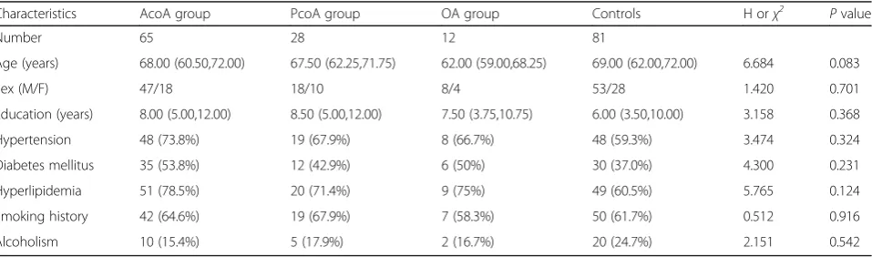
Among the 126 patients with severe unilateral ICA sten-osis, 105 patients and all the normal controls completed all the examinations and tests. Based on their results, 65 patients were assigned to the AcoA group, 28 patients to the PcoA group, and 12 patients to the OA group. Demographic data and risk factors were summarized (Table 1). There was no significant difference in these demographic characteristics and risk factors among the four groups (all P> 0.05). In the AcoA group, the inci-dence of diabetes mellitus (χ2= 4.125, P= 0.042) and hyperlipidemia (χ2= 5.395, P= 0.02) were slightly higher than those in controls after pairwise comparisons.
Influence of collateral circulation on BFV
The ipsilateral BFV parameters (Vs, Vm, and Vd) in pa-tients with ICA stenosis were significantly lower than those in normal controls (P< 0.0001). These BFV parame-ters from high to low in order were AcoA group > PcoA group > OA group (allP< 0.05). PI in patients was signifi-cantly lower than that in normal controls, and PI from high to low in order was OA group > PcoA group > AcoA group (all P< 0.05). The statistical results were presented in Table 2. The contralateral BFV parameters (Vs, Vm, and Vd) only in the AcoA group were significantly higher than those in the other groups (allP< 0.0001). There was no significant difference in contralateral PI among the groups (P> 0.05) (Table3).
Influence of collateral circulation on neuropsychological tests
Word Fluency Test scores of the AcoA group and con-trols were significantly higher than those in the PcoA and OA groups (AcoA vs PcoA, P= 0.005; AcoA vs OA,
P= 0.012; PcoA vs controls, P< 0.0001; OA vs controls,
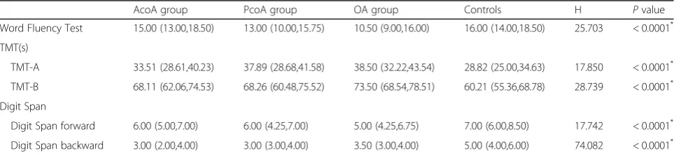
P= 0.001), and there was no significant difference be-tween the AcoA group and controls. All patients re-quired significantly longer time to finish TMT (Part A
and Part B) than controls (for Part A: AcoA vs controls,P= 0.013; PcoA vs controls:P= 0.018; OA vs controls, 0.023; for Part B: AcoA vs controls,P< 0.0001; PcoA vs controls,P= 0.002; OA vs controls,P= 0.001). The scores of Digit Span (forward and reverse) in all patients were significantly lower than those of the control participants (for forward: AcoA vs controls, P= 0.01; PcoA vs controls, P= 0.009; OA vs controls, P= 0.016; for backward: every patient group vs controls, P< 0.0001). There was no significant dif-ference among the three patient groups in TMT (Part A and Part B) and Digit Span (P> 0.05) (Table 4).
There was no significant difference in RTSCWT A or RTSCWT B among the four groups (P> 0.05). RTSCWT C and RT for SIE in all patient groups were significantly longer than those in normal controls (for SCWTC: AcoA vs controls, P< 0.0001; PcoA vs controls, P= 0.002; OA vs controls, P= 0.043; for RT for SIE: AcoA vs controls,P< 0.0001; PcoA vs controls,P= 0.021; OA vs controls, P= 0.005), and the AcoA group had higher score than the PcoA groups (for SCWT C: P= 0.001; for RT for SIE:P= 0.02) and OA groups (for SCWT C:
P= 0.046; for RT for SIE: P= 0.03). There was no sig-nificant difference in ARSCWT A or ARSCWT B among the four groups (P> 0.05). ARSCWT C in the OA group was significantly lower than that in the normal controls (P= 0.004), while there was no significant difference in
Table 1Demographic data of patients with severe ICA stenosis and normal controls
Characteristics AcoA group PcoA group OA group Controls H orχ2 Pvalue
Number 65 28 12 81
Age (years) 68.00 (60.50,72.00) 67.50 (62.25,71.75) 62.00 (59.00,68.25) 69.00 (62.00,72.00) 6.684 0.083
Sex (M/F) 47/18 18/10 8/4 53/28 1.420 0.701
Education (years) 8.00 (5.00,12.00) 8.50 (5.00,12.00) 7.50 (3.75,10.75) 6.00 (3.50,10.00) 3.158 0.368
Hypertension 48 (73.8%) 19 (67.9%) 8 (66.7%) 48 (59.3%) 3.474 0.324
Diabetes mellitus 35 (53.8%) 12 (42.9%) 6 (50%) 30 (37.0%) 4.300 0.231
Hyperlipidemia 51 (78.5%) 20 (71.4%) 9 (75%) 49 (60.5%) 5.765 0.124
Smoking history 42 (64.6%) 19 (67.9%) 7 (58.3%) 50 (61.7%) 0.512 0.916
Alcoholism 10 (15.4%) 5 (17.9%) 2 (16.7%) 20 (24.7%) 2.151 0.542
χ2
value, by Chi-squared test.HandPvalue, by non-parametric Kruscal-Wallis tests within the four groups
Table 2ThePvalues of ipsilateral BFV parameters between different groups
Vs Vm Vd PI
AcoA vs PcoA 0.017 < 0.0001 < 0.0001 0.034
AcoA vs OA < 0.0001 < 0.0001 < 0.0001 0.032
AcoA vs controls < 0.0001 < 0.0001 0.036 < 0.0001
PcoA vs OA 0.003 0.002 0.007 0.997
PcoA vs controls < 0.0001 < 0.0001 < 0.0001 < 0.0001
OA vs controls < 0.0001 < 0.0001 < 0.0001 < 0.0001
ARSCWT Camong AcoA group, PcoA group and normal controls (P> 0.05). AR for SIE in PcoA group and OA group was significantly higher than that in AcoA group (for PcoA group, P= 0.03; for OA group, P= 0.026) and controls (for PcoA group,P= 0.001; for OA group,
P= 0.015), which also caused differences among the four groups (P< 0.0001) (Table5).
Correlation between BFV parameters and neuropsychological scores
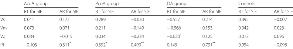
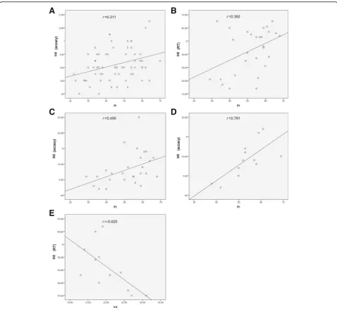
There was no significant correlation between BFV param-eters and Word Fluency Test or Digit Span. The ipsilateral PI was positively correlated with AR for SIE in patients with ICA stenosis (P< 0.05), especially in PcoA group and OA group (P< 0.01). The ipsilateral PI was positively cor-related with RT for SIE in the PcoA group (P< 0.05). There was a negative correlation between Vd and RT for SIE in the OA group (P< 0.05) (Table6) (Fig.1).
Discussion
In the present study, we investigated whether cerebral BFV and frontal lobe cognitive function would be influenced by the different type of collateral circulation in patients with ICA stenosis. Previous studies have proposed that BFV mea-sured by TCD was a major indicator reflecting cerebral hemodynamics [18], and BFV of MCA is the most reliable parameter [19]; herein, we measured BFV in bilateral MCA.
On the other hand, Word Fluency Test, TMT, Digit Span, and SCWT were also included in the frontal lobe cognitive function assessment [20]. Our findings indi-cated that SCWT was affected by collaterals more than other neurological function tests. Moreover, we found the ipsilateral PI was significantly associated with impaired selective attention. In brief, the current study provides a new insight into the correlations among collateral circulation, cerebral perfusion and selective attention.
Relationship between CBFV and collateral circulation Generally, ICA stenosis mainly affects the blood perfusion of ipsilateral anterior circulation. We found the BFV of ipsi-lateral MCA was significantly reduced in patients with ICA stenosis. Especially, the decrease of BFV in PcoA group was more apparent than that in AcoA group, suggesting collat-eral circulation through the AcoA may provide a better compensation for the impaired cerebral perfusion. However, Gfusi et al. proposed that collateral circulation through PcoA to be important for cerebral perfusion because the absence of PcoA collaterals was found to be associated with poor prognosis [21]. Thus, we also speculate collateral circu-lation through the PcoA may be necessary in the absence of AcoA collaterals. Additionally, the BFV of contralateral MCA in AcoA group was higher than that in other groups, which may be attributed to the compensation.
Table 3TCD parameters in patients with severe ICA stenosis and normal controls
Parameters AcoA group PcoA group OA group Controls H Pvalue
Vs (Affected side) 50.00 (44.50,56.00) 44.00 (36.25,52.25) 31.50 (27.25,41.25) 79.00 (74.00,85.00) 143.610 < 0.0001*
Vs (Unaffected side) 123.00 (102.00,136.50) 80.50 (68.25,96.00) 96.00 (69.00,107.25) 102.901 < 0.0001*
Vm (Affected side) 39.00 (35.00,43.50) 32.50 (28.25,38.00) 22.50 (20.25,30.50) 52.00 (46.00,57.00) 117.469 < 0.0001*
Vm (Unaffected side) 73.00 (63.00,86.50) 47.50 (42.25,61.50) 55.50 (41.25,67.25) 78.395 < 0.0001*
Vd (Affected side) 33.00 (28.00,37.00) 28.00 (23.25,31.75) 18.50 (17.00,25.50) 37.00 (32.75,40.00) 55.753 < 0.0001*
Vd (Unaffected side) 51.00 (42.00,64.50) 31.50 (28.00,43.75) 35.50 (28.50,44.50) 57.931 < 0.0001*
PI (Affected side) 0.45 (0.35,0.54) 0.55 (0.42,0.60) 0.53 (0.50,0.59) 0.85 (0.75,1.03) 131.688 < 0.0001*
PI (Unaffected side) 0.88 (0.74,1.11) 0.96 (0.85,1.13) 0.99 (0.87,1.15) 6.445 0.092
Vs, stolic flow velocity;Vm, mean flow velocity;Vd, diastolic velocity;PI, pulsatility index;HandPvalue, by non-parametric Kruscal-Wallis tests within the four groups *
P< 0.0001 indicate statistically significant differences
Table 4Comparison of cognitive assessment in patients with severe ICA stenosis and normal controls
AcoA group PcoA group OA group Controls H Pvalue
Word Fluency Test 15.00 (13.00,18.50) 13.00 (10.00,15.75) 10.50 (9.00,16.00) 16.00 (14.00,18.50) 25.703 < 0.0001*
TMT(s)
TMT-A 33.51 (28.61,40.23) 37.89 (28.68,41.58) 38.50 (32.22,43.54) 28.82 (25.00,34.63) 17.850 < 0.0001*
TMT-B 68.11 (62.06,74.53) 68.26 (60.48,75.52) 73.50 (68.54,78.51) 60.21 (55.36,68.78) 28.739 < 0.0001*
Digit Span
Digit Span forward 6.00 (5.00,7.00) 6.00 (4.25,7.00) 5.00 (4.25,6.75) 7.00 (6.00,8.50) 17.742 < 0.0001*
Digit Span backward 3.00 (2.00,4.00) 3.00 (3.00,4.00) 3.50 (3.00,4.00) 5.00 (4.00,6.00) 74.082 < 0.0001*
TMT, Trail-Making Test;HandPvalue, by non-parametric Kruscal-Wallis tests within the four groups *
The effect of OA collaterals on hemispheric hemodynamics in patients with severe ICA stenosis is still controversial. Telman et al. reported that collateral circulation through OA had no influence on hemispheric hemodynamics [22]; nevertheless, Henderson et al. found that OA collat-erals may be associated with a lower risk of stroke and TIA [23]. In our study, the BFV of ipsilateral MCA was the lowest in the OA group. The possible reason for these inconsistent results may be the dif-ferent inclusion and grouping criteria. Besides AcoA and PcoA, there may be other collaterals (e.g. lepto-meningeal artery) which have little effect on cerebral perfusion. Cheng et al. found cerebral perfusion to be significantly impaired in patients with collateral circu-lation via secondary collaterals, which was consistent with our findings [24].
Relationship between frontal lobe cognitive function and collateral circulation
Some studies have proposed that cerebral BFV can re-flect the brain perfusion as well as cognitive dysfunctions [25]. Wang et al. reported significant CBF reduction in patients with anterior circulation TIA who had vascular cognitive impairment [26]. Patients with carotid athero-sclerosis showed lower scores in verbal fluency test even when the atherosclerosis were subclinical [27], and
verbal fluency was improved after carotid artery stenting or carotid endarterectomy for patients with carotid ar-tery stenosis [28]. These findings indicated that CBF was associated with verbal fluency performance. In the present study, decreased performance of Word Fluency Test in the PcoA and OA groups indicated their im-paired sematic memory and language ability. The undif-ferentiated results between the AcoA and control group implied a potential for preseverd language function among AcoA collateral patients. It has been reported that patients with greater carotid artery intima-media thickness had worse TMT performance [29], which is consistent with our findings. Digit Span is a measure of working memory and is not associated with emotional lability [30]. The worse results of TMT and Digit Span in patients with ICA stenosis indicated impaired execu-tive function and working memory included in frontal lobe cognitive function, and the type of collaterals had no significant impact on these subfunctions.
In SCWT, significant difference was only found in RT and AR of SCWT C and SIE, suggesting a higher sensitivity for detection of selective attention in patients with severe ICA stenosis. In SCWT C, subjects needed to suppress the habitual response to the meaning of the words and shift their attention to its incongruent color. The incongruent task in Stroop requires response inhibition, which may be Table 5SCWT assessment in patients with severe ICA stenosis and normal controls
SCWT parameters AcoA group PcoA group OA group Controls H Pvalue
Response time (ms)
SCWT A 30.00 (21.50,36.50) 29.00 (20.00,37.50) 28.00 (21.75,34.00) 28.00 (18.50,33.00) 2.209 0.566
SCWT B 49.00 (41.00,61.50) 49.00 (42.25,52.75) 47.50 (42.00,54.25) 46.00 (38.50,52.50) 4.842 0.184
SCWT C 104.00 (94.50,117.50) 96.00 (82.50,106.50) 92.50 (86.75,102.75) 82.00 (70.00,91.00) 65.925 < 0.0001**
Response time for SIE 55.00 (48.00,63.00) 52.00 (32.00,57.00) 43.50 (37.75,52.25) 36.00 (27.50,41.00) 70.771 < 0.0001**
Accuracy rate (%)
SCWT A 98.00 (92.50,100.00) 99.00 (96.25,99.75) 97.00 (95.25,99.75) 98.00 (96.00,99.50) 1.134 0.726
SCWT B 85.00 (79.00,96.50) 90.00 (85.00,97.75) 83.50 (78.75,98.25) 89.00 (86.00,95.00) 4.924 0.177
SCWT C 81.00 (75.50,90.00) 81.00 (75.25,90.75) 78.50 (69.75,86.00) 85.00 (80.00,90.00) 11.579 0.009*
Accuracy rate for SIE 4.00 (2.50,6.00) 7.00 (5.25,11.75) 9.00 (5.50,11.75) 4.00 (2.00,5.00) 35.621 < 0.0001**
SIE, Stroop interference effects;HandPvalue, by non-parametric Kruscal-Wallis tests within the four groups *
P< 0.01,**
P< 0.001 indicate statistically significant differences
Table 6Correlation coefficient between TCD parameters (side of stenosis) and SIE scores
AcoA group PcoA group OA group Controls
RT for SIE AR for SIE RT for SIE AR for SIE RT for SIE AR for SIE RT for SIE AR for SIE
Vs 0.041 0.172 0.289 −0.030 −0.557 0.214 0.095 −0.007
Vm 0.073 0.071 0.211 −0.149 −0.566 0.153 0.042 0.023
Vd 0.084 −0.015 0.034 −0.234 −0.620* 0.125 0.013 0.096
PI −0.103 0.311* 0.392* 0.490** 0.143 0.791** 0.054 −0.098
RT, response time;AR, accuracy rate;SIE, Stroop interference effects; Vs, systolic flow velocity;Vm, mean flow velocity;Vd, diastolic flow velocity;PI, pulsatility index *
linked with anterior cingulate gyrus [21]. Better perform-ance in SCWT requires more blood flow in frontal lobe [31], and worse performance in patients with ICA stenosis may be explained due to the altered blood flow. Carter et al. hypothesized that the poor performance in SCWT may be associated with the anterior cingulate gyrus dys-function as evaluated by positron emission tomography [32]. Moreover, Benabarre et al. found the SCWT score to be well correlated with the CBF in striatal, temporo-medial, and parietal cortices [33]. Taylor et al. found the ac-tivation of left inferior frontal gyrus on positron emission
tomography to reflect the Stroop processing [34]. All of the regions mentioned can be affected by reduced blood supply of ICA. Furthermore, the dorsal anterior cingulate cortex receives top-down information from the dorsolat-eral prefrontal cortex and bottom-up information from the left caudate nucleus [35]. Hypoperfusion may disrupt the interaction of these grey-matter areas, leading to poor per-formance in SCWT. AcoA has been verified to supply the frontal lobe in patients with ICA occlusion [36]. We also noted patients with collaterals via AcoA exhibited a better performance in SCWT than the patients in other groups.
A
B
C
E
D
Fig. 1Correlation between TCD parameters (side of stenosis) and SIE scores.aScatter plot demonstrating correlation between pulsatility index (PI) and SIE accuracy in the AcoA group.bScatter plot demonstrating correlation between PI and reaction time of SIE in the PcoA group.c
Of note, RTSCWT Cand RT for SIE in AcoA group were longer than those of other groups. He et al. found the Chinese language functional areas were located in both frontal and temporal lobes [37]. Therefore, patients in AcoA group may suffer less impairment in language func-tion because of the well-preserved anterior circulafunc-tion. The normal results of the AcoA group in Word Fluency Test also demonstrated inconsistency. In SCWT, they could be easier to be interfered by the incongruent meaning of the words, and they might spend more time in judging the color of the words. However, there was no difference in AR for SIE only between the AcoA group and controls. We speculate the patients in AcoA group had relatively intact language function manifesting as normal AR at a cost of prolonged RT. Both RT and AR were increased in PcoA and OA groups, indicating that collaterals via PcoA or OA to be associated with a relatively severe impairment of se-lective attention. Additionally, ARSCWT Cin the OA group was significantly lower than in normal controls, suggesting the patients in the OA group had more severe selective at-tention impairment. Reinhard et al. proposed that the cere-bral blood autoregulation in patients with collaterals via secondary pathways was worse than that in patients with collaterals via primary pathways [38], and cerebral autoreg-ulation to be crucial for preserving the cognitive function, which may help explain our findings. In general, the frontal lobe cognitive function is impaired to various extents in patients with ICA stenosis. Being different from other frontal lobe cognitive assessment, SCWT, especially SCWT C and SIE reflected respective position and function for different collaterals more precisely.
Relationship between selective attention and TCD parameters
We noted a negative correlation between Vd of MCA and RT for SIE in the OA group, indicating that BFV may be insensitive in reflecting selective attention be-cause we only found statistically significant correlation between Vd and RT for SIE in the OA group. A possible reason is that the inconstant blood flow may be influ-enced by blood pressure, temperature and psychological state, and cerebral BFV may reflect selective attention only when cerebral perfusion is severely impaired.
The positive correlation between ipsilateral PI and SIE suggests that PI could be a potential indicator for evaluat-ing selective attention impairment. In our patients with severe ICA stenosis, PI of ipsilateral MCA decreased in all patient groups. In previous studies, the relationship be-tween PI and cognitive function remains controversial. Altmann et al. demonstrated that elevated PI was associ-ated with impairment in several cognitive domains [39]. However, Shim et al. found that PI in patients with cogni-tive impairment did not differ from that in normal con-trols [40]. We speculate the impaired selective attention
may be partly due to the abnormal resistance of cerebral vessels and low cerebral perfusion. Moreover, the more general correlation of PI and AR for SIE indicated AR may be a more valuable indicator in reflecting cognitive function in patients with ICA stenosis.
Limitations
There are several limitations to the current study. First, the slight statistical difference in diabetes mellitus and hyperlipidemia between the AcoA group and the con-trols may be due to the limited sample size. However, the worse results of the PcoA and OA group showed the association of diabetes mellitus and hyperlipidemia with the impairment of frontal lobe selection may be nonexis-tent. Second, the present study was limited by the small sample size of other types of collaterals as only collateral circulation via AcoA, PcoA and OA was analyzed. Third, drugs (especially statins) may be confounding factor on vasomotor reactivity [41]. In the future, we will enlarge the sample size via initiating a multi-center study and take the drug-related confounding factors into account.
Conclusions
To our knowledge, this study is the first to analyze the influence of different collaterals on CBF and frontal lobe cognitive function in patients with severe ICA stenosis. The frontal lobe cognition function is impaired to differ-ent extdiffer-ents according to the type of collaterals. Most notably, selective attention impairment is correlated with the type of collaterals. Collateral circulation via AcoA is associated with a relatively intact selective attention, and collateral circulation via OA is associated with a severe impairment of selective attention. Furthermore, PI may be a potential indicator for identifying selective attention dysfunction in patients with severe ICA stenosis.
Abbreviations
AcoA:Anterior communicating artery; ANOVA: Analysis of variance; AR: Accuracy rate; BFV: Blood flow velocity; CBF: Cerebral blood flow; DSA: Digital subtraction agiography; ICA: Internal carotid stenosis; MCA: Middle cerebral artery; MMSE: Mini-Mental State Examination; NASCET: North American Symptomatic Carotid Endarterectomy Trail; OA: Ophthmaltic artery; PcoA: Posterior communicating artery; PI: Pulsatility index; RT: Response time; SCWT: Stroop Color Word Test; SD: Standard deviation; SIE: Stroop interference effects; TCD: Transcranial Doppler; TIA: Transient ischemic attack; TMT: Trail-Making Test; Vd: Diastolic flow velocity; Vm: Mean flow velocity; Vs: Systolic flow velocity
Acknowledgements
Not applicable.
Authors’contributions
HL and WW designed the study. XD and ZL contributed samples collection. WW and JR collected, analysed and interpreted the data. WW and HL wrote the initial paper and XY revised the paper. All authors gave intellectual input to the study and approved the final version of the manuscript.
Funding
declare that they have no financial relationship with the organization that sponsored the research, and the funding body was not involved in study design, data collection, analysis and writing of the study.
Availability of data and materials
The datasets used and/or analysed during the current study are available from the corresponding author on reasonable request.
Ethics approval and consent to participate
This study was approved by the Ethics Committee of the Affiliated Hospital of Southwest Medical University. All procedures performed in studies involving human participants were in accordance with the ethical standards of the institutional and national research committee and with the 1964 Helsinki declaration and its later amendments or comparable ethical standards. Written informed consent was obtained from all individual participants included in the study.
Consent for publication
Not applicable.
Competing interests
The authors declare that they have no competing interests.
Author details 1
Department of Neurology, the Affiliated Hospital of Southwest Medical University, No. 25 Taiping Road, Luzhou 646000, Sichuan, China.2Department of Neurology, People’s Hospital of Deyang City, Deyang 618000, Sichuan, China.3Department of Rehabilitation medicine, the Affiliated Hospital of Southwest Medical University, Luzhou 646000, Sichuan, China.
Received: 23 November 2018 Accepted: 26 June 2019
References
1. Lee JM, Park JM, Min KS, Oh YJ, Kim CJ, Kim YJ. The ameliorative effects of exercise on cognitive impairment and white matter injury from blood-brain barrier disruption induced by chronic cerebral hypoperfusion in adolescent rats. Neurosci Lett. 2017;638:83–9.
2. Zhao JH, Tian XJ, Liu YX, Yuan B, Zhai KH, Wang CW, et al. Executive dysfunction in patients with cerebral hypoperfusion after cerebral angiostenosis/occlusion. Neurol Med Chir (Tokyo). 2013;53:141–7. 3. Sussman ES, Kellner CP, Mergeche JL, Bruce SS, Mcdowell MM, Heyer EJ,
et al. Radiographic absence of the posterior communicating arteries and the prediction of cognitive dysfunction after carotid endarterectomy. J Neurosurg. 2014;121:593–8.
4. Zavoreo I, BašićKV, Lisak M, MaršićN, Ciliga D, Trošt BT. Cognitive decline and cerebral vasoreactivity in asymptomatic patients with severe internal carotid artery stenosis. Acta Neurol Belg. 2013;113:453.
5. Everts R, Wapp M, Burren Y, Kellner-Weldon F, El-Koussy M, Jann K, et al. Cognitive and emotional effects of carotid stenosis. Swiss Med Wkly. 2014; 144:w13970.
6. Sztriha LK, Nemeth D, Sefcsik T, Vecsei L. Carotid stenosis and the cognitive function. J Neurol Sci. 2009;283:36.
7. Warkentin S, Passant U. Functional imaging of the frontal lobes in organic dementia. Regional cerebral blood flow findings in normals, in patients with frontotemporal dementia and in patients with Alzheimer's disease, performing a word fluency test. Dement Geriatr Cogn Disord. 1997;8:105–9. 8. Reitan RM. Validity of the trail making test as an Indicator of organic brain
damage. Percept Motor Skills. 1958;8:271–6.
9. Strasser ES, Haffner P, Fiebig J, Quinlivan E, Adli M, Stamm TJ. Behavioral measures and self-report of impulsivity in bipolar disorder: no association between Stroop test and Barratt impulsiveness Scale. Int J Bipolar Disord. 2016;4:16.
10. Pardo JV, Pardo PJ, Janer KW, Raichle ME. The anterior cingulate cortex mediates processing selection in the Stroop attentional conflict paradigm. Proc Natl Acad Sci U S A. 1990;87:256–9.
11. Saalmann YB, Pinsk MA, Liang W, Xin L, Sabine KJS. The pulvinar regulates information transmission between cortical areas based on attention demands. Science. 2012;337:753–6.
12. Zhang Y, Lin X, Zhou H, Xu J, Du X, Dong GJFiP. Brain Activity toward Gaming-Related Cues in Internet Gaming Disorder during an Addiction Stroop Task. 2016;7.
13. Collaborators NASCET. Beneficial effect of carotid endarterectomy in symptomatic patients with high-grade carotid stenosis. N Engl J Med. 1991; 325:445–53.
14. Xu J, Zhu XY, Sun H, Xu XQ, Xu SA, Suo Y, et al. Low vitamin D levels are associated with cognitive impairment in patients with Hashimoto thyroiditis. BMC Endocr Disord. 2018;18:87.
15. Spreen O, Strauss E. A compendium of neuropsychological tests: administration, norms, and commentary. Cogn Behav Neurol. 1991;41:1856. 16. Wechsler D. Manual for the WECHSLER adult intelligence SCALE. 1981;70. 17. Wang C, An Y, Yu H, Feng L, Liu Q, Lu Y, et al. Association between
exposure to the Chinese famine in different stages of early life and decline in cognitive functioning in adulthood. Front Behav Neurosci. 2016;10:146. 18. Giller CA, Bowman G, Dyer H, Mootz L, Krippner W. Cerebral arterial
diameters during changes in blood pressure and carbon dioxide during craniotomy. Neurosurgery. 1993;32:737.
19. Lindegaard KF, Lundar T, Wiberg J, Sjøberg D, Aaslid R, Nornes H. Variations in middle cerebral artery blood flow investigated with noninvasive transcranial blood velocity measurements. Stroke. 1987;18:1025–30. 20. Shin MK. Effects of an exercise program on frontal lobe cognitive function
in elders. J Korean Acad Nurs. 2009;39:107–15.
21. Guler S, Utku U, Aynaci O. Early clinical signs, lesion localization, and prognostic factors in unilateral symptomatic internal carotid artery occlusion. J Stroke Cerebrovasc Dis. 2014;23:1908–14.
22. Telman G, Kouperberg E, Sprecher E, Hoffman A, Yarnitsky D. Assessment of ophthalmic artery collateral pathway in the hemispheric cerebral hemodynamics in patients with severe unilateral carotid stenosis. Neurol Res. 2003;25:309–11. 23. Henderson RD, Eliasziw M, Fox AJ, Rothwell PM, Barnett HJ.
Angiographically defined collateral circulation and risk of stroke in patients with severe carotid artery stenosis. North American symptomatic carotid endarterectomy trial (NASCET) group. Stroke. 2000;31:128–32.
24. Cheng XQ, Tian JM, Zuo CJ, Liu J, Zhang Q, Lu GM. Quantitative perfusion computed tomography measurements of cerebral hemodynamics: correlation with digital subtraction angiography identified primary and secondary cerebral collaterals in internal carotid artery occlusive disease. Eur J Radiol. 2012;81:1224–30.
25. Becker A, Mandell AR, Tangney JP, Chrosniak LD, Shaw TH. The effects of self-control on cognitive resource allocation during sustained attention: a transcranial Doppler investigation. Exp Brain Res. 2015;233:2215–23. 26. Wang J, Li Y, Bo Z, Jian W, Wang Z, Dan D, et al. Computed tomography
perfusion imaging may predict cognitive impairment in patients with first-time anterior circulation transient ischemic attack. Int J Cardiovasc Imaging. 2016;32:671–7.
27. Wu DM, Zhang JJ, Guo N, Zheng SJ, Zhu QF, Feng YQ, et al. Cognitive impairment correlates with serum carbonyl compound profiles in subclinical carotid atherosclerosis. Neuroreport. 2018;29:1550–7.
28. Kougias P, Collins R, Pastorek N, Sharath S, Barshes NR, Mcculloch K, et al. Comparison of domain-specific cognitive function after carotid endarterectomy and stenting. J Vasc Surg. 2015;62:355–61.
29. Suemoto CK, Santos IS, Bittencourt MS, Pereira AC, Goulart AC, Rundek T, et al. Subclinical carotid artery atherosclerosis and performance on cognitive tests in middle-aged adults: baseline results from the ELSA-Brasil. Atherosclerosis. 2015;243:510–5.
30. Jensen DA, Hovik MF, Monsen NJN, Eggen TH, Eichele H, Adolfsdottir S, et al. Keeping emotions in mind: the influence of working memory capacity on parent-reported symptoms of emotional lability in a sample of children with and without ADHD. Front Psychol. 2018;9:1846.
31. Tsuchiya K, Mitsui S, Fukuyama R, Yamaya N, Fujita T, Shimoda K, et al. An acute bout of housework activities has beneficial effects on executive function. Neuropsychiatr Dis Treat. 2018;14:61–72.
32. Carter CS, Mintun M, Nichols T, Cohen JD. Anterior cingulate gyrus dysfunction and selective attention deficits in schizophrenia: [15O]H2O PET study during single-trial Stroop task performance. Am J Psychiatry. 1997;154:1670. 33. Benabarre A, Vieta E, Martínezarán A, Garciagarcia M, Martín F, Lomeña F,
et al. Neuropsychological disturbances and cerebral blood flow in bipolar disorder. Aust N Z J Psychiatry. 2010;40:375–6.
35. Ridderinkhof KR, Wp VDW, Segalowitz SJ, Carter CS. Neurocognitive mechanisms of cognitive control: the role of prefrontal cortex in action selection, response inhibition, performance monitoring, and reward-based learning. Brain Cogn. 2004;56:129–40.
36. de Boorder MJ, Van dGJ, van Dongen AJ, Klijn CJ, Jaap KL, Van Rijk PP, et al. Spect measurements of regional cerebral perfusion and carbondioxide reactivity: correlation with cerebral collaterals in internal carotid artery occlusive disease. J Neurol. 2006;253:1285–91.
37. He C, Andre VG, Gloria G, Pamela T, Andrea H, Duncan JS. Mandarin functionalMRILanguage paradigms. Brain Behav. 2016;6:1–11.
38. Reinhard M, Müller T, Guschlbauer B, Timmer J, Hetzel A. Dynamic cerebral autoregulation and collateral flow patterns in patients with severe carotid stenosis or occlusion. Ultrasound Med Biol. 2003;29:1105–13.
39. Altmann M, Thommessen B, Rønning OM, Benth JŠ, Reichenbach AS, Fure B. Middle cerebral artery Pulsatility index is associated with cognitive impairment in lacunar stroke. J Neuroimaging. 2016;26:431–5.
40. Shim Y, Yoon B, Shim DS, Kim W, An JY, Yang DW. Cognitive correlates of cerebral vasoreactivity on transcranial Doppler in older adults. J Stroke Cerebrovasc Dis. 2015;24:1262–9.
41. Giannopoulos S, Katsanos AH, Tsivgoulis G, Marshall RS. Statins and cerebral hemodynamics. J Cereb Blood Flow Metab. 2012;32:1973–6.
Publisher’s Note