Vol. 7, Number 1, Winter/Spring 2017/1-9 - DOI: 10.22059/pbs.2018.206895.1217
Identification and characterization of pigmented
photoreceptor-producing microorganisms using FTIR
spectroscopy
Maryam Fanaei1, Giti Emtiazi1,*
1
Department of Biology, Faculty of Science, University of Isfahan, Hezarjarib street, Zip code: 8174673441, Isfahan, Iran
Received: November 9, 2016; Accepted: May 6, 2017
The identification and classification of microorganisms have been subjects of research for many years. Recently, Fourier transform infrared (FTIR) spectroscopy techniques have gained attention in the characterization and classification of microorganisms based on biochemical profiles and cell structure characteristics. In the present study, the characterization and identification of pigmented photoreceptor-producing microorganisms using FTIR spectroscopy were carried out. For this purpose some microorganisms were isolated from different environments, of which three pigment producing bacteria were selected to limit the scope of the study to one phenotypic characteristic. Genomic relatedness among the isolated strains was investigated. It was shown that these strains had similarities to Kushneria marisflavi, Halobacillus halophilus and Halobacillus faecis. In addition, Halobacterium salinarum was investigated as a typical representative pigment producing archaeon. Spectra (500-4000 cm-1) of the intact cells and crude extracted pigments were recorded on an FTIR spectrometer and compared with each other. The similarities among the spectra were evaluated using hierarchical cluster analysis and compared with the phylogenetic tree based on the genomic study. Our results demonstrate that hierarchical clustering based on extracted pigments shows discrimination of strains more distinctly than those based on intact cells. The results of the present study suggest that FTIR analysis of bacterial pigments is an easy and economic method comparable to phylogenetic markers, for identification and characterization of bacteria.
Keywords: Archaea; Classification; Fourier transform infrared (FTIR) spectroscopy; Phylogenetic analysis; Pigment production
Introduction
Microbial pigments often have a significant phenotypic effect in such a way that in certain genera,
their identification. Many efforts are directed towards the phenotypic detection of microorganisms. Consequently, researchers make computer-assisted models to predict the properties and behavior of microbial cells. They predict the behavior of these cells using detailed information obtained from other similar cells. So far, analysis of cell proteins (using two-dimensional protein gel electrophoresis) and membrane lipids have been carried out (1, 2). Another approach to the phenotypic detection of microorganisms is inferring the phylogenetic position of bacteria based on their phenotypic characteristics, since microbial phylogeny based on the 16S rRNA gene cannot exactly describe the phenotypic properties such as microorganism morphology and structure. Some routine methods for phenotypic microbial identification rely on information obtained from the analysis of cell structure, cellular metabolism and identification of cell components such as fatty acids, pigments and cell walls (3).
Spectroscopy is recognized as an important technique for the rapid identification of organic and inorganic compounds. IR analysis is one of the spectroscopic methods that have attracted the attention of researchers because it is rapid and uses high informative content of vibrational spectra. The infrared spectrum of a given sample is expectable when a beam of infrared light is passed through the sample. The absorbed energy at each wavelength is determined through examination of the transmitted light. FTIR is a type of IR spectroscopy proposed to be useful for classification of microorganisms based on analysis of intact cell surface. This method is based on wavelength measurement and determination of the intensities of infrared absorption bands. This kind of spectroscopy appears to have high potential for investigating the functional groups on bacterial surface, detecting the change in the structure of bacteria during growth phases (4), inferring the secondary structure of peptide (5) and for bacterial identification in culture and food (6).
Recently, some microbial compounds have been investigated by FTIR to determinate the microorganisms. Kim et al. (7) successfully applied the FTIR technique to recognize six different serotypes of Salmonella enterica according to their lipopolysaccharides (LPS). Similarly, the IR spectra of crude phenol-phase extracts containing bacterial lipopolysaccharides have been used to differentiate five strains of Escherichia coli (8). In addition, in previous studies, FTIR was applied to analyze the
pigment compositions of microorganisms (9). In the present study, identification and classification of four pigmented microorganisms using FTIR spectra were carried out and the biochemical properties of the extracted pigments were compared with those of intact cells.
Microbial pigments are an important class of natural metabolites which have a wide variety of activities. A small group of pigmented organisms can produce photoreceptors responsive to light irradiation. We focused on the pigmented microorganisms capable of producing photoreceptors to limit the scope of the study to one phenotypic characteristic and investigate whether there were any phenotypic similarities among the members of this group of microorganisms.
Materials and Methods
Isolation of bacteriaWater and soil samples were collected from different regions of Iran. Water (1 mL) and soil (0.1 g) samples were transferred to a medium containing 0.5% yeast extract, 0.5% Casamino acid, 0.1% peptone, 0.2% KCl, 0.3% sodium citrate, 2% MgSO4, 20% NaCl,
0.0036% FeCl2 and 2% agar (pH was adjusted to 7.5)
(10). The samples were incubated at room temperature and were exposed to light and darkness during the day and night, respectively. The colored colonies obtained after 4 weeks of incubation, were selected and spread on an agar plate using the streak plate technique in order to obtain pure bacterial colonies. A strain of
Halobacterium salinarum was obtained from the Iranian biological resource center (Strain Number: IBRC-M 10715) as the control bacterial strain.
Screening of photoreceptor-producing bacteria
plates were used to investigate the interaction of the TTC solution with lysed cells in conditions of both light and darkness. 150 µl of the lysed cells was transferred to each of the microtiter plates and 150 µl TTC solution was added to each one (TTC and bacterial solutions were examined separately as negative controls). One of the plates was immediately placed in darkness and the other in light to investigate the effect of light on photoactive protein activity. Samples were incubated at 37°C for 16 hrs. TTC reduction was determined by colorimetric assay at a wavelength of 492 nm and the reactions were carried out in triplicates.
Identification of the isolates
Analysis of bacterial morphology and growth condition
Bacterial staining was performed by applying an improved technique for halophilic bacteria using samples fixed with acetic acid as described by Dussault (11). An optical microscope was used to study the bacterial morphology.
DNA extraction and polymerase chain reaction (PCR) processing
The boiling extraction method was carried out to prepare the DNA template from fresh colonies which had been suspended in 750 µl sterile distilled water, vortexed for a few mins and boiled in a water bath (100°C) for 15 min. The samples were centrifuged at 7000 rpm for 10 min. 200 µl of the supernatant was collected from the specimen as the DNA template and stored at ‒20°C until the next step.
Identification of the isolates was performed using the modification method described by Yang et al. (12). A partial sequence of the 16S ribosomal RNA
gene was amplified using the universal primer pairs RW01, 5′-AAC TGG AGG AAG GTG GGG AT-3′ and DG74, 5′-AGG AGG TGA TCC AACCGC A-3′. The PCR mixtures (50 µl) contained 2 µl of each primer, 4 µl DNA extraction product, 0.8 µl Taq DNA polymerase, 1 µl dNTP, 1.4 µl MgCl2 and 5 µl buffer.
The PCR reaction was run using the following temperature profile: preliminary denaturation, 5 min at 95ºC, 30 cycles of 45 s denaturation at 94ºC, 30 s annealing at 58ºC, 45 s elongation at 72ºC and 7 min final elongation at 72ºC. The PCR products were
separated by electrophoresis in 1.3% agarose gel and stained with ethidium bromide. The gel was run at 85 V for 45 min. Gel purification of the PCR product from agarose gel was conducted and the PCR fragments were sequenced using an automated sequencer supplied by Bioneer (South Korea). Sequence blast analysis against the 16S ribosomal RNA sequences (Bacteria and Archaea) in the NCBI database was performed to find the similarity between the query partial sequences and the sequences in the NCBI database. Hierarchical analysis based on the partial sequences of the 16S rRNA was done using CLC software version 6.1.
Organisms and growth conditions
The bacterial strains used in this study were
Halobacterium salinarum, Kushneria marisflavi strain ZF, Halobacillus faecis strain HF and Halobacillus halophilus strain HA. Halobacterium salinarum is a well-known pigment-producing organism which was obtained from the Iranian Biological Resource Center.
Kushneria marisflavi, Halobacillus faecis and
Halobacillus halophilus were isolated using a method for screening photoactive protein-producing bacteria. Each cell was sub-cultured in a medium containing 0.5% yeast extract, 0.5% Casamino acid, 0.1% peptone, 0.2% KCl, 0.3% sodium citrate, 2% MgSO4,
20% NaCl, 0.0036% FeCl2 and 2% agar (pH was
adjusted to 7.5) at 37ºC.
Extraction of pigments
The crude pigments were extracted using acetone/methanol solvents by a method used by Olson
et al. and modified slightly for the purpose of this study (13). The colonies were harvested from the solid medium and transferred to distilled water. After spontaneous cell lysis in distilled water, the pigments were extracted with acetone/methanol (7:3 v/v) at ambient temperature with continuous shaking for 2 hrs. The samples were centrifuged at 4000 rpm for 10 min and the supernatants were collected as pigment solutions. The supernatants were analyzed by scanning in the wavelength region 350-600 nm using a UV-visible spectrophotometer.
Sample preparation for FTIR analysis
the agar surface to prepare the intact cells. The resultant samples were air-dried at ambient temperature to remove the excess water molecules before infrared analysis. The dried intact cells and the crude extracted pigments were prepared separately for spectral analysis. The samples were milled with potassium bromide (KBr) until they were thoroughly mixed and were then analyzed using Fourier transform infrared spectroscopy.
FTIR analysis
The data was obtained over the mid IR region (500 to 4000 cm-1) at a spectral resolution of 4 cm-1 (JASCO, FT/IR-6300; Japan). The baseline of the spectra was adjusted using Spekwin 32 software to avoid dissimilarity of spectra due to shift in baseline. In addition, the spectra were normalized to eliminate the differences between each spectrum due to difference in sample amount. The second derivatives of the spectra were calculated to obtain the most significant peaks of absorption (spectral region 600–2000 cm-1). Hierarchical cluster analysis was carried out by SPSS Software version 16 based on the similarities among the FTIR spectra of the samples.
Results
In this study tree pigment-producing bacteria were
selected among 75 pigmented microorganisms. All the isolated bacteria were halophilic, pigment producing strains and their colored colonies appeared on the plate after 2-3 days of incubation at 25°C. The partial sequences of the 16S rRNA gene from the new isolates were blasted with the GenBank database. Sequence blast analysis showed that the isolates have similarity to a partial sequence of the 16S rRNA gene of Kushneria marisflavi, Halobacillus halophilus and Halobacillus faecis strains. The related nucleotide sequences of the 16S rRNA gene were deposited in the NCBI Genbank under accession numbers KC906182, KF523277 and KC906181, respectively.
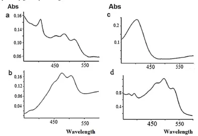
Spectral measurement of pigments demonstrates that the action spectrum for the extracted pigments from Kushneria marisflavi and the Halobacteium salinarum are characterized by three-fingered peaks; Halobacillus faecis is characterized by four-fingered peaks and the extracted pigments from Halobacillus halophilusis are characterized by one peak (Fig. 1). The TTC reduction of all three pigment-producing strains was higher when they were incubated under light, because these strains have a light-driven protein which can reduce TTC to red-colored triphenyl formazan (TPF) in a lighted environment.
FTIR analysis
The spectra of the intact cells and the crude pigment extracted from each strain were aligned and compared to each other within the mild-infrared range (500‒ 4000 cm-1) (Fig. 2).
Identification of the strains using Fourier transform infrared spectroscopy
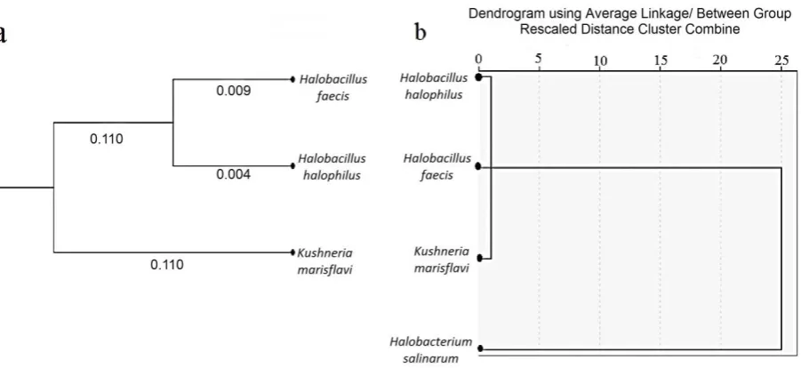
The similarity between the spectra of the intact cells and those of the extracted pigments were evaluated using hierarchical cluster analysis. The information obtained from each spectrum was considered as input data for the calculation of spectral distances. The clusters were produced by the Average Linkage algorithm and arranged by drawing a dendrogram using SPSS software (Fig. 3a). The hierarchical clustering achieved by the FTIR data indicates that the three strains show different placement based on the FTIR analysis (Fig. 3b).
Figure 2. FT-IR spectra (4000–500 cm−1) of intact cells and crude pigment extracted from each strain were aligned to each other.
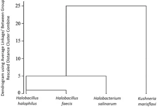
The FTIR spectra of the extracted pigments from the four pigment-producing microorganisms were also recorded and the dendrogram was drawn to arrange the clusters produced by the Average Linkage algorithm. The dendrogram illustrates that the branches related to the extracted pigments from the Halobacillus halophilus and Halobacillus faecis resemble each other and the branch related to the
extracted pigments from the Kushneria marisflavi was isolated from the Halobacillis halophilus and Halobacillus faecis by a relatively large distance as in the case of the Halobacterium salinarum (Fig. 4). The results indicate there was no direct relationship among the extracted pigments and the intact cell patterns of the microorganisms investigated in this study.
Figure 4. Differentiation of extracted pigments related to four pigment-producing microorganisms by hierarchical cluster analysis based on FTIR spectral data.
Discussion
The spectra of the intact cells illustrate six important regions that are representatives of the physicochemical properties of the cell surfaces. The 2800‒3000 cm-1
region is related to fatty acids. The 1637‒1695 cm-1
spectral region is the amide I band of proteins that characterize the C═O- stretch vibration. The position of the amide II absorption band is 1520‒ 1550 cm-1 which correlates with the bending vibration of the N—H bond in proteins (14). The 1200‒1500 cm-1 spectral region is a mixed region of fatty acid bending vibrations, proteins and phosphate-carrying compounds. Carbohydrates are located at about 900‒ 1200 cm-1, and 700‒900 cm-1 is the fingerprint region (15). Both amide I and amide II bonds are generally employed for the structural analysis of proteins (5). As shown in Fig. 2, the significant difference between the aligned absorption spectra is in the amide II region. In all the spectra of the intact cells both amide
I and amide II bonds can be observed, but in the spectra of the extracted pigments the amide II bonds are eliminated or weak in intensity. The major factor responsible for conformational sensitivity of amide II is the hydrogen-deuterium exchange in protein. During this process a hydrogen atom involved in a covalent bond is replaced by a deuterium atom or vice versa (16). The main factors affecting the amide hydrogen/deuterium exchange are pH, temperature, solvent accessibility, pKa and neighboring amino acids (17). A proton in a protein can be exchanged with the solvent; therefore, solvent accessibility causes conformational change of protein (18). In the present study the solvent used for pigment extraction may have influenced the amid II regions of the pigments. Another possible reason is the difference among the natural components of the intact cell pigments.
(Fig. 3a) compared to the hierarchical clustering achieved by the FTIR data indicates that the three strains were misclassified using FTIR analysis
.
The hierarchical clustering of the strains based on FTIR spectral data (Fig. 3b) indicates that theHalobacterium salinarum was totally isolated from the other microorganisms. Since the Halobacterium salinarum is an archaeon and the other strains are members of the Bacteria group, this result is satisfactory in terms of taxonomic classification. The
Kushneria marisflavi, Halobacillus halophilus and
Halobacillus faecis were clustered in the same branch.
The position of the amide I bond is related to the secondary structure of a protein and differences in overall amid I absorption is due to the different amounts of the secondary structural elements in the protein. The α-helix is at 1648‒1660 cm-1
, the β sheet is at 1625‒1640 and the turns are at 1660‒1685 cm-1
. The position of the amide I in all intact cells in this research was at 1655 cm-1 which indicated that α helix was the dominant structural element in the cell. The position of the amide I bond in the extracted pigment from the Halobacterium salinarum is similar to that of its intact cells; therefore, the proteins of the two components have the same secondary structure. The position of the amide I bond in the extracted pigments from the Halobacillus halophilus, Halobacillus faecis
and Kushneria marisflavi shifted to 1627 cm-1, 1636 cm-1 and 1640 cm-1, respectively indicating that the dominant structural element in the pigments is the β sheet
.
In addition, comparison of spectra shows that carbohydrate content (900-1200 cm-1) in the surface of the Halobacillus halophilus, Halobacillus faecis andKushneriam arisflavi cells is significantly different from that of their extracted pigments.
Spectroscopic techniques permit the study of some particular compounds of a given sample. Spectroscopic analysis of bacterial intact cells was introduced for the analysis of the chemical composition of bacteria (19, 20). Thereafter FTIR spectroscopy was successfully used for the classification of bacteria (21). Intact microbial cells have various chemical compounds which are different from those of other microorganisms. The spectra of intact microbial cells have fingerprint-like patterns which give general information concerning the physico-chemical properties of a strain (22). The
detection and characterization of microorganisms based on cellular biochemical profile is a valuable method since it is rapid, sensitive and economic. Recently FTIR analysis of the species in some genera such as Salmonella (23), Escherichia coli (6), Bacillus
(24), and Staphylococcus (25) has been applied for their identification and classification. In the present study the properties of a group of various species of microorganism showing a degree of phenotypic similarity were investigated. For this purpose, pigmented-producing organisms were selected.
Two significant roles have been suggested for the pigments: enabling strains to adapt to their environmental conditions and providing them with protection against environmental damages such as light irradiation (24). Microorganisms coexisting in the same ecological niche encounter similar environmental challenges; therefore they introduce similar functional groups on their cell surfaces. It seems that related pigments are produced for the same purpose. Accordingly, we focused on pigmented microorganisms living in environments of high salt concentrations in order to investigate microorganisms with similar phenotypic features
.
The result of the hierarchical clusters based on FTIR spectral data demonstrated that the intact cells of the microorganisms under study were not as clearly separated as the crude extracted pigments. Hierarchical clustering based on crude extracted pigment spectral data offered a good strain resolution, but was very different from classification based on intact cell spectral data and 16S rRNA partial sequences; therefore hierarchical classification based on the spectral data of crude extracted pigments can constitute a new kind of classification by which microorganisms are well separated. However, further research is required to determine the photoreceptor type of each cell and investigate the correlation of photoreceptor type with bacterial clustering based on pigment.
Acknowledgements
1. Movahedi, S., and Waites, W. (2000) A two-dimensional protein gel electrophoresis study of the heat stress response of Bacillus subtilis cells during sporulation. J. Bacteriol., 182, 4758-4763.
2. Quinn, P.J., Brain, A.P.R., Stewart, L.C., and Kates, M. (1986). The structure of membrane lipids of the extreme halophile, Halobacterium cutirubrum, in aqueous systems studied by freeze-fracture. BBA-Biomembranes, 863, 213-223.
3. Bochner, B.R. (2009) Global phenotypic characterization of bacteria. FEMS Microbiol. Rev., 33, 191-205. 4. Ede, S.M, Hafner, L.M., and Fredericks, P.M. (2004) Structural changes in the cells of some bacteria during population growth: a Fourier transform infrared-attenuated total reflectance study. Appl. Spectrosc.,
58, 317-322.
5. Adochitei, A., and Drochioiu, G. (2011) Rapid characterization of peptide secondary structure by FT-IR spectroscopy. Rev. Roum. Chim., 56, 783-791.
6. Mauer, L.J., and Reuhs, B.L. (2008) Mid‐infrared sensors for the rapid analysis of select microbial food borne pathogens. Wiley Handbook of Science and Technology for Homeland Security. John Wiley & Sons, Inc., New York.
7. Kim, S, Reuhs, B.L., and Mauer, L.J. (2005) Use of Fourier transform infrared spectra of crude bacterial lipopolysaccharides and chemometrics for differentiation of Salmonella enterica serotypes. J. Appl. Microbiol. 99, 411-417.
8. Kim, S., Burgula, Y., Ojanen‐Reuhs, T., Cousin, M.A, Reuhs, B.L., and Mauer, L.J. (2006) Differentiation of crude lipopolysaccharides from Escherichia coli strains using Fourier transform infrared spectroscopy and chemometrics. J. Food Sci., 71, M57-M61
9. Tripathi, B.K., and Agarwal, M.K. (2014) Detection of carotenoids in psychrotrophic bacteria by spectroscopic approach. J. BioSci. Biotech., 3, 253-260.
10. Fanaei, M., and Emtiazi, G. (2014) Isolation of a new Pseudomonas halophila strain possess bacteriorhodopsin-like protein by a novel method for screening of photoactive protein producing bacteria.
WORLD J. Microb. Biot., 30, 585-594.
11. Dussault, H.P. (1955) An improved technique for staining red halophilic bacteria. J. Bacteriol.70, 484. 12. Yang, J., Wang, M., Cheng, A., Pan, K., Li, C., and Deng, S. (2008) A simple and rapid method for
extracting bacterial DNA from intestinal microflora for ERIC-PCR detection. WORLD J. Gastroentero.,
14, 2872-2876.
13. Le O., Tien, V., Allison M.L, Francis, J.N., Pierson, B.K., and Blankenship, R.E. (2007) Pigment analysis of “Candidatus Chlorothrix halophila,” a green filamentous anoxygenic phototrophic bacterium. J. Bacteriol. 189, 4187-4195.
14. Barth, A., and Zscherp, C. (2002) What vibrations tell about proteins? Q. Rev. Biophys., 35, 369-430. 15. Davis, R., and Mauer, L.J. (2010) Fourier transform infrared (FT-IR) spectroscopy: a rapid tool for
detection and analysis of foodborne pathogenic bacteria. Curr. Res. Tech. Ed. Top. App. Microbiol. Microbial Biotech., 2, 1582-1594.
16. Finucane, M.D, and Jardetzky, O. (1996) The pH dependence of hydrogen‐deuterium exchange in trp repressor: The exchange rate of amide protons in proteins reflects tertiary interactions, not only secondary structure. Protein Sci., 5, 653-662.
17. Katta, V., Chait, B.T., and Carr, S. (1991) Conformational changes in proteins probed by hydrogen‐
exchange electrospray‐ionization mass spectrometry. Rapid Commun. Mass Sp., 5, 214-217.
19. Levine, S., Stevenson, H.J.R., Chambers, L.A., and Kenner, B.A. (1953) Infrared spectrophotometry of enteric bacteria. J. Bacteriol., 65, 10.
20. Rebuffo-Scheer, C.A., Schmitt, J., and Scherer, S. (2007) Differentiation of Listeria monocytogenes
Serovars by using artificial neural network analysis of Fourier-transformed infrared spectra. Appl. Environ. Microb., 73, 1036-1040.
21. Helm, D., Labischinski, H., Schallehn, G., and Naumann, D. (1991) Classification and identification of bacteria by Fourier-transform infrared spectroscopy. Microbiology. 137, 69-79.
22. Burgula, Y., Khali, D., Kim, S., Krishnan, S.S., Cousin, M.A., Gore, J.P., and Mauer, L.J. (2006). Detection of Escherichia coli O157: H7 and Salmonella typhimurium using filtration followed by Fourier-transform infrared spectroscopy. J. Food Protect., 69, 1777-1784.
23. Filip, Z., F., Herrmann, S., and Kubat, J. (2004) FT-IR spectroscopic characteristics of differently cultivated Bacillus subtilis. Microbiol. Res., 159, 257-262.://dx http.doi.org/ 10.1016/j.micres.2004.05.002. 24. Azamjon S.B., Kakushi, H., and Keiichi, E. (2011). Bioactive pigments from marine bacteria: applications
and physiological roles. Evidence-Based Complementary and Alternative Medicine, 2011(3), 331-366. 25. Amiali, Nassim M, Mulvey, Michael R, Berger-Bächi, Brigitte, Sedman, Jacqueline, Simor, Andrew E, and