*For correspondence:sdnibley@ gmail.com
Competing interests:The authors declare that no competing interests exist.
Received:25 November 2016 Accepted:08 December 2016 Published:26 July 2017 Author Keywords:ultrasound, general practitioner, GP, pocket, tendon
CopyrightsBJGP Open 2017; DOI:10.3399/
bjgpopen17X100893
GP-confirmed complete Achilles tendon
rupture using pocket-sized ultrasound: a
case report
SJ Davis,
MBChB1*, A Lott,
MBBS2, E Besada,
MD31
GP & University Lecturer, Department of General Practice, Institute of Community
Medicine, University of Tromsø, Tromsø, Norway;
2Junior Radiologist, Department
of Radiology, Institute of Clinical Medicine, University of Tromsø (UiT) The Arctic
University of Norway, Tromsø, Norway;
3Rheumatologist & University Lecturer,
Department of Rheumatology, Institute of Clinical Medicine, University of Tromsø
(UiT) The Arctic University of Norway, Tromsø, Norway
Introduction
The incidence of complete Achilles tendon rupture is 18 per 100 000 patient-years1 and is usually
diagnosed clinically by GPs. The extent of clinical misdiagnosis is unknown in Norway, but may be high.2This is important as delayed treatment has unfavourable consequences.1,3We report how a
GP, with no clinical ultrasound experience, recorded images with a pocket-sized ultrasound device (PSUD) under supervision to confirm a complete Achilles tendon rupture. This could present a new indication for GP ultrasound.
Case report
A 36-year-old man experienced acute pain above the right heel accompanied by an audible snap while sprinting. He immediately had difficulty walking and 3 hours later consulted an on-call GP. Pos-terior ankle swelling with a tender depression 3 cm proximal to the calcaneum was found. Active plantar flexion against resistance was weak and Simmonds–Thompson test was ‘partially positive’ on applying a strong calf-squeeze. Based on these findings, calf muscle rupture was diagnosed as the Achilles tendon was thought to be intact. The patient was advised to elevate the foot and wait 2 weeks for improvement. Two days later a second GP, who was aware of a history of an audible snap, considered complete tendon rupture and reexamined the patient. Findings included an absent right heel raise due to weakness, minimal active plantar flexion against gravity and lying prone, significant right ankle swelling without bruising, and an altered angle of declination. Palpation elicited no ankle bony tenderness, yet a painful gap was identified 6 cm proximal from the calcaneal attachment, along the line of the Achilles tendon. Simmonds–Thompson’s test was clearly positive. The positive Simmond’s triad indicated a clinical diagnosis of complete rupture of the Achilles tendon.
A 3.4–8 MHz linear array probe PSUD (VScan dual probe, GE Healthcare), set at a depth of
3.5 cm, was used under the supervision of a rheumatologist experienced in ultrasound. The tendon was enlarged from 1 cm to 6 cm above the calcaneal insertion, where a clear gap was seen (Figure 1). Two hours later a radiologist-performed ultrasound (LOGIQ E9, GE Healthcare)
and reported an enlarged distal tendon and a complete rupture at 5–6 cm from the calcaneal attach-ment, creating a 2.7 cm blood-filled gap (Figure 2). Surgical exploration 8 days post-injury found a complete Achilles tendon rupture ‘5–10 cm above the ankle joint’.
Discussion
Tromsø Hospital serves a large area with a population of approximately 160 000. Between 2010– 2014 an average of 21 patients per year were referred by their GP for suspected Achilles rupture.
PRACTICE & POLICY
*For correspondence:sdnibley@ gmail.com
Competing interests:The authors declare that no competing interests exist.
Received:25 November 2016 Accepted:08 December 2016 Published:26 July 2017 Author Keywords:ultrasound, general practitioner, GP, pocket, tendon
CopyrightsBJGP Open 2017; DOI:10.3399/
bjgpopen17X100893
GP-confirmed complete Achilles tendon
rupture using pocket-sized ultrasound: a
case report
SJ Davis,
MBChB1*, A Lott,
MBBS2, E Besada,
MD31
GP & University Lecturer, Department of General Practice, Institute of Community
Medicine, University of Tromsø, Tromsø, Norway;
2Junior Radiologist, Department
of Radiology, Institute of Clinical Medicine, University of Tromsø (UiT) The Arctic
University of Norway, Tromsø, Norway;
3Rheumatologist & University Lecturer,
Department of Rheumatology, Institute of Clinical Medicine, University of Tromsø
(UiT) The Arctic University of Norway, Tromsø, Norway
Introduction
The incidence of complete Achilles tendon rupture is 18 per 100 000 patient-years1 and is usually
diagnosed clinically by GPs. The extent of clinical misdiagnosis is unknown in Norway, but may be high.2This is important as delayed treatment has unfavourable consequences.1,3We report how a
GP, with no clinical ultrasound experience, recorded images with a pocket-sized ultrasound device (PSUD) under supervision to confirm a complete Achilles tendon rupture. This could present a new indication for GP ultrasound.
Case report
A 36-year-old man experienced acute pain above the right heel accompanied by an audible snap while sprinting. He immediately had difficulty walking and 3 hours later consulted an on-call GP. Pos-terior ankle swelling with a tender depression 3 cm proximal to the calcaneum was found. Active plantar flexion against resistance was weak and Simmonds–Thompson test was ‘partially positive’ on applying a strong calf-squeeze. Based on these findings, calf muscle rupture was diagnosed as the Achilles tendon was thought to be intact. The patient was advised to elevate the foot and wait 2 weeks for improvement. Two days later a second GP, who was aware of a history of an audible snap, considered complete tendon rupture and reexamined the patient. Findings included an absent right heel raise due to weakness, minimal active plantar flexion against gravity and lying prone, significant right ankle swelling without bruising, and an altered angle of declination. Palpation elicited no ankle bony tenderness, yet a painful gap was identified 6 cm proximal from the calcaneal attachment, along the line of the Achilles tendon. Simmonds–Thompson’s test was clearly positive. The positive Simmond’s triad indicated a clinical diagnosis of complete rupture of the Achilles tendon.
A 3.4–8 MHz linear array probe PSUD (VScan dual probe, GE Healthcare), set at a depth of
3.5 cm, was used under the supervision of a rheumatologist experienced in ultrasound. The tendon was enlarged from 1 cm to 6 cm above the calcaneal insertion, where a clear gap was seen (Figure 1). Two hours later a radiologist-performed ultrasound (LOGIQ E9, GE Healthcare)
and reported an enlarged distal tendon and a complete rupture at 5–6 cm from the calcaneal attach-ment, creating a 2.7 cm blood-filled gap (Figure 2). Surgical exploration 8 days post-injury found a complete Achilles tendon rupture ‘5–10 cm above the ankle joint’.
Discussion
Tromsø Hospital serves a large area with a population of approximately 160 000. Between 2010– 2014 an average of 21 patients per year were referred by their GP for suspected Achilles rupture.
PRACTICE & POLICY
*For correspondence:sdnibley@ gmail.com
Competing interests:The authors declare that no competing interests exist.
Received:25 November 2016 Accepted:08 December 2016 Published:26 July 2017
GP-confirmed complete Achilles tendon
rupture using pocket-sized ultrasound: a
case report
SJ Davis,
MBChB1*, A Lott,
MBBS2, E Besada,
MD31
GP & University Lecturer, Department of General Practice, Institute of Community
Medicine, University of Tromsø, Tromsø, Norway;
2Junior Radiologist, Department
of Radiology, Institute of Clinical Medicine, University of Tromsø (UiT) The Arctic
University of Norway, Tromsø, Norway;
3Rheumatologist & University Lecturer,
Department of Rheumatology, Institute of Clinical Medicine, University of Tromsø
(UiT) The Arctic University of Norway, Tromsø, Norway
Introduction
The incidence of complete Achilles tendon rupture is 18 per 100 000 patient-years1 and is usually
diagnosed clinically by GPs. The extent of clinical misdiagnosis is unknown in Norway, but may be high.2This is important as delayed treatment has unfavourable consequences.1,3We report how a
GP, with no clinical ultrasound experience, recorded images with a pocket-sized ultrasound device (PSUD) under supervision to confirm a complete Achilles tendon rupture. This could present a new indication for GP ultrasound.
Case report
A 36-year-old man experienced acute pain above the right heel accompanied by an audible snap while sprinting. He immediately had difficulty walking and 3 hours later consulted an on-call GP. Pos-terior ankle swelling with a tender depression 3 cm proximal to the calcaneum was found. Active plantar flexion against resistance was weak and Simmonds–Thompson test was ‘partially positive’ on applying a strong calf-squeeze. Based on these findings, calf muscle rupture was diagnosed as the Achilles tendon was thought to be intact. The patient was advised to elevate the foot and wait 2 weeks for improvement. Two days later a second GP, who was aware of a history of an audible snap, considered complete tendon rupture and reexamined the patient. Findings included an absent right heel raise due to weakness, minimal active plantar flexion against gravity and lying prone, significant right ankle swelling without bruising, and an altered angle of declination. Palpation elicited no ankle bony tenderness, yet a painful gap was identified 6 cm proximal from the calcaneal attachment, along the line of the Achilles tendon. Simmonds–Thompson’s test was clearly positive. The positive Simmond’s triad indicated a clinical diagnosis of complete rupture of the Achilles tendon.
A 3.4–8 MHz linear array probe PSUD (VScan dual probe, GE Healthcare), set at a depth of
3.5 cm, was used under the supervision of a rheumatologist experienced in ultrasound. The tendon was enlarged from 1 cm to 6 cm above the calcaneal insertion, where a clear gap was seen (Figure 1). Two hours later a radiologist-performed ultrasound (LOGIQ E9, GE Healthcare)
and reported an enlarged distal tendon and a complete rupture at 5–6 cm from the calcaneal attach-ment, creating a 2.7 cm blood-filled gap (Figure 2). Surgical exploration 8 days post-injury found a complete Achilles tendon rupture ‘5–10 cm above the ankle joint’.
Discussion
Tromsø Hospital serves a large area with a population of approximately 160 000. Between 2010– 2014 an average of 21 patients per year were referred by their GP for suspected Achilles rupture.
PRACTICE & POLICY
Author Keywords:ultrasound, general practitioner, GP, pocket, tendon
CopyrightsBJGP Open 2017; DOI:10.3399/
bjgpopen17X100893
GP-confirmed complete Achilles tendon
rupture using pocket-sized ultrasound: a
case report
SJ Davis,
MBChB1*, A Lott,
MBBS2, E Besada,
MD31
GP & University Lecturer, Department of General Practice, Institute of Community
Medicine, University of Tromsø, Tromsø, Norway;
2Junior Radiologist, Department
of Radiology, Institute of Clinical Medicine, University of Tromsø (UiT) The Arctic
University of Norway, Tromsø, Norway;
3Rheumatologist & University Lecturer,
Department of Rheumatology, Institute of Clinical Medicine, University of Tromsø
(UiT) The Arctic University of Norway, Tromsø, Norway
Introduction
The incidence of complete Achilles tendon rupture is 18 per 100 000 patient-years1 and is usually
diagnosed clinically by GPs. The extent of clinical misdiagnosis is unknown in Norway, but may be high.2This is important as delayed treatment has unfavourable consequences.1,3We report how a
GP, with no clinical ultrasound experience, recorded images with a pocket-sized ultrasound device (PSUD) under supervision to confirm a complete Achilles tendon rupture. This could present a new indication for GP ultrasound.
Case report
A 36-year-old man experienced acute pain above the right heel accompanied by an audible snap while sprinting. He immediately had difficulty walking and 3 hours later consulted an on-call GP. Pos-terior ankle swelling with a tender depression 3 cm proximal to the calcaneum was found. Active plantar flexion against resistance was weak and Simmonds–Thompson test was ‘partially positive’ on applying a strong calf-squeeze. Based on these findings, calf muscle rupture was diagnosed as the Achilles tendon was thought to be intact. The patient was advised to elevate the foot and wait 2 weeks for improvement. Two days later a second GP, who was aware of a history of an audible snap, considered complete tendon rupture and reexamined the patient. Findings included an absent right heel raise due to weakness, minimal active plantar flexion against gravity and lying prone, significant right ankle swelling without bruising, and an altered angle of declination. Palpation elicited no ankle bony tenderness, yet a painful gap was identified 6 cm proximal from the calcaneal attachment, along the line of the Achilles tendon. Simmonds–Thompson’s test was clearly positive. The positive Simmond’s triad indicated a clinical diagnosis of complete rupture of the Achilles tendon.
A 3.4–8 MHz linear array probe PSUD (VScan dual probe, GE Healthcare), set at a depth of
3.5 cm, was used under the supervision of a rheumatologist experienced in ultrasound. The tendon was enlarged from 1 cm to 6 cm above the calcaneal insertion, where a clear gap was seen (Figure 1). Two hours later a radiologist-performed ultrasound (LOGIQ E9, GE Healthcare)
and reported an enlarged distal tendon and a complete rupture at 5–6 cm from the calcaneal attach-ment, creating a 2.7 cm blood-filled gap (Figure 2). Surgical exploration 8 days post-injury found a complete Achilles tendon rupture ‘5–10 cm above the ankle joint’.
Discussion
Tromsø Hospital serves a large area with a population of approximately 160 000. Between 2010– 2014 an average of 21 patients per year were referred by their GP for suspected Achilles rupture.
Davis Set al. BJGP Open 2017;DOI: 10.3399/bjgpopen17X100893 1 of 3
*For correspondence:gc.island@ gmail.com pollybrandon@mac. com
Competing interests:The authors declare that no competing interests exist.
Received:14 August 2016 Accepted:16 August 2016 Published:09 January 2017
This article is Open Access:
https:// creativecommons.org/licenses/ by/4.0/)
sBJGP Open 2017; DOI:10.3399/ bjgpopen17X100557
Primary care in the Calais Jungle
Gerry Clare,
BSc, MSc, PhD, FRCOphth1*, Polly Nyiri,
MBBChir, DTM&H, MA International Health2*
1
Consultant ophthalmologist, Western Eye Hospital, London, UK;
2GP, Health
Inclusion Clinic, Guy’s and St Thomas’ Hospital NHS Foundation Trust, London, UK
Introduction
Last summer our small medical team visited the Calais ’Jungle’. Since that time much has changed and the camp is being demolished and by the time this article is read, it will probably be long gone. Some youngsters are finally being brought to the UK under the ’Dubs’ amendment. However, once this camp is cleared it will not solve the ongoing flight of refugees from war torn areas: other camps are already appearing.
July 2016
A young Afghan man caught his finger on a sharp point while trying to cross a barbed wire fence. The finger was partially degloved. He attended the local hospital, where they placed a few sutures, but now, 2 weeks later, the skin is necrotic and the underlying tissue looks infected. He is in danger of losing his finger.
A middle-aged Sudanese man has been having rigors and is generally unwell. He says it is similar to when he last had malaria.
A young Ukrainian woman complains of lower back pain and urinary frequency.
The paths of these three people may never have crossed; yet here they are, denizens of the Calais Jungle. They turn up to a makeshift primary care ‘clinic’ that we set up in the heart of the unofficial refugee camp one weekend in July 2016.
With only basic medical supplies, we are immediately challenged by what we see. How can we arrange secondary care for the young Afghan in danger of losing his finger? We try to persuade him to return to the original local hospital, but he is reluctant. It was not a good experience for him the first time round.
With the other two patients, it is easier. They can attend the Salam clinic run by a local association during weekdays. Later, we receive word that malaria has been confirmed in our Sudanese patient.
More people arrive, presenting with scabies, rat bites, tinea, chest infections, and wheezing from inhaling smoke from fires lit to cook and keep warm in their tents at night. We examine a severely malnourished 2-year-old boy. We meet several of the camp’s 600 unaccompanied children, at grave risk of sexual exploitation. We learn that there is inadequate safeguarding in place to protect them. A young Eritrean man comes in worried about his eye. He has sustained direct ocular trauma from a rubber bullet, and will never see normally again out of that eye. We see haematomas from police batons, and hear about children being exposed to tear gas again and again (Figure 1).
The reality
These are no ordinary patients. They have travelled far from home to escape war, poverty, and mis-ery. They have endured personal odysseys to get here, experienced untold hardships, and suffered unimaginable privations. Many have survived the loss of their families, torture, and rape. Their jour-neys over, for the moment at least, they must make their homes in the Calais Jungle. Their new shel-ters are in many cases mere tarpaulin covers, and their new beds just rugs on the ground. They own next to nothing. There is little for them to do, besides use their ingenuity to cross the English Chan-nel in search of a better life. They are vulnerable to exploitation, crime, injury, and disease. Poten-tially violent clashes with local police, with other ethnic groups resident in the Jungle, or local far
Clare G and Nyiri P. BJGP Open 2017;DOI: 10.3399/bjgpopen17X100557 1 of 5
PRACTICE & POLICY
CC BY license (
The Authors;
*For correspondence:peter. [email protected]
Competing interests:The authors declare that no competing interests exist.
Received:15 March 2018
Accepted:16 March 2018
Published:27 June 2018
Author Keywords:Asthma, peak expiratory flow, peak expiratory flow meter, technique, primary care, general practice
Copyrights2018, DOI:10.3399/ bjgpopen18X101592
Does peak expiratory flow measured
sitting differ from that measured
standing? A cross-over study in primary
care in Barbados
O Peter Adams,
BSc, MBBS, DM1*, Khatija AS Mangera,
MBBS, DM2,
Ian R Hambleton,
BA, MSc, PhD3, Euclid H Morris,
MBBS, MSc, MRCGP, DoccMed4,
Joanne L Paul-Charles,
MBBS, DM51
Dean, Faculty of Medical Sciences, University of the West Indies Cave Hill Campus,
St Michael, Barbados;
2Registrar, General Practice Unit, Edgar Cochrane Polyclinic,
Wildey, St. Michael, Barbados;
3Professor in Biostatistics, George Alleyne Chronic
Disease Research Centre, The Caribbean Institute for Health Research, The
University of the West Indies, Bridgetown, Barbados;
4Lecturer in Family Medicine,
Faculty of Medical Sciences, University of the West Indies, Cave Hill Campus, St
Michael, Barbados;
5Lecturer in Family Medicine, Faculty of Medical Sciences,
University of the West Indies, Cave Hill Campus, St Michael, Barbados
Abstract
Background: Several authorities recommend measuring peak expiratory flow (PEF) standing. Limited evidence suggests that PEF obtained sitting is similar in magnitude but there are no studies in African populations.
Aim:To determine in adults aged 18–60 years if PEF measured sitting differs from that measured standing.
Design & setting: Crossover design with alternating position of initial measurement in people attending primary care clinics in Barbados.
Method: Quota sampling by age, sex, and clinic of adults aged 18–60 years was done and an interviewer-administered questionnaire was completed. PEF sitting and standing was measured with an European Union (EU) scale Mini-Wright meter. The highest of three readings in each
position was used and the difference in means tested for significance using the paired samplet -test.
Results: Characteristics of the 199 participants were 44% male; 96.5% of African descent; mean age 37 years (standard deviation [SD] 12.8); 22% with an asthma diagnosis; 23% tobacco users; and 22% marijuana users. Mean PEF standing was 438.4 versus 429.7 lmin–1sitting, mean difference 8.7 (95% confidence interval [CI] = 3.6 to 13.8). For men, mean PEF standing was 518.7 versus 506.3 lmin–1 sitting, mean difference 12.4 (95% CI = 3.3 to 21.5). For women, mean PEF was 374.7 standing versus 368.9 lmin–1sitting, mean difference 5.8 (95% CI = 0.11 to 11.5). A Bland-Altman plot accounting for trend and a Lin’s correlation coefficient of 0.935 demonstrated good agreement between standing and sitting PEF.
Conclusion: PEF measurements are reduced when performed sitting compared to standing. The difference is small and unlikely to alter clinical management in most cases.
How this fits in
PEF readings are used to help guide the management of asthma. Most authorities recommend that measurement be done standing but limited available evidence suggests that readings in the sitting position are similar in magnitude. In this study, PEF readings are reduced on average by 8.7 lmin-1 (95% CI = 3.6 to lmin-13.8) when performed sitting compared to standing. This difference is unlikely to alter clinical management.
Introduction
About 334 million people worldwide have asthma, with about 14% of children and 8.6% of adults aged 18–45 years experiencing symptoms.1
Patients and physicians do not always accurately recognise asthma symptoms or their severity. It is recommended that lung function measurements such as the PEF be included in both self and phy-sician assessment.2PEF is the maximum flow achieved during an expiration delivered with maximal force starting from the level of maximal lung inflation.3It is measured with a PEF meter that can be used in the home, clinic, and hospital setting to evaluate severity of airflow limitation and the response to treatment, and help guide therapeutic decisions.
In order to obtain accurate and reproducible PEF readings, a standardised technique should be followed. Several authorities recommend that measurements be done while standing. These authori-ties include the Global Initiative for Asthma (GINA),4American Lung Association,5American Acad-emy of Allergy Asthma and Immunology,6 National Asthma Education and Prevention Program (NAEPP),7 the National Institutes of Health Medline Plus,8 and the Caribbean Health Research Council.2However, the British guideline on the management of asthma recommends that the patient can be sitting or standing.9Further predicted peak flow values for adult that accompany two com-monly used PEF meters (Mini-Wright and TruZoneÒ)3,10are based on a study by Nunn and Gregg which does not specify the position in which the measurements were done.11 These values were derived from a white European population.
In a busy primary care setting, and especially with patients who may have difficulty standing, it may be quicker and more convenient to do the measurement sitting rather than standing. To the authors’ knowledge, only three published studies12–14 have addressed the question of whether it matters if PEF is measured sitting upright in a chair or standing; the first study of 33 healthy people (39% white and 61% Asian American, aged 18–58 years) found no difference in PEF (mean PEF 461 lmin–1sitting versus 469 lmin–1standing,P>0.1).12The second study was done on 211 healthy Col-lege of Pharmacy students (aged 20–43 years) from the University of Tennessee of various but unspecified ethnicities; the university is, however, 82% white.15The study found that PEF did not dif-fer by position (506 lmin–1sitting versus 508 lmin–1standing,P= 0.45).13The third study was done in white European brass players with a mean age of 21 years attending music and drama colleges in Wales. This study used spirometry rather than a PEF meter and did not find a statistical difference in percentage of predicted PEF achieved (85.6±1.76% sitting upright versus 88.0±1.94% standing,P
= 0.117).14None of the studies were carried out in a population of predominantly African-descent. It has been recognised that in addition to age, sex, and height that there may be difference in lung function between people of different ethnicities.16–17
While none of the studies showed a statistical difference in PEF, the findings may not be general-isable to adults of all ages and ethnicities. None of the previous studies were performed among patients in a primary care setting, a situation where PEF is commonly measured to guide decisions with regards clinical care. The objective of this study is to determine whether a difference between sitting and standing PEF exists in the Barbadian population aged 18–60 years, 92% of whom are of African origin.18
Method
Setting
Health, is provided through nine polyclinics strategically located across the island. Each polyclinic serves people from a catchment geographic area and provides multidisciplinary services. This study was conducted in four of these polyclinics. The polyclinics chosen spanned rural and urban areas and included two where study authors worked.
Study design and participants
This study had a crossover (within-subjects) design and was conducted on people attending four of the eight public sector polyclinics in the Barbados. PEF meters are often used in the management of asthma in this setting. Quota sampling was used to achieve an approximately equal distribution by sex, by age group (18–30 years, 31–44 years, and 45–60 years), and by polyclinic. Inclusion criteria were that participants should be aged 18–60 years, and be a patient or accompanying person in the waiting room of the clinic. Exclusion criteria were the presence of a respiratory tract infection, cough, wheezing, febrile illness, pregnancy, and inability to follow verbal instructions.
Sample size
A previous study reported SDs for PEF readings of 60 lmin–1 for women and 71 lmin–1 for men.13Assuming a PEF SD of 71 lmin–1
and a correlation between sitting and standing PEF of 0.5, a sample size of 101 was needed to be 95% certain that a difference of 20 lmin–1between sitting and
standing PEF was not due to chance, and to have an 80% power to detect such a difference.
Recruitment
All patients and accompanying persons in the waiting room of the clinic on data collection days were given a brief description of the study. Those considered initially eligible were taken to a private area, eligibility was confirmed, and informed consent obtained. Participants were given the option of not signing the consent form and giving verbal consent only since information on marijuana use, an illegal activity, was being collected. Data collection forms were labelled with a code, and kept separately and securely from the consent forms.
Questionnaire and examination
An interviewer-administered questionnaire collected demographic data including age, ethnicity, sex, and medical conditions and habits that may affect lung function including asthma, tobacco use, mari-juana use, and inhaled medication use. The study was piloted on six participants. A small adjustment was made to the questionnaire and these participants were not included in the study.
PEF measurement
A Mini-Wright (standard range) EU scale PEF meter (part reference: 3103387) designed for multiple patient uses and one-way valve disposable mouthpieces were used.
Fieldworkers were trained on the use of the PEF meter and on the study protocol. The first partic-ipant performed the PEF measurement sitting and then standing. With each subsequent particpartic-ipant, the order of the starting position (sitting or standing) was alternated. After one to three trial attempts to familiarise participants with PEF meter use, the following instructions based on the NAEPP7and the American Lung Association were followed:5
1. Sit with back straight in a chair and feet flat on the floor, or stand straight. 2. Remove any food or gum from your mouth.
3. Make sure the marker on the peak flow meter is at the bottom of the scale. 4. Breathe in slowly and deeply. Hold that breath.
5. Place the mouthpiece on your tongue and close lips around it to form a tight seal (do not put tongue in the hole).
6. Blow out as hard and fast as possible. Blow a ’fast hard blast’ rather than ’slowly blowing’ until nearly all of the air has been emptied from the lungs.
7. If the participant coughs or makes a mistake, discard reading and do it over again. 8. Repeat two more times.
9. Rest briefly then repeat in alternate position — standing or sitting.
Analysis
After examining PEF for distributional normality, the mean difference in PEF between sitting and standing positions was calculated and formal statistical significance examined using the paired sam-plet-test. The Pearson’s correlation coefficient (precision) and the bias correction factor (a measure of how far each PEF observation deviates from the 45
˚
degree line through the origin) were calcu-lated and their product was used to estimate Lin’s concordance coefficient, a measure of agreement. A Bland-Altman plot accounting for trend was used to demonstrate agreement between standing and sitting PEF through the whole range of values. The influence of selected patient characteristics on mean PEF was explored using linear regression, including age, asthma diagnosis, height, body mass index (BMI), and sex as potential model predictors. For all formal comparisons, statistical signif-icance was assumed at the 5% level, with exactP-values and 95% CIs presented at all times to clarify the extent of any relationship. All analyses used the Stata statistical software (release 15).Results
Of the 209 people approached, eight were deemed ineligible due to a respiratory tract infection or wheezing. One person declined to participate and complete data was not obtained for one person. Complete PEF data was available on 199 people and these were included in the analysis. The sample consisted of 88 (44%) men and 111 (56%) women. The mean age was 37.2 years (SD 12.8), and the population was 96% of African descent; 22% had asthma, 23% were current tobacco users, and 22% were marijuana users (Table 1). All the people who reported a diagnosis of asthma reported using prescription inhalers, with 32% reporting beta agonist use alone and 68% beta agonist plus steroid use.
Mean PEF was 8.7 lmin–1(95% CI = 3.6 to 13.8) higher in the standing compared to the sitting
position. For men, the difference was 12.4 lmin–1(95% CI = 3.3 to 21.5) and women 5.8 (95% CI Table 1.Demographic characteristics of study participants
Total Men Women
Sex,n% 199 88 (44.2) 111 (55.8)
Ethnicity,n(%)
African descent 192 (96.5) 86 (97.7) 106 (95.5)
Mixed 6 (3.0) 1 (1.1) 5 (4.5)
East Indian 1 (0.5) 1 (1.1) 0 (0)
Mean age, years (SD) 37.2 (12.8) 37.9 (13.1) 36.7 (12.6)
Asthma diagnosis,n(%) 44 (22.1) 14 (15.9) 30 (27.0)
Mean height, cm (SD),N= 197 168.5 (9.8) 174.7 (7.2) 163.6 (8.8)
BMI, kg/m2(SD),N= 195 28.2 (7.4) 26.1 (6.3) 29.8 (7.7)
Current tobacco user,n(%) 29 (23.4) 20 (29.8) 9 (15.8)
Marijuana use in last month,n(%) 44 (22.2) 29 (33.3) 15 (13.5)
BMI = body mass index. SD = standard deviation.
Table 2.Mean peak expiratory flow rate measurements sitting and standing
Standing PEF lmin–1(SD) Sitting PEF (SD) Difference (95% CI) P-value
Total (n= 199) 438.4 (121) 429.7 (118) 8.7 (3.6 to 13.8) 0.001
Men (n= 88) 518.7 (111) 506.3 (107) 12.4 (3.3 to 21.5) 0.008
Women (n= 111) 374.7 (86) 368.9 (86) 5.8 (0.11 to 11.5) 0.046
Asthma diagnosis (n= 44) 395.3 (115) 393.0 (103) 2.4 (-6.7 to 11.5) 0.600
No asthma (n= 153) 451.6 (121) 441.2 (120) 10.4 (4.3 to 16.5) 0.001
= 0.11 to 11.5) (Table 2). There was no difference between sitting and standing PEF for people who reported a diagnosis of asthma.
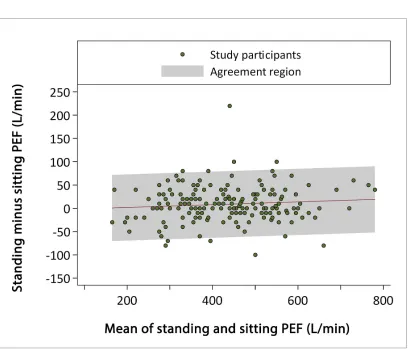
Agreement between readings done in the sitting and standing positions was good, with a Lin’s correlation coefficient of 0.95 (95% CI = 0.94 to 0.96) (Table 3). The SD of the distribution of the dif-ference in standing and sitting PEF values was 36.4 lmin–1. A Bland-Altman plot accounting for trend
(Figure 1) showed good agreement between standing and sitting PEF measurements except that the mean standing values were on average 8.7 lmin–1higher than the sitting values. The agreement
appeared to be maintained for the whole range of PEF values.
The mean PEF of men was higher than women in both the sitting (mean difference 137 lmin–1, 95% CI = 110 to 164,P<0.001) and standing (mean difference 144 lmin–1, 95% CI = 116 to 171,
P<0.001) positions, and was also higher for non-asthmatics compared to those with asthma in both the sitting (mean difference 48, 95% CI = 9 to 88,P= 0.02) and the standing (mean difference 56,
Table 3.Correlation and agreement between measurement of PEF when standing and when sitting
Women Men All
Pearson’s correlation coefficient 0.94 0.92 0.95
Bias between readings (slope, intercept) 1.00 (1.00, 5.68) 0.99 (1.04, 6.11) 1.00 (1.03, 4.12)
Agreementa
(95% CI) 0.94 (0.912 to 0.959) 0.92 (0.88 to 0.95) 0.95 (0.938 to 0.964)
Limits of agreement (95% CI) 5.81 ( 53.60 to 65.22) 12.39 ( 71.51 to 96.28) 8.72 ( 62.66 to 80.10)
aLin’s concordance coefficient (Pearson’s correlation coefficient x bias between readings). PEF = peak expiratory flow.
95% CI 16 to 97, P = 0.01) positions. After adjusting for age, height, and BMI, sex and asthma remained statistically significant predictors of standing PEF (sex: men higher than women, mean dif-ference 114 lmin–1, 95% CI = 81 to 148,P<0.001; asthma: PEF higher among those without asthma, mean difference 33 lmin–1, 95% CI = 1 to 67,P= 0.05). Using the same adjusting factors, only sex remained a statistically significant predictor of sitting PEF (sex: men higher than women, mean differ-ence 109 lmin–1, 95% CI = 77 to 142,P<0.001; asthma: PEF higher among those without asthma,
mean difference 26 lmin–1, 95% CI = 7 to 59, P= 0.12). Adjusted models explained between 36
and 37% of data variation.
Discussion
Summary
This study found that in a population largely of African descent, PEF measured standing is on aver-age higher than that measured sitting. However, the difference is small and may not make a differ-ence to clinical management. Furthermore, for people with a history of asthma the differdiffer-ence in PEF was not significantly different by position of measurement. Agreement between readings done in the sitting and standing positions was good over the whole range of PEF values. Linear regression indicated that sex was a significant predictor of PEF in both the standing and sitting positions after adjusting for age, height, and BMI.
Strengths and limitations
Strengths of this study were that participants were selected over a wide age range, were typical of those attending primary care, and that bias was reduced by alternating the initial position of mea-surement. A limitation of this study is that it did not include people who were wheezing and there-fore may not be generalisable to this situation. It was decided not include such people to avoid subjecting them to a study protocol that was unnecessary for their treatment and that might cause discomfort. However, the study did include people with asthma who were being prescribed medica-tion and this is also a populamedica-tion in which PEF estimamedica-tion is indicated in practice.
Comparison with existing literature
All three previous published studies examining the effect of sitting upright versus standing that were identified have shown a small but statistically insignificant higher PEF when measured standing com-pared to sitting.12–14None of the previous studies involved a population of predominantly African descent.
All 22% of the sample who reported a diagnosis of asthma also reported using prescription inhalers for the condition. A previous study in Barbados published 30 years ago estimated an asthma prevalence of 16% in children aged 13–14 years.20Adult prevalence data for Barbados are not avail-able. It is likely that people on prescription medicine for a chronic condition will be overrepresented in a clinic population.
Implications for research and practice
The basis of the recommendation of several authorities to use the standing position as opposed to a sitting position when measuring PEF is not clear, and the often quoted normal adult PEF range is based on a study that did not specify the position used in the measurement.10,11The present study therefore adds information that may be useful to practice guideline writers and for individual physi-cians making decisions in the clinical setting.
Performing the test sitting in a primary care setting would usually be more convenient and quicker for patients. A few patients, for example frail and older people, may have trouble standing and the impact of position on these subpopulations can be investigated by future research. In cases where standing is difficult, it may be reasonable to do the measurement sitting and note that the reading may underestimate that done in the standing position by about 9 lmin–1or about 2%. To
Funding
A staff research grant from the University of the West Indies, Cave Hill Campus funded this study.
Ethical approval
Ethical approval was obtained from the Institutional Review Board of the University of the West Indies, Cave Hill campus. Permission to carry out the study was obtained from the Ministry of Health.
Provenance
Freely submitted; externally peer reviewed.
References
1. Global Asthma Network. The global asthma report 2014. 2014.http://www.globalasthmareport.org/burden/ burden.php(accessed 1 Jun 2018).
2. Caribbean Health Research Council.Managing asthma in the Caribbean. St Augustine, Trinidad Caribbean Health Research Council: 2009.
3. Quanjer PH, Lebowitz MD, Gregg I,et al. Peak expiratory flow: conclusions and recommendations of a Working Party of the European Respiratory Society.Eur Respir J Suppl1997;24:2S–8.
4. Global Initiative for Asthma. Global Initiative for Asthma GINA Patient Guide. You Can Control Your Asthma. 2007.http://www.ginasthma.org/local/uploads/content/files/GINA_PatientGuide2007.pdf
5. American Lung Association. Measuring your peak flow rate Chicago: American Lung Association.http:// www.lung.org/lung-disease/asthma/taking-control-of-asthma/measuring-your-peak-flow-rate.html(accessed 1 Jun 2018).
6. American Academy of Allergy AI. Peak flow meter: tips to remember: American Academy of Allergy, Asthma & Immunology. http://www.aaaai.org/conditions-and-treatments/library/at-a-glance/peak-flow-meter.aspx
7. National Asthma Education and Prevention Program. How to use a peak flow meter: National Asthma Education and Prevention Program. 2013.http://www.nhlbi.nih.gov/files/docs/public/lung/asthma_tipsheets. pdf(accessed 1 Jun 2018).
8. National Institutes of Health. How to use your peak flow meter. 2015.http://www.nlm.nih.gov/medlineplus/ ency/patientinstructions/000043.htm(accessed 1 Jun 2018).
9. British guideline on the management of asthma. A national clinical guideline. Edinburgh and London Scottish Intercollegiate Guidelines Network and British Thoracic Society. 2014. https://www.brit-thoracic.org.uk/document-library/clinical-information/asthma/btssign-asthma-guideline-2014/(accessed 1 Jun 2018).
10. Clement Clarke International. Mini-wright peak flow meter. Predictive normal values (Nomogram, EU scale). 2004.http://www.peakflow.com/top_nav/normal_values/(accessed 1 Jun 2018).
11. Nunn AJ, Gregg I. New regression equations for predicting peak expiratory flow in adults.BMJ1989;298 (6680):1068–1070.doi: 10.1136/bmj.298.6680.1068
12. Vaswani R, Moy R, Vaswani SK. Evaluation of factors affecting peak expiratory flow in healthy adults: is it necessary to stand up?J Asthma2005;42(9):793–794.doi: 10.1080/02770900500308528
13. McCoy EK, Thomas JL, Sowell RS,et al. An evaluation of peak expiratory flow monitoring: a comparison of sitting versus standing measurements.J Am Board Fam Med2010;23(2):166–170.doi: 10.3122/jabfm. 2010.02.090120
14. Price K, Schartz P, Watson AH. The effect of standing and sitting postures on breathing in brass players. SpringerPlus2014;3:210.doi: 10.1186/2193-1801-3-210
15. College Data. University of Tennessee: college data your online college advisor. 2015. http://www. collegedata.com/cs/data/college/college_pg01_tmpl.jhtml?schoolId=1545(accessed 1 Jun 2018).
16. Braun L. Race, ethnicity and lung function: a brief history.Can J Respir Ther2015;51(4):99–101.
17. Ghazal-Musmar S, Musmar M, Minawi WA. Comparison of peak expiratory flow rates applying European and Iranian equations to Palestinian students.East Mediterr Health J2010;16(4):386–390.
18. Barbados Statistical Service. 2010 Barbadian Population and Housing Census: Barbados Statistical Service, Government of Barbados. 2010.http://www.barstats.gov.bb/files/documents/PHC_2010_Census_Volume_1. pdf(accessed 1 Jun 2018).
19. Wallace JL, George CM, Tolley EA,et al. Peak expiratory flow in bed? A comparison of 3 positions.Respir Care2013;58(3):494–497.doi: 10.4187/respcare.01843