OR
IGI
NA
L
A
R
T
IC
L
E
Volume 11 Number 4 (August 2019) 313-319
Antibacterial activity of self-adhesive resin cements against
Streptococcus
mutans
at different time intervals
Amir-Ahmad Ajami1, Sahand Rikhtegaran1, Mahmoud Bahari1,2*, Sayeh Hamadanchi1
1Department of Operative Dentistry, Faculty of Dentistry, Tabriz University of Medical Sciences, Tabriz, Iran 2Dental and Periodontal Research Center, Faculty of Dentistry, Tabriz University of Medical Sciences, Tabriz,
Iran
Received: September 2018, Accepted: May 2019
ABSTRACT
Background and Objectives: Self-adhesive resin cements release fluoride and have cytotoxic and preventive monomers
against the bacteria in their composition. They have acidic property before their complete setting too. The antibacterial ac- tivity of three different self-adhesive resin cements against Streptococcus mutans at different time intervals was investigated in this study.
Materials and Methods: The modified direct contact test was used to evaluate the antibacterial effect of Max-Cem, G-Cem
and Bis-Cem on S. mutans after aging the samples in phosphate-buffered saline solution for one hour, 24 hours and 1 week. Data were analyzed using one-way ANOVA, repeated measurement ANOVA and Tukey HSD tests (P<0.05).
Results: The differences in the mean bacterial counts between all the study groups and between the study groups and the
corresponding control groups were significant at 1-hour and 24-hour intervals (P<0.001). At 1-week, only the differenc- es between Bis-Cem and G-Cem, between Max-Cem and Bis-Cem, and between Bis-Cem and the corresponding control group were significant (P<0.001). There were significant differences between G-Cem and Max-Cem at all the time intervals (P<0.001). In addition, with the use of Bis-Cem there were significant differences between 1-hour and 1-week (P=0.01) and 24-hour and 1-week (P<0.001).
Conclusion: All the cements exhibited antibacterial activity after 1 hour and 24 hours. However, after 1 week, only Bis-Cem
retained its antibacterial activity.
Keywords: Anti-bacterial agents; Resin cements; Self-etch primer; Dental caries; Bacteria
*Corresponding author: Mahmoud Bahari, Ph.D, Depart-
ment of Operative Dentistry, Faculty of Dentistry, Tabriz Uni- versity of Medical Sciences, Tabriz, Iran; Dental and Peri- odontal Research Center, Faculty of Dentistry, Tabriz University of Medical Sciences, Tabriz, Iran.
Tel: +98 914 102 5982 Fax: +98 41 3334 6977
Email: [email protected]
INTRODUCTION
which is considered a common clinical phenomenon. Subsequently, oral liquids, ions, molecules and oral bacteria percolate between the tooth and restorative interface. Then an appropriate space for the growth of these cariogenic bacteria, especially Streptococ- cus mutans (S. mutans), will be created, resulting in recurrent caries. In order to prevent this detrimental effect, it is of vital importance to choose a proper cement (1).
Cements that are used in indirect restorations are divided, based on the dominant setting reaction type, into types that include acid-base reaction (Glass Ion- omer, Resin modified Glass Ionomer, Zinc Oxide Eugenol, Zinc Poly Carboxylate and Zinc Phosphate) and resin types with their setting is done with po- lymerization (2). Previous studies revealed that con- ventional acid-base cements have limited or some amount of antibacterial activity against S. mutans. While incorporating chlorhexidine/Certimide mix- tures to their formulation may provide greater anti- bacterial effect (3). According to Feoz et al. (4) Zinc Phosphate and Zinc Oxide Eugenol exhibited highest amount of antibacterial activity. While, Glass Iono- mer cement was the weakest of all. Conversely, in vitro studies have shown that both conventional and resin-modified glass-ionomers can decrease artificial caries; they can also remineralize carious lesions in vivo (5) and enhance fluoride uptake by underlying dentin (6). This property might be as attributed to the presence of fluoride and zinc in their structure and their initial acidic pH. Both fluoride and zinc exert several effects on dental plaque bacteria, with both inhibiting a variety of enzymes in intact cells; as a result, they are both widely used as antimicrobials in oral hygiene products, predominantly as anticar- iogenic agents (7).
Currently, adhesive resin cements are used for ce- mentation of indirect restorations. Adhesive resin cements have etch-and-rinse, self-etch and self-ad- hesive types. In self-adhesive generation, which are the most recent type, all of the etching, bonding and luting steps are carried out in one step and all to- gether (8). This generation has some advantages such as decreasing dissolution in oral liquids, decreasing working time, less working sensitivity and presence of fluoride in its structure (9).
In self-adhesive resin cements, the pH of the self-etching primer is sufficiently low to demineral- ize the smear layer and the underlying dentin surface so that etching and priming of dentin can be accom-
plished simultaneously (8). Therefore, the separate acid-etching step is generally omitted. However, due to the non-rinsing procedure, residual bacteria may remain at the interface between the tooth and the restorative material. The dentin primer is the com- ponent that comes into contact and reacts with the dentin substrate at the first stage of restoration. Fur- thermore, if tooth conditioners, such as primers pres- ent in the composition of these luting agents, possess antibacterial activity, these bacteria could be elim- inated, thereby preventing secondary caries. Thus, the antibacterial activity of these self-etching prim- ers that are directly applied to the dentin plays an important role in the longevity of the restoration (9). Considering the release of fluoride and the primary low pH and acidic property of the self-adhesive resin cements and the presence of cytotoxic and preventive monomers against the bacteria in their composition, the aim was to assess the antibacterial activity of three self-adhesive resin cements against S. mutans
at different time intervals.
MATERIALS AND METHODS
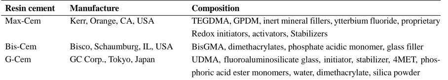
For the purpose of the present study, three differ- ent self-adhesive resin cements, including Bis-Cem (Bisco, Schaumburg, IL, USA), G-Cem (GC Cor- poration, Itabashi-ku, Tokyo, Japan) and Max-Cem Elite (Kerr, Orange, CA, USA) were used as exper- imental groups. Compositions and manufacturer in- formation for these cements are provided in Table 1.
Samples grouping. Since it is necessary for the bacteria floating in the liquid culture media to be ex- posed to the resin cement, the modified direct contact test was used for the purpose of this study. Based on the results of a pilot study, 58 microplates were used for each experimental group. In addition, 58 micro- plates with the bacterial solution but without cements (positive control) and 5 microplates with the cements under study but without any bacteria (negative con- trol) were used as the negative control. Another five microplates without cements and bacteria, with only the culture media, was prepared in order to control the sterility of microplates.
Table 1. Composition and manufacturers of Self-adhesive resin cements
Resin cement
Max-Cem
Bis-Cem G-Cem
Manufacture
Kerr, Orange, CA, USA
Bisco, Schaumburg, IL, USA GC Corp., Tokyo, Japan
Composition
TEGDMA, GPDM, inert mineral fillers, ytterbium fluoride, proprietary Redox initiators, activators, Stabilizers
BisGMA, dimethacrylates, phosphate acidic monomer, glass filler UDMA, fluoroaluminosilicate glass, initiator, stabilizer, 4MET, phos- phoric acid ester monomers, water, dimethacrylate, silica powder
TEGDMA; Triethylene glycol dimethacrylate, GPDM; Glycerophosphoric acid dimethacrylate, BisGMA; Bisphenol glycidyl methacrylate, UDMA; Urethane dimethacrylate, 4MET; 4-Methacryloxyethyl trimellitic acid
tion was carried out according to the manufacturer’s instructions. Then the microplates underwent an ag- ing process by storage in phosphate-buffered saline solution at 37°C with 95% atmospheric moisture for 1 hour, 24 hours and 1 week. During the aging pro- cess for 1 week, the physiologic serum was refreshed every 24 hours. At the end of the each aging period, the physiologic serum contents of the microplates were retrieved and 10 μL of S. mutans bacterial sus- pension (approximately 106 bacteria) were added to each microplate. The microplates were kept at 37°C for 60 minutes in a moist environment.
During this period, the bacteria came into direct contact with the free surface of the cements. Then 240 μL of Brain Heart Infusion (BHI) culture me- dium were added to each microplate and mixed for 2 minutes. In the final stage, serial dilutions were prepared from the content of each micro tube in the BHI culture medium and 20 µL of each dilution was cultured on BHI culture plate using the spreading technique. The bacterial counts were described as CFU/mL.
Statistical analysis. One-way ANOVA was used to analyze data obtained for each time interval sep- arately. Post-hoc Tukey HSD tests were used for two-by-two comparisons of the groups in cases of significant differences. Furthermore, repeated mea- surement ANOVA was used to analyze bacterial counts at three time intervals for each group, sepa- rately. In this study, P<0.05 was considered statisti- cally significant.
RESULTS
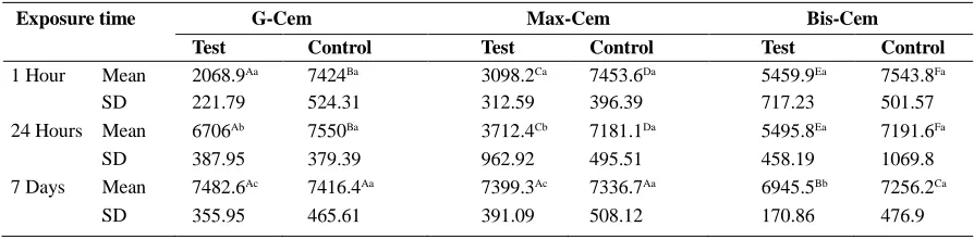
The means, standard deviations and standard errors
of bacterial counts (CFU/mL) in the study groups are presented in Table 2. The results of Kolmogor- ov-Smirnov test showed normal distribution of data in the study groups (P>0.05). One-way ANOVA showed significant differences in mean bacterial counts between the study groups (P<0.001).
Two-by-two comparisons of the groups with post hoc Tukey tests showed significant differences in mean bacterial counts between all the study groups and between the study groups and the correspond- ing control groups at 1-hour and 24-hour intervals (P<0.001). At 1-week interval, the differences be- tween Bis-Cem and G-Cem and between Max-Cem and Bis-Cem were significant (P<0.001). In addi- tion, the difference between the Bis-Cem group and the corresponding control group was significant (P<0.001). In other cases, no significant differences were detected between the study groups and between the study and control groups at this time interval (P>0.05).
Repeated measurement ANOVA was used to compare the mean bacterial counts with the use of each cement at different time intervals. The results showed significant differences between G-Cem and Max-Cem cements at all the intervals (P<0.001), with an increase in bacterial counts over time. In this con- text, there were significant differences in Bis-Cem cement between 1-hour and one-week (P=0.01) and 24-hour and 1-week (P<0.001) intervals; however, the difference between 1-hour and 24-hour intervals was not significant (P>0.05).
DISCUSSION
Table 2. Mean colony count (cfu/mL) and standard deviations (SD) of Streptococcus mutans growth after exposure to three different cements after different time intervals.
Exposure time G-Cem Max-Cem Bis-Cem
Test Control Test Control Test Control
1 Hour Mean 2068.9Aa 7424Ba 3098.2Ca 7453.6Da 5459.9Ea 7543.8Fa
SD 221.79 524.31 312.59 396.39 717.23 501.57
24 Hours Mean 6706Ab 7550Ba 3712.4Cb 7181.1Da 5495.8Ea 7191.6Fa
SD 387.95 379.39 962.92 495.51 458.19 1069.8
7 Days Mean 7482.6Ac 7416.4Aa 7399.3Ac 7336.7Aa 6945.5Bb 7256.2Ca
SD 355.95 465.61 391.09 508.12 170.86 476.9
In each row, different upper letters show statistically significant differences between resin cements (p<0.05). In each column, different lower letters show statistically significant differences between time intervals (p<0.05).
which is a localized lesion affecting restoration mar- gins, believed to be associated with residual bacteria and microleakage; its etiology and histology is sim- ilar to those of primary caries. It is difficult to diag- nose secondary caries and it cannot be permanently managed by operative strategies. One technique to decrease the frequency and severity of this issue is to use fluoride-containing restorative materials and luting agents (8).
The agar diffusion test (ADT) was the most fre- quently used method for the evaluation of antimi- crobial activity of various cements. However, its disadvantages semi-quantitative results that rely on solubility and diffusion characteristics of the test material - and the medium used are well recog- nized (12). Weiss et al. introduced a direct contact test (DCT) that circumvents many of the problems of ADT (13). It is a quantitative assay allowing the use and testing of water-insoluble materials. It uses direct and close contact between the test microor- ganisms and materials, being almost independent of the diffusion properties of both the tested material and the media. Apart from its reproducibility and quantitative nature, DCT is relatively insensitive to the size of the inoculate brought into contact with the test material, facilitating simultaneous standardized measurements of a large number of specimens and their controls on the same micro-titer plate to mon- itor the microbial growth, both in the presence and absence of the test material (13). As a result, in this study a modified DCT was used for the assessment of antibacterial properties of three commercially avail- able self-adhesive resin cements (G-Cem, Max-Cem and Bis-Cem) at different time intervals (1 hour, 24
hours, 1 week).
All the cements evaluated in the present study ex- hibited significant antibacterial activities for after 1 hour and 24 hours compared with the positive con- trol group. Similarly, Magalhaes et al. (14) showed that RelyX ARC a conventional resin cement and RelyX U200 a self-adhesive one exhibit significant antibacterial activity against S. mutans for 24 hours. Previous studies have shown that the formation of S. mutans colonies significantly decreased at pH values <5.1, being completely inhibited at pH values ≤4.8 (15, 16). The most probable reason for this finding might be the low primary pH of these cements. These cements have an acidic pH value due to the acidic monomers in their structure that are responsible for self-etching capacity (17). According to manufac- turers, the self-etching capacity is provided by the presence of different monomers in the luting agent formulation: GPDM in Maxcem, the hydrophilic monomer 4-META and phosphoric acid ester mono- mers in G-Cem, and phosphoric acid ester monomers in Bis-Cem.
G-Cem, and 4.0 for Bis-Cem, corresponding to the antibacterial activities of these cements at 1- and 24- hour intervals, respectively (6, 19).
In contrast, at 1-week interval the antibacterial ac- tivity of Bis-Cem was higher than those of G-Cem and Max-Cem are, which exhibited similar antibac- terial properties at this time interval. Based on a literature review, the antibacterial activity of restor- ative materials might also be attributed to the fluo- ride content. A large number of studies have shown that fluoride ion is safe and effective in preventing and controlling caries at certain doses. Fluoride can inhibit the growth of oral streptococci in vitro at a concentration range of 0.16-0.31 mmol/L, also (24, 25).
The composition, solubility and permeability of the resin matrix, and the source, size and concentra- tion of the fluoride ions are important characteristics of the fluoride-releasing materials (26-29). Aguiar et al. (9), reported that Max-Cem released less fluo- ride in water compared to Bis-Cem. Max-Cem resin cement contains fluoroaluminosilicate particles that bear resemblance to the glass powder in G-Cem. The similarity in the fluoride source of both materials might explain the similarity in antibacterial activity of G-Cem and MaxCem. However, Bis-Cem self-ad- hesive resin cement contains glass fillers composed of fluoride glass. The glass fillers are supplied in both base and catalyst pastes of Bis-Cem cement, releas- ing fluoride in water. Furthermore, the glass powder serves as a reservoir for fluoride (30).
As shown in previous studies, all the resin cements released high levels of ion on the initial days for all the resin cements, demonstrating that the release of fluoride was not uniform over time (9, 21, 26, 31). According to Aguiar et al. (9), after 15 days, fluoride release from Bis-Cem was 10 folds greater than its release from Max-Cem, possibly a further evidence of higher anti-bacterial activity of Bis-Cem com- pared to Max-Cem and G-Cem, which have similar fluoride particles.
A number of different mechanisms are involved in the anti-cariogenic effects of fluoride on teeth. It can act directly or it forms of metal complexes that inhibit many enzymes (32). However, it appears its main action that leads to the inhibition of acid pro- duction by intact bacterial cells at low pH is relat- ed to its capacity to enhance proton permeability of cell membranes by acting in the form of protonated fluoride (HF) as a trans-membrane proton carrier.
Fluoride prevents proton extrusion by F-ATPases by returning excreted proton back into the cell through movements of HF; the cell is approximately 10 times more permeable to HF compared to fluoride. HF in the relatively alkaline cytoplasm dissociates to yield the enzyme poison F and H1, which acidifies the cy- toplasm and inhibits glycolytic enzymes. A decrease in ΔpH by fluoride has a negative effect on the ener- getic status of the cell because by increasing re-entry of protons across the cell membrane it increases the demand on ATP for acid–base regulation. The final result is increased acidification and starvation stress- es on the cell (33).
Self-adhesive resin cements seem to provide prom- ising antibacterial effects. Although an initial low pH value has a great role in antibacterial effects and etching of enamel and dentin, if the low pH lasts for a long time, it might exert negative effects on the adhesion of the cement to dentin (6, 19). Despite the antibacterial properties of fluoride, its activi- ty is still to be elucidated. However, GICs and res- in-modified GICs have been reported to be the only materials that release the highest amount of fluoride among the luting agents, but even those products severely reach the inhibitory release level of fluoride (29).
The available data are derived from studies that assessed only a limited number of these cements currently available. Furthermore, bacteria other than
S. mutans are also responsible for caries, should be investigated in future studies. In addition, long-term clinical antibacterial and anti-cariogenic effects of these materials should be assessed before making general recommendations.
CONCLUSION
REFERENCES
1. Daugela P, Oziunas R, Zekonis G. Antibacteri- al potential of contemporary dental luting cements.
Stomatologija 2008; 10: 16-21.
2. Villat C, Tran XV, Pradelle-Plasse N, Ponthiaux P, Wenger F, Grosgogeat B, et al. Impedance methodol- ogy: A new way to characterize the setting reaction of dental cements. Dent Mater 2010; 26: 1127-1132. 3. Korkmaz FM, Tuzuner T, Baygin O, Buruk CK, Durkan
R, Bagis B. Antibacterial activity, surface roughness, flexural strength, and solubility of conventional luting cements containing chlorhexidine diacetate/cetrimide mixtures. J Prosthet Dent 2013; 110: 107-115.
4. Feroz S, Bhoyar A, Khan S. Comparative evaluation of antibacterial effect of dental luting cements on Strep- tococcus mutans and Lactobacillus acidophilus: An in vitro study. J Contemp Dent Pract 2016; 17: 973-977. 5. Zhang H, Shen Y, Ruse ND, Haapasalo M. Antibacte-
rial activity of endodontic sealers by modified direct contact test against Enterococcus faecalis. J Endod
2009; 35: 1051-1055.
6. Han L, Okamoto A, Fukushima M, Okiji T. Evalua- tion of physical properties and surface degradation of self-adhesive resin cements. Dent Mater J 2007; 26: 906-914.
7. Koo H, Sheng J, Nguyen PT, Marquis RE. Co-opera- tive inhibition by fluoride and zinc of glucosyl trans- ferase production and polysaccharide synthesis by mu- tans streptococci in suspension cultures and biofilms.
FEMS Microbiol Lett 2006; 254: 134-140.
8. Runnacles P, Correr GM, Baratto Filho F, Gonzaga CC, Furuse AY. Degree of conversion of a resin cement light-cured through ceramic veneers of different thick- nesses and types. Braz Dent J 2014; 25: 38-42. 9. Aguiar TR, Pinto CF, Cavalli V, Nobre-dos-Santos M,
Ambrosano GM, Mathias P, et al. Influence of the cur- ing mode on fluoride ion release of self-adhesive resin luting cements in water or during pH-cycling regimen.
Oper Dent 2012; 37: 63-70.
10. Ebrahimi Chaharom ME, Ajami AA, Abed Kahnamouei M, Jafari Navimipour E, Tehranchi P, Zand V, et al. Antibacterial effect of all-in-one self-etch adhesives on Enterococcus faecalis. J Dent Res Dent Clin Dent Prospects 2014; 8: 225-229.
11. Huang Q, Huang S, Liang X, Qin W, Liu F, Lin Z, et al. The antibacterial, cytotoxic, and flexural properties of a composite resin containing a quaternary ammonium monomer. J Prosthet Dent 2018; 120: 609-616. 12. Lewinstein I, Matalon S, Slutzkey S, Weiss EI. Anti-
bacterial properties of aged dental cements evaluated by direct-contact and agar diffusion tests. J Prosthet Dent 2005; 93: 364-371.
13. Weiss EI, Shalhav M, Fuss Z. Assessment of antibac-
terial activity of endodontic sealers by a direct contact test. EndodDent Traumatol 1996; 12: 179-184. 14. Magalhaes AP, Moreira FC, Alves DR, Estrela CR,
Estrela C, Carriao MS, et al. Silver nanoparticles in resin luting cements: Antibacterial and physiochemical properties. J Clin Exp Dent 2016; 8(4): e415-e422. 15. Tay FR, Pashley DH. Aggressiveness of contemporary
self-etching systems. I: Depth of penetration beyond dentin smear layers. Dent Mater 2001; 17: 296-308. 16. Piwowarczyk A, Bender R, Ottl P, Lauer HC. Long-
term bond between dual-polymerizing cementing agents and human hard dental tissue. Dent Mater 2007; 23: 211-217.
17. Abo-Hamar SE, Hiller KA, Jung H, Federlin M, Friedl KH, Schmalz G. Bond strength of a new universal self-adhesive resin luting cement to dentin and enamel.
Clin Oral Investig 2005; 9: 161-167.
18. Zicari F, Couthino E, De Munck J, Poitevin A, Scotti R, Naert I, et al. Bonding effectiveness and sealing ability of fiber-post bonding. Dent Mater 2008; 24: 967-977. 19. Wang Y, Spencer P. Continuing etching of an all-in-one
adhesive in wet dentin tubules. J Dent Res 2005; 84: 350-354.
20. Carvalho AS, Cury JA. Fluoride release from some dental materials in different solutions. Oper Dent 1999; 24: 14-19.
21. Attar N, Turgut MD. Fluoride release and uptake ca- pacities of fluoride-releasing restorative materials.
Oper Dent 2003; 28: 395-402.
22. Mukai Y, ten Cate JM. Remineralization of advanced root dentin lesions in vitro. Caries Res 2002; 36: 275- 280.
23. Levy SM. An update on fluorides and fluorosis. J Can Dent Assoc 2003; 69: 286-291.
24. Tenuta LM, Zamataro CB, Del Bel Cury AA, Tab- choury CP, Cury JA. Mechanism of fluoride dentifrice effect on enamel demineralization. Caries Res 2009; 43: 278-285.
25. Van Dijken J, Kalfas S, Litra V, Oliveby A. Fluoride and mutans streptococci levels in plaque on aged resto- rations of resin-modified glass lonomer cement, com- pomer and resin composite. Caries Res 1997; 31: 379- 383.
26. Hara AT, Queiroz CS, Freitas PM, Giannini M, Serra MC, Cury JA. Fluoride release and secondary caries inhibition by adhesive systems on root dentine. Eur J Oral Sci 2005; 113: 245-250.
27. Burke FM, Ray NJ, McConnell RJ. Fluoride-contain- ing restorative materials. Int Dent J 2006; 56: 33-43. 28. Yoda A, Nikaido T, Ikeda M, Sonoda H, Foxton RM,
ride-releasing restorative materials--fluoride release and uptake characteristics, antibacterial activity and influence on caries formation. Dent Mater 2007; 23: 343-362.
30. Itota T, Okamoto M, Sato K, Nakabo S, Nagamine M, Torii Y, et al. Release and recharge of fluoride by re- storative materials. Dent Mater J 1999; 18: 347-353. 31. de Araujo FB, Garcia-Godoy F, Cury JA, Conceicao
EN. Fluoride release from fluoride-containing materi- als. Oper Dent 1996; 21: 185-190.
32. Li L. The biochemistry and physiology of metallic flu- oride: action, mechanism, and implications. Crit Rev Oral Biol Med 2003; 14: 100-114.
33. Svensater G, Sjogreen B, Hamilton IR. Multiple stress responses in Streptococcus mutans and the induction of general and stress-specific proteins. Microbiology