Sickle Cell Beta Thalassemia: A Rare Entity OMPJ
Sickle Cell Beta Thalassemia: A Rare Entity
1Rashmi E Mathen, 2Kamala Rawson, 3Anu Vijayan, 4Pretty Prince P, 5Sonia S PhilipOMPJ
CASE REPORT
10.5005/jp-journals-10037-1148
ABSTRACT
Introduction: Haemoglobinopathies are the disorders which affect the structure of hemoglobin. Most common hemoglobin-opathies are sickle cell disorders and thalassemia. Sickle cell disorders are genetic disorders characterized by the predomi-nance of hemoglobin S whereas thalassemia is the disorder of hemoglobin synthesis with decreased production of globin chains of hemoglobin molecules.
Case report: We present a case of a rare hereditary disease in a 19-year-old female patient with both sickle cell and beta thalas-semia traits along with the clinical, radiological manifestations, diagnosis, management, and dental considerations.
Conclusion: Oral physicians and oral pathologists should be vigilant enough to identify any hemoglobinopathy, with adequate radiological and hematological investigations, although it has a rare occurrence. If diagnosed, a multidisciplinary approach involving dental surgeon, and orthodontist is mandatory for these patients
Keywords: Beta-thalassemia, Genetic disorder, Hemoglobin-opathies, Sickle cell disease.
How to cite this article: Mathen RE, Rawson K, Vijayan A, Prince PP, Philip SS. Sickle Cell Beta Thalassemia: A Rare Entity. Oral Maxillofac Pathol J 2019;10(1):35-40.
Source of support: Nil Conflict of interest: None
INTRODUCTION
Hemoglobinopathies are a group of genetic disorders of hemoglobin (Hb) in which there is abnormal produc-tion or structure of the hemoglobin molecule. Sickle cell disease is a generic term for a group of genetic disorders characterized by the predominance of Haemoglobin S (HbS). The disorders include sickle cell anemia, the sickle cell beta thalassemia syndromes and hemoglobinopathies in which HbS is in association with abnormal hemoglobin like sickle cell hemoglobin C disease (hemoglobin SC
disease), sickle cell hemoglobin D disease (hemoglobin SD disease) and hemoglobin OArab disease.1 Sickle cell beta thalassemia is a disorder in which both sickle beta-globin gene (βS) and beta thalassemia gene are present.2 The sickle cell disorders are found in people of African, Mediterranean, Indian and Middle Eastern heritage.3 The highest prevalence of hemoglobinopathies in India has been recorded in the Central Eastern area, especially in the state of Orissa (1–44%) followed by Madhya Pradesh (1–40%), Kerala (1–30%) and the least in Karnataka (1–8%).4 The overall prevalence of sickle cell beta thalassemia in India is 0.02%.5
CASE REPORT
A 19-year-old female patient, a native of Orissa, reported to the Department of Oral Medicine with a chief com-plaint of forwardly placed upper front teeth since 7 years. Her medical history revealed that she is a sickle cell beta thalassemia patient diagnosed during her childhood, and had undergone multiple blood transfusions till the age of 18 years. No other family member except her youngest brother was affected with this disease, who expired at the age of 10 years. The patient gave a history of extrac-tion of teeth in the lower left and right back tooth region 3 years back due to decay. On physical examination, the patient had a normal gait, and she was moderately built and nourished. Examination of the systems revealed hepatosplenomegaly, whereas CVS, CNS and respiratory system were normal.
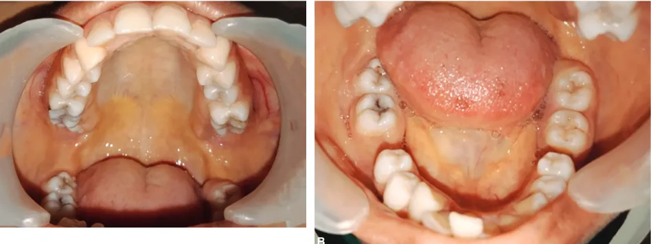
On extra oral examination, pallor and yellowish discoloration of the skin over the face and yellowish tint of both the sclera were noted. Other findings included bimaxillary protrusion, lip incompetence and features of chipmunk facies like slanting of the eyes, frontal bossing, hypertelorism, saddle nose, increased malar prominence (Figs 1A and B). Intraorally, hard tissue examination revealed bilateral Class I canine relation with an overjet of 5mm, missing teeth 36 and 46 and discolored 42. On soft tissue examination, yellowish discoloration of the mucosa was noted over the gingiva, hard and soft palate, tongue, buccal mucosa, and floor of the mouth (Figs 2A and B). Thus, based on clinical examination, a provisional diag-nosis of Angles class I malocclusion was given. Since the patient was a known case of sickle cell beta thalassemia, we proceeded with the radiological and hematological investigations before the treatment.
1,4Postgraduate Student, 2Professor and HOD, 3Associate
Professor, 5Senior Lecturer
1-3,5Department of Oral Medicine and Radiology, Mar Baselios
Dental College, Kerala, India
4Department of Oral and Maxillofacial Pathology, Mar Baselios
Dental College, Kerala, India
On radiographic investigations, intraoral periapical radiographs of the maxillary and mandibular anterior region (Figs 3A and B) and panoramic view showed the presence of enlarged marrow spaces, large and coarse trabeculae. There was also the presence of missing teeth 36 and 46 with a well-defined unilocular radiolucency in the missing tooth 36 region extending from the distal of 35 to mesial of 37 suggestive of residual cyst in relation to missing 36 region (Fig. 4). Lateral skull radiograph
showed increased diploic space (Fig. 5). Lateral cepha-logram showed maxillary and mandibular prognathism, frontal bossing, and lip incompetence (Fig. 6).
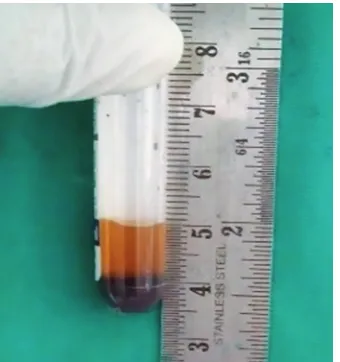
The centrifuged blood of the patient showed more plasma proportion than formed elements and the color was yellowish brown (Fig. 7) The complete hemogram showed hemoglobin of 7.5 gm/dl. Hematocrit value was 37.5% and had a lower MCV, MCH values 45 fl and 15 pg respectively, MCHC value = 333g/dL and a low Mentzer index of 9. Blood investigations revealed RBC count 5 million/cu.mm, normal WBC counts (6600cells/ cu.mm) with neutrophil count 74%, ESR (5mm/hour)
Figs 1A and B: (A) Extraoral photograph depicting the facial features; (B) Icterus in both eyes
Figs 2A and B: (A) Yellowish discoloration on the palate and buccal mucosa; (B) Yellowish discoloration in the floor of mouth
A B
Figs 3A and B: (A) Intraoral periapical radiograph of maxillary anterior region; (B) Intraoral periapical radiograph of mandibular anterior region
Fig. 4: Panoramic radiograph showing enlarged marrow spaces and residual cyst irt missing 36
A B
Sickle Cell Beta Thalassemia: A Rare Entity OMPJ
Thus the case presented with features of both sickle cell disease and beta thalassemia suggestive of sickle cell beta thalassemia, which has a unique presentation. The patient was referred for cyst enucleation and orthodon-tic treatment, and she was advised to take hematologist consent before the procedure.
DISCUSSION
The hemoglobinopathies are genetic disorders characteri-zed by the production of structurally defective hemoglo-bin (Hb), and in thalassemia, there is reduction in rate of production of normal Hb due to absent or decreased synthesis of one or more types alpha(α) or beta (β) of globin polypeptide chains.2 Sickle cell disorders (SCD) and β-thalassemia are autosomal recessive inherited disorders affecting the structure and synthesis of Hb.1
SCD result from the polymerization of HbS in red blood cells (RBCs) into the characteristic sickle shape under hypoxic conditions, which results in the occlu-sion of blood vessels.6 SCD results when one β-globin
Fig. 5: Lateral skull radiograph showing increased diploic space Fig. 6: Lateral cephalogram showing bimaxillary protrusion and lip incompetence
Fig. 7: The centrifuged blood with yellowish brown plasma
Fig. 8: Peripheral blood picture characterized by microcytic, hypochromic RBCs, with target cells and sickle cells
Fig. 9: The peripheral smear red blood cell showing sickling
β-globin gene such as mutations associated with HbC, Hb β-thalassemia, HbD, and HbOArab.7
The main hemoglobinopathies causing sickle cell disorder (SCD) include:1
• Sickle cell anemia (HbSS),
• Sickle cell β-thalassemia (Sβ0 thal, Sβ+ thal) • Sickle hemoglobin C disease (HbSC) • Sickle hemoglobin D disease (HbSD) • Sickle hemoglobin E disease (HbSE), • Other rare sickle-cell disorders.
Sickle cell beta-thalassemia affected individuals have a sickle beta-globin gene (βS) and a gene for beta thalassemia; and is classified into two: one charac-terised by the complete absence of Hb-A due to the
(Sβ+ thalassemia).2
Clinical Features
The two cardinal pathophysiologic features of sickle cell disorders include chronic hemolytic anemia and vaso-occlusion.3 Other features include painful crises, acute chest syndrome, the hand-foot syndrome, aseptic necrosis of bone, and hepatosplenomegaly.2 Pallor and yellowish discoloration of the skin is noted due to reduced Hb levels.
The characteristic facial feature is the Chipmunk facies
or rodent facies, characterized by malar prominence, frontal bossing, saddle nose, and hypertelorism. Skeletal class II malocclusion is found due to maxillary
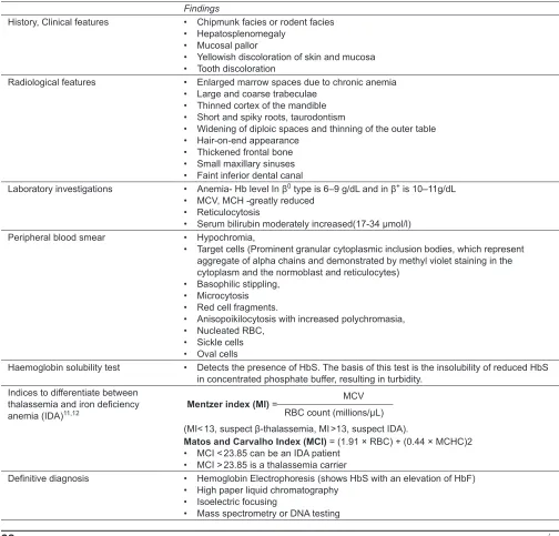
protru-Table 1: Diagnostic criteria
Findings
History, Clinical features • Chipmunk facies or rodent facies • Hepatosplenomegaly
• Mucosal pallor
• Yellowish discoloration of skin and mucosa • Tooth discoloration
Radiological features • Enlarged marrow spaces due to chronic anemia • Large and coarse trabeculae
• Thinned cortex of the mandible • Short and spiky roots, taurodontism
• Widening of diploic spaces and thinning of the outer table • Hair-on-end appearance
• Thickened frontal bone • Small maxillary sinuses • Faint inferior dental canal
Laboratory investigations • Anemia- Hb level In β0 type is 6–9 g/dL and in β+ is 10–11g/dL
• MCV, MCH -greatly reduced • Reticulocytosis
• Serum bilirubin moderately increased(17-34 µmol/l) Peripheral blood smear • Hypochromia,
• Target cells (Prominent granular cytoplasmic inclusion bodies, which represent aggregate of alpha chains and demonstrated by methyl violet staining in the cytoplasm and the normoblast and reticulocytes)
• Basophilic stippling, • Microcytosis • Red cell fragments.
• Anisopoikilocytosis with increased polychromasia, • Nucleated RBC,
• Sickle cells • Oval cells
Haemoglobin solubility test • Detects the presence of HbS. The basis of this test is the insolubility of reduced HbS in concentrated phosphate buffer, resulting in turbidity.
Indices to differentiate between thalassemia and iron deficiency anemia (IDA)11,12
MCV Mentzer index (MI) =
RBC count (millions/µL) (MI< 13, suspect β-thalassemia, MI >13, suspect IDA).
Matos and Carvalho Index (MCI) = (1.91 × RBC) + (0.44 × MCHC)2 • MCI < 23.85 can be an IDA patient
• MCI > 23.85 is a thalassemia carrier
Definitive diagnosis • Hemoglobin Electrophoresis (shows HbS with an elevation of HbF) • High paper liquid chromatography
• Isoelectric focusing
Sickle Cell Beta Thalassemia: A Rare Entity OMPJ
sion and mandibular atrophy.8 The present case showed features such as icterus, yellowish discoloration of the skin, hepatosplenomegaly, bimaxillary protrusion, lip incompetence, saddle nose, malar prominence, frontal bossing and hypertelorism, all favoring the features of thalassemia.
Oral Manifestations
In affected individuals, decreased hemoglobin levels manifest as mucosal pallor and atrophic glossitis. Severe gingivitis is seen if a splenectomy is done. Iron deposits may lead to teeth discoloration and inflammation of salivary glands. High ferritin levels in blood lead to dark-colored gingiva.8 Those affected are prone to high caries index with increased overjet and anterior open bite.9 Our case showed findings such as mucosal pallor, yellowish discoloration of the oral mucosa, increased overjet, and tooth discoloration.
Diagnosis
Various features which help in diagnosis of the disease has been compiled and enumerated in Table 1.
Our case showed radiographic features such as enlarged marrow spaces, increased diploic space and maxillary protrusion. Laboratory investigations and peripheral blood smear revealed reduced hemoglobin count, reduced MCV,10 reticulocytosis, increased serum bilirubin, RBC with anisocytosis and microcytosis, sickle cells, and target cells, and low Mentzer index.
The differences between the two types of sickle cell beta-thalassemia, Sβ0 thalassemia and Sβ+ thalassemia based on hematological findings and hemoglobin elec-trophoresis are described in Table 2.2.3
Management
Management of patients with sickle cell beta thalas-semia primarily begins with education and psychosocial support, genetic counseling,followed by immediate treatment of acute complications of vaso-occlusive crisis by the supplemental oxygen therapy, correction of acidosis and aggressive treatment of associated
infec-tions. Prophylactic penicillin prophylaxis is used until 5 years of age.7 Blood transfusions are indicated only during acute severe exacerbations of anemia, as during splenic crisis or aplastic crises. Chelation therapy can be administered with iron chelators such as deferoxamine, deferasirox, and deferiprone to reduce the iron overload after multiple blood transfusions. Bone marrow trans-plantation and gene therapy are definitive therapies to cure the disease.8
Dental Considerations
Factors precipitating stress, acidosis, and hypoxia should be avoided. Liver function, Hb level (should be >10 mg%) and coagulation tests should be done before any surgical procedure.8 Any invasive procedure should be done under antibiotic prophylaxis and immediately after transfusion.9 In less severe type, the orofacial defects and malocclusion can be treated surgically followed by an orthodontic correction. If the malocclusion is minimal, it can be corrected at an early stage by preventive and interceptive orthodontics with low forces.8
Drugs like aspirin, tetracycline, metronidazole, eryth-romycin estolate, and other hepatotoxic drugs should be avoided.9 In cases who underwent splenectomy, admin-istration of antiplatelet drugs requires monitoring of bleeding time and hematologist consultation.8 Sedation should be used with caution in patients with thalassemia due to the risk of respiratory depression and the presence of chronic, potentially severe anemia.
CONCLUSION
Oral physicians and oral pathologists should be vigilant enough to identify any hemoglobinopathy, with adequate radiological and hematological investigations, although it has a rare occurrence. If diagnosed, a multidisciplinary approach involving dental surgeon, and orthodontist is mandatory for these patients.
ACKNOWLEDGMENTS
Authors would like to express their sincere gratitude to Dr. Sheeba Padiyath, Department of Oral Medicine and
Table 2: Comparison of the types of sickle cell beta-thalassemia (Sβ0 thalassemia and of Sβ+ thalassemia)
Type
Hematologic values Hemoglobin electrophoresis
Hb (g/dL) Reticulo cyte (%) MCV (fl) RBC Morphology Interacting genes HbS (%) HbF (%) HbA(%)2 HbA (%)
Sβ0
thalassemia 6-10 5-20 <80 Sickle cells, nucleated rbc, hypochromia microcytosis, anisocytosis, poikilocytosis, target cells
βS and β0
gene 70–90 5–30 4–8 0
Sβ+
thalassemia 9-12 5-10 <75 No sickle cells, hypochromia, microcytosis, anisocytosis, poikilocytosis, target cells
βS and β+
Authors would also like to thank Dr.B.S. Sreenivasan and Dr. Soma Susan Varghese, Department of Oral and Maxillofa-cial Pathology, Mar Baselios Dental College for their guidance.
REFERENCES
1. Duggal MS, Bedi R, Kinsey SE, Williams SA. The dental man-agement of children with sickle cell disease and β-thalassemia: a review. Int J Paediatr Dent. 1996;6:227-234.
2. Firkin F, Chesterman C, Penington D, Rush B. de Gruchy’s Clinical Hematology in Medical Practice. 5th Ed. 2010 Black-well publishing. Wiley India Pg 143-150.
3. Reid CD, Charace S, Lubin B. Management and Therapy of Sickle cell disease. 3rd Ed. 1995. National Heart, Lung and Blood
Institute p. 15-18.
4. Balgir RS. Spectrum of Hemoglobinopathies in the state of Orissa, India: A ten years cohort study. J Assoc Physicians India. 2005;53:1021-1026.
5. Niwas JK, Reddy R, Udayashankar D, Krishnan D. Sickle Beta+ Thalassemia.Chettinad Health City Medical Journal 2014;3:133-135.
Sickle Cell Anemic Patients: A Comparative Study. J Int Oral Health. 2013;5:53-58.
7. Shrestha B, Karmacharya K, Singh J, Kotwal J, Devgan A. Compound Heterozygous Sickle and Thalassemia Trait: A Case Report. J Nep PaedtrSoc2011;31:130-133.
8. Helmi N, Bashir M, Shireen A, Ahmed IM. Thalassemia review: features, dental considerations and management. Electronic Physician 2017;9:4003-4008.
9. Madhok S, Madhok S. Dental considerations in Thalassemic patients. Journal of Dental and Medical Sciences 2014;13: 57-62.
10. Trent RJ. Diagnosis of the haemoglobinopathies. Clin Biochem Rev. 2006 Feb;27(1):27-38.
11. Vehapoglu A, Ozgurhan G, Demir AD, Uzuner S, Nursoy MA, Turkmen S, Kacan A. Hematological Indices for Differential Diagnosis of beta thalassemia trait and Iron deficiency anemia. Anemia 2014; Hindawi Publishing Corporation. Article ID 576738. http://dx.doi.org/10.1155/2014/576738.