Solar Retinopathy Following
the Eclipse of March 7, 1970*
WALTER J. GEERAETS, THOMAS W. NOONEY, JOSEPH R. SVOBODA AND FLORENCIO C. CHING
Department of Ophthalmology, Medical College of Virginia,
Richmond 23219.
In spite of extensive warnings by the news media prior to the solar .eclipse on March 7, 1970, retinal burn injuries were expected to occur either in children too young to comprehend the dangers involved or in persons ignoring or disbelieving these warnings.
Eclipse burns have been described since ancient times, dating back to the fourth century BC. We read in Plato's Phaedo of Rouse's translation "I must be careful not to be affected like people who observe and watch an eclipse of the sun. What happens to them is that some lose their sight, unless they look at his reflection in water or something of that sort." Probably the greatest number of retinal burns from eclipse watching have been reported in the 20th century. Several hundred cases of more or less severe ocular injuries were observed following the eclipse of April 17, 1912; as noted by Wendenburg (1914), Birch-Hirschfeld (1912), Bohm (1913), and others. Central scotomata of various degrees and severity were the usual findings; but, in addition, ring scotomata for colors were described for a series of cases by Jess ( 1913). The significance of metamorphopsia in patients with solar retinitis was discussed by Verhoeff, Bell, and Walker (1916). Work concerning intensities re-quired for production of retinal lesions during sun watching had already been reported during the 19th and early 20th centuries [Czerny (1867); Deutschmann (1882); Herzog (1903); Verhoeff, Bell, and Walker (19.16); and others]. More recent descriptions of solar retinitis have been reported by Rosen ( 1948), who I isled a total of 23 cases.
About 40 per cent of the radiant solar energy reach-ing the earth is within the visible spectral range, and 55 per cent is in the nonvisible infrared region. Under normal conditions, the bright sunlight causes maximal constriction of the pupil; hence, the light intensity incident on the retina is greatly reduced. Moreover, painful photophobia experienced during direct ob-servation of the sun elicits the blink-reflex ( approxi-mately 150 ms), thus providing further protection. The
*
This work was supported in part by a Special Fellow-ship (9 F03 EY20986-03 VSN) from the National Eye Institute of the U. S. Public Health Service, National Institutes of Health.total amount of light entering the eye during observa-tion of an eclipse is proporobserva-tional to the percentage of the solar surface not obscured by the moon. How-ever, the energy density per unit area on the retina remains the same; ie, although the image diameter of the sun on the retina is smaller, related to the por-tion of the sun hidden by the moon, the light remains equally intense over the image area the sun casts on the retina. Since during eclipse watching the pupil may not be constricted to the same degree as in look-ing at the sun under normal circumstances, the situa-tion becomes even worse. This factor plays a partic-ular role if inadequate filters are used, which, because of their darkness, may decrease the amount of visible light entering the eye and, thus, permit the pupil to dilate. At the same time, however, it would allow more nonvisible infrared radiation to enter the eye, possibly resulting in retinal burn injury.
Case Reports
Case I
A 14-year-old Caucasian girl was examined for the first time five days after the eclipse on March 7, 1970. She gave a history of having watched the eclipse with her right eye in the Richmond, Virginia area shortly before 1: 00 PM, estimating the time of view-ing as havview-ing been about 15 to 30 seconds. No pro-tective goggles or other filters were used. The patient initially noted a bright glare which after a fe~ sec-onds became bluish and allowed for good and clear visualization of the eclipse. She discontinued her di-rect observation to answer a telephone call, at which time she noted that "Everything looked orange," ( erythropsia) when viewed with the right eye. She then continued to watch the progress of the eclipse by using projection techniques. Following the eclipse the vision in her right eye was very hazy, and the next day she noted a dense black spot in the center of fixation. This spot increased somewhat in size over the next few days but had remained about the same for a period of 24 hours preceding consultation.
SOLAR RETINOPATHY
of the right macula and covering the entire foveal
region. The lesion appeared to be of a light orange
color with some scarce pigmentation and a grayish
cen-ter. The area immediately surrounding the lesion
ap-peared somewhat "wet" and of darker coloration (Fig
I ) . No other pathology was noted.
Visual acuity. 20/200 OD improving to 20/70
by "scanning" single letters, and 20/20 OS. Normal
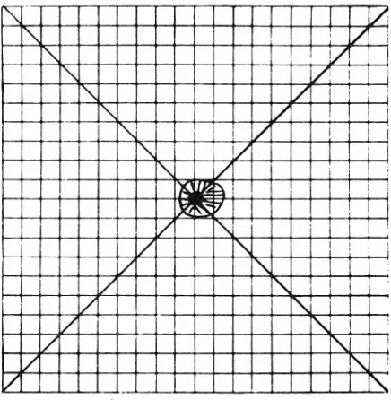
pupil reaction. Pupillary diameter (daylight): 3.8 mm. Visual field plot using the Amsler Grid charts
re-vealed a central scotoma 2 degrees in diameter with
the aid of diagonal lines to maintain fixation (Fig 2).
This size reduced to about 1 degree over the next six
days. At this time her visual acuity for near at direct
steady fixation was 20/200 distance equivalent, but
equal to 20/20 in scanning individual digits.
Case 2
A Caucasian male, 30-years-old, was examined
seven days after the eclipse on March 7, 1970. The
patient stated that he had viewed the eclipse in
Pow-hatan county near Richmond, Virginia intermittently
with both eyes unprotected, about one minute at a
time as the phenomenon developed. He continued
observation to the completion of the eclipse. Estimated
total time of viewing was three to five minutes. After total eclipse was reached he attempted to read but
"could not make out the words exactly right." When
looking at a person wearing a red garment from a
dis-tance of about 200 yards, he noticed fading of the red
color to grey or even black. There was a sensation of
slight film over his eyes.
Ocular findings. OD: Center of the macula showed
a diffuse reflex near the nasal margin of the fovea
with increased scattered pigmentation around the
fovea, giving the small lesion a dark red appearance.
All other ocular findings were normal. OS: A small
circular lesion (about 1/10 disc diameter) covered
the fovea, with some pigmentation, though less intense
than in OD.
Visual acuity. 20/30 at distance, and 20/30-1 at
near, for both OD and OS which improved to 20/25
over several more days. Pupil reactions were normal
and the pupillary diameter (daylight) measured 3 mm
in diameter OU. Visual field plots (Amsler Grid
charts) indicated presence of dense central scotomata
(diameter 1/2 degree) with a bluish outer edge
sur-rounded by a whitish halo extending to 1 degree (Fig
3). Color vision was normal. There was. some
indica-tion of metamorphopsia.
Case3
A Caucasian male, 23-years-old, was examined seven
days after the recent eclipse of March 7, 1970. The
patient stated that he looked steadily at the eclipse
with eyes unprotected, for a period of three to four
minutes when about "Y<i of the sun was left,"
avert-Fig 1-Fundusphotograph of macular region of the right
eye. Note the small circular lesion in the center of the
macula. (Case I)
'
/
'
/
'
/
'
I"'-.v
v
'
/
'
/
'
v
•
/
I'-/
'
:/'
/
"
/
I'-I/
I'-I/
I'-/
I'.
/
'
Fig 2- Central positive scotoma as plotted by the
W. J. GEERAETS, T. W. NOONEY, J. R. SVOBODA AND F. C. CHING
ing his eyes then for awhile. Total viewing time was estimated at five to ten minutes. Beginning about
15 minutes after culmination of the eclipse, until late
the same evening, the patient saw everything "red" ( erythropsia). When reading he could only see the last few letters of words. The following morning he
noted a black spot, about "the size of an orange"
when looking toward the sky.
Ocular findings. Ophthalmoscopic examination
re-vealed bilateral parafoveal hyperpigmentation and poor foveal reflex OU. All other ocular findings were
normal.
Visual acuity. 20/50 OD for distance and 20/50-1
for near, improving to 20/30 over the next few days.
OS was 20/30-1 for distance and 20/30-1 for near.
Pu-pil reactions were normal in both eyes. Pupillary
diam-eter (daylight) measured 2.7 mm at the time of
ex-amination. Visual field plots (Amsler Grid charts)
indicated presence of a dense black central scotoma
(diameter 0.8 degree OD and 0.6 degree OS) with a gray outer ring extending to about 2 degrees (Fig 4).
The adjacent black lines of the grid appeared somewhat
fuzzy and distorted (metamorphopsia). No defect in
color vision was noted.
Discussion
With an assumed irradiance of 71.7 mW/cm2 at
sea level, the energy density entering the eye with a
pupil diameter of 3 mm can be calculated to be 5.1
mW/cm2. This value would be 14.1 mW/cm2 for a
pupil diameter of 5 mm, increasing to 36.2 mW /cm2
for a diameter of 7 mm. Based on calculations that
1.3 mW entering the eye will cause a IC temperature
rise in the retina, the corresponding temperature
elevations upon viewing the sun for the above given
three pupil diameters would be approximately 3.9,
10.7, and 28C respectively (Clarke, Geeraets and
Ham, 1969). Expressed in power density on the retina
for a given image diameter of the sun on the human
retina of 158 µ, these values correspond to 21.8, 59.9, and 156.8 W /cm2; taking into account the ocular spectral characteristics for specific absorption in and
reflection from the various structures ( Geeraets and
Berry, 1968) (Fig 5).
Eccles and Flynn (1944) gave a value above 50
caljcm2/min for the production of a retinal solar burn
in rabbits at an exposure time of 30 seconds, however, they used a telescope to enlarge the sun's image on the retina about ten times. For the given retinal image
size of the sun ( 158 µ in diameter) and an exposure time of 30 seconds, experimental, data obtained in
our own laboratories indicate that a power density of
approximately 40 W /cm2 is required for the
produc-tion of retinal burn injury in the rabbit eye (Clarke,
Ham, Geeraets, et al, 1969) (Fig 6). On the basis of
these calculations and an estimated exposure time of
1"'-
v
·)<\.
/
I'><-
/ '1x._
x
I~
K"
I
X:
..
/
I
Ix
/ '
I'><
l?
I"><-
v<
-· ~ tr'Lii ~
>-··+
v
"">'
' Iv
1->Z-
-
·-··IX
~x
I\.
I>'
I'\LX
~I/
''\,l)<
I'>.;
v
- I~Fig 3-Dense central scotoma approximately 0.5 degree
in diameter with bluish outer edge and a whitish halo of
about 1 degree. Diagonal lines appeared somewhat blurred,
OU. (Case 2)
~ -
I /
I"'-
V1
ij'\, ! I
v
I!
I"-
v
' Ii
I"-
]/
II j
I"-
I/]
II"-,
I
i
i
/
i II
"-
I!
/
i !I'\
VI
""'
~ ~ 1.R'2
~v I/
1""'-
!I/
'\.
I Ii
v
"""'
/
"""'
I I
I
/
1"'-/
"'\.
'
i
I/
"""'
:
v
!
""'-/
-"'\.
Fig 4-Dense central scotoma approximately 0.6-0.8 de
SOLAR RETINOPATHY
PERCENT ABSORPTION IN RETINAL PIGMENT EPITHELIUM AND CHOROID
I
I I
I
I
f----t--+--+--+--+-+-+--1-t--+--+--+--+-+- -+-+-+----<
I 00 f----t--+--+--+--+-+-+--1-t--+--+-
-+--+-+-90f----t--+--+--+--+-+-+--1-t--+
--+--+--+-+-~BOri--f--j--::;;P+--F-t:::it-t-t---t---t--t-t
~701-t-+.~'t'-<;"-•f-'~~~,rl---+----ir-l-t
l'-.,-'1'..-t---t--t-t-(i)
,1
-
- ....
..
~' ,
~ 6Q I • '"'..,J ·"'1---t-+--1-t-'l,rr---
+--f-~ 5Q f -
1
:
..
..
MEAN VALUES
fU;ht 0!1~<10!
int111sit>' onti
111cict111tont111 corn10) (011!cll~Chin
cllillo}
- •Monhy (Rllu11s)
~ : £ .. , ••• \
~
40 r : .... -,.l
,
_/ '
,
:
....
--30 r->;~ ~.+-+--1-t--+--+--+--+'<~~~~t-i--+--+--+--+--+--+-f----I--+--+---<
t-+t-=t+--+--+--+-+-+-1--+--+--.':.,,.__+--f----t--+--+--+--+-+-+--f----<
20
I 0
~:tt-
--+---t--+-+--
t----i--+-+--+-
,
---<
,~~·"1~7~~
t
·~·~~;;::;j:;::j:
:::J
--t-TI
...
•••••15•;~.
.
.
.
.
.
~.
.
400 500 600 700 800 900 1000 1100 1200 1300 1400 1500
WAVE LENGTH (nm)
Fig 5-Per cent of spectral absorption in retina and
choroid for light incident on the cornea.
30 sec during which the patient (Case 1) may have
watched the eclipse with the unprotected eye, the
pupil diameter should have been about 4 mm or
larger to allow sufficient energy to be incident on the retina to result in a thermal lesion. The degree of
ocular fundus pigmentation is, of course, another
factor influencing the required energy for production
of a retinal burn.
At the time the eclipse was observed by this pa-tient (Case 1), approximately 60 per cent of the sun
was still uncovered by the moon. However, as stated
in the introduction, the energy density on the retina for the area on which the sun's image impinged re-mained the same as if the entire sun had been ob-served. Only the size of the image was smaller, ie, was proportional to the percentage of the unobscured por
-tion of the sun.
The funduscopic examination and fundus
photo-graphs indicate that the lesion diameter is
approxi-mately 1.5 degrees or 400-450 µ. The larger lesion
size in comparison to the calculated image size of
the sun on the retina can be due, in part, to heat con-duction from the site of exposure into the surrounding tissues, and to unsteady fixation. The central scotoma plotted by the patient (Case 1) at an observation
dis-tance of 28 cm on the Amsler Grid chart corresponds
to a lesion size of approximately 540 .µ in diameter. The difference between the first and second figure can be explained by reactive edema surrounding the lesion which was still present at the time of her first examina-tion. This factor explains, as well, the decrease in
visual acuity to 20/70 OD at that time. With
subse-quent disappearance of the edema and the remaining
permanent foveal lesion, one may predict an
improve-10'
10'
10'
10'
10'
LOG-LOG PLOT FOR MINIMAL OPHTHALMOSCOPIC VISIBLE
RETINAL LESIONS
OLAS£RPULSES
Wovtlt"Qlll 694.lm,u
10° l__,_u.,,JL...w...J.~"""-~.L~d.u~.l_,_~Lw..wui~="-~""'-~,
101 107 10' 105 10'4 163 162 161 10° 101 102. 10
TIME-SECONDS
Fig 6-Power density required to produce minimal
oph-thalmoscopically observable lesions for retinal image
diam-eters of 160 and 800µ.
ment in visual acuity to 20/ 40 or better, provided a
macular hole with concomitant central retinal
de-tachment does not develop.
The relatively less severe macular lesions of the other two patients can be explained by the factor of pupillary diameter and possible variations in degree of pigmentation of the retina and choroid.
Summary
A case of unilateral retinal burn injury of the fovea and two cases of binocular injuries have been reported, in connection with the viewing of the eclipse of March 7, 1970. Some calculations of energy densities
in-volved have also been presented.
References
BOHM H: Blendungsretinitis infolge der Beobachtung der Sonnenfinsternis am 17.4.1912. Klin Mbl Augenhk, April
1913
BIRCH-HIRSCHFELD A: Zurn Kapitel der Sonnenblendung
des Auges. Zeitschr f Augenhk 28, 1912
CLARKE AM, GEERAETS WJ, HAM WT: An equilibrium
thermal model for retinal injury from optical sources. Ap-plied Optics 8: 1051, 1969
CLARKE AM, HAM WT, GEERAETS WJ, WILLIAMS RC,
MUELLER HA: Laser effects on the eye. Arch Environ
W. J. GEERAETS, T. W. NOONEY, J. R. SVOBODA AND F. C. CHING
CZERNY D: Ober Blendung der Netzhaut <lurch direktes
Sonnenlicht. K Akad d Wissensch, Oct. 6, 1867
DEUTSCHMANN W: Ober die Blendung der Netzhaut <lurch
direktes Sonnenlicht. Arch f Ophthal 28: 241, 1882
ECCLES JC, FLYNN AJ: Experimental photo-retinitis. Med
J Aust I: 339, 1944
GEERAETS WJ, BERRY ER: Ocular spectral characteristics
as related to hazards from lasers and other light sources.
Am J Ophth 66: 15, 1968
HERZOG H: Diskussion zum Vortrag Birch-Hirschfeld.
Bericht d ophth Ges Heidelberg, 1903, p 1964
JESS L: Die Ringskotome nach Sonnenblendung, Arch f
Augenhk 68: 78, 1913
Plato's Phaedo: translation by Rouse WMD in Great
Dia-logues of Plato. New York, Mentor Book, The American
Library of World Literature, 1956, p 503
RosEN E: Solar retinitis. Brit J Ophth 32: 23, 1948
VERHOEFF FH, BELL L, WALKER CB: The pathological
ef-fects of radiant energy on the eye. Proc Am Acad Arts &
Sciences 51: 627, 1916
WENDENBERG K: Schadigungen des Sehorgans <lurch Blend-ung bei Sonnenfinsternis Beobachtungen. Berlin, S. Karger,