M
AGDALENAA. S
OKALSKA−J
URKIEWICZ1, M
ARTAM
ADEJ1, B
EATAN
OWAK1, A
NNAC
ZARNY2,
E
WAZ
ACZYŃSKA2, P
IOTRW
ILAND3Activation of STAT−1, −3, and −6 Proteins
in Early Arthritis*
Aktywacja białek STAT−1, −3 i −6 we wczesnym zapaleniu stawów
1 Department of Rheumatology and Internal Diseases Silesian Piasts University of Medicine in Wrocław, Poland 2 Ludwik Hirszfeld Institute of Immunology and Experimental Therapy, Polish Academy of Sciences, Wrocław,
Poland
3 Department of Balneology Silesian Piasts University of Medicine in Wroclaw, Poland Adv Clin Exp Med 2008, 17, 4, 399–404
ISSN 1230−025X
ORIGINAL PAPERS
© Copyright by Silesian Piasts University of Medicine in Wrocław
Abstract
Background. Early arthritis is the inflammation of one or more joints which appears for the first time and lasts
shortly. The role of STAT proteins, which belong to a family of latent cytoplasmic transcription factors activated in a response to cell stimulation through various cytokine receptors, in autoimmune diseases is being widely dis− cussed. It is postulated that STATs play a vital role in rheumatoid arthritis. Their exact function in its pathogene− sis, especially in its early stage, is not known. The aim of the study was to investigate STAT activation levels in the leukocytes of early arthritis patients.
Material and Methods. The levels of STAT−1, −3, and −6 in peripheral blood leukocytes from 31 early arthritis pa−
tients and 30 healthy individuals were examined by an immunocytochemical method. Clinical characteristics and the final diagnoses of the patients were assessed.
Results. STAT−3 and −6 activation was increased in the leukocytes of early arthritis patients compared with the he−
althy controls. There was no statistically significant difference in STAT−1 activation between these groups. The sub− groups of early arthritis (rheumatoid arthritis and undifferentiated arthritis) did not differ in STAT−1, −3, or −6 acti− vation.
Conclusions. The increased activation levels of STAT−3 and STAT−6 in the leukocytes of early arthritis patients
confirms the importance of STATs in the pathogenesis of autoimmune arthritis. The lack of differences in STAT−1 activation may result from the early stage of the disease, the heterogeneity of the patient group, the fact that STAT−1 was predominantly expressed in the cells not migrating to peripheral blood (present only in synovial tissue), or in− hibition by c−fosgene. The lack of differences in STAT−1, −3, and −6 activation between the subgroups of early ar− thritis (rheumatoid arthritis and undifferentiated arthritis) supports the hypothesis of the undifferentiated character of the onset of these types of inflammatory arthritis (Adv Clin Exp Med 2008, 17, 4, 399–404).
Key words:arthritis, STAT transcription factors.
Streszczenie
Wprowadzenie.Wczesne zapalenie stawów jest definiowane jako stan zapalny minimum jednego stawu o krót−
kim czasie trwania, pojawiający się po raz pierwszy u danego pacjenta. Rola białek STAT należących do grupy cy− toplazmatycznych czynników transkrypcyjnych, aktywowanych w odpowiedzi na pobudzenie komórki przez ago− nistów błonowych receptorów cytokinowych, w chorobach autoimmunologicznych jest obecnie często dyskutowa− na. Postuluje się, że białka te odgrywają ważną rolę w reumatoidalnym zapaleniu stawów. Dokładne ich znaczenie w patogenezie zapalenia stawów, a zwłaszcza w jego początkowym okresie, nie jest jeszcze znane.
Cel pracy.Określenie stopnia aktywacji białek STAT w leukocytach pobranych od chorych na wczesne zapalenie
stawów.
Early arthritis is the inflammation of one or more joints which appears for the first time and lasts shortly. The early symptoms of arthritis are often very slight and gradually increase in intensi− ty over time. After more symptoms are revealed, the disease may eventually be diagnosed and an appropriate therapy administered. From the onset of symptoms up to that moment, the patient awaits the proper diagnosis. Traditionally, early arthritis patients were treated only with non−steroidal anti− inflammatory drugs (NSAIDs), which diminished symptoms but rarely stopped the inflammatory process [1]. In many cases the disease developed and differentiated into rheumatoid arthritis or other inflammatory disorder. According to some research, very early and aggressive treatment, espe− cially in rheumatoid arthritis, diminishes function− al disability [2]. Insight into the pathological inflammatory process at the very beginning of arthritis could provide an opportunity for early dif− ferentiation between diverse disorders, which could radically improve the effectiveness of med− ication.
Signal transducer and activator of transcrip− tion (STAT) proteins are a family of latent cyto− plasmic transcription factors that are activated in response to cell stimulation by various cytokine receptors. STATs play a vital role in mediating the intracellular effects of many cytokines involved in the pathogenesis of autoimmune arthritis, such as interferons (IFNs) and many interleukins (ILs) [3]. Seven members of the mammalian STAT family have been identified: STAT−1, −2, −3, −4, −5A, −5B, and −6. Most of them are widely expressed in diverse cells and may be activated by multiple lig− ands, depending on the cell type and the local environment, but certain cytokines activate partic− ular STATs preferentially [4]. In the case of the cytokines involved in the pathogenesis of arthritis,
IFN-γ is known to activate STAT−1, STAT−3 is
stimulated by IL−6 and IL−10, and IL−4 activates STAT−6.
STAT−1 transduces signals of IFNs during cell defense against viruses and other intracellular pathogens. The process results in the transcription of the major histocompatibility complex (MHC), chemokine, nitric oxide synthase, and complement genes. STAT−1 is also responsible for the antiprolif− erative and proapoptotic effects of interferons. STAT−3 can be activated by many ligands, including IL−6, IL−10, and IFN-αand −β, and exerts different actions depending on the activator and cell type [5]. In bone−marrow progenitor cells, STAT−3 negatively regulates granulopoiesis, while in T cells it is impor− tant in IL−6−mediated suppression of apoptosis and stimulation of proliferation and it is also necessary for correct T−cell reaction to IL−2. On the other hand, the effect of STAT−3 activation due to IL−10 receptor binding observed in macrophages and neutrophils is mainly anti−inflammatory. STAT−6 mediates mainly IL−4 and IL−13 signals and regulates the Th2 response by stimulating Th2 differentiation, allow− ing B−cell class−switching and IgE production and enabling IL−4−induced cell proliferation.
The roles of STAT proteins in autoimmune inflammatory arthritis are not fully understood, but preliminary research on STAT activation dur− ing synovitis has been performed. The first reports of STAT activation in arthritis described STAT−3− −DNA binding in freshly isolated synovial fluid mononuclear cells from rheumatoid arthritis patients [6, 7]. Later it was shown that STAT−1 expression and activity was increased in the syn− ovium of patients suffering from active rheuma− toid arthritis [8, 9]. In subsequent reports it was demonstrated that not only STAT−1, but also STAT−4 and STAT−6 expressions were increased during RA synovitis [10, 11] and that STAT−3 pro− moted the survival of synovial fibroblasts collect− ed from rheumatoid arthritis patients [10].
Materiał i metody. Do badania zakwalifikowano 31 chorych na wczesne zapalenie stawów oraz 30 zdrowych
ochotników. Przeprowadzono kliniczną charakterystykę chorych oraz wykonano badania diagnostyczne w celu po− stawienia ostatecznego rozpoznania. Stopień aktywacji STAT−1, −3 i −6 w leukocytach krwi obwodowej określono za pomocą metody immunocytochemicznej.
Wyniki.Stopień aktywacji białek STAT−3 i STAT−6 był znacząco większy u chorych w porównaniu z grupą kon−
trolną. Nie stwierdzono istotnej statystycznie różnicy w stopniu aktywacji białka STAT−1 między grupami. Podgru− py wyodrębnione z grupy chorych na wczesne zapalenie stawów (chorzy na reumatoidalne zapalenie stawów i nie− zróżnicowane zapalenie stawów) nie różniły się pod względem stopnia aktywacji białek STAT.
Wnioski.Zwiększona aktywacja STAT−3 i STAT−6 w leukocytach chorych na wczesne zapalenie stawów potwier−
dza znaczenie tych czynników transkrypcyjnych STAT w patogenezie autoimmunologicznego zapalenia stawów. Brak zmian w stopniu aktywacji STAT−1 może wynikać z bardzo wczesnego etapu choroby, niejednorodności gru− py chorych, obecności komórek z aktywowanymi białkami STAT−1 poza układem krążenia (tylko w obrębie bło− ny maziowej) lub hamowania aktywacji w wyniku wzmożonej ekspresji genu c−fos. Brak różnicy w aktywacji STAT−1, STAT−3 i STAT−6 między podgrupami chorych na wczesne zapalenie stawów wskazuje na niezróżnicowa− ny charakter procesu patologicznego w początkowym okresie zapalenia stawów (Adv Clin Exp Med 2008, 17, 4,
399–404).
The present study was undertaken to investi− gate the role of the Jak/STAT pathway in the impact of cytokines on target cells in early arthri− tis. In particular, the levels of activation of STAT−1, −3, and −6 in peripheral blood leukocytes of early arthritis patients were compared with those of healthy individuals.
Material and Methods
Thirty−one patients (35% males) and thirty healthy individuals (34% males) as a control group were recruited. The patients reported swelling and pain in one or more joints lasting up to 12 months. After physical examination they were all initially diagnosed with early arthritis. Individuals previ− ously treated with disease−modifying anti− rheumatic drugs (DMARDs), except sulfasalazine, were excluded. The patients were invited to attend a second visit after 6 months to check for a final diagnosis. In the case of the individuals who failed to come for the second visit, the first diagnosis was considered valid. Patients were diagnosed with a particular disorder according to established crite− ria, for example an RA diagnosis was based on the 1987 ARA criteria. The study was approved by the Silesian Piasts University of Medicine in Wrocław Bioethics Committee and all subjects gave their written informed consent.
In previous studies on STAT activation in the leukocytes of arthritis patients, the cells were iso− lated from synovial tissue or fluid during routine puncture, synovectomy, or other procedure. However, patients recruited in those studies were characterized by diagnosed and, mostly, long−last− ing disease. In early arthritis, biopsy of the syn− ovial tissue would be technically very difficult and in most cases unjustified in terms of medical necessity, thus presumably unethical. Since autoimmune arthritis (especially rheumatoid arthritis) is a systemic disease, increased STAT activation in leukocytes not only from synovial tis− sue, but also from the peripheral blood of arthritis patients was postulated.
To examine the presence of active STAT−1, −3, and −6 in peripheral blood leukocytes, an immuno− cytochemical method was applied. The leukocytes were isolated from the blood samples by density gradient centrifugation (1.115 g/cm3 Gradisol G)
and deposited onto glass slides by cytospin cen− trifugation. The specimens were fixed by a 4% formaldehyde solution and washed in phosphate− buffered saline (PBS). The activity of endogenous peroxidase was inhibited using a 3% H2O2solution
in methanol. Subsequently, normal blocking serum was applied and removed after 30 minutes. Then
the primary antibodies anti−STAT−1 (Abcam, Cambridge, UK), anti−STAT−3, or anti−STAT−6 (Chemicon International Inc., Temecula, CA) were applied for 1 hour at room temperature. After washing in PBS, the complex of primary antibody and STAT was detected using a Vectastain ABC Universal Kit and DAB Peroxidase Substrate Kit (Vector Laboratories, Ltd., Peterborough, UK).
The staining evaluation was performed using a Nikon type 120 microscope. Based on the assumption that the Jak/STAT pathway activation results in the presence of STAT protein in the cell nucleus, stained cell nuclei were counted. The level of the particular STAT activation was esti− mated as the number of the stained cells per 100 cells analyzed.
Results
Patient Characteristics
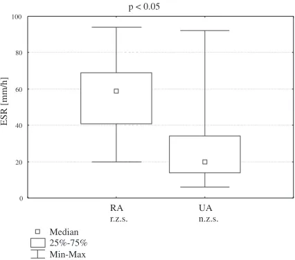
The clinical characteristics of the patients with early inflammatory arthritis are shown in Table 1. One patient was diagnosed with hepatitis B virus (HBV) infection. No hepatitis C virus (HCV) or human immunodeficiency virus (HIV) infection was detected. Based on urethral swab, Chlamydia trachomatisinfection was diagnosed in 3 patients. Of the 31 patients with early arthritis, 12 had rheumatoid arthritis (RA), 11 had undifferentiated arthritis (UA), 3 developed reactive arthritis, 2 were diagnosed with gout, and the other 2 with paraneoplastic syndrome, 1 having undifferentiat− ed spondyloarthropathy. The clinical parameters of the two most important groups (RA and UA patients) were compared. Mean DAS−28 score and ESR were significantly higher in the RA patients than in the UA patients (Figs. 1 and 2).
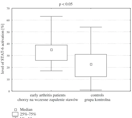
STAT Activation Levels
correlations between each pair of STATs were similar.
In the early arthritis patient group, correlations between STAT activation levels and clinical para− meters were observed. There was a positive corre− lation between STAT−3 activation and the number of swollen joints (r= 0.50, p< 0.05) and negative correlations between STAT−1 activation and the blood parameters erythrocyte count (r = –0.46,
p < 0.05), hemoglobin concentration (r = –0.43,
p< 0.05), and hematocrit (r= –0.40, p< 0.05).
Table 1.Clinical characteristics of the patients with early inflammatory arthritis
Tabela 1.Charakterystyka kliniczna pacjentów chorych na wczesne zapalenie stawów
Symptoms duration – months 6.50 ± 0.71 (Czas trwania objawów – miesiące)
DAS−28 5.20 ± 0.29
OB (mm/h) 44.8 ± 5.4
CRP (mg/dl) 3.00 ± 0.79
RF ≥30 IU/ml (n) 10
Anti−CCP antibodies ≥5 RU/ml (n) 8 (Przeciwciała anty−CCP≥5 RU/ml
Leukocytes per mm3 8000 ± 480 (Liczba leukocytów – mm3)
Erythrocytes per mm3
(Liczba erytrocytów – mm3) (4.20 ± 0.08) ⋅106 Haemoglobin concentration (g/dl) 13.30 ± 0.31 (Stężenie hemoglobiny)
Hematocrit (%) (Hematokryt) 39.40 ± 0.75
MCV (fl) 93.6 ± 1.0
Platelets per mm3 255.000 ± 13.000 (Liczba płytek – mm3)
Uric acid (mg/dl) 4.90 ± 0.32
(Stężenie kwasu moczowego)
Initial NSAID use (n) 22
(Liczba chorych stosujących leki z grupy n.p.l.z. przed włączeniem do badania)
Initial sulfasalazine use (n) 10 (Liczba chorych stosujących sulfa− zynę przed włączeniem do badania) Initial steroid use (n) 8 (Liczba chorych stosujących steroidy przed włączeniem do badania)
p < 0.05
RA r.z.s.
UA n.z.s.
3 4 5 6 7 8 9
D
A
S
−28
sc
or
e
Fig. 1.Mean disease activity scores (DAS−28) for ear−
ly arthritis patients ultimately diagnosed with rheuma− toid arthritis (RA) or undifferentiated arthritis (UA)
Ryc. 1.Średni wynik wskaźnika aktywności choroby
DAS−28 dla chorych na wczesne zapalenie stawów z ostatecznym rozpoznaniem reumatoidalnego zapale− nia stawów (r.z.s.) i niezróżnicowanego zapalenia sta− wów (n.z.s.)
p < 0.05
Median 25%−75% Min−Max
RA r.z.s.
UA n.z.s. 0
20 40 60 80 100
ESR [mm/h]
Fig. 2. Mean ESR scores for early arthritis patients
ultimately diagnosed with rheumatoid arthritis (RA) or undifferentiated arthritis (UA)
Ryc. 2. Średni wynik OB dla chorych na wczesne za−
palenie stawów z ostatecznym rozpoznaniem reumato− idalnego zapalenia stawów (r.z.s.) i niezróżnicowane− go zapalenia stawów (n.z.s.)
p < 0.05
Median 25%−75% Min−Max
early arthritis patients chorzy na wczesne zapalenie stawów
controls grupa kontrolna 0
10 20 30 40 50 60 70 80
le
ve
l
o
f
S
TA
T−
3
ac
ti
v
at
io
n
[%
]
Fig. 3.STAT−3 activation levels in the early arthritis
patients and the control group
Ryc. 3. Stopień aktywacji STAT−3 w grupie chorych
Discussion
It is known that many cytokines are involved in the pathogenesis of autoimmune inflammatory arthritis and that the JAK/STAT pathway is responsible for signal transduction and the activa− tion of transcription of target genes under stimula− tion by a number of cytokines. The objective of this study was to investigate STAT−1, STAT−3, and STAT−6 activation at the beginning of the inflam− matory process. The most important finding of this study was increased activation of STAT−3 and STAT−6 in peripheral blood leukocytes of early
arthritis patients. This confirms the importance of the JAK/STAT pathway in the pathogenesis of autoimmune arthritis. The results of previous investigations, which showed increased STAT−3 and STAT−6 activation in cells collected during synovial biopsy from rheumatoid arthritis patients [10–12], are concordant with these findings, even though cell origin and disease stage were slightly different. The increased STAT−3 and STAT−6 acti− vation in both early and rheumatoid arthritis sug− gests a potential therapeutic target for arthritis treatment. Blocking STAT−3 or STAT−6 at early stages of arthritis would perhaps stop disease evo− lution and differentiation.
In the case of STAT−1 activation in peripheral blood leukocytes, no statistically significant differ− ence between the early arthritis group and the con− trol group was detected. According to previous pub− lications, data on STAT−1 activation in arthritis are diverse. Most of the studies confirm increased STAT−1 activation or expression in cells isolated from synovial tissue of patients with established rheumatoid arthritis [8, 9]. However, on the basis of analysis of RNA separated from synovial cells of rheumatoid arthritis patients, two distinct subgroups of these patients were revealed [13]. The first was characterized by a high expression of inflammatory genes and activation of STAT−1, but activation of STAT−1 in the second subgroup was insignificant and tissue remodeling was observed instead of inflammation. Moreover, decreased STAT−1 phos− phorylation in the lymphocytes of rheumatoid arthritis patients was caused by the c−fos gene, which is overexpressed in this disease [14]. Eventually it was revealed that STAT−1 was pre− dominantly expressed in synovial CD55+ cells [11], which do not migrate to the blood. The apparent lack of difference in STAT−1 activation between early arthritis patients and healthy individuals may be explained by the early stage of the disease, the heterogeneous group of patients, the fact that STAT−1 was predominantly expressed in cells not present in peripheral blood, or inhibition by the c−fosgene.
The lack of differences between the subgroups of early arthritis (rheumatoid arthritis and undif− ferentiated arthritis) in STAT−1, STAT−3, or STAT−6 activation supports the hypothesis of the undiffer− entiated character of onset of these types of inflammatory arthritis.
Table 2.Correlations between STAT activation levels (Pearson’sr) for the early arthritis patients and controls
Tabela 2.Korelacje między stopniem aktywacji białek STAT u chorych na wczesne zapalenie stawów i u osób zdrowych
Early arthritis Controls
patients (Grupa
(Chorzy na kontrolna) wczesne zapalenie
stawów)
STAT−1 and STAT−3 0.46 0.55
STAT−1 and STAT−6 0.42 0.40
STAT−3 and STAT−6 0.52 0.42
p < 0.05
Median 25%−75% Min−Max
early arthritis patients chorzy na wczesne zapalenie stawów
controls grupa kontrolna 0
10 20 30 40 50 60 70
le
v
el
o
f
S
T
A
T
−6
act
iv
at
io
n
[%
]
Fig. 4. STAT−6 activation levels in the early arthritis
patients and the control group
Ryc. 4. Stopień aktywacji STAT−6 w grupie chorych
na wczesne zapalenie stawów i w grupie kontrolnej
References
[1] Pincus T, Smolen JS: Early arthritis Introduction. Clin Exp Rheumatol 2003, 21, Suppl. 31, S1.
[2] Emery P, Salmon M: Early rheumatoid arthritis: time to aim for remission? Ann Rheum Dis 1995, 54, 944–947.
[3] Firestein GS, Manning AM: Signal transduction and transcription factors in rheumatic disease. Arthritis Rheum
[4] Ivashkiv LB, Hu X:The JAK/STAT pathway in rheumatoid arthritis: pathogenic or protective? Arthritis Rheum 2003, 48, 2092–2096.
[5] Ivashkiv LB, Hu X: Signaling by STATs. Arthritis Res Ther 2004, 6, 159–168.
[6] Sengupta TK, Chen A, Zhong Z, Darnell JE Jr, Ivashkiv LB:Activation of monocyte effector genes and STAT
family transcription factors by inflammatory synovial fluid is independent of interferon gamma. J Exp Med 1995, 181, 1015–1025.
[7] Wang F, Sengupta TK, Zhong Z, Ivashkiv LB:Regulation of the balance of cytokine production and the signal
transducer and activator of transcription (STAT) transcription factor activity by cytokines and inflammatory syno− vial fluids: J Exp Med 1995, 182, 1825–1831.
[8] Van der Pouw Kraan TCTM, van Gaalen FA, Kasperkovitz PV, Verbeet NL, Smeets TJ, Kraan MC, Fero M,
Tak PP, Huizinga TW, Pieterman E, Breedveld FC, Alizadeh AA, Verweij CL: Rheumatoid arthritis is a he−
terogeneous disease: evidence for differences in the activation of the STAT−1 pathway between rheumatoid tissu− es. Arthritis Rheum 2003, 48, 2132–2145.
[9] Kasperkovitz PV, Verbeet NL, Smeets TJ, van Rietschoten JG, Kraan MC, van der Pouw Kraan TC, Tak PP,
Verweij CL: Activation of the STAT1 pathway in rheumatoid arthritis. Ann Rheum Dis 2004, 63, 3, 233–239.
[10] Krause A, Scaletta N, Ji J, Ivashkiv LB:Rheumatoid arthritis synoviocyte survival is dependent on Stat3. J Im−
munol 2002, 169, 6610–6616.
[11] Walker JG, Ahern MJ, Coleman M, Weedon H, Papangelis V, Beroukas D, Roberts−Thomson PJ, Smith MD:
Expression of Jak3, STAT1, STAT4, and STAT6 in inflammatory arthritis: unique Jak3 and STAT4 expression in dendritic cells in seropositive rheumatoid arthritis. Ann Rheum Dis 2006, 65, 149–156.
[12] Shouda T, Yoshida T, Hanada T, Wakioka T, Oishi M, Miyoshi K, Komiya S, Kosai K, Hanakawa Y, Ha−
shimoto K, Nagata K, Yoshimura A:Induction of the cytokine signal regulator SOCS3/CIS3 as a therapeutic
strategy for treating inflammatory arthritis. J Clin Invest 2001, 108, 1781–1788.
[13] Van der Pouw Kraan TCTM, van Gaalen FA, Kasperkovitz PV, Verbeet NL, Smeets TJ, Kraan MC, Fero M,
Tak PP, Huizinga TW, Pieterman E, Breedveld FC, Alizadeh AA, Verweij CL: Rheumatoid arthritis is a he−
terogeneous disease: evidence for differences in the activation of the STAT−1 pathway between rheumatoid tissu− es. Arthritis Rheum 2003, 48, 2132–2145.
[14] Hikasa M, Yamamoto E, Kawasaki H, Komai K, Shiozawa K, Hashiramoto A, Miura Y, Shiozawa S:
p21waf1/cip1 is down−regulated in conjunction with up−regulation of c−Fos in the lymphocytes of rheumatoid ar− thritis patients. Biochem Biophys Res Commun 2003, 25, 304, 1, 143–147.
Address for correspondence:
Magdalena A. Sokalska−Jurkiewicz
Department of Rheumatology and Internal Diseases Silesian Piasts University of Medicine
Borowska 213 50−556 Wrocław Poland
Tel.: +48 693 444 295, +48 71 343 39 68 E−mail: [email protected]
Conflict of interest: None declared