M
ARCINF
RĄCZEK1, B
OŻENAJ
AŹWIEC2, M
ARCINM
ASALSKI1, B
EATAN
ADOLSKA1,
T
OMASZK
RĘCICKI1Circulating Soluble Vascular Cell Adhesion Molecule−1
and Vascular Endothelial Growth Factor
in Patients with Laryngeal Squamous Cell Cancer
Ocena stężenia rozpuszczalnych form
naczyniowej cząsteczki adhezyjnej oraz naczyniowo−śródbłonkowego
czynnika wzrostu w surowicy pacjentów
chorych na raka płaskonabłonkowego krtani
1 Department of Otolaryngology, Silesian Piasts University of Medicine in Wrocław, Poland 2 Department of Hematology, Silesian Piasts University of Medicine in Wrocław, Poland
Adv Clin Exp Med 2007, 16, 3, 389–394 ISSN 1230−025X
ORIGINAL PAPERS
© Copyright by Silesian Piasts University of Medicine in Wrocław
Abstract
Background.Tumor growth and subsequent metastatic spread of cancer cells are multistep processes accompanied by changes in the expressions of many growth factors and vascular adhesion molecules.
Objectives.Investigation whether serum levels of soluble vascular cell adhesion molecule−1 (sVCAM−1) and vascu− lar endothelial growth factor (VEGF) are related to clinicopathological variables of laryngeal squamous cell cancer (LSCC) patients.
Material and Methods.The serum concentrations of sVCAM−1 and VEGF165were investigated in 41 patients with
LSCC and in 27 controls by an enzyme−linked immunosorbent assay (ELISA).
Results. In LSCC cases the mean ± SD serum concentration of sVCAM−1 was 542.1 ± 164.9 ng/ml (range: 388–1284 ng/ml) and of VEGF 519.4 ± 335.9 pg/ml (82–1374 pg/ml). The serum levels of both sVCAM−1 and VEGF were significantly elevated in LSCCs compared with the healthy controls (p< 0.05 and p< 0.001, respec− tively). In the control group the mean sVCAM−1 concentration was 434.8 ± 76.3 ng/ml (314.7–648.8 ng/ml) and VEGF level 263.6 ± 166.4 pg/ml (70.5–702.9 pg/ml). There were no significant differences between the serum lev− els of sVCAM−1 and VEGF and clinicopathological variables of the patients with LSCC. In neither the cancer cases nor the control group was a linear correlation between sVCAM−1 and VEGF serum levels found.
Conclusions.The results suggest that the serum levels of sVCAM−1 and VEGF165are not clinically useful bio−
markers for predicting tumor progression or for identifying the metastatic potential of LSCC (Adv Clin Exp Med 2007, 16, 3, 389–394).
Key words:laryngeal cancer, sVCAM−1, VEGF.
Streszczenie
WprowadzenieWzrost guzów nowotworowych oraz tworzenie przerzutów to wieloetapowe procesy, w których często dochodzi do nadekspresji wielu czynników wzrostu i cząstek adhezyjnych.
Cel pracy.Ocena poziomu naczyniowej cząsteczki adhezyjnej−1 (sVCAM−1) oraz naczyniowo−śródbłonkowego czynnika wzrostu (VEGF) w surowicy pacjentów chorych na raka płaskonabłonkowego krtani oraz korelacja uzy− skanych wyników ze wskaźnikami klinicznymi.
Materiał i metody.Stężenie rozpuszczalnych postaci sVCAM−1 i VEGF165oceniono w surowicy krwi obwodo−
wej pacjentów chorych na raka płaskonabłonkowego krtani oraz 27 osobowej grupie kontrolnej za pomocą meto− dy immunoenzymatycznej (ELISA).
The metastatic spread of cancer cells is a key event in tumor progression and, therefore, in deter− mining the prognosis of patients with malignant disease. The development of metastasis includes malignant cells detachment from the primary tu− mor, penetration of the blood or lymph vessels, cellular arrest in the capillary endothelium, extra− vasation, and secondary lesion formation. The process is accompanied by changes in the expres− sions of many cell adhesion molecules, including vascular cell adhesion molecule−1 (VCAM−1), a 110−kDa glycoprotein which is critical in cell−to− cell interactions. VCAM−1 expression has been demonstrated on the endothelial cells of small ves− sels at the invasive margin of tumors, suggesting possible interactions between endothelial and tumor cells involved in metastatic spread [1]. It has been shown that endothelial cells expressing VCAM−1 bind melanoma cell lines, which indi− cates the function of VCAM−1 as an adhesion mol− ecule facilitating metastasis [2].
The phenomenon prerequisite to both tumor growth and metastasis is angiogenesis. The induc− tion of tumor vascularization is mediated in part by the release of angiogenic peptides from tumor cells, macrophages, and the extracellular matrix. Among these angiogenic factors, vascular endo− thelial growth factor (VEGF) is thought to be one of the most important [6]. VEGF is a dimeric gly− coprotein with four spliced variants containing 121, 165, 189, and 206 amino−acid residues, expressing almost identical biological activities as endothelial cell−specific mitogen and vascular per− meability factor. Furthermore, VEGF promotes endothelial cell proliferation and stimulates VCAM−1 expression on endothelial cells [3]. The most frequently expressed isoform is VEGF165.
Although there is abundant evidence showing that VEGF and VCAM−1 play central roles in the development and growth of malignant tumors, information regarding the clinical utility of serum VEGF and sVCAM−1 levels in laryngeal squa− mous cell cancer (LSCC) is limited. Secretion of these biomarkers was detected in a large variety of human solid tumors, including head and neck malignancies [4–7]. Due to the high heterogeneity of head and cancers it seems reasonable to sepa− rate particular subgroups of tumors to enhance the
reliability of the results. According to the present authors’ knowledge, the number of studies on cir− culating soluble molecules in LSCC is restricted. On the basis of recent studies it could be suspect− ed that the biological roles of VEGF165 and
sVCAM−1 serum levels may also be important in the progression and metastatic process of laryn− geal cancer. Thus the aim of this study was to investigate whether serum levels of soluble VEGF165 and sVCAM−1 are related to clinico−
pathological variables of LSCC patients.
Material and Methods
Subjects
The concentrations of sVCAM−1 and VEGF165
were investigated in 41 patients (40 male, 1 fe− male) with histologically proven LSCC diagnosed and treated at the Department of Otolaryngology, Silesian Piasts University of Medicine in Wrocław. The patients’ ages ranged from 40 to 81 years (mean: 58.7 years). Tumor staging was made in accordance with the TNM staging system as fol− lows: pT1 4 cases, pT2 9 cases, pT3 12 cases, and pT4 16 cases. Cervical nodal metastases were observed in 19 (46%) patients. Three cases were in stage I of clinical advancement, 4 in stage II, 17 in stage III, and 22 in stage IV. No patient had received chemo− or radiotherapy or blood transfu− sion prior to surgery.
The control group consisted of 27 age− −matched healthy subjects (25 male and 2 female). The absence of LSCC was assessed by clinical his− tory and endoscopic examination. The presence of a history of rheumatoid arthritis, recent pregnancy, trauma, or surgery (within 1 month) was excluded.
Blood Samples and Assays
Peripheral venous blood samples were drawn into sterile glass tubes in the morning. The sam− ples were allowed to coagulate at room tempera− ture for 30 min and then centrifuged at 2000 ×g
for 10 min. The serum was separated, aliquoted, and stored at –70°C until assay. Before analysis,
434,8 ng/ml; SD 76,3), a stężenie VEGF 70,5–702,9 pg/ml (średnia 263,6 pg/ml; SD 166,4). Nie stwierdzono istot− nych zależności między stężeniem sVCAM−1 i VEGF a wskaźnikami klinicznymi i patologicznymi. Zarówno w grupie pacjentów chorych na raka krtani, jak i w grupie kontrolnej nie wykazano liniowej zależności między stę− żeniami obu badanych markerów.
Wnioski. Uzyskane wyniki sugerują, że ocena stężenia sVCAM−1 oraz VEGF w surowicy pacjentów chorych na raka płaskonabłonkowego krtani nie ma klinicznego znaczenia w przewidywaniu progresji raka i ocenie możliwości tworzenia przerzutów (Adv Clin Exp Med 2007, 16, 3, 389–394).
the samples were slowly thawed and gently mixed. Serum VEGF165 and sVCAM−1 concentrations
were determined using a solid phase ELISA kit (R&D Systems, Minneapolis, MN, USA). The assays use the quantitative sandwich enzyme immunoassay technique using antibodies raised against recombinant human VEGF and sVCAM−1. For each analysis, 100 µl of sample was used. The measurements were carried out as described by the supplier. To correct for optical imperfections in the plate, the reading was repeated as recommended by the supplier at a different wavelength and these results were subtracted. Each serum sample con− centration was calculated automatically from stan− dard curves. All of the analyses and calibrations were carried out in duplicate. VEGF and sVCAM− 1 concentrations are reported in pg/ml and ng/ml, respectively.
Statistical analysis using the chi−squared test was performed. Differences were considered sta− tistically significant at p< 0.05.
Results
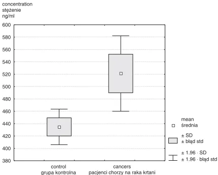
Both sVCAM−1 and VEGF were detectable in all cancer patients and control subjects. Among the cancer patients the mean ± SD serum concentra− tion of sVCAM−1 was 542.1 ± 164.9 ng/ml (range: 388–1284 ng/ml) and of VEGF 519.4 ± 335.9 pg/ml (82–1374 pg/ml). The serum levels of the both sVCAM−1 and VEGF were significantly elevated in patients with LSCC compared with the healthy control volunteers (p< 0.05 and p< 0.001, respec− tively) (Figs. 1 and 2). In the control group the mean sVCAM−1 concentration was 434.8 ± 76.3 ng/ml (range: 314.7–648.8 ng/ml) and VEGF level 263.6 ± 166.4 pg/ml (70.5–702.9 pg/ml).
There were no significant differences between the serum levels of sVCAM−1 and VEGF and the clinicopathological variables of the patients with LSCC (age, tumor size, nodal status, and clinical
Fig. 1.Comparison of sVCAM−1 concentrations in the sera of patients with LSCC and controls (p< 0.05)
Ryc. 1.Porównanie stężeń cząsteczki sVCAM−1 w surowicy pacjentów chorych na raka krtani i w grupie kontrolnej (p < 0,05)
mean średnia
± SD ± błąd std
control grupa kontrolna
cancers pacjenci chorzy na raka krtani 380 400 420 440 460 480 500 520 540 560 580 600
± 1.96 SD 1.96 b
⋅ ⋅
± łąd std
concentration stężenie ng/ml
Fig. 2.Comparison of VEGF concentrations in the sera of patients with LSCC and controls (p< 0.001)
Ryc. 2.Porównanie stężeń VEGF w surowicy pacjentów chorych na raka krtani i w grupie kontrolnej (p < 0,001)
150 200 250 300 350 400 450 500 550 600 650 mean średnia ± SD ± błąd std
control grupa kontrolna
cancers pacjenci chorzy na raka krtani
± 1.96 SD 1.96 b
⋅ ⋅
± łąd std
concentration stężenie ng/ml
Table 1.Characteristics of the cancer patients and associated VEGF and sVCAM−1 levels Tabela 1.Stężenie cząsteczki sVCAM−1 i VEGF w surowicy pacjentów chorych na rakakrtani
Variables n sVCAM−1 ng/ml VEGF pg/ml
(Wskaźniki) (mean – range) (mean – range)
Patient age – years < 58 21 560.5 (403–1284) 571.1 (142–1198)
(Wiek pacjenta – lata) ≥58 20 503.6 (388–680) 418.7 (82–1374)
Tumor size pT1 + pT2 13 472.6 (388–680) 527.8 (219–1198)
(Wielkość guza) pT3 + pT4 28 600.3 (429–1284) 511.3 (82–1374)
Nodal status N0 22 501.9 (388–680) 470.6 (82–1198)
(Stan węzłów chłonnych) N+ 19 560.9 (403–1284) 586.6 (123–1374)
Clinical stage I + II 12 496.7 (388–680) 549.6 (202–1198)
stage) (Tab. 1). However, elevated serum levels of sVCAM−1 were insignificantly associated with locally advanced tumors (pT3–pT4). The levels of circulating VEGF were slightly higher in the sub− group of patients with lymph node metastases (N+). These changes were not statistically significant (p= 0.1439 and p= 0.2796, respectively). In both the cancer cases and the control group, no linear correlation between sVCAM−1 and VEGF serum levels was found.
Discussion
Laryngeal carcinoma is the second most com− mon malignant neoplasm of the respiratory tract after lung cancer. Despite novel combined adjuvant and neoadjuvant therapeutic approaches, the sur− vival rate is still unsatisfactory. A connection between circulating biological markers and disease prognosis may add interesting insight into the biol− ogy of the tumor phenotype and help to identify rational targets for novel therapeutic approaches.
It is now widely accepted that an increase in the tumor cell population must be preceded by an increase in microvessels supplying the neoplasm. It is speculated that the induction of angiogenesis is one of the first events in the transition to a malig− nant state which further enables the growth of solid tumors and the formation of metastases [2, 8]. In early breast cancers it has been demonstrated that serum sVCAM−1 levels correlated with tumor microvessel density and were an accurate marker for tumor angiogenesis [9].
In the present study, a significant difference in serum sVCAM−1 and VEGF levels between patients with LSCC and healthy controls was demonstrated. The serum concentrations of these markers have also been shown to be elevated in many other malignancies [10]. In some locations, such as in gastric, breast, ovarian, and colorectal cancer, high circulating levels of these markers are generally associated with clinical progression and poor prognosis [5, 6, 11, 12]. In patients with gas− tric cancer, a positive correlation was observed between the level of soluble VCAM−1 and tumor stage and invasion depth. The concentration of sVCAM−1 in patients with lymph node metastasis was significantly higher than in patients without [12]. In contrast, the levels of sVCAM−1 were higher in healthy controls than in patients with localized prostate cancer; the levels were highest in patients with skeletal metastases [13]. In the same group, circulating levels of VEGF were greatly elevated in patients with regional and distant metastases as compared with those with nonmetastatic prostate cancer or healthy subjects.
It has been stated that preoperative assessment of circulating VEGF may improve early identifi− cation of patients which harbor lymph node metas− tases, thereby providing surgeons with the oppor− tunity of intensive and meticulous lymphadenecto− my or early systemic intervention [13]. Similarly, serum VEGF appears to be a useful marker for monitoring the clinical course of ovarian cancers after surgery [11].
In the present study no statistically significant differences in the serum levels of VEGF and sVCAM−1 and clinicopathological parameters were found. These data are consistent with find− ings reported in the sera of patients with head and neck cancers. In those studies the serum levels before treatment were also significantly higher than in the control groups [4, 7, 14].
In the present study the mean level of VEGF in the sera of LSCC patients was 519.4 pg/ml, which is plainly higher than the findings recorded by Riedel et al. (142 pg/ml) [4]. In that study the serum levels of VEGF grew from 114 pg/ml in oropharyngeal to 181 pg/ml in hypopharyngeal cancer cases. The serum levels of VEGF in the control group of the present study were similar to those obtained by Hyodo et al. [15], who reported a concentration of VEGF in healthy controls as 238 ± 125 pg/ml. In contrast, the mean level of sVCAM−1 in the present study’s patient group was lower than Liu’s et al. data (1155 ng/ml) [7]. In healthy controls the sVCAM−1 concentrations were similar to those previously reported by Alexiou et al. [5] with a mean level of 413 ng/ml. The results of the present study suggest that serum VEGF and sVCAM−1 might simply not be markers of the extent of disease. The correlation between the serum concentrations of these mole− cules and clinicopathological parameters has been a subject of controversy in various human malig− nancies, which is in line with the present data. The sources of soluble VCAM−1 and VEGF are also not fully known. Some studies have reported that solu− ble VCAM−1 may be related to the inflammation process reflected by white cell count. To eliminate that possibility, only LSCC patients with normal blood tests were included in further investigations. Among head and neck SCC patients, very common factors influencing VEGF serum level are smoking and coronary artery disease [16, 17].
VEGF levels because clot formation induces platelet activation and subsequent abundant VEGF release. Circulating VEGF in patients with cancer originates from many sources and reflects the degree of mitogenicity of serum on endothelial cells, which suggests that platelets and leukocytes may scavenge biologically active VEGF [21]. It is also known that not all of the VEGF stored in blood cells is endogenously synthesized, but may origi− nate primarily from plasma [22]. Consequently it is possible that VEGF secreted by neoplastic cells is transmitted to the circulation and accumulates in blood cells. For these reasons, in the present
authors’ opinion the measurement of VEGF in the serum of patients with malignancy appears more appropriate than its measurement in plasma.
In conclusion, the results of the present study suggest that the serum levels of the investigated antigens may be helpful in distinguishing invasive cancers. However, circulating sVCAM−1 and VEGF165 are not clinically useful biomarkers for
predicting tumor progression or identifying metastatic potential in LSCC. The possible roles of soluble molecules in the prognosis of LSCC deserve further elucidation and evaluation with long−term patient follow−up.
References
[1] Nelson H, Ramsey PS, Donohue JH:Cell adhesion molecule expression within the microvasculature of human colorectal malignancies. Clin Immunol Immunopathol 1994, 72, 129–136.
[2] Osborn L, Hession C, Tizard R, Vassallo C, Luhowskyj S:Direct expression cloning of vascular cell adhesion molecule 1, a cytokine−induced endothelial protein that binds to lymphocytes. Cell 1989, 59, 1203–1211.
[3] Ferrara N, Davis−Smyth T:The biology of vascular endothelial growth factor. Endocrine Rev 1997, 18, 4–25.
[4] Riedel F, Götte K, Schwalb J, ·Wirtz H, Bergler W, Hörmann K: Serum levels of vascular endothelial growth factor in patients with head and neck cancer. Eur Arch Otorhinolaryngol 2000, 257, 332–336.
[5] Zhang GJ, Adachi I:Serum levels of soluble intercellular adhesion molecule−1 and E−selectin in metastatic breast carcinoma: Correlations with clinicopathological features and prognosis. Int J Oncol 1999, 14, 71–77.
[6] Alexiou D, Karayiannakis AJ, Syrigos KN:Serum levels of E−selectin, ICAM−1 and VCAM−1 in colorectal can− cer patients: Correlations with clinicopathological features, patient survival and tumour surgery. Eur J Cancer 2001, 37, 2392–2397.
[7] Liu C−M, Sheen T−S, Shun C−T: Circulating intercellular adhesion molecule 1 (ICAM−1), E−selectin and vascu− lar cell adhesion molecule 1 (VCAM−1) in head and neck cancer. Br J Cancer 1999, 79(2), 360–362.
[8] Liotta LA, Steeg PS, Stetler−Stevenson WG:Cancer metastasis and angiogenesis: an imbalance of positive and negative regulation. Cell 1991, 64, 327–336.
[9] Byrne GJ, Ghellal A, Iddon J:Serum soluble vascular cell adhesion molecule−1: role as a surrogate marker of angiogenesis. J Natl Cancer Inst 2000, 92, 1329–1336.
[10] Kamezaki S, Kurozawa Y, Iwai N, Hosoda T:Serum levels of soluble ICAM−1 and VCAM−1 predict pre−clin− ical cancer. Eur J Cancer 2005, 41, 2355–2359.
[11] Yamamoto S, Konishi I, Mandai M, Kuroda H, Komatsu T, Nanbu K:Expression of vascular endothelial growth factor (VEGF) in epithelial ovarian neoplasms: correlation with clinicopathology and patient survival and analysis of serum VEGF levels. Br J Cancer 1997, 76, 1221–1227.
[12] Ding Y−B, Chen G−Y, Xia J−G, Zang X−W, Yang H−Y:Association of VCAM−1 overexpression with oncogen− esis, tumor angiogenesis and metastasis of gastric carcinoma. World J Gastroenterol 2003, 9(7), 1409–1414.
[13] Shariat SF, Anwuri VA, Lamb DJ, Shah NV, Wheeler TM: Association of preoperative plasma levels of Vascular Endothelial Growth Factor and soluble Vascular Cell Adhesion Molecule−1 with lymph node status and biochemical progression after radical prostatectomy. J Clin Oncol 2004, 22, 1655–1663.
[14] Kawano T, Yanoma S, Nakamura Y, Shiono O, Kokatu T, Kubota A:Evaluation of soluble adhesion mole− cules CD44 (CD44st, CD44v5, CD44v6), ICAM−1, and VCAM−1 as tumor markers in head and neck cancer. Am J Otolaryngol 2005, 26(5), 308–313.
[15] Ohydo I, Doi T, Mendo H, Hosokawa H, Nishikawa Y, Tanimizu M:Clinical significance of plasma Vascular Endothelial Growth Factor in gastrointestinal cancer. Eur J Cancer 1998, 34(13), 2041–2045.
[16] Inoue M, Itoh H, Ueda M, Naruko T, Kojima A, Komatsu R, Doi K:Vascular endothelial growth factor (VEGF) expression in coronary artherosclerotic lesions: possible pathophysiological significance of VEGF in pro− gression of artherosclerosis. Circulation 1998, 98, 2108–2116.
[17] Wasada T, Kawahara R, Katsumori K, Naruse M, Omori Y:Plasma concentrations of immunoreactive vas− cular endothelial growth factor and its relation to smoking. Metabolism 1998, 47, 27–30.
[18] Freeman MR, Schneck FX, Gagnon ML, Corless C, Soker S:Peripheral blood T−lymphocytes infiltrating human cancers express vascular endothelial growth factor: a potential role for T cells in angiogenesis. Cancer Res 1995, 55, 4140–4145.
[19] Berse B, Brown LF, Van de Water L, Dvorak HF, Senger DR:Vascular permeability factor (vascular endothe− lial growth factor) gene is expressed differentially in normal tissues, macrophages and tumors. Mol Biol Cell 1992, 3, 211–220.
[21] Vermeulen PB, Salven P, Benay I, Gasparini G, Dirix LY:Blood platelets and serum VEGF in cancer patients. Br J Cancer Lett 1999, 79, 370–371.
[22] Harrison P, Wilbourn B, Debili N, Vainchenker W, Breton J, Gorius J:Uptake of plasma fibrinogen into the alpha granules of human megakaryocytes and platelets. J Clin Invest 1989, 84, 1320–1324.
Address for correspondence:
Marcin Frączek
Department of Otolaryngology Silesian Piasts University of Medicine Chałubińskiego 2
50−368 Wrocław Poland
Tel.: +48 71 78 42 512 E−mail: [email protected]
Conflict of interest: None declared Received: 8.09.2006