Alicja Kędzia
1, Ewa Wałek
1, Katarzyna Podleśny
1, Krzysztof Dudek
2Musculus Sartorius Metrology in the Fetal Period
Metrologia
musculus sartorius
w okresie płodowym
1 Normal Anatomy Department, Wroclaw Medical University, Poland
2 Institute of Machines Design and Operation, Technical University of Wrocław, Poland
Abstract
Objectives. The goal of the study was a musculus sartorius metrological analysis in the fetal period.
Material and Methods. The experiment material consisted of sartorius muscle specimens derived from 71 fetuses (25 females), aged 14–28 weeks of fetal life. The following methods were used: anatomical dissection, anthropologi-cal, computer and acquisition methods as well as statistical methods.
Results. The sartorius muscle’s symmetry and sexual dimorphism as well as growth rate were analyzed. The exam-ined material revealed statistically significant sexual dimorphism of thigh length and sartorius muscle length
(big-ger sizes were observed in female fetuses, respectively by 2.7 mm and 4.3 mm) although v-tub and v-pl lengths did
not reveal significant difference.
Conclusions. Significant asymmetry in thigh length was detected (the left side longer than the right one by 1.1 mm on average). Thigh length and sartorius muscle length rate was stable and amounted to 0.8 mm/week and 1.2 mm/ /week respectively. However, age classes IV and VII were scarce (Adv Clin Exp Med 2011, 20, 5, 567–574). Key words: sartorius muscle, fetal period, anatomy, morphometry.
Streszczenie
Cel pracy. Analiza metrologii musculus sartorius w okresie płodowym.
Materiał i metody. Materiał badawczy stanowiły preparaty mięśnia krawieckiego 71 płodów (w tym 25 żeńskich), w wieku 14–28 tygodni. Zastosowano następujące metody: preparacyjną, antropologiczną, komputerową, akwizy-cyjną oraz metody statystyczne. Pomiary były wykonywane w programie komputerowym ImageJ.
Wyniki. Analizowano symetrię, dymorfizm płciowy oraz tempo wzrostu mięśnia krawieckiego. W badanym mate-riale zaobserwowano statystycznie istotny dymorfizm płciowy dla długości uda i długości mięśnia krawieckiego (dłuższe wymiary wystąpiły u płodów żeńskich, odpowiednio o 2,7 mm i 4,3 mm), mimo że nie było istotnej
róż-nicy w długościach ciała v-tub i v-pl.
Wnioski. Stwierdzono istotną asymetrię długości uda (strona lewa dłuższa od prawej średnio o 1,1 mm). Tempo wzrostu długości uda i mięśnia krawieckiego było stałe i wynosiło odpowiednio 0,8 mm/tydz. i 1,2 mm/tydz. Należy jednak podkreślić, że klasy wiekowe IV i VII były nieliczne (Adv Clin Exp Med 2011, 20, 5, 567–574).
Słowa kluczowe: m. sartorius, okres płodowy, anatomia, morfometria.
Adv Clin Exp Med 2011, 20, 5, 567–574 ISSN 1230-025X
OrIgINAl PAPErS
© Copyright by Wroclaw Medical University
The sartorius muscle, also called the longest thigh muscle, belongs to the thigh muscle anterior group. The origin is situated on the anterosuperior iliac spine. In its course, the muscle wraps spirally around the thigh running towards the thigh me-dian plane were its distal attachment along with semitendinosus and gracilis muscles’ tendons form a “duckfoot shovel”. It consists of two layers – a su-perficial one (formed of sartorius muscle tendon) and a deep one (formed from semitendinosus and gracilis muscles) [6]. The literature provides
accom-panied by nerves forming neurovascular bundles. The muscle innervation comes from the femoral nerve [16].
The goal of the study was the evaluation of topography, metrology, growth rate and morpho-logical variants of the sartorial muscle with special consideration to muscle symmetry in the two limbs as well as of sexual dimorphism.
Material and Methods
The following methods were incorporated into the study: anatomical dissection, anthropological and computer methods with digital image acqui-sition [7, 8]. The measurements were made with the ImageJ computer program [5] as well as with statistical methods (STATISTICA program).
Symmetry, sexual dimorphism and sartorial muscle growth rate were analyzed. The muscle width was taken in 3 sites as well as its length and femoral length (Fig. 1).
The material consisted of sartorial muscle specimens from 71 fetuses (25 females) aged 14–28 weeks (x = 21.2; SD = 2.1) of v-tub length from 78 to 207 mm (x = 162; SD = 21). Table 1 presents the fetuses’ basic somatic feature statistics.
The male and female fetus groups were homo-geneous with respect to age and body mass and sizes which were checked with a Student’s t-test at the level of p < 0.05. Basic statistics for sartorial muscle and thigh sizes were evaluated in different sex groups and these features’ mean values were compared in male and female fetuses (F vs. M) applying the Student’s t-test for related variables. Earlier, a Shapiro-Wilk’s test was used to verify the measurable features’ empiric distribution
normal-ity. The distribution of all parameters were close to normal (P > 0.05). regression and correlation analysis was used to assess the size growth rate. linear regression function parameters were esti-mated and significance tests were done for corre-lation coefficients as well as for regression straight line directional coefficients. The correlation dia-grams present the dispersion of the results. They also include mathematical models of their increase in fetal age function (in weeks).
Results
The analyzed structures’ results were statisti-cally analyzed. Mean value and standard deviation (x ± SD), median value (Me) as well as extreme values (min ÷ max) were calculated for each pa-rameter (separately and then collectively for males and females). Table 2 presents the calculations and test results.
Female fetus right thigh length was longer than male fetus length on average by 2.8 mm (Fig. 2) and this difference was statistically signifi-cant (P < 0.05). Also, left thigh length was greater in females (by 2.7 mm) but the difference was not statistically significant (p > 0.05). After female and male fetus result integration, a “no asymmetry” hypothesis was verified with a Student’s t-test for paired samples (Fig. 3). left limb thigh length was greater than those of the right limb on average by 1.1 mm and the difference was statistically signifi-cant (P < 0.05).
Also, female fetus left limb sartorial muscle length was significantly longer (by 4.3 mm) (Fig. 4). The difference in sartorial muscle right limb length in males and females (2.8 mm) as well as in left and
Table 1. Descriptive statistics (x ± SD) of examined somatic features Tabela 1. Statystyki opisowe cech somatycznych badanych płodów
Female fetuses (Płody żeńskie) n = 25
Male fetuses (Płody męskie) n = 46
Comparison result (Wynik porównania) F vs. M
Age – week of fetal life
(Wiek – tydzień życia płodowego) 21.1 ± 1.7 21.3 ± 2.3 P = 0.789 Crown-rump length v-tub – mm
(Długość ciemieniowo-siedzeniowa – mm) 163.1 ± 17.2 162.0 ± 23.6 P = 0.835 Total length v-pl – mm
(Długość całkowita – mm) 235.0 ± 30.9 231.0 ± 36.4 P = 0.648
Body mass m – g
(Masa ciała – g) 301 ± 103 280 ± 101 P = 0.401
n – number; x – mean value; SD – standard deviation; P – Student’s t-test significance.
Table 2. Basic statistics (mean value ± standard deviation; median; min–max) of thigh and sartorial muscle of female (F) and male (M) fetuses on the right (r) and left (l) side
Tabela 2. Podstawowe statystyki (średnia ± odchylenie standardowe; mediana; min.–maks.) wymiarów uda i mięśnia krawieckiego płodów żeńskich (F) i męskich (M) po stronie prawej (r) i lewej (l)
Size (Wymiar)
mm gender (Płeć) Total (razem) (F + M)
(N = 71)
Comparison result (Wynik porównania) female (F)
(N = 25) male (M)(N = 46) F vs. M* l vs. r**
lFL: x ± SD Me min–max
46.5 ± 6.5 48.0 31.5–54.6
43.7 ± 6.4 42.7 31.2–59.2
44.7 ± 6.5 44.0 31.2–59.2
P = 0.091 P = 0.035
lFR: x ± SD Me min–max
45.4 ± 5.3 45.3 35.1–56.9
42.6 ± 5.3 42.8 31.0–58.2
43.6 ± 5.4 43.6 31.0–58.2
P = 0.039
lMSL: x ± SD Me min–max
46.1 ± 7.2 47.0 30.4–56.7
41.8 ± 7.5 41.3 24.5–60.9
43.3 ± 7.6 42.3 24.5–60.9
P = 0.023 P = 0.112
lMSR: x ± SD Me min–max
44.0 ± 5.1 43.7 33.7–54.7
41.3 ± 6.6 41.2 26.9–54.7
42.3 ± 6.2 42.2 26.9–54.7
P = 0.072
bMS1L: x ± SD Me
min–max
3.3 ± 1.2 3.3 1.3–7.4
3.4 ± 1.1 3.2 1.7–6.2
3.4 ± 1.1 3.3 1.3–7.4
P = 0.850 P = 0.778
bMS1R: x ± SD Me
min–max
3.3 ± 0.9 3.1 1.5–5.2
3.4 ± 0.9 3.2 1.4–5.5
3.3 ± 0.9 3.2 1.4–5.5
P = 0.744
bMS2L: x ± SD Me
min–max
4.0 ± 0.9 3.9 2.6–5.7
3.7 ± 1.0 3.6 1.9–7.0
3.8 ± 1.0 3.8 1.9–7.0
P = 0.205
P = 0.761
bMS2R: x ± SD Me
min–max
3.8 ± 0.9 3.7 2.4–6.0
3.7 ± 0.8 3.7 2.1–6.0
3.8 ± 0.9 3.7 2.1–6.0
P = 0.726
bMS3L: x ± SD Me
min–max
2.8 ± 1.0 2.8 1.2–6.0
2.4 ± 0.6 2.3 0.7–3.6
2.5 ± 0.8 2.4 0.7–6.0
P = 0.024
P = 0.799
bMS3R: x ± SD Me
min–max
2.7 ± 0.7 2.6 1.8–4.6
2.5 ± 0.8 2.3 0.9–4.7
2.6 ± 0.8 2.5 0.9–4.7
P = 0.299
* Student’s t-test (independent samples t-test); ** Student’s t-test (paired samples t-test).
lFL – left thigh length; lFR – right thigh length; lMSL – left muscle length; lMSR – right muscle length; bMS1L – left muscle width at level 1; bMS1R- right muscle width at level 1; bMS2L – left muscle width at level 2; bMS2R– right muscle width at level 2, szerokość mięśnia prawego na poziomie 2; bMS3L – left muscle width at level 3; bMS3R– right muscle width at level 3.
lFL – długość uda po stronie lewej; lFR – długość uda po stronie prawej; lMSL – długość mięśnia po stronie lewej; lMSR – długość mięśnia po stronie prawej; bMS1L – szerokość mięśnia lewego na poziomie 1; bMS1R – szerokość mięśnia prawego na poziomie 1; bMS2L – szerokość mięśnia lewego na poziomie 2; bMS2R – szerokość mięśnia prawego na poziomie 2; bMS3L – szerokość mięśnia lewego na poziomie 3; bMS3R – szerokość mięśnia prawego na poziomie 3.
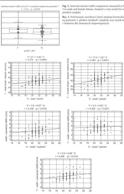
right limb in all fetuses (1.1 mm) were statistically non significant (P > 0.05). Sartorial muscle width measured at level III in females was bigger on av-erage by 0.5 mm and the difference is statistically significant (P < 0.05) (Fig. 5).
Other analyzed sizes differed significantly (p > 0.05) neither between left and right sides nor between males and females (Tab. 2).
well as left and right side results were collected. Table 3 presents the size variation analysis by age group (monthly). Correlation diagrams (Fig. 6) present these sizes’ linear regression analysis with respect to age, shown in weeks. Thigh and sarto-rial muscle growth is characteristic for their linear character. These lengths’ weekly increases amount to 0.8 and 1.2 mm. In turn, in the examined mate-rial, the muscle widths taken at levels I and III can be regarded as stable (1.8 and 3.3 mm). The muscle width at level 3 increases linearly 0.08 mm/week.
Discussion
The application of a method of preparation along with image acquisition and analysis with the use of computer programs enables the evaluation of anatomical structure geometry while avoiding au-topsical material damage. The available literature discussing sartorial muscles is not very detailed. It considers the musculus sartorius muscle’s sig-nificance in adult life, especially in reference to its clinical use in plastic and reconstructive surgery.
Fig. 1. Sartorial muscle. Defined sizes: A – width at primary attachment, B – width in the central part, C – width at the final attachment, D – muscle length, E – thigh length
Ryc. 1. Mięsień krawiecki, oznaczenia: A – szerokość
w części początkowej bMS1, B – szerokość w części
środkowej bMS2, C – szerokość w części końcowej bMS3,
D – długość mięśnia lMS, E – długość uda lF
Fig. 2. right thigh length comparison in male and
female fetuses, Student’s t-test results for independent
samples
Ryc. 2. Porównanie długości uda prawego płodów
żeńskich i męskich oraz wynik testu t-Studenta dla
zmiennych niepowiązanych
t = 2.108; p = 0.039
right thigh length / długość uda prawego [mm]
55 50 45 40 35 30 25
F
gender / płeć M
Fig. 3. left and right thigh length collective
compari-son in male and female fetuses and Student’s t-test
result for paired samples
Ryc. 3. Porównanie długości uda lewego i prawego
płodów żeńskich oraz wynik testu t-Studenta dla
zmiennych powiązanych
t = 2.154; p = 0.0347 thighs length / długość uda [mm]
60 55 50 45 40 35 30
L
side / strona R
Fig. 4. Sartorial muscle length (mm) comparison in
female fetuses left limb and Student’s t-test result for
independent samples
Ryc. 4. Porównanie długości (mm) mięśnia krawieckie-go lewekrawieckie-go płodów żeńskich i męskich oraz wynik testu
t-Studenta dla zmiennych powiązanych
t = 2.322; p = 0.0232
sartorial muscle length / długość mięśnia krawieckiego
65
55
45
35
25
F
Fig. 5. Sartorial muscle width comparison measured at level
3 in male and female fetuses, Student’s t-test result for
inde-pendent samples
Ryc. 5. Porównanie szerokości [mm] mięśnia krawieckiego na poziomie 3. płodów żeńskich i męskich oraz wynik testu
t-Studenta dla zmiennych niepowiązanych
t = 2.311; p = 0.0238
sartoriar muscle width on level 3 / szerokość mięśnia na poziomie 3
6
5
4
3
2
1
0
F
gender / płeć M
Fig. 6. Correlation diagrams and regression models of dependence between thigh length as well as sartorial muscle length and width and fetus age
Ryc. 6. Diagramy korelacyjne i modele regresyjne długości i szerokości mięśnia krawieckiego w zależności od wieku płodu
r = 0.279 (p = 0.0008)Y = 27.1 + 0,80 * X
14 16 18 20 22 24 26 28 X – week / tydzień
80 70 60 50 40 30 20
Y
– thigh lenght / długość uda [mm]
r = 0.362 (p < 0.0001)Y = 17.3 + 1,20 * X
14 16 18 20 22 24 26 28 X – week / tydzień
70
60
50
40
30 25
Y
–
muscle lenght / długość mięśnia [mm]
r = 0.150 (p = 0.074)Y = 1.8 + 0.07 * X
14 16 18 20 22 24 26 28 X – week / wydzień
8 7 6 5 4 3 2 1
width / szerokość bMS1 [mm]
r = 0.052 ( p = 0.537)Y = 3.3 + 0.02 * X
14 16 18 20 22 24 26 28 X – week / tydzień
7 6 5 4 3 2 1
Y
–
width / szerokość bMS2 [mm]
r = 0.208 (p = 0.014)Y = 0.9 + 0.08 * X
14 16 18 20 22 24 26 28 X – week / tydzień
6 5 4 3 2 1 0
Y
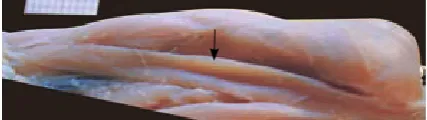
–
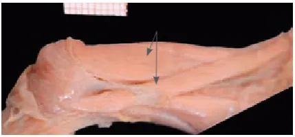
Two types of morphological varieties were observed: rectangular (classical type) (Fig. 7) and cone-shaped. Also, some anomalies were recog-nized sporadically and they included: the presence of an extra tendon (Fig. 8), the start of muscle du-plication in the form of a hollow (Fig. 9), partial duplication of the muscle distal fragment (Fig. 10) and ‘discontinuous’ muscle (Fig. 11). In sites of the first anomaly observation, the muscle belly was normally formed and the extra tendon branched in the inferior area at the muscle’s 1/3 length. In turn, partial bifurcation had started in the muscle’s median part and gave the beginning of two mus-cle bellies of the same size. That is why musculus sartorius had two distal attachments: the posterio-medial was an element of the “duckfoot shovel” whereas the anteriolateral part was parallel to the former one and adhered laterally. Duplication is described in the literature. M. g. El-Badawi [2] as well as M. Melling, K. Zweymueller [10] described the same type of bifurcation. In both cases, the de-scribed muscle lateral part was relatively smaller in comparison to the median one the course of which was typical for a sartorial muscle as it ended in the pes anserinus. In M.g. El-Badawi’s observations, the lateral belly’s attachment was situated in the femoral bone median condyle upper part, whereas in Melling’s and Zweymueller’s studies, it attached to the median meniscus. Available reports also
discuss cases of sartorial muscle proximal ending bifurcation (Bhatnagar and Narayan 3 anomalies of this type [2]) or this muscle’s absolute absence (Williams and Warwick [2]). Initial muscle bifur-cation in the form of a hollow (little pan) along the muscle’s long axis was another anomaly. In turn, discontinuous muscle consisted of three muscular parts and two connecting tendinous parts.
The literature provides numerous descriptions of musculus sartorius’ clinical use. Due to its rich vascularization and segmental structure, the mus-cle can be used in flap formation, widely applied in both reconstructive and plastic surgery in the groin, genual, cluneal and abdominal areas. Mus-culus sartorius flaps are used not only in vascular transplants covering the groin area and limiting infection danger or anastomosis breakdown, but
Table 3. Thigh and sartorial average muscle sizes in age groups, variation analysis result
Tabela 3. Średnie wartości wymiarów uda i mięśnia krawieckiego w grupach wiekowych i wynik analizy wariancji
Sizes (Wymiary) mm Month of fetal life
(Miesiąc życia płodowego) ANOVA
IV V VI VII
Thigh length (Długość uda) lF 45.5 ± 3.1 41.7 ± 6.5 45.1 ± 5.6 46.5 ± 5.4 P = 0.012 Sartorial muscle length (Długość mięśnia) lMS 38.1 ± 8.9 40.7 ± 7.8 43.8 ± 6.0 46.8 ± 8.0 P = 0.021 Muscle width (Szerokość mięśnia) bMS1 4.1 ± 0.9 3.1 ± 1.2 3.4 ± 0.9 4.3 ± 1.3 P = 0.016 Muscle width (Szerokość mięśnia) bMSI2 4.6 ± 0.7 3.6 ± 0.8 3.9 ± 0.9 3.5 ± 0.9 P = 0.048 Muscle width (Szerokość mięśnia) bMS3 2.9 ± 0.8 2.2 ± 0.6 2.7 ± 0.8 2.8 ± 0.8 P = 0.015
Fig. 7. Sartorial muscle (classical type) Ryc. 7. Mięsień krawiecki (typ klasyczny)
Fig. 8. Sartorial muscle with extra tendon present Ryc. 8. Mięsień krawiecki z dodatkowym ścięgnem
Fig. 9. Initial process of muscle bifurcation manifested by antrum presence
also in the treatment of already infected grafts. After lymphadenectomy procedures, necrotic ar-eas are filled with the flaps in order to cover the vessels and protect the healing process [9, 12–14]. In the genual area, the flaps are used in soft tissue reconstruction and knee rigidity treatment [1, 4]. Musculus sartorius flaps are also used in extensive reparative surgical procedures. In the case of the abdominal wall, such a defect may include the wall total thickness [15]. In the cluneal area, the flaps are used when musculus tensor fasciae latae flaps prove to be insufficient [3].
The authors concluded that statistically sig-nificant sexual dimorphism of thigh length and sartorial muscle length were observed (sizes longer by 2.7 mm and 4.3 mm respectively were found in female fetuses although no statistically significant difference was found in body lengths v-tub and v-pl). Thigh length significant asymmetry was de-tected (left side longer than the right by 1.1 mm on average). Thigh length and sartorial muscle length growth rates were stable and amounted to 0.8 mm/ /week and 1.2 mm/week respectively. However, IV and VII age groups were scarce.
Fig. 10. Muscle fragment partial duplication Ryc. 10. Częściowa duplikacja dystalnego fragmentu
mięśnia Fig. 11. Discontinuous muscleRyc. 11. Mięsień „przerywany”
References
[1] Chen QS, Zhu LX, Chen X: Application of sartorius muscle in the quadricepsplasty. Zhongguo Xiu Fu Chong Jian Wai Ke Za Zhi 1999 Nov, 13, 355–358.
[2] El-Badawi MG: An anomalous bifurcation of the sartorius muscle. Anat Anz 1987, 163, 79–82.
[3] Gu B, Fan QY, Lu YP, Goldenberg B: repair of a huge defect of the gluteal region by rotation of a combined ten-sor fasciae latae-sartorius myocutaneous flap. Plast reconstr Surg 1990, 86, 983–986.
[4] Hong JP, Lee HB, Chung YK, Kim SW, Tark KC: Coverage of difficult wounds around the knee joint with pre-fabricated, distally based sartorius muscle flaps. Ann Plast Surg 2003 May, 50, 484–490.
[5] Image J: Image Processing and Analysis in Java, http://rsbweb.nih.gov/ij/.
[6] Ivey M, Prud’homme J: Anatomic variations of the pes anserinus: a cadaver study. Orthopedics 1993 May, 16, 601–606.
[7] Kędzia A, Woźniak J, Ziajkiewicz M, Dudek K, Derkowski W: Ocena wieku płodu na podstawie wybranych para-metrów tułowia i głowy. Komputerowe wspomaganie badań naukowych: The computer-aided scientific research. XVI Kowban 2009, 241–246.
[8] Kędzia A, Ziajkiewicz M, Seredyn A, Dudek K: Computer morphometric analysis of the Palmaris longus muscle in fetal period. Adv Clin Exp Med 2009, 18, 5, 437–447.
[9] Laustsen J, Bille S, Christensen J: Transposition of the sartorius muscle in the treatment of infected vascular grafts in the groin. Eur J Vasc Surg 1988, 2, 111–113.
[10] Melling M, Zweymueller K: Musculus sartorius bicaudatus. Acta Anat (Basel) 1996, 155, 215–218.
[11] Mojallal A, Wong C, Shipkov C, Hocuoq C, Recchiuto J, Brown S, Rohrich RJ, Saint–Cyr M: redefining the vascular anatomy and clinical applications of the sartorius muscle and myocutaneous flap. Plast reconstr Surg 2011, 127, 1946–1957.
[12] Petrasek PF, Kalman PG, Martin RD: Sartorius myoplasty for deep groin wounds following vascular reconstruc-tion. Am J Surg 1990 Aug, 160, 175–178.
[13] Pu LL, Jahania MS, Mentzer RM Jr: Successful management of recalcitrant groin lymphorrhoea with the combi-nation of intraoperative lymphatic mapping and muscle flap. J Plast reconstr Aesthet Surg 2006, 59, 1363–1366. Epub 2006 May 11.
[14] Scher KS: Sartorius transposition to protect vascular grafts in the groin. Am Surg 1989 Mar, 55, 158–161. [15] Sensöz O, Ustüner TE, Taner OF: Use of a sartorius myofasciocutaneous flap for reconstruction of a large,
full-thickness abdominal wall defect. Ann Plast Surg 1990, 25, 193–196.
[17] Wysocki J, Krasuski P, Czubalski A: Vascularization of the sartorius muscle. Folia Morphol (Warsz) 1996, 55, 115–120.
[18] Yang D, Morris SF, Sigurdson L: The sartorius muscle: anatomic considerations for reconstructive surgeons. Surg radiol Anat 1998, 20, 307–310.
Address for correspondence:
Alicja Kędzia
Normal Anatomy Department Wroclaw Medical University Chałubińskiego 6a
50-368 Wrocław Poland
Conflict of interest: None declared