© 2016 Amel Amrani et al. This is an open access article distributed under the terms of the Creative Commons Attribution License -NonCommercial-ShareAlikeUnported License (http://creativecommons.org/licenses/by-nc-sa/3.0/).
Journal of Applied Pharmaceutical Science Vol. 6 (04), pp. 067-071, April, 2016 Available online at http://www.japsonline.com
DOI: 10.7324/JAPS.2016.60409 ISSN 2231-3354
Impact of Chrysanthemum fontanesii extract on sodium valproate
mediated oxidative damage in mice kidney
Amel Amrani
1,2*, Ouahiba Benaissa
1, Nassima Boubekri
1,2, Kenza Biod
2, Radja Djabari
2, Nawal Beroal
2, Djamila
Zama
1,2, Fadila Benayache
1, Samir Benayache
11
Unité de Recherche Valorisation des Ressources Naturelles, Molécules Bioactives, Analyses Physicochimiques et Biologiques (VARENBIOMOL), Université frères Mentouri Constantine, Route de Aïn El Bey, 25000 Constantine, Algérie. 2Departement de Biologie Animale; Faculté des SNV. Université frères Mentouri Constantine, Route de Aïn El Bey, 25000 Constantine, Algérie.
ARTICLE INFO ABSTRACT
Article history:
Received on: 04/09/2015 Revised on: 11/11/2015 Accepted on: 05/02/2016 Available online: 30/04/2016
The objective of this study was to evaluate the potential of n-butanol extract obtained from flowers of
Chrysanthemum fontanesii (Cf) and vitamin E against kidney oxidant injury induced by sodium valproate in
mice. The evaluation was made through Histopathological examination and measuring kidney lipid peroxidation (LPO) and antioxidant parameters: reduced glutathione (GSH), glutathione peroxidase (GPx) and catalase. Severe alterations in all biomarkers were observed after injury with VPA. Treatment with Cf extract and vitamin E resulted in markedly decreased levels of LPO while increased levels of GSH and antioxidant enzymes activities were observed. In conclusion, n-butanol extract of Cf may be an interesting candidate for the treatment of kidney injury induced by VPA through enhancing endogenous antioxidant defenses and normalizing the kidney histopathological architecture.
Key words: Valproic acid;
Chrysanthemum fontanesii;
kidney oxidant injury; Antioxidant; Renoprotective effect.
INTRODUCTION
Valproic acid (VPA) is an effective and widely used anticonvulsant, associated with metabolic adverse effects such as hepatotoxicity and nephrotoxicity (Gossrau and Graf, 1989; Raza
et al., 2000; Rahman et al., 2006). Although the exact
biochemical mechanisms of VPA toxicity to kidney have not been well defined, several hypothesis have been proposed. The recent hypothesis suggests an involvement of peroxidative injury
in VPA induced renal tubular disorder (Raza et al., 1997); free
radical scavengers like vitamin E supplements provided adequate
protection against VPA toxicity in kidney (Aktas et al., 2010).
Chrysanthemum (Asteraceae) is a large genus of about 300
species (Kumar et al., 2005) from which Chrysanthemum
fontanesii growing in algeria. A series of studies have
demonstrated that Chrysanthemum species possesses antioxidants
(Duh et al., 1999; Lin et al., 2010; Liu et al., 2012; Amrani et al.,
2013a), anticancer (Bi et al., 2008; Jin et al., 2009), antimicrobial
.
* Corresponding Author Email: [email protected]
(Shunying et al., 2005), anti-inflammatory (Lee et al., 2009; Kim
et al., 2012), immunomodulatory (Cheng et al., 2005),
Hepatoprotective (Amrani et al., 2013b) and neuroprotective
effects (Chun et al., 2008).Many chemical compounds have been
isolated and identified from species of the Chrysanthemum
(Benaissa et al., 2011, Lograda et al., 2013). Based on these effects
this study was designed to determine the possible protective effect
of the n-butanol extract obtained from flowers of Chrysanthemum
fontanesii against oxidative damage of kidneys induced by VPA.
MATERIAL AND METHODS
Plants
Chrysanthemum fontanesii which is endemic to North
Africa was collected during the flowering stage in the area of bijaia, north east of Algeria. Flowers (1516 g) were powdered and
macerated at room temp with EtOH–H2O (8:2 v/v) for 48 h three
times. The filtrates were combined, concentrated and successively
extracted with chloroform, ethyl acetate and n-butanol. The
organic layers were dried with Na2SO4. Removal of solvents under
reduced pressure, CHCl3 (7.20 g), EtOAc (30.50 g), n-butanol
Animals and treatment
Female Albino Swiss mice aging from 6-8 weeks were purchased from Pasteur Institut Algiers and were maintained on 12h light/dark cycle. The animals were kept in12h light/ dark cycles, maintained in an air-conditioned room at 22–25 °C, with
free access to food and water ad libitum. The general guidelines
for the use and care of living animals in scientific investigations were followed (Council of European Communities, 1986).
Mice were divided in to five groups (Gr) consisting of 8 animals each. All substances were given for 12 days.
1-Gr.1 served as control.
2-Gr.2 received VPA intraperitoneally (ip) at single dose of 300 mg/kg body weight for 12 days.
3-Gr.3 received plant extract (100mg/kg by gavage). 4-Gr.4 received plant extract (100mg/kg) prior to VPA administration.
5-Gr.5 received vitamin E orally (100mg/kg) prior to VPA administration.
After 12 days of study, all mice (eight animals in each of five groups) were killed. Kidneys were removed, weighed, and homogenized in 5 ml of ice-cold KCl 1.55%. Homogenates (10%) were centrifuged at 3000 rpm for 15 min at 4°C, and the supernatant was kept on ice until assayed.
Determination of kidney lipid peroxidation
Lipid peroxidation, an indicator of tissue injury induced by reactive oxygen species will be measured as thiobarbituric acid reactive substance (TBARS). The amount of kidney TBARS will be measured by the thiobarbituric acid assay (TBA) as previously described by (Uchiyama and Mihara, 1978). Briefly, 0.5 ml of kidney homogenate will be reacted with 1 ml of TBA reagent containing 0.67% TBA and 3 ml of 1% phosphoric acid. Samples will be boiled for 45 minutes, cooled and centrifuged. Absorbance of the supernatants will be spectrophotometrically measured at 532 nm. TBARS concentrations will be calculated by the use of 1,3,3,3 tetra-ethoxypropane as a standard. The results will be expressed as nmol/g tissue weight.
Measurement of glutathione
Reduced glutathione (GSH) content in the kidney was measured according to the method described by (Ellman, 1959) using Ellman's reagent. This method is based on the reactive cleavage of 5, 5′-dithiobis-(2-nitrobenzoic acid) by sulfohydryl group to yield a yellow colour with maximum absorbance at 412 nm against reagent blank with no homogenate.
Evaluation of GPx activity
GPx activity in the kidney was measured according to the
method described by (Flohe and Gunzler, 1984).Based on H2O2
reduction in the medium by GPx in the presence of GSH. Briefly 0.2ml supernatant obtained from kidneys, 0.4ml GSH (0.1 mM), 0.2ml TBS solution (Tris 50mM, NaCl 150mM PH 7.4) were added to the tubes and mixed. After 5 min incubation at 25 °C, 0.2
ml of HO (1.3mM) was added in the mixture. The reaction was
stopped after 10 min by addition of 1ml trichloroacetic acid (TCA 1 %, w/v), and then the tubes maintained at 0-5°C in an ice bath for 30min, centrifuged for 10 min at 3000 rpm. After this, 0.48ml supernatant was taken and added to each tube, and then 2.2 ml TBS solution and 0.32 ml DTNB (1mM) were added. The optical density was measured at 412 nm in the spectrophotometer after 5 min.
Evaluation of catalase activity
The enzymatic activity of catalase was measured by
determining the decomposition of its substrate H2O2as described
by(Claiborne,1985).The homogenate was centrifuged at 10000 rpm for 45 min at 4° C; the final supernatant is the source used for the evaluation of the activity of catalase. The disappearance of
H2O2 was determined spectrophotometrically at 240 nm. Catalase
activity was expressed as U/mg of protein,
In order to express the antioxidant enzyme (GPx, catalase) activities per gram of protein, total protein concentration was determined colorimetrically by using the method of(Lowry, 1951).
Histopathological examination
Kidney tissue fragments were taken and fixed in formalin 10 %. The fixed specimens were then trimmed, washed and dehydrated. These specimens were then embedded in paraffin, cut into 5μm thick sections and stained with Harris hematoxylin and eosin and examined microscopically.
Statistical analysis
Data are expressed as the mean ± SD. Statistical interferences were based on student's t-test for mean values comparing control and treated animals.
RESULTS
Effect of VPA, plant extract and vitamin E on LPO, GSH levels and GPx activityin mice kidney
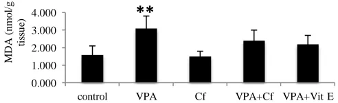
Treated mice with VPA have developed kidney damage as was evident from the significant increase (P<0.01) of LPO,
which is reversed in animals treated with both Cf and vitamin E
(Fig.1). VPA were significantly decline in the activities of GPx(P<0.01), catalase and GSH level (P<0.001). The treatment
with Cf/ or Vit E showed significant improvement in the
antioxidant elements (Fig. 2, 3, 4).
Fig. 1: Effect of VPA, plant extract and vitamin E on lipid peroxidation (TBARs content) in kidney Data are reported as means ± SD. Treatment groups were statistically compared to control group. (Students t-test, **P<0.01)
0.000 1.000 2.000 3.000 4.000
control VPA Cf VPA+Cf VPA+Vit E
M
D
A
(n
m
o
l/
g
ti
ss
u
Histopathological examination
In the slides of kidney sections of the control group stained with Hematoxylene-Eosine, the appearance of the kidney was histologically normal (figure 5A). In kidney sections of valproic acid group; necrotic areas, inflammatory cell infiltrates, oedema and congestion were observed (fig. 5 B, C). The kidney
.
section of plant extract treated mice, showed a histopathological picture that closely approximate that of the control group(figure 5D). In the groups of VPA+ Vitamin E and butanolic extract +VPA, sections belonging to these groups showed relatively normal ultra-structure when compared to VPA group. We noted some minor changes like Congestion (figure 5 E, F).
Fig. 2: Effect of VPA, plant extract and vitamin E on GSH levels in mice kidney after. 12 days of treatment. Data are reported as means ±
SD.Treatment groups were statistically compared to control group(Students t-test, ***P<0.001).
Fig. 3: Effect of VPA, plant extract and vitamin E on the antioxidant enzyme (GPx). activitiesin mice kidney after 12 days treatment. Data are reported as means ± SD., Treatment groups were statistically compared to control group. (Students t-test, **P<0.01).
Fig. 4: Effect of VPA, plant extract and vitamin E on the antioxidant enzyme (Catalase) activities in mice kidney after 12 days treatment. Data are reported as means ± SD. Treatment groups were statistically compared to control group. (Students t-test, ***P<0.001). Fig. 1, 2, 3 and 4: Control; VPA, treated with 300 mg/kg valproic acid; VPA + Cf, treated with 300 mg/kg valproic acid and plant extract of Chrysanthemum fontanesii100 mg/kg ; VPA+Vit E, treated with 300 mg/kg valproic acid and 100mg/kg vitamin E ; VPA intraperitoneal injection. Plant extract and vitamin E were given by gavage. *: Significant p<0.05, **: Highly significant p<0.01***: Very highly significant p<0.001.
A B C
D E F
Fig. 5: Histopathologic changes affecting the kidneys of Swiss albino mice. (A)Normal kidney of the control group; (B) and(C)kidneys of VPA treated group showed necrosis, inflammatory cell infiltrates and oedema respectively; (D)kidney of Cf group showed a histopathological picture that closely approximate that of the control group; (E) and (F)kidneys of VPA+ Vitamin E and VPA+ Plant extract treated groups respectively showed congestion.
0 2 4 6 8
Control VPA Cf Cf+VPA Vit E+VPA
n
m
o
l/
g
k
id
n
ey
***
0 0.05 0.1 0.15
Control VPA Cf Cf+VPA Vit E+VPA
m
m
o
l
G
S
H
/m
g
p
ro
te
in
k
id
n
ey
**
0.00 0.20 0.40 0.60 0.80
Control VPA Cf VPA+Cf VPA+Vit E
U
/m
g
p
r
o
te
in
DISCUSSION
Results of the present study show that catalase, GPx activities and GSH level were significantly decreased in the VPA treated mice. Several studies have reported that reactive oxygen species (ROS) are considered to be important mediators of VPA
induced kidney injury (Raza et al, 1997). The increased production
of ROS causes inactivation of antioxidant enzymes. Normally GSH contributed to the intracellular antioxidant defense system as it is powerful consumer of free radicals. Glutathione (GSH) is one of the most abundant tripeptide, non-enzymatic biological antioxidant present in tissues. It removes free radical species such as hydrogen peroxide, superoxide radicals and maintains membrane protein thiols. Also it is a substrate for glutathione peroxidase (GPx) and detoxifies foreign compounds and biotransformation drugs.
CAT widely distributed in all animal tissues which decomposes hydrogen peroxide and protects the tissues from highly reactive hydroxyl radicals (Mates and Sanddiez-jimenez, 1999). Therefore, reduction in the activity of these enzymes may result in a number of alterations do to the accumulation of
superoxide radicals and H2O2 (Halliwell, 1994). The suppression
of MDA content and the over expression of the antioxidant
enzymes in mice treated with Cf implies that this potential
antioxidant defense is reactivated by the plant extract of Cf with a
resulting increase in the capacity of detoxification through enhanced scavenging of free radicals and acted by up regulating endogenous antioxidant defenses.
Most of these effects are considered to be due to the presence of wide range of well defined phytochemical phenolic compounds and flavonoids which have been known to be
responsible of health promoting (Benaissa et al., 2011, Amrani et
al., 2013b; 2014).
CONCLUSION
The present study revealed that the oral administration of
n-butanol extract of Chrysanthemum fontanesii to valproic acid
intoxicated mice exhibited significant nephroprotective effect.
Hence, we suggest that Chrysanthemum fontanesii may be
developed as successful chemotherapeutic agents.
REFERENCES
Aktas A, Nergız Y, Akkus M, Nasır Y. The effects of valproic acid on renal corpuscle of pregnant rats and protective role of folic acid and vitamin E. Afr J Biotechnol, 2010; 9(34): 5605-5610.
Amrani A, Zama D, Boubekri N, Benaissa O, Meraihi Z, Benayache F, Benayache S, Bettuzzi S. The protective effect of
Chrysanthemum fontanesii extract, vitamin E and C on sodium
valproate-induced embryotoxicity in pregnant mice. J Med PlantsRes, 2012; 6: 3535-3544.
Amrani A, Boubekri N, Benaissa O, Zama D, Benayache F, Benayache Sa. In vitro antioxidant, antibacterial and membrane stabilizing activity of plant extract from Chrysanthemum fontanesii. International journal of phytomedcine, 2013; 5(4): 493-498.
Amrani A, BenaissaO, BoubekriN, ZamaD, BenayacheF, BenayacheSb. Valproic acid induced liver toxicity and oxidative damage in pregnant mice: The protective effect of butanolic extract from flowers
of Chrysanthemum fontanesii. Ann Biol Res, 2013; 4(12): 6-14.
Amrani A, BoubekriN, BenaissaO, ZamaD, BenayacheF, BenayacheS. Ethanol induced toxicity and lipid peroxidation in pregnant mice: Protective effects of butanolic extract from leaves of
Chrysanthemum fontanesii, vitamin E and C. J. stress physiol. biochem,
2014; 10(2): 35-43.
Benaissa O, Bentamene A, Bicha S, Benayache F, Brouard I, Garcia VP, Leon F, Bermejo J., Benayache S. Flavonoid aglycones and sterol from Chrysanthemum fontanesii. Chem. Nat. Comp, 2011; 47 (Issue 1): 107-108
Cheng W, Li J, You T, Hu C. Anti-inflammatory and immunomodulatory activities of the extracts from the inflorescence of
Chrysanthemum indicum Linne. J Ethnopharmacol, 2005; 101:334–337.
Chun HS, Kim JM, Choi EH, Chang N. Neuroprotective effects of several korean medicinal plants traditionally used for stroke remedy. J Med Food, 2008; 11:246–251.
Claiborne A. Catalase activity In CRC Handbook of Methods for Oxygen Radical Research, ed. Greenwald RA, 1985; pp. 283–284.
DuhP-D, Tu Y-Y, Yen G-C. Antioxidant activity of water extract of harngjyur (Chrysanthemum morifolium Ramat).LWT-Food SciTechnol, 1999; 32(Issue 5): 269–277
Ellman GL. Plasma antioxidants. Arch. Biochem Biophyis, 1959; 82: 70-77.
Flohe L, Gunzler WA. Analysis of glutathione peroxidase, Methods Enzymol 1984; 105: 114-121.
Gossrau R, Graf R. Lesions and repair of cells of maternal mice after valproic acid treatment on day 8 pregnancy: an enzyme histochemical analysis. Acta Histochem, 1989; 86(1): 23–32.
Halliwell B. Free radicals, antioxidants, and human disease: curiosity, cause, or consequence? Lancet,1994; 344 (8924): 721–724.
Jin SR, Zhu BD, Qin XH. The effect of Chrysanthemum
indicum on SMMC7721, PC3 and HL60 cell lines. ZhongyaoYaoli Yu
Linchuang, 2005; 21:39–41.
Kim Y, Han J, Sung J, Sung M, Choi Y, Jeong H-S, Lee, J Anti-inflammatory activity of Chrysanthemum zawadskii var. latilobum leaf extract through haem oxygenase-1 induction. Journal of Functional Foods, 2012; 4 (Issue 2): 474–479
Kumar A, Singh SP,Bhakuni R. S. Secondary metabolites of
Chrysanthemum genus and their biological activities. Curr.Sci, 2005; 89
(9):1489-1501
Lee Y, Choi G, Yoon T, Cheon MS, Choo BK, Kim HK. Anti-inflammatory activity of Chrysanthemum indicum extract in acute and chronic cutaneous inflammation. J Ethnopharmacol, 2009; 123:149–154.
Li ZF, Wang ZD, Ji YY, Zhang S, Huang C, LiJ, and Xia X-M.Induction of apoptosis and cell cycle arrest in human HCC MHCC97H cells with Chrysanthemum indicum extract. World J Gastroenterol, 2009; 15(36): 4538–4546.
Lin GH, Lin L, Liang HW, Ma X, Wang JY, Wu LP, Jiang HD, Bruce IC, Xia Q.Antioxidant action of a Chrysanthemum morifolium
extract protects rat brain against ischemia and reperfusion injury.J Med Food, 2010; 13(2):306-11.
Liu Q, Liu H, Yuan Z, Wei D, Ye Y.Evaluation of antioxidant activity of Chrysanthemum extracts and tea beverages by gold nanoparticles-based assay.Colloids Surf B.Biointerfaces, 2012; 92:348-52. Lograda T, Ramdani M, Chalard P, Figueredo G, Silini H and Kenoufi M. Chemical composition, antibacterial activity and chromosome number of Algerian populations of two chrysanthemum species. J App Pharm Sci, 2013; 3 (8Suppl 1): S6-S11.
Lowry OH, Rosegrough NJ, Farr AL, Randall RJ.Protein measurement with the folin phenol reagent.J. Biol. Chem, 1951; 193: 265-275.
Rahman MH, Haqqie SS, McGoldrick MD.Acute hemolysis with acute renal failure in a patient with valproic acid poisoning treated with charcoal hemoperfusion. HemodialInt, 2006; 10(3):256-9.
Raza M, Al-Bekaire AM, Ageel AM, Qureshi S. Biochemical basis of sodium valproate hepatotoxicity and renal tubular, time dependence of peroxidative injury. Pharmacol Res, 1997; 35(2): 153-7.
Raza M, Al-shabanah OA, Al-bekairi AM,Qureshi S. Pathomorphological changes in mouse liver and kidney during prolonged valproate administration.Int J. Tissues React, 2000; 22(1):15-21.
Shunying Z, Yang Y, Huaidong Y, Yue Y, Guolin Z. Chemical composition and antimicrobial activity of the essential oils of Chrysanthemum indicum. J Ethnopharmacol, 2005; 96:151–158
Uchiyama M, Mihara M.Determination of malonaldehyde precurssor in tissues by thiobarbituric acid test. Anal. Biochem, 1978; 86: 271-278.
How to cite this article:
Amrani A, Benaissa O, Boubekri N, Biod K, Djebari R, Beroal N, Zama D, Benayache F, Benayache S. Impact of Chrysanthemum
fontanesii extract on sodium valproate mediated oxidative damage