R E S E A R C H
Open Access
Lack of transient receptor potential vanilloid 1
channel modulates the development of
neurogenic bladder dysfunction induced by
cross-sensitization in afferent pathways
Qi Lei
1, Xiao-Qing Pan
1, Antonio N Villamor
1, Tirsit S Asfaw
2, Shaohua Chang
3, Steven A Zderic
4and Anna P Malykhina
1*Abstract
Background:Bladder pain of unknown etiology has been associated with co-morbid conditions and functional abnormalities in neighboring pelvic organs. Mechanisms underlying pain co-morbidities include cross-sensitization, which occurs predominantly via convergent neural pathways connecting distinct pelvic organs. Our previous results showed that colonic inflammation caused detrusor instability via activation of transient receptor potential vanilloid 1 (TRPV1) signaling pathways, therefore, we aimed to determine whether neurogenic bladder dysfunction can develop in the absence of TRPV1 receptors.
Methods:Adult male C57BL/6 wild-type (WT) and TRPV1−/−(knockout) mice were used in this study. Colonic inflammation was induced by intracolonic trinitrobenzene sulfonic acid (TNBS). The effects of transient colitis on abdominal sensitivity and function of the urinary bladder were evaluated by cystometry, contractility and relaxation of detrusor smooth muscle (DSM)in vitroto various stimuli, gene and protein expression of voltage-gated sodium channels in bladder sensory neurons, and pelvic responses to mechanical stimulation.
Results:Knockout of TRPV1 gene did not eliminate the development of cross-sensitization between the colon and urinary bladder. However, TRPV1−/−mice had prolonged intermicturition interval and increased number of
non-voiding contractions at baseline followed by reduced urodynamic responses during active colitis. Contractility of DSM was up-regulated in response to KCl in TRPV1−/−mice with inflamed colon. Application of Rho-kinase inhibitor caused relaxation of DSM in WT but not in TRPV1−/−mice during colonic inflammation. TRPV1−/−mice demonstrated blunted effects of TNBS-induced colitis on expression and function of voltage-gated sodium channels in bladder sensory neurons, and delayed development of abdominal hypersensitivity upon colon-bladder cross-talk in genetically modified animals.
Conclusions:The lack of TRPV1 receptors does not eliminate the development of cross-sensitization in the pelvis. However, the function of the urinary bladder significantly differs between WT and TRPV−/−mice especially upon development of colon-bladder cross-sensitization induced by transient colitis. Our results suggest that TRPV1 pathways may participate in the development of chronic pelvic pain co-morbidities in humans.
Keywords:Chronic pelvic pain, Bladder sensory neurons, Neurogenic bladder, Detrusor contractility
* Correspondence:[email protected] 1
Department of Surgery, Division of Urology, University of Pennsylvania, 500 South Ridgeway Avenue, Glenolden, PA 19036, USA
Full list of author information is available at the end of the article
Background
Chronic pelvic pain (CPP) is a common symptom of many urologic and gastrointestinal disorders, including interstitial cystitis/bladder pain syndrome (IC/BPS), irritable bowel syndrome (IBS), and non-bacterial prostatitis/chronic pelvic pain syndrome. A high level of co-morbidities among CPP disorders is well documented in the clinical setting [1-3]. The etiology of IC/BPS is often complicated by visceral hypersensitivity arising from the gastrointestinal (GI) tract [1,4]. Similarly, a significant number of patients with IBS complain of urinary symptoms, including nocturia [5], frequency and urgency of micturition [2] and incomplete bladder emptying [6]. Viscero-visceral reflexes between the lower GI and urinary tracts are controlled by both autonomic and central nervous systems (CNS), suggesting the dominant role of neural pathways in pelvic organ co-morbidities.
The latest research efforts aimed at understanding the mechanisms underlying complex CPP disorders provided evidence that cross-sensitization in afferent pathways may initiate the development of neurogenic inflammation in the pelvis and functional chronic pelvic pain [7,8]. Cross-sensitization among pelvic organs implies the transmission of noxious stimuli from a diseased pelvic organ to an adja-cent normal structure resulting in the occurrence of func-tional changes in the latter [9]. A pathological condition developed in one of the pelvic organs may cause initial sensitization of peripheral afferent fibers and sensory neu-rons. These primary changes then lead to amplification of nociceptive signaling in the CNS followed by descending modulatory input from the CNS to the periphery [8-10].
The use of animal models provides insight into investi-gation of functional changes in nerve fibers and neurons as research of these in humans has certain challenges. Of major interest for the studies of functional co-morbidities are animal models in which an initial acute stimulus (inflammation, infection, noxious distension, trauma, and so on) is transient but powerful enough to cause long-lasting abdominal hypersensitivity and hyperexcitability of visceral afferents [11-13]. Several independent investiga-tions established that transient inflammation of the distal colon in animal models induces the occurrence of neuro-genic cystitis due to cross-sensitization in neural pathways [14-19]. After recovery from transient colitis, neither the colon nor the urinary bladder demonstrated detectable histological/biochemical changes suggestive of active in-flammation. However, the bladder develops many signs of neurogenic dysfunction evaluated by cystometry [14], hyperexcitability of bladder sensory [13,16] and spinal [20] neurons, release of pro-inflammatory neuropeptides in the bladder [21], and altered detrusor contractility [14,22].
Recent functional and molecular studies from our labora-tory identified the changes in a number of nociception-related genes and an increased release of pro-inflammatory
neuropeptides in the urinary bladder following transient inflammation of the distal gut [16,21]. We also established that these changes are associated with activation of intracolonic transient receptor potential vanilloid 1 (TRPV1) receptors [21]. TRPV1 is a non-specific cation channel activated by heat, protons, and vanilloids [23]. It is expressed in many visceral organs including the colon and urinary bladder [24,25] with the highest level of expression in primary sensory neurons innervating the visceral and somatic structures [23]. A growing body of evidence has led to the emergence of TRPV1 as a key player in sensory transduction. The role of TRPV1 in somatic pain has been extensively studied during the last decade, however, much less is known about the involvement of TRPV1 in viscero-visceral hyperalgesia and chronic viscero-visceral pain. To address the role of TRPV1 in nociceptive signal transmission from the inflamed colon to the urinary bladder and development of neurogenic bladder dysfunction, we evaluated the effects of TRPV1 gene knockout on the function of the urinary bladder, detrusor contractility and associated signaling
in vivo and in vitro in a model of colon-bladder cross-sensitization using TRPV1 knockout mice.
Materials and methods Animals and experimental groups
Adult male C57BL/6 wild-type (WT) and TRPV1 gene-deleted (TRPV1−/−, KO) mice (10 to 12 wks, 20 to 25 g, Jackson Laboratories, Bar Harbor, ME, USA; N = 56) were used in this study. Animals were housed in a regulated environment, with free access to food and water and main-tained on a 12:12-h light/dark cycle. There were no overt differences in feeding behavior, litter size, growth rate and body weight between the WT and TRPV1−/− mice. Animals were divided into four experimental groups: 1) WT control group; 2) WT group with colonic inflamma-tion; 3) TRPV1−/− control group; and 4) TRPV1−/− mice with colonic inflammation. Animals from each group were used forin vivoandin vitroexperiments at 3 to 5 days after intracolonic treatments. All protocols were approved by the University of Pennsylvania Institutional Animal Care and Use Committee and adhered to the guidelines for experimental pain in animals published by the International Association for the Study of Pain.
Animal model of colon-bladder cross-sensitization induced by colonic inflammation
Transient colonic inflammation was induced by a single administration of 2,4,6-trinitrobenzene sulfonic acid (TNBS, 60 mg/kg, in 25% C2H5OH), a chemical irritant
syringe. Mice received 0.3 ml of either vehicle (25% ethanol, control groups) or TNBS (inflammation groups) solution. To assess the severity of induced colonic inflamma-tion in vivo, the daily disease activity index (DAI) was calculated by scoring changes in animal weight, occult blood positivity, gross bleeding, and stool consistency as previously described [16].
Histological and biochemical evaluation of inflammation in vitro
Segments of the colon and the entire urinary bladder were isolated from WT and TRPV1−/− mice at the end of physiological experiments. One part of each tissue was fixed in 4% paraformaldehyde for histological evaluation. Fixed samples of the colon and urinary bladder were em-bedded in paraffin and sectioned at 10μm thickness. The samples were stained with hematoxylin and eosin (H&E staining kit, Richard-Allan Scientific, Kalamazoo, MI, USA) and assessed for the signs of inflammation under a light microscope. Another part of each specimen was snap-frozen in liquid nitrogen and stored at −80°C for running a myeloperoxidase (MPO) assay using MPO ELISA kit (Alpco, Salem, NH, USA) as previously described [16].
Surgical procedure to catheterize the urinary bladder for cystometry
Mice included in the groups for urodynamic evaluation of the urinary bladder function (awake cystometry) underwent survival surgical procedure for catheter inser-tion. An animal was anesthetized with isoflurane (VEDCO, St. Joseph, MO, USA), and a polytetrafluoro-ethylene catheter with a blunted end (Catamount Research, St. Albans, VT, USA) was sutured in place at the bladder dome and tunneled out the abdomen to the nape of the neck where it was then inserted into the end of a 22-gauge angiocath intravenous catheter. Upon determination of the optimal length, the catheter was affixed to the angiocath with super glue. The angiocath was first tested with a gentle saline infusion to reveal no leak at the bladder, then capped and the abdomen was closed in layers. The angiocath was anchored to the fascia and skin of the neck using two to three 3–0 Vicryl sutures. After recovery from anesthesia, animals were returned to the animal facility and kept in individual cages to avoid possible damage to the catheters by their cage mates. Mice were allowed to recover from surgery for 4 days followed by cystometric evaluation of bladder function under normal physiological conditions without anesthesia (baseline cystometry). After initial urody-namic evaluation, mice received a single intracolonic instillation of either vehicle or TNBS, as described above, followed by repeated cystometric evaluation at 3 to 5 days post-treatment.
Assessment of urodynamic parameters of bladder function
Conscious mice were placed in cystometry cages (16 cm width, 12 cm height, and 24 cm length) without any restraint and allowed to acclimate for 30 min. The tip of the exteriorized bladder catheter located at the base of the mouse neck was connected to a pressure transducer and an infusion pump of the cystometry station (Small Animal Laboratory Cystometry, Catamount Research and Development, St. Albans, VT, USA) using a T-shaped valve. Room temperature saline solution (0.9% NaCl) was infused in the bladder at a rate of 10 μl/min. Voided urine was collected in the tray connected to a force displacement transducer integrated into the data acquisition system. Each animal was observed for up to six to eight voiding cycles. Urodynamic values were recorded continuously using data acquisition software (Small Animal Laboratory Cystometry, Catamount Research and Development, St. Albans, VT, USA). The following cystometric parameters were recorded and analyzed in this study: bladder capacity, pressure at the start of micturition, micturition rate, intra-vesical pressure, inter-micturition interval, and the number of non-micturition contractions. Non-micturition contrac-tions were defined as increased values in detrusor pressure from baseline that had amplitudes of at least a third of maximal pressure at the start of micturition. Cystometric parameters were uploaded from the acquisition software into analysis software (SOF-552 Cystometry Data Analysis, Version 1.4, Catamount Research and Development Inc., St. Albans, VT, USA) for statistical analysis.
In vitromeasurements of detrusor contractility
For in vitro recordings of detrusor contractility, animals were euthanized with overdose of sodium pentobarbital (150 mg/kg). Midline laparotomy was performed to remove the urinary bladder, which was divided in two halves longi-tudinally and weighed. Full-thickness strips of the bladder wall were tied to silk threads and suspended from L-shaped hooks in 15-ml organ bath chambers. The chambers were filled with the Tyrode buffer (in mM: 125 NaCl, 2.7 KCl, 23.8 NaHCO3, 0.5 MgCl2·6H2O, 0.4 NaH2PO4·H2O, 1.8
CaCl2, and 5.5 dextrose), maintained at 37°C, and perfused
continuously with a mixture of 95% O2and 5% CO2. After
a 30-min equilibration period, the length of optimal force development (L0) was determined by increasing the length
drugs. One strip from each bladder was stimulated with muscarinic receptor agonist carbachol (CCh) whereas the second strip underwent stimulation with a protein kinase C (PKC) activator followed by a Rho kinase (ROK) inhibi-tor. Cumulative doses of CCh (10−7to 10−4M) were used to trigger muscle contractions and assess the contractile response to muscarinic receptor activation. Phorbol-12,13-dibiturate (PDBU, 1 μM) was used as a PKC activator followed by application of Y27632 (20μM), a ROK inhibi-tor. Additional groups of mice (WT and TRPV1−/−) were used to evaluate the effects of ROK inhibitor on detrusor contractility induced by KCl. Contraction parameters were measured using PowerLab Lab-Chart version 7.1.2 software (ADinstruments, Colorado Springs, CO, USA). The raw traces were analyzed manually and then exported into SigmaPlot 11 Software (Systat Software, San Jose, CA, USA).
Surgical procedure for labeling urinary bladder sensory neurons
Mice were anesthetized with 2% isoflurane and held on a warming pad inside the designated hood to minimize an investigator`s exposure to the anesthetic. A midline laparotomy was performed under the sterile conditions to gain access to the pelvic organs. The distal colon was exposed and DiI (1,1'-dioctadecyl-3,3,3'3'-tetramethylin-docarbocyanine perchlorate; Molecular Probes, Eugene, OR, USA; 1.5% w/v in methanol) was injected into the colonic wall using a Hamilton syringe with 26 gauge needle at six to ten sites. The colon was placed back into the abdominal cavity and the urinary bladder was exposed for injections. Fast Blue (Polysciences Inc., Warrington, PA, USA; 1.5% w/v in water) was injected into the urinary bladder wall (detrusor) using the same approach as described for the colon. We intentionally performed double labeling to exclude cells receiving dual afferent input from the distal colon and urinary bladder as these convergent neurons would be directly affected by colonic treatments [13]. The total volume of dye injected into each organ was 20 to 25μl. Adjacent pelvic organs were isolated with gauze to soak up any spills and prevent the labeling of adjacent structures during dye injections. Additionally, the needle was kept in place for 30 s after each injection. Any leaked dye was removed with a cotton swab before placing the organ into the pelvic cavity. Incisions were sutured in layers under the sterile conditions followed by subcutaneous injection of buprenorphine (0.5 mg/kg). Animals were allowed to recover on a warm blanket until they gained full consciousness and then were returned to their cages. Mice underwent subsequent treatments with either vehicle or TNBS 7 to 10 days after the labeling of dorsal root ganglion (DRG) neurons.
Isolation of single sensory neurons for patch-clamp experiments
Dorsal root ganglia were dissected and removed bilaterally at L6-S2 levels. Tissues were treated with collagenase (Worthington, type 2, Biochemical Corp., Lakewood, NJ, USA) in F-12 medium (Invitrogen, Carlsbad, CA, USA) for 90 min in an incubator with 95% O2and 5% CO2at
37°C. Isolated ganglia were then rinsed in phosphate-buffered saline and incubated for 15 min in the presence of trypsin (Sigma-Aldrich, St. Louis, MO, USA; 1 mg/ml) at room temperature. The enzymatic reaction was termi-nated in Dulbecco’s modified Eagle’s medium (DMEM) containing 10% of fetal bovine serum. Single neurons were obtained by gentle trituration using fire-polished Pasteur pipettes in DMEM with trypsin inhibitor (2 mg/ml; Sigma-Aldrich, St. Louis, MO, USA) and deoxyribonuclease (DNase 1 mg/ml; Sigma-Aldrich, St. Louis, MO, USA). The cell suspension was centrifuged for 10 mins at 700 rpm (4°C), and supernatant was discarded. The pellet, containing sensory neurons, was resuspended in 2 ml of DMEM containing 10% of fetal bovine serum. Neurons were plated on poly-L-ornithine-coated 35 mm Petri dishes. Isolated cells were maintained overnight in an incuba-tor at 37°C with 95% O2/5% CO2 and were used for
electrophysiological experiments within 24 hours.
Electrophysiological recordings of voltage gated Na+ currents from bladder DRG neurons
Bladder-labeled neurons were identified using specific filter for Fast Blue (UV-2A, Nikon, Tokyo, Japan) under an inverted fluorescent microscope (Ti E2000-5, Nikon). Only neurons exhibiting bright blue fluorescence (Fast Blue labeled) were used for Na+current recordings using perfo-rated whole-cell patch clamp technique. Neurons with red (colon projecting) and pink (convergent colon-bladder neurons) fluorescence were excluded from the study. For voltage clamp experiments the external solution contained (in mM): NaCl 45, TEA Chloride 30, Choline Chloride 60, KCl 5.4, MgCl2 1, CaCl2 1, HEPES 5, D-glucose 5.5,
adjusted with NaOH to pH 7.4. Pipette solution for these experiments consisted of (in mM): L-aspartic acid 100, CsCl 30, MgCl22, Na-ATP 5, EGTA 5, HEPES 5 adjusted
series resistance resulted in a maximum voltage error >5 mV or if the seal or access resistance were unstable. Recordings and analysis of kinetic parameters of voltage-gated Na+ channels (VGSC) were performed using previ-ously established protocols described in [16]. pCLAMP software (Axon Instruments, Union City, CA, USA) was used for data acquisition and analysis.
Gene expression of voltage gated Na+channels in lumbosacral DRG
A separate set of WT (N = 5) and TRPV1−/−mice (N = 5) was used for RNA isolation and real-time PCR analysis of gene expression of VGSC including Nav1.7, Nav1.8, and Nav1.9 members. These channels were selected due to their exclusive expression in sensory ganglia and estab-lished participation in nociceptive signaling [26,27]. L6-S2 sensory ganglia were isolated bilaterally and snap-frozen in liquid nitrogen. Total RNA was extracted using Trizol reagent following the protocol from Invitrogen (Carlsbad, CA, USA) as previously described [21]. First strand cDNA was synthesized from 2μg of the total RNA with 200 U of the superscript III reverse transcriptase (Invitrogen, number 18080–051) in the presence of 40 U RNaseOUT, 10 mM DTT, dNTP mix at 10 mM and 50 μM of Oligo (dT)20. Real-time PCR was run on 7500 Fast Real Time
PCR system (Applied Biosystems, Foster City, CA, USA). TaqMan Gene Expression Master Mix (4369016-PEC) and TaqMan™ primer/probe kits were used for mouse Nav1.7
(Mm00450762-s1), Nav1.8 (Mm00501467-m1), and
Nav1.9 (Mm00449377-m1) channels (all from Applied Bio-systems, Carlsbad, CA, USA). Glyceraldehyde-3-phosphate dehydrogenase (GAPDH) gene served as an endogenous control for the quantification of gene expression. The data were analyzed using comparative Ct values as previously described [21]. For example, the Ct value for each gene of interest (Nav1.7, Nav1.8, or Nav1.9) in the control group was subtracted from that of GAPDH (housekeeping gene) to obtain theΔCt value. The same subtraction was done in all treated groups to obtain the ΔCt. To compare the changes in the expression levels of Nav1.7, Nav1.8, and Nav1.9 genes between control and experimental tissues, these twoΔCt values were subtracted to obtain theΔΔCt. The fold change was measured as 2−ΔΔCt.
Assessment of abdominal sensitivity using von Frey filaments
Inflammation in the pelvic viscera is associated with enhanced abdominal sensitivity due to convergence of visceral and somatic inputs in the CNS. This phenomenon is known as viscerosomatic referred hyperalgesia and could be measured by using mechanical stimulation with von Frey filaments on the lower abdominal area [28]. In order to follow the dynamic changes in pelvic sensitivity of mice during the occurrence of experimental colitis, we
tested animals right before the TNBS instillation (day 0 or baseline) followed by daily measurements until the peak of colonic inflammation developed (3 days post-instilla-tion). The same approach was applied to mice in the groups with intracolonic vehicle treatments. Mice were tested in individual Plexiglas chambers (6 × 10 × 12 cm) with a stainless steel wire grid floor (acclimation period was 30 min before testing). Frequency of withdrawal responses was tested using five individual fibers with forces of 0.04, 0.16, 0.4, 1, and 4 g (Stoelting, Kiel, WI, USA). Each filament was applied for 1 to 2 s with an inter-stimulus interval of 5 s for a total of 10 times, and the hairs were tested in ascending order of force. All tests were performed by the same person who was unaware of the phenotype/treatment of animals. Stimulation was confined to the lower abdominal area in the general vicin-ity of the bladder, and care was taken to stimulate different areas within this region to avoid desensitization or‘ wind-up’ effects. Retraction of the abdomen, licking or scrat-ching of the area of filament stimulation, and jumping-like behavior were considered as positive responses to filament stimulation as previously described [28].
Chemicals and drugs
All chemicals were obtained from Sigma-Aldrich with the exception of DiI (Molecular Probes, Invitrogen, Eugene, OR, USA) and Fast Blue (Polysciences, Warrington, PA, USA). Isoflurane was purchased from VEDCO Inc. (St. Joseph, MO, USA) and Amphotericin B from ACROS (Organics, Morris Plains, NJ, USA). Trizol kit for RNA/protein isolation and SuperScript III Reverse Transcriptase were obtained from Invitrogen (Carlsbad, CA, USA).
Statistical analysis
All data are expressed as the mean ± standard error of the mean (SEM). N reflects the number of animals per group and n corresponds to the number of samples in each group. Statistical significance between the groups was assessed by one-way repeated measures ANOVA followed by Bonferroni`s post hoc test when appropriate (Systat Software Inc., San Jose, CA, USA). Difference between the groups and treatments was considered statistically significant at P ≤0.05.
Results
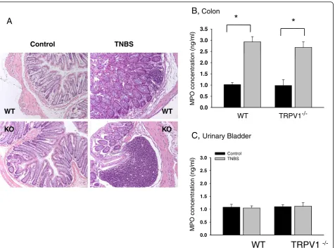
Absence of TRPV1 receptors does not eliminate the development of colonic inflammation
detectable structural changes in the colonic wall (Figure 1A). However, TNBS treatment induced crypt segmentation, local infiltration and thickening of the muscle layer in the distal colon in both groups of animals (Figure 1A). The cytoarchitecture of the bladder wall was not affected by either treatment in all tested animals (data not shown). The MPO assay is a validated biochemical method of grading inflam-mation in the tissue and measures the amount of MPO enzyme released by neutrophils at the site of inflammation [29]. The concentration of the enzyme was increased 3-fold in the colon of both WT (N = 10) and TRPV1−/−(N = 10) mice in TNBS groups (Figure 1B) but was unchanged in the urinary bladder (Figure 1C). Taken together, these data demonstrate substantial inflam-matory changes in the distal colon during acute colitis in both WT and TRPV1−/−groups suggesting that the lack of
TRPV1 receptors does not prevent the colon from the development of acute inflammatory reaction.
Urodynamic analysis of bladder function in awake unrestrained mice
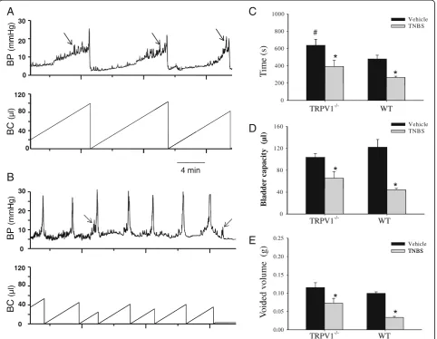
To evaluate the effects of colon-bladder cross-sensitization on the micturition parametersin vivo, we performed cysto-metric assessment of bladder function during slow continu-ous bladder filling in conscicontinu-ous mice. Cystometrograms were recorded under control conditions in WT (N = 4) and TRPV1−/−(N = 5) mice followed by second evaluation at 3 to 5 days post-TNBS. Figure 2 shows cystometric traces obtained from a TRPV1−/− mouse before (A) and after TNBS application (B). Urodynamic parameters were first compared to the baseline to evaluate the effects of TNBS-induced colitis on the voiding reflex in each animal fol-lowed by comparisons between the treatments and groups.
B,
Colon*
*
Control TNBS
2.0 2.5 3.0 3.5
0 0 0.5 1.0 1.5
MPO concentration
(ng/ml)
O K O
K
.
WT TRPV1
-/-C,
Urinary Bladder2.0 2.5
3.0 ControlTNBS
0.5 1.0 1.5
MPO concentration
(ng/ml)
0.0
WT
TRPV1
-/-WT
A
WT
Analysis of cystometrograms in WT and TRPV1−/−mice at baseline showed that the average intermicturition interval was increased by 32.9% in KO animals in comparison to WT littermates (P≤0.05, Figure 2C). However, non-voiding contractions were observed more frequently in TRPV1−/− mice (3.1 ± 1.2 vs 1.1 ± 0.3 in WT group,P≤0.05). Other urodynamic characteristics were not different between WT and KO animals.
Intracolonic TNBS had a profound effect on the function of the urinary bladder in both WT (N = 4) and TRPV1−/− (N = 5) groups. At 3 to 5 days after TNBS treatment, inter-micturition interval was reduced from 638 ± 70.7 s to 393.3 ± 70.8 s in KO group (P≤0.05 to vehicle) and from 480.3 ± 43.1 s to 265.9 ± 14.8 s in WT group (P≤0.05 to respective vehicle, Figure 2C). Likewise, bladder capacity
was reduced by 64% in WT mice and by 37% in TRPV1−/− littermates during colitis (P ≤0.05 to vehicle, Figure 2D). Occurrence of bladder dysfunction was also associated with decreased voided volume in both groups of animals (Figure 2E). In WT mice, the average voided volume per cycle was reduced by 67% during active colitis, however, in KO group, a decrease was only 36.4% (P≤0.05 to vehicle, Figure 2E). Taken together, these results suggest that the effects of colitis-induced cross-sensitization on bladder function were attenuated in TRPV1−/−mice.
Differential effects of experimental colitis on the contractility of DSM in WT and TRPV1−/−mice
We performed a series of in vitroexperiments using iso-lated urinary bladder strips to identify the effects of TRPV1
800 1000
Vehicle TNBS
C
30
A
mHg) #
200 400 600
T
im
e (s)
120 0 10 20
BP
(mm
*
*
0
TRPV1-/- WT
160 Vehicle
TNBS
0 40 80
D
BC
(μl)
(μl)
40 80 120
30
4 min
B
Hg)
dder
capacity
(
*
*
0
0 10 20
E
BP
(mmH
Bla
TRPV1-/- WT
0.25
Vehicle TNBS
40 80 120
BC
(μl)
ded volume (g)
0.10 0.15
0.20 TNBS
*
0
Vo
id
0.00 0.05
TRPV1-/- WT
*
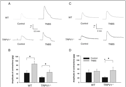
gene knockout on the mechanisms of detrusor smooth muscle (DSM) contractility. Isometric contractions in response to EFS at 32 Hz were evaluated in muscle strips isolated from WT and TRPV1−/− mice (Figure 3A). This frequency was chosen based on our previous studies that showed significant alterations in detrusor contractility at higher frequencies during acute colonic inflammation [14,22]. The maximal amplitude of the normalized con-tractile response to EFS was 44.4 ± 10.8 g/g in WT group under control conditions and increased 2-fold after TNBS treatment (85.9 ± 12.3 g/g,P≤0.05 to control, Figure 3B). The contractility of DSM isolated from TRPV1−/− mice reached 24.1 ± 4.5 g/g in the control group, which was lower than in respective WT group (P = 0.11 to WT, Figure 3B). However, colonic inflammation had similar effect on the detrusor of TRPV1−/−animals causing 2-fold increase in the contractile response (P ≤0.05 to vehicle, Figure 3B).
Stimulation of DSM stripsin vitrowith KCl (Figure 3C) allowed evaluation of the effects of TRPV1 gene knockout on DSM contractility due to depolarizing effect of KCl on smooth muscle cells [30]. The normalized amplitude of the contractile response to KCl was 22.7 ± 3.3 g/g in TRPV1−/− group, which was lower than in WT group (44.5 ± 12.2 g/g), however, this difference did not reach statistical significance (P = 0.12 to WT, Figure 3D). Intracolonic treatment with TNBS had no effect on KCl-induced contractility of the detrusor in WT animals but caused a significant up-regulation of the response in TRPV1−/− strips (P ≤0.05 to respective control, Figure 3D).
We further investigated the effects of TRPV1 gene knockout on cholinergic regulation of DSM by studying the concentration-contractility response relationship upon application of carbachol (CCh). Cumulative addition of 10-7 to 10-4 M CCh to the bath solution resulted in
C
A
WT
Control TNBS
WT
Control TNBS
/
TNBS
TRPV1
-/-0.5 min
1 g
2 min
1 g
B
D
TRPV1
-Control TNBS
/
Control TNBS
80 100 120
80 100 120
Control TNBS
0 20 40 60
0 20 40 60
0
WT TRPV1
-/-0
WT TRPV1
-/-Amplitude of
contractions
(g/g)
Amplitude of
contractions
(g/g)
concentration-dependent contractions in vehicle-treated WT animals (Figure 4A). Acute colonic inflammation diminished the response of the detrusor muscle to stimulation with CCh in both WT and TRPV1−/− groups, however, the difference did not reach statistical significance (Figure 4B).
Comparison of PKC and ROK dependent contractions in DSM upon colon-bladder cross-sensitization
Increased sensitivity of DSM to KCl stimulation induced by colonic inflammation in TRPV1−/− mice suggested potential modulation of the signaling changes in muscle contractile apparatus. Therefore, we tested the effects of PKC activator PDBU on detrusor contractility and Rho-kinase inhibitor Y27632 on relaxation of PDBU-induced contractile response in DSM isolated from WT and TRPV1−/− mice (Figure 5A). The normalized amplitude of contractile response to PDBU was 6.5 ± 1.8 g/g in WT group (n = 6) and 3.2 ± 1.1 g/g in TRPV1−/− strips (n = 8, P >0.05, Figure 5B). Colonic inflammation increased responses to PKC activator by 110% in WT
group and by 133% in TRPV1−/−group (P≤0.05 to respec-tive baseline, Figure 5B). Maximal contractile response to PDBU was taken as 100% before the application of Rho-kinase inhibitor Y27632. Application of Y27632 caused relaxation of DSM in vehicle-treated WT mice by 76%, whereas the effect on the strips from KO animals was around 40% (Figure 5C). Colonic inflammation significantly reduced the relaxation of DSM strips in WT group but had no effect in TRPV1−/−mice (Figure 5C).
To further clarify the role of ROK pathway in the response of isolated detrusor to KCl stimulation, we per-formed an additional series of experiments using four groups of WT and TRPV1−/− mice with and without colonic inflammation (N = 4, n = 8 in each group). Experi-mental design included initial stimulation of detrusor strips with KCl followed by wash and subsequent incubation with Rho-kinase inhibitor Y27632 for 30 min. After incubation with the drug, the contractile response of the same strips to KCl was tested again. The normalized amplitude of the ini-tial contractile response to KCl in all tested groups was similar to that presented in Figure 3D. However, incubation
0.1 μM 1 μM 10 μM 100.0uM
A
WT
B
80 100 120
WT +TNBS
40 60 80
- WT
TRPV1
-/-Amplitude ofcontractions
(g/g)
-7 -6 -5 -4
0
20 - WT+TNBS
- KO - KO+TNBS
TRPV1-/-+TNBS
1 g
1 min
-Log (Carbachol)
with Y27632 compound did not significantly affect the amplitude of KCl-induced contractions in either group (Figure 5D) suggesting that the ROK-dependent compo-nent of the detrusor response to KCl was not directly affected by the knockout of TRPV1 gene.
Colonic inflammation up-regulates voltage-gated sodium channels in bladder sensory neurons from WT but not TRPV1−/−mice
In the next set of experiments we aimed to determine if knockdown of TRPV1 gene would affect excitability of bladder sensory neurons via effects on voltage-gated so-dium channels (VGSC) as we previously reported for a rat model of cross-sensitization [16,31]. Retrograde la-beling of lumbosacral sensory neurons revealed three populations of isolated DRG neurons: colon-projecting, bladder-projecting and colon-bladder convergent neu-rons as previously detected [13,15]. We intentionally
performed double labeling to exclude from the experi-ments neurons receiving dual afferent input from the distal colon and urinary bladder as these neurons would be directly affected by colonic treatments. The number of double labeled cells was around 10% as previously reported [13,15]. Only neurons labeled with Fast Blue (bladder projecting) underwent electrophysiological evaluation and were used for data analysis.
The current–voltage (I-V) relationship of total Na+ current recorded upon membrane depolarization in bladder afferent neurons from WT mice is shown in Figure 6A. Acute colonic inflammation increased the peak amplitude of total Na+current by 2-fold from −153.4 ± 17.4 pA/pF at−20 mV in the control group (N = 6, n = 9) to−256.5 ± 24.1 pA/pF in the acute colitis group (N = 5, n = 7,P≤0.05, Figure 6A). The amplitude of total Na+current recorded in bladder DRG neurons from TRPV1−/−mice (N = 6, n = 8) with vehicle treatment was enlarged in comparison to
12 14 16 18
B
#
A
WT
Control TNBS
2 4 6 8 10
0.2 g
TRPV1
-/-Control TNBS
2 min
0 2
Amplitude of contractions (g/g)
Amplitude of contractions (g/g)
WT
TRPV1
-/-C
#
Control TNBS
D
0.6 0.8 1.0
esponse
Control TNBS
80 100 120
0.2 0.4
Ratio of
r
20 40 60
0.0
WT
TRPV1
-/-0
TRPV1
-/-WT
respective WT group (N = 5, n = 7), however, intracolonic TNBS had no effect on Na+ current in KO animals (Figure 6B).
We next assessed the kinetic parameters of Na+currents after the induction of experimental colitis. The steady-state activation was studied by using a three-pulse protocol with a negative pre-pulse to −110 mV and a series of short pulses (10 ms duration) to activate Na+currents as previ-ously described [16]. The amplitude of steady-state activa-tion was measured at the peak of tail current upon the voltage step to −70 mV, normalized and plotted as I/Imax
against the voltage. The amplitude of steady-state in-activation was measured at a series of membrane de-polarizing steps ranging from −100 mV to +70 mV. In the group of WT mice, neither rate of activation,
nor steady-state inactivation kinetics of total Na+ current in bladder sensory neurons were affected by acute colitis (Figure 6C). However, in TRPV1−/− neurons, acute colitis induced a rightward shift in steady-state inacti-vation by 7 mV (V1/2=−39.7 ± 1.9 mV in the vehicle
group vs −32.7 ± 1.7 mV in TNBS group, P ≤0.05, Figure 6D).
Up-regulation of gene expression of VGSC in lumbosacral DRG by colitis is attenuated in TRPV1 KO mice
To correlate the observed functional changes of the recorded VGSC in TRPV1−/− mice with potential effects on gene expression of VGSC in sensory neurons involved in nociceptive signaling, we performed quantitative RT-PCR analysis. This set of experiments was carried out on
-80 -60 -40 -20 0 20 40 60 80 0
Voltage (mV)
-80 -60 -40 -20 0 20 40 60 80 0
Voltage (mV)
B
A
-150 -100 -50
nsity (pA/pF) -150
-100 -50
nsity (pA/pF)
-300 -250 -200
WT WT+TNBS
Current Den
-300 -250 -200
TRPV1-/-Current Den
-350 -350
TRPV1-/-+TNBS
1.0 1.0
D
C
0.6 0.8
WT
I/Imax
0.6 0.8
TRPV1
-/-I/Imax
0.0 0.2
0.4 WT-TNBS
WT WT-TNBS
0.0 0.2
0.4 TRPV1-/--TNBS
TRPV1
-/-TRPV1-/--TNBS
-100 -80 -60 -40 -20 0 20 40 60
Voltage (mV)
-100 -80 -60 -40 -20 0 20 40 60
Voltage (mV)
the whole L6-S2 ganglia due to the presence of a limited number of retrogradely labeled bladder neurons. The DRG from both vehicle and TNBS groups were harvested 3 days post-treatment. The comparison of mRNA levels of VGSC between WT (N = 4) and TRPV1−/−(N = 4) mice did not reveal significant differences under control physio-logical conditions (data not shown). mRNA expression of VGSC in L6-S2 DRG isolated from WT mice showed 3-fold up-regulation for Nav1.7 channel and 2-3-fold increase for Nav1.8 channel by experimental colitis (N = 5,P≤0.05 to respective vehicle, Figure 7A). The Nav1.9 mRNA level was also increased in WT group, but did not reach the level of statistical significance. The effects of colonic inflammation on gene expression of Nav1.7, Nav1.8 and Nav1.9 channels were diminished in TRPV1−/− ganglia showing 2-fold up-regulation for Nav1.7 channel only (N = 5, P ≤0.05 to vehicle, Figure 7B).
Lack of TRPV1 receptors delays the development of abdominal hypersensitivity during colon-bladder cross-talk
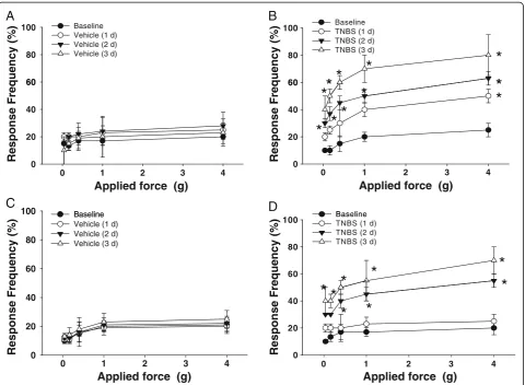
Abdominal sensitivity was daily tested in WT and TRPV1−/− mice at the baseline and after intracolonic treatments with either vehicle or TNBS. Figure 8 sum-marizes the frequency of responses in the lower abdo-minal area in all tested mice. Mechanical stimulation of the pelvic area under control conditions (vehicle treat-ment) in both WT (N = 4, Figure 8A) and TRPV1−/− mice (N = 4, Figure 8C) resulted in a response frequency that correlated with the applied force, reaching around 20% at the maximal tested force of 4 g during the entire tested period . In the group with TNBS treatment, WT mice showed increased sensitivity to the filament testing and responses reached 40% at applied forces of 1 and 4 g (Figure 8B, N = 5,P≤0.05 to baseline). An increase in pelvic sensitivity became most significant at 3 days after
the treatment with statistically higher percentage of responses at all tested forces (Figure 8B, N = 6, P≤0.05 to baseline). Mice in TRPV1−/− group with had similar baseline sensitivity to von Frey filaments when compared to WT group. TNBS application did not evoke signifi-cant changes in withdrawal response 1 day after the treatment but increased abdominal sensitivity at days 2 and 3 up to 40% and 65%, respectively, for 4 g filaments (P ≤0.05 to vehicle, Figure 8D). These results provide evidence that colonic inflammation leads to an increased viscerosomatic response associated with abdominal hyperalgesia and discomfort, and the lack of TRPV1 receptors delays the development of viscerosomatic hyperalgesia in KO mice.
Discussion
This is the first report which evaluated viscero-visceral cross-reflexes in TRPV1 KO mice and confirmed previ-ously reported data that transient inflammation of the distal colon triggers the development of neurogenic bladder dys-function via TRPV1-related pathways [14,16,18,19,21,32]. Our results provide direct evidence that the lack of TRPV1 receptors does not prevent the occurrence of colon-bladder cross-sensitization induced by transient colitis, however, several important physiological characteristics of bladder function are altered by the knockout of TRPV1 gene. The major differences included prolonged intermicturition interval in KO animals followed by reduced urodynamic responses during active colitis; up-regulation of DSM con-tractility in response to KCl in TRPV1−/− mice with inflamed colon; diminished relaxation of DSM in transgenic animals in the presence of ROK inhibitor; attenuated effects of colonic inflammation on expression and function of VGSC in bladder sensory neurons from TRPV1−/− mice; and delayed development of abdominal hypersensitivity
A
B
4.0
5.0 Control
TNBS
4.0 5.0
Control TNBS
* *
2.0 3.0
2.0 3.0
*
0.0 1.0
NaV1.7 NaV1.8 NaV1.9
mRNA
(fold
change)
NaV1.7 NaV1.8 NaV1.9
mRnNA
(fold
chage)
0.0 1.0
upon colon-bladder cross-talk in genetically modified animals.
An increasing number of clinical reports support animal studies that peripheral and central cross-sensitization may underlie unknown etiology of functional gastrointestinal and urologic disorders associated with abdominal discom-fort, visceral hypersensitivity, and chronic pelvic pain [3,4,33]. While it is understood that animal models cannot fully mimic human co-morbid conditions, they provide valuable insight to study the underlying mechanisms. In the present study, we established that the absence of TRPV1 did not affect the severity of TNBS-induced colitis in transgenic mice. Several previous reports presented contradictory results on the role of TRPV1 in inflamma-tion suggesting that the impact of TRPV1 involvement is model-, dose-, and species-dependent [34]. For instance,
Okayama et al. [35] established that desensitization of TRPV1 receptors with potent agonists elevated histo-logical changes in the colon in dextran sulfate sodium (DSS) model of colitis, whereas other groups demon-strated a diminished level of the developed inflammatory reaction in the distal colon of rats [21,36-38]. Experiments on TRPV1 KO mice using a single dose of dinitrobenzene sulfonic acid reported an increased susceptibility of the colon to the applied inflammatory agent [39], however, the effects of DDS on TRPV1 KO mice were clearly dose-dependent. In 2% DSS-treated group, the lack of TRPV1 receptors decreased the severity of the induced colitis, however, this difference was not observed for 5% DSS, when much severe inflammatory reaction developed [40]. While the severity of inflammatory response could be modulated by activation of TRPV1 receptors, it is also
B
A
60 80
100 BaselineTNBS (1 d) TNBS (2 d) TNBS (3 d)
*
*
*
*
*
60 80
100 Baseline Vehicle (1 d) Vehicle (2 d) Vehicle (3 d)
20 40 60
*
*
*
*
*
*
*
*
20 40 60
0 1 2 3 4
0
Applied force (g)
Response Frequency
(%
)
Response Frequency
(%
)
Response Frequency
(%
)
Response Frequency
(%
)
D
Baseline
0 1 2 3 4
0
Applied force (g)
100 Baseline
C
60 80
100 BaselineTNBS (1 d) TNBS (2 d) TNBS (3 d)
*
*
*
*
*
60 80
Baseline Vehicle (1 d) Vehicle (2 d) Vehicle (3 d)
0 20 40
*
* *
*
0 20 40
0 1 2 3 4
Applied force (g) Applied force (g)
0 1 2 3 4
possible that nerve fibers sensitive to capsaicin can be activated by non-TRPV1-dependent pathways during pathological conditions [40]. In a model of cutaneous inflammation, the lack of TRPV1 receptors did not alter the leukocyte accumulation suggesting participation of both neurogenic and non-neurogenic mechanisms [41]. Importantly, the activation of sensory nerves was still observed in TRPV1 KO mice providing evidence that neurogenic inflammation may also develop via TRPV1 independent pathways [41].
Our analysis of urodynamic parameters in awake mice confirmed previously reported involvement of TRPV1 in regulation of urinary bladder function under both normal and pathophysiological conditions. The intermicturition interval in TRPV1−/− mice was increased along with the number of non-voiding contractions at the baseline, and the effects of experimental colitis on bladder capacity and voided volume were diminished in comparison to WT littermates. Our results are in line with the previous studies which reported increased bladder capacity and non-micturition contractions [42] as well as attenuated bladder distension-induced afferent discharge in TRPV1−/− mice [43]. Participation of TRPV1 in bladder sensitivity and pain was also demonstrated in several animal models of direct bladder inflammation [44-46]. These combined results sug-gest that TRPV1 pathways are involved in inflammatory and neurogenic bladder dysfunctions, and also contribute to the development of pelvic organ cross-sensitization.
Previous studies from our [14,16,20] and other [12,15,17] laboratories established that colon-bladder cross-sensitization develops predominantly, but not exclusively, via neural pathways. It is well established that TRPV1 channels are expressed not only on sen-sory neurons but also on peripheral afferents supplying visceral organs of the gastrointestinal and genitourinary tracts [24,25,47]. Our data from isolated DSM strips clearly demonstrate that the absence of TRPV1 expression on bladder afferents did not cause significant changes in con-tractile responses of the detrusor upon normal physio-logical conditions. However, some modulatory changes in the signaling cascades became more evident during add-itional stimulation like active colonic inflammation or PKC activation. Specifically, we found that the contractile response of DSM to KCl stimulation was enhanced by experimental colitis in KO animals. KCl is used as a tool to bypass G protein-coupled receptor stimulation and activate smooth muscle by changing potassium equilibrium poten-tial and triggering membrane depolarization (reviewed in [30]). Activation of TRPV1, a cation channel permeable mostly to Ca2+, has the same depolarizing effect on the cell but the major mechanism includes massive influx of Ca2+ through the pore of the channel [48]. Recent studies estab-lished that activation of TRPV1 by capsaicin, a potent TRPV1 agonist, is associated with PKC activation [49,50],
and the opening of TRPV1 channels is promoted by channel phosphorylation [51]. PKC modulation of TRPV1 channel function was previously shown to occur under conditions of chronic pain resulting from nerve damage or inflammation [50].
Rho kinase (ROK) pathway in bladder detrusor smooth muscle was shown to be involved in the contractile response of the isolated DSM to KCl [30]. ROK pathway is critical for the maintenance of basal tone in DSM and serves as a common final pathway of various contractile stimuli in many species [52]. ROK directly phosphorylates and inactivates myosin light chain (MLC) phosphatase, ultimately increasing the phosphorylation state of myosin and facilitating contraction [53]. Y27632, a ROK inhibitor, was shown to reduce KCl-induced contractile force without inhibition of KCl-induced increases in intracellular Ca2+ [54-57], but with concomitant inhibition of MLC phosphorylation [55,56].
The relationship between TRPV1 and ROK in the detrusor muscle is currently not established, but activation of TRPV1 resulted in inhibition of ROK in vascular smooth muscle [58]. Fujimoto et al. previously investi-gated the effects of capsaicin-induced relaxation on KCl-induced contractions and phosphorylation of MLC in ileal smooth muscle of rats [59]. They established that capsa-icin relaxed intestinal smooth muscle after KCl-induced contraction and this effect was accompanied by a decrease in MLC20 phosphorylation [59]. To clarify the effects of
Sensory neurons receiving afferent input from the pelvic viscera play a key role in the development of peripheral cross-sensitization [13,15]. They express several types of ion channels including TRPV1 and VGSC, which are well-known transducers of nociceptive processing in pain path-ways [26,27,48]. Excitability of sensory afferents is defined by expression and function of VGSC [26,27]. Single cell analysis of Na+ channel transcripts indicated that sensory neurons innervating the pelvic viscera express a combin-ation of tetrodotoxin-sensitive and -resistant Na+channels, which consist mostly of Nav1.6, Nav1.7, Nav1.8 and Nav1.9
members [62]. In this study, the peak amplitude of total sodium current in neurons from vehicle-treated TRPV1−/− mice was greater than that of neurons from the vehicle-treated WT mice. Multiple sodium channel isoforms are expressed in DRG neurons and play a central role in neuronal electrogenesis. However, a number of other ion channels also confer electrical excitability of neurons including voltage-gated calcium and potassium channels. Interactions between different ion channels and co-expressed molecules lead to the occurrence of multiple ionic conductances, which shape the cells' firing patterns, and confirm that regulation of neuronal excitability is a complex process. Under non-pathological conditions, the firing properties of DRG neurons are usually main-tained within a circumscribed range. This is a result of homeostatic regulation of ion channel expression, post-translational modification, and/or interaction with binding partners or modulators [63]. While variations in the level of expression of any VGSC isoform present within DRG neurons could affect their level of excitability, computa-tional models have demonstrated that multiple, distinct sets of membrane parameters can produce similar levels of activity. For instance, it has been proposed that changes in expression of channel ‘B' can compensate for changes in expression of channel‘A' to maintain excitability within a particular range [64]. Indeed, it was demonstrated that overexpression of Shal potassium channel gene in somatogastric ganglion neurons caused a large increase in potassium current but little change in the neuron's firing properties due to a compensatory increase in expression of a hyperpolarization-activated inward current [65]. Likewise, in Purkinje neurons isolated from NaV1.6−/−
mice, sodium current density was reduced, however, an up-regulation of calcium channels maintained excitability near its normal level [66]. Therefore, while we observed enhanced amplitude of VGSC current in TRPV1−/− neurons, we could not confirm that these neurons were hyperexcitable in comparison to WT phenotype. Additional extensive studies are warranted to perform comprehensive evaluation of neuronal excitability in genetically modified animals.
Studies utilizing animal models of acute and chronic in-flammation in different parts of gastrointestinal [11,67,68]
and genitourinary [44,69] systems showed an increased excitability of DRG neurons receiving direct input from the inflamed organs. Previously published data from our group also established that colonic inflammation increased excitability of bladder DRG neurons via an up-regulation of VGSC [16]. However, cross-channel interac-tions between TRPV1 and VGSC at the membrane level have not been fully investigated. Underin vitroconditions, application of capsaicin usually leads to a significant block of VGSC [70], but intraluminal application of TRPV1 agonists in vivo causes an up-regulation of VGSC on the cell soma and is associated with increased excitability of sensory neurons [16]. Additional in vitro studies revealed that capsaicin-induced blockade of VGSC is concentration-dependent [71]. It was also established that the block of VGSC by low concentrations of capsaicin was reversed in TRPV1−/− mice [71]. There are a few suggested mecha-nisms underlying functional interconnection between TRPV1 and VGSCs. Indirect pathways include intercros-sing of TRPV1 and VGSC signaling pathways via modula-tion of the same transcripmodula-tional/translamodula-tional factors or recruitment of second messengers (for example cAMP or intracellular Ca2+). Direct pathways involve TRPV1-VGSC interactions within a complex as it was proven for other channels [72-74]. It is possible that genetic knockdown of TRPV1 gene in sensory neurons could lead to compensa-tory changes by up- or down-regulation of other ion chan-nels. Since VGSCs are the main source of positive charge influx into sensory neurons, they could have been affected by TRPV1 gene knockout during development. The future research steps should determine if one of the VGSCs expressed in DRG has some sort of capsaicin receptor and whether modulatory subunits of both channels are involved in TRPV1-VGSC‘cross-talk’.
Inflammation in the pelvic viscera is associated with enhanced abdominal sensitivity due to convergence of vis-ceral and somatic inputs in the nervous system. This phenomenon is known as viscerosomatic referred hyper-algesia and could be measured by using mechanical stimu-lation with von Frey filaments on the lower abdominal area [28]. The referred hyperalgesia is proportional to the inten-sity of the spontaneous visceral pain-related behavior expressed by the animals. Increased responses of the lower abdominal wall to mechanical stimulation are reflective of and correlate with pain and discomfort arising from inflamed pelvic organs [75]. Our behavioral results estab-lished that the development of abdominal hypersensitivity induced by colitis upon mechanical stimulation of the lower pelvic region was delayed in TRPV1−/−mice. This observa-tion is in line with the previously published studies con-firming participation of TRPV1 in the development of abdominal hyperalgesia and inflammatory pain [76]. Wang
either cyclophosphamide- or acrolein-induced models of bladder inflammation [46]. This group also established that abdominal hyper-reactivity and cutaneous allodynia were abolished in TRPV1−/− mice. Likewise, selective TRPV1 antagonists attenuated mechanical allodynia and hyperalge-sia in rat models of neuropathic pain [76-78]. However, TRPV1−/−mice did not reveal a significantly altered pheno-type in the partial sciatic nerve ligation model of neuro-pathic pain [79]. The use of RNA interference technologies further confirmed model- and species-associated differ-ences in test results from genetically and functionally modi-fied animals. Functional knockdown of TRPV1 using shRNA showed diminished behavioral responses to intra-plantar injection of capsaicin, enhanced paw withdrawal la-tencies to heat, and diminished tactile hypersensitivity in an injury-induced neuropathic pain model [80,81]. siRNA treatment also diminished spontaneous visceral pain behav-ior induced by capsaicin application to the rectum of mice [77]. Interestingly, spinal nerve injured TRPV1 knockout but not shRNA-treated animals developed mechanical allo-dynia and hypersensitivity [80]. These results indicate that neither the complete loss nor a profound reduction in TRPV1 channels can completely eliminate the effects of visceral or cutaneous noxious stimulation on behavioral responses suggesting that TRPV1 is not the only player in nociceptive signaling.
Conclusions
In summary, the results of our extensive study clarified the role of TRPV1 in the development of colon-bladder cross-sensitization and associated dysfunction in the lower urinary tract. Knockout of TRPV1 gene changed several important physiological characteristics of bladder function including prolonged intermicturition interval along with reduced urodynamic responses during active colitis in genetically modified mice; up-regulation of DSM contractility in response to KCl in TRPV1−/−mice with inflamed colon; diminished relaxation of DSM in transgenic animals in the presence of ROK inhibitor, and attenuated effects of colonic inflammation on VGSC in bladder sensory neurons. The mechanisms underlying neurogenic bladder dysfunctions due to pelvic organ cross-interactions require further studies to provide valuable information for the development of new pharmacological approaches and therapeutic strategies for the treatment of co-morbid conditions characterized by functional pelvic pain arising from gastrointestinal or genitourinary systems.
Abbreviations
CCh: carbachol; CNS: central nervous system; CPP: chronic pelvic pain; DAI: disease activity index; DMEM: Dulbecco’s modified Eagle’s medium; DRG: dorsal root ganglia; DSM: detrusor smooth muscle; DSS: dextran sulfate sodium; EFS: electric field stimulation; ELISA: enzyme-linked immunosorbent assay; GI: gastrointestinal; IBS: irritable bowel syndrome; IC/BPS: interstitial cystitis/bladder pain syndrome; KC1: potassium chloride; KO: knockout;
MLC: myosin light chain; MPO: myeloperoxidase; PDBU: phorbol-12,13-dibiturate; PKC: protein kinase C; ROK: Rho kinase; PCR: polymerase chain reaction; TNBS: 2,4,6-trinitrobenzene sulfonic acid; TRPV1: transient receptor potential vanilloid 1; TRPV1−/−: TRPV1 knockout; VGSC: voltage-gated sodium channels; WT: wild type.
Competing interests
The authors declare that they have no competing interests.
Authors’contributions
APM and QL designed the study; QL, X-QP, ANV, TSA, SC and SAZ performed the experiments; QL, ANV, TSA, X-QP, and APM analyzed the data; QL X-QP, ANV and APM prepared the figures; QL and APM edited and finalized the manuscript. All authors approved the final version of the paper.
Acknowledgements
Preliminary results of this work were presented in an abstract form at the AUA 2012 meeting. This study was supported by the NIH/NIDDK grants DK077699 (APM) and DK 077699-S2 (APM).
Author details
1Department of Surgery, Division of Urology, University of Pennsylvania, 500
South Ridgeway Avenue, Glenolden, PA 19036, USA.2Department of Obstetrics and Gynecology, Weill Cornell Medical College, 1305 York Avenue, New York, NY 10065, USA.3Department of Surgery, Cooper University, One Cooper Plaza, Camden, NJ 08103, USA.4John W. Duckett Center for Pediatric Urology, Children’s Hospital of Philadelphia, 34th Street and Civic Center Blvd, Philadelphia, PA 19104, USA.
Received: 20 August 2012 Accepted: 21 December 2012 Published: 11 January 2013
References
1. Alagiri M, Chottiner S, Ratner V, Slade D, Hanno PM:Interstitial cystitis: unexplained associations with other chronic disease and pain syndromes.Urology1997,49:52–57.
2. Francis CY, Duffy JN, Whorwell PJ, Morris J:High prevalence of irritable bowel syndrome in patients attending urological outpatient departments.Dig Dis Sci1997,42:404–407.
3. Rodriguez MA, Afari N, Buchwald DS:Evidence for overlap between urological and nonurological unexplained clinical conditions.J Urol2009,
182:2123–2131.
4. Cory L, Harvie HS, Northington G, Malykhina A, Whitmore K, Arya L:
Association of neuropathic pain with bladder, bowel and catastrophizing symptoms in women with bladder pain syndrome.J Urol2012,
187:503–507.
5. Terruzzi V, Magatti F, Quadri G, Tenore C, Minoli G, Belloni C:Bladder dysfunction and irritable bowel syndrome.Am J Gastroenterol1992,
87:1231–1232.
6. Whorwell PJ, Lupton EW, Erduran D, Wilson K:Bladder smooth muscle dysfunction in patients with irritable bowel syndrome.Gut1986,
27:1014–1017.
7. Giamberardino MA, Costantini R, Affaitati G, Fabrizio A, Lapenna D, Tafuri E, Mezzetti A:Viscero-visceral hyperalgesia: characterization in different clinical models.Pain2010,151:307–322.
8. Latremoliere A, Woolf CJ:Central sensitization: a generator of pain hypersensitivity by central neural plasticity.J Pain2009,10:895–926. 9. Malykhina AP:Neural mechanisms of pelvic organ cross-sensitization.
Neuroscience2007,149:660–672.
10. Brumovsky PR, Gebhart GF:Visceral organ cross-sensitization - an integrated perspective.Auton Neurosci2010,153:106–115.
11. Bielefeldt K, Lamb K, Gebhart GF:Convergence of sensory pathways in the development of somatic and visceral hypersensitivity.Am J Physiol Gastrointest Liver Physiol2006,291:G658–G665.
12. Brumovsky PR, Feng B, Xu L, McCarthy CJ, Gebhart GF:Cystitis increases colorectal afferent sensitivity in the mouse.Am J Physiol Gastrointest Liver Physiol2009,297:G1250–G1258.
13. Malykhina AP, Qin C, Greenwood-van MB, Foreman RD, Lupu F, Akbarali HI:
14. Asfaw TS, Hypolite J, Northington GM, Arya LA, Wein AJ, Malykhina AP:
Acute colonic inflammation triggers detrusor instability via activation of TRPV1 receptors in a rat model of pelvic organ cross-sensitization.Am J Physiol Regul Integr Comp Physiol2011,300:R1392–R1400.
15. Christianson JA, Liang R, Ustinova EE, Davis BM, Fraser MO, Pezzone MA:
Convergence of bladder and colon sensory innervation occurs at the primary afferent level.Pain2007,128:235–243.
16. Lei Q, Malykhina AP:Colonic inflammation up-regulates voltage-gated sodium channels in bladder sensory neurons via activation of peripheral transient potential vanilloid 1 receptors.Neurogastroenterol Motil2012,
24:575–e257.
17. Pezzone MA, Liang R, Fraser MO:A model of neural cross-talk and irritation in the pelvis: implications for the overlap of chronic pelvic pain disorders.Gastroenterology2005,128:1953–1964.
18. Ustinova EE, Fraser MO, Pezzone MA:Colonic irritation in the rat sensitizes urinary bladder afferents to mechanical and chemical stimuli: an afferent origin of pelvic organ cross-sensitization.Am J Physiol Renal Physiol2006,
290:F1478–F1487.
19. Xia CM, Gulick MA, Yu SJ, Grider JR, Murthy KS, Kuemmerle JF, Akbarali HI, Qiao LY:Up-regulation of brain-derived neurotrophic factor in primary afferent pathway regulates colon-to-bladder cross-sensitization in rat.
J Neuroinflammation2012,9:30.
20. Qin C, Malykhina AP, Akbarali HI, Foreman RD:Cross-organ sensitization of lumbosacral spinal neurons receiving urinary bladder input in rats with inflamed colon.Gastroenterology2005,129:1967–1978.
21. Pan XQ, Gonzalez JA, Chang S, Chacko S, Wein AJ, Malykhina AP:
Experimental colitis triggers the release of substance P and calcitonin gene-related peptide in the urinary bladder via TRPV1 signaling pathways.Exp Neurol2010,225:262–273.
22. Noronha R, Akbarali H, Malykhina A, Foreman RD, Greenwood-van MB:
Changes in urinary bladder smooth muscle function in response to colonic inflammation.Am J Physiol Renal Physiol2007,293:F1461–F1467. 23. Caterina MJ, Schumacher MA, Tominaga M, Rosen TA, Levine JD, Julius D:
The capsaicin receptor: a heat-activated ion channel in the pain pathway.Nature1997,389:816–824.
24. Avelino A, Cruz F:TRPV1 (vanilloid receptor) in the urinary tract: expression, function and clinical applications.Naunyn Schmiedebergs Arch Pharmacol2006,373:287–299.
25. Birder LA, Birder LA, Kanai AJ, de Groat WC, Kiss S, Nealen ML, Burke NE, Dineley KE, Watkins S, Reynolds IJ, Caterina MJ:Vanilloid receptor expression suggests a sensory role for urinary bladder epithelial cells.
Proc Natl Acad Sci USA2001,98:13396–13401.
26. Dib-Hajj SD, Cummins TR, Black JA, Waxman SG:Sodium channels in normal and pathological pain.Annu Rev Neurosci2010,33:325–347. 27. Waxman SG:Channel, neuronal and clinical function in sodium
channelopathies: from genotype to phenotype.Nat Neurosci2007,
10:405–409.
28. Rudick CN, Chen MC, Mongiu AK, Klumpp DJ:Organ cross talk modulates pelvic pain.Am J Physiol Regul Integr Comp Physiol2007,293:R1191–R1198. 29. Krawisz JE, Sharon P, Stenson WF:Quantitative assay for acute intestinal
inflammation based on myeloperoxidase activity. Assessment of inflammation in rat and hamster models.Gastroenterology1984,
87:1344–1350.
30. Ratz PH, Berg KM, Urban NH, Miner AS:Regulation of smooth muscle calcium sensitivity: KCl as a calcium-sensitizing stimulus.Am J Physiol Cell Physiol2005,288:C769–C783.
31. Malykhina AP, Qin C, Foreman RD, Akbarali HI:Colonic inflammation increases Na+ currents in bladder sensory neurons.Neuroreport2004,
15:2601–2605.
32. Ravnefjord A, Brusberg M, Kang D, Bauer U, Larsson H, Lindstrom E, Martinez V:Involvement of the transient receptor potential vanilloid 1 (TRPV1) in the development of acute visceral hyperalgesia during colorectal distension in rats.Eur J Pharmacol2009,611:85–91. 33. Chelimsky G, Safder S, Chelimsky T:FGIDs in children are associated with
many nonpsychiatric comorbidities: the tip of an iceberg?J Pediatr Gastroenterol Nutr2012,54:690–691.
34. Storr M:TRPV1 in colitis: is it a good or a bad receptor? - a viewpoint.
Neurogastroenterol Motil2007,19:625–629.
35. Okayama M, Tsubouchi R, Kato S, Takeuchi K:Protective effect of lafutidine, a novel histamine H2-receptor antagonist, on dextran sulfate
sodium-induced colonic inflammation through capsaicin-sensitive afferent neurons in rats.Dig Dis Sci2004,49:1696–1704.
36. Goso C, Evangelista S, Tramontana M, Manzini S, Blumberg PM, Szallasi A:
Topical capsaicin administration protects against trinitrobenzene sulfonic acid-induced colitis in the rat.Eur J Pharmacol1993,249:185–190. 37. Kihara N, de la Fuente SG, Fujino K, Takahashi T, Pappas TN, Mantyh CR:
Vanilloid receptor-1 containing primary sensory neurones mediate dextran sulphate sodium induced colitis in rats.Gut2003,52:713–719. 38. Kimball ES, Wallace NH, Schneider CR, D'Andrea MR, Hornby PJ:Vanilloid
receptor 1 antagonists attenuate disease severity in dextran sulphate sodium-induced colitis in mice.Neurogastroenterol Motil2004,16:811–818. 39. Massa F, Sibaev A, Marsicano G, Blaudzun H, Storr M, Lutz B:Vanilloid
receptor (TRPV1)-deficient mice show increased susceptibility to dinitrobenzene sulfonic acid induced colitis.J Mol Med (Berl)2006,
84:142–146.
40. Szitter I, Pozsgai G, Sandor K, Elekes K, Kemeny A, Perkecz A, Szolcsanyi J, Helyes Z, Pinter E:The role of transient receptor potential vanilloid 1 (TRPV1) receptors in dextran sulfate-induced colitis in mice.J Mol Neurosci2010,42:80–88.
41. Banvolgyi A, Pozsgai G, Brain SD, Helyes ZS, Szolcsanyi J, Ghosh M, Melegh B, Pinter E:Mustard oil induces a transient receptor potential vanilloid 1 receptor-independent neurogenic inflammation and a non-neurogenic cellular inflammatory component in mice.Neuroscience2004,
125:449–459.
42. Birder LA, Nakamura Y, Kiss S, Nealen ML, Barrick S, Kanai AJ, Wang E, Ruiz G, de Groat WC, Apodaca G, Watkins S, Caterina MJ:Altered urinary bladder function in mice lacking the vanilloid receptor TRPV1.Nat Neurosci2002,
5:856–860.
43. Daly D, Rong W, Chess-Williams R, Chapple C, Grundy D:Bladder afferent sensitivity in wild-type and TRPV1 knockout mice.J Physiol2007,
583:663–674.
44. Charrua A, Cruz CD, Cruz F, Avelino A:Transient receptor potential vanilloid subfamily 1 is essential for the generation of noxious bladder input and bladder overactivity in cystitis.J Urol2007,177:1537–1541. 45. Dinis P, Charrua A, Avelino A, Yaqoob M, Bevan S, Nagy I, Cruz F:
Anandamide-evoked activation of vanilloid receptor 1 contributes to the development of bladder hyperreflexia and nociceptive transmission to spinal dorsal horn neurons in cystitis.J Neurosci2004,24:11253–11263. 46. Wang ZY, Wang P, Merriam FV, Bjorling DE:Lack of TRPV1 inhibits
cystitis-induced increased mechanical sensitivity in mice.Pain2008,139:158–167. 47. Yu W, Hill WG, Apodaca G, Zeidel ML:Expression and distribution of
transient receptor potential (TRP) channels in bladder epithelium.Am J Physiol Renal Physiol2011,300:F49–F59.
48. Caterina MJ:Vanilloid receptors take a TRP beyond the sensory afferent.
Pain2003,105:5–9.
49. Xu YP, Zhang JW, Li L, Ye ZY, Zhang Y, Gao X, Li F, Yan XS, Liu ZG, Liu LJ, Cao XH:Complex regulation of capsaicin on intracellular second messengers by calcium dependent and independent mechanisms in primary sensory neurons.Neurosci Lett2012,517:30–35.
50. Mandadi S, Armati PJ, Roufogalis BD:Protein kinase C modulation of thermo-sensitive transient receptor potential channels: Implications for pain signaling.J Nat Sci Biol Med2011,2:13–25.
51. Studer M, McNaughton PA:Modulation of single-channel properties of TRPV1 by phosphorylation.J Physiol2010,588:3743–3756.
52. Zhang X, Disanto ME:Rho-kinase, a common final path of various contractile bladder and ureter stimuli.Handb Exp Pharmacol2011,
202:543–568.
53. Sward K, Dreja K, Susnjar M, Hellstrand P, Hartshorne DJ, Walsh MP:
Inhibition of Rho-associated kinase blocks agonist-induced Ca2+ sensitization of myosin phosphorylation and force in guinea-pig ileum.
J Physiol2000,522(Pt 1):33–49.
54. Janssen LJ, Tazzeo T, Zuo J, Pertens E, Keshavjee S:KCl evokes contraction of airway smooth muscle via activation of RhoA and Rho-kinase.Am J Physiol Lung Cell Mol Physiol2004,287:L852–L858.
55. Mita M, Yanagihara H, Hishinuma S, Saito M, Walsh MP:Membrane depolarization-induced contraction of rat caudal arterial smooth muscle involves Rho-associated kinase.Biochem J2002,364:431–440.