Open Access
Review
Enzymes: An integrated view of structure, dynamics and function
Pratul K Agarwal*
Address: Computational Biology Institute, and Computer Science and Mathematics Division, Oak Ridge National Laboratory, Oak Ridge, Tennessee 37831, USA
Email: Pratul K Agarwal* - [email protected] * Corresponding author
Abstract
Microbes utilize enzymes to perform a variety of functions. Enzymes are biocatalysts working as highly efficient machines at the molecular level. In the past, enzymes have been viewed as static entities and their function has been explained on the basis of direct structural interactions between the enzyme and the substrate. A variety of experimental and computational techniques, however, continue to reveal that proteins are dynamically active machines, with various parts exhibiting internal motions at a wide range of time-scales. Increasing evidence also indicates that these internal protein motions play a role in promoting protein function such as enzyme catalysis. Moreover, the thermodynamical fluctuations of the solvent, surrounding the protein, have an impact on internal protein motions and, therefore, on enzyme function. In this review, we describe recent biochemical and theoretical investigations of internal protein dynamics linked to enzyme catalysis. In the enzyme cyclophilin A, investigations have lead to the discovery of a network of protein vibrations promoting catalysis. Cyclophilin A catalyzes peptidyl-prolyl cis/trans isomerization in a variety of peptide and protein substrates. Recent studies of cyclophilin A are discussed in detail and other enzymes (dihydrofolate reductase and liver alcohol dehydrogenase) where similar discoveries have been reported are also briefly discussed. The detailed characterization of the discovered networks indicates that protein dynamics plays a role in rate-enhancement achieved by enzymes. An integrated view of enzyme structure, dynamics and function have wide implications in understanding allosteric and co-operative effects, as well as protein engineering of more efficient enzymes and novel drug design.
Introduction
Microbial cell factories operate as a collection of efficient molecular machines. The success of these factories depends on the efficiency of a particular class of biomole-cules – protein enzymes. Enzymes are responsible for cat-alyzing reactions in a variety of biological processes in all living cells. It is well known that enzymes are highly effi-cient catalysts as they can accelerate reactions by as many as 17 orders of magnitude [1,2]. The factors that enable enzymes to provide the large enhancement of reaction
rates; however, still remain a matter of discussion [3,4]. For more than a century, the activity of enzymes has been related to their structure; the "lock-and-key" and "induced-fit" hypotheses have suggested that the struc-tural interactions between enzymes and the substrates play a role in enzyme catalysis [5,6]. Such a view is incom-plete as it fails to explain allosteric and cooperative effects, as well as the detailed mechanism of the large rate-enhancement achieved by enzymes. Enzymes catalyze reactions on a wide range of time-scales, which are similar Published: 12 January 2006
Microbial Cell Factories 2006, 5:2 doi:10.1186/1475-2859-5-2
Received: 08 November 2005 Accepted: 12 January 2006
This article is available from: http://www.microbialcellfactories.com/content/5/1/2
© 2006 Agarwal; licensee BioMed Central Ltd.
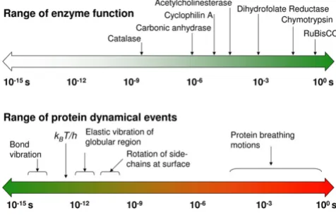
to the time-scales for various events of internal protein dynamics, raising the question whether dynamics and enzyme catalysis are interrelated or not (see Figure 1) [7-12]. It is known that protein dynamics plays a role in many aspects of enzyme function, including substrate/ cofactor binding or release. Its connection to the substrate turnover step, however, has been challenging to ascertain.
An integrated view of protein structure, dynamics, and function is emerging, where proteins are considered as dynamically active machines and internal protein motions are closely linked to function such as enzyme catalysis. Currently there is wide interest, both from exper-imental and computational groups, in investigating this interconnection. A number of investigations have pro-vided fascinating details about the movement of protein parts and their involvement in enzyme function. Tech-niques such as X-ray crystallography and small-angle scat-tering [13,14], NMR studies [15-17], hydrogen-deuterium exchange [18], neutron scattering [19], biochemical and mutational analysis [7,20,21] have provided vital clues at individual time-scales; however, the detailed understand-ing of protein dynamics requires information over a broad range of time-scales. Moreover, the hydration-shell and bulk solvent fluctuations have been suggested to impact protein dynamics, and therefore, protein function [22,23]. Theoretical studies and computational modeling are playing a vital role in investigating the link between protein dynamics, solvent fluctuations and enzyme catal-ysis at multiple time-scales [8,10-12].
In this review, we describe recent biochemical and theo-retical/computational studies that have investigated the link between protein dynamics and enzyme catalysis. In particular, we describe the recent investigations of the peptidyl-prolyl cis/trans isomerization activity of the enzyme cyclophilin A, followed by a discussion on similar evidence from other enzyme reactions, namely the hydride transfer reactions catalyzed by dihydrofolate reductase and by liver alcohol dehydrogenase. There are wide implications of understanding the interconnection between protein structure, dynamics and function such as enzyme catalysis. It is known that enzymes catalyzing the same reactions belong to a protein "fold" family, where the overall characteristic shape of the protein is similar. Also, enzymes catalyzing mechanistically similar reac-tions often belong to the same super-family of protein fold. The benefits of better understanding of enzyme "folds" and dynamics include the possibility of improving the efficiency of microbial factories by engineering of enzymes, as well as designing new enzymes with novel functionalities. Further, there are medical implications of allosteric and cooperative effects for enzyme activity in novel drug design.
Cyclophilin A
The peptidyl-prolyl cis/trans isomerase (PPIase) activity of cyclophilin A (CypA) has been investigated in detail for the link between protein dynamics and enzymatic cataly-sis, both by biochemical experiments and theoretical methods [10-12,15,16]. CypA is a ubiquitously expressed cytosolic protein, which was discovered as the major intracellular receptor protein for the immunosuppressive drug cyclosporin A [24,25]. CypA belongs to the cyclophi-lin class of enzymes, which are involved in many biologi-cal reactions including protein folding, intracellular protein transport and signaling [26,27]. CypA acts as a PPIase, catalyzing the isomerization of peptidyl-prolyl amide bonds that are N-terminal to proline residues in a wide variety of peptides and protein substrates (see Figure 2) [26,28]. Human CypA is a single peptide chain with 165 amino acids. Its molecular architecture consists of an eight-stranded anti-parallel β-barrel with hydrophobic residues forming a core at the center and the active-site located on one face of the molecule (see Figure 3) [29-31]. In addition to the β-strands and α-helices, there are sev-eral flexible surface loop regions as indicated by large tem-perature factors from X-ray crystallographic studies.
A number of factors make CypA an attractive system for investigating the link between internal protein dynamics and enzymatic activity; it is a small protein and does not require metal ions or cofactors for PPIase activity and it catalyzes peptide bond isomerization in a wide variety of substrates. Further, there is also biomedical interest in CypA; cyclophilins are of interest as drug targets because of their likely involvement in the broad spectrum, anti-infective activities of cyclosporin A and
non-immunosup-The range of time-scales involved in substrate turnover step of enzyme catalyzed reactions and internal protein dynamics are similar
Figure 1
pressive derivatives thereof [32,33]. In addition, Gag-encoded capsid protein (CA) from human immunodefi-ciency virus type 1 (HIV-1), is a naturally occurring bio-logically relevant substrate for CypA [16]. The protein-protein complex between CypA and CA has been the sub-ject of many experimental studies [16,34-37]. There is
medical interest in CypA-CA complex, as incorporation of CypA is required for infectious activity of HIV-1 [38,39].
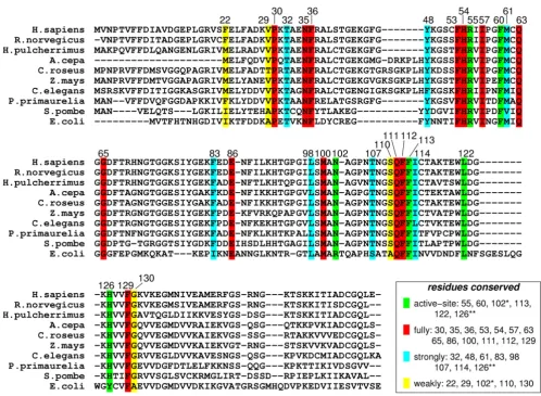
Genomic analysis based on multiple sequence alignment has identified conserved residues in the CypA active-site and also distal to the active-site. This analysis was based on aligning 50 PPIase sequences from 25 diverse organ-isms, ranging from bacteria to human [10]. The results from this analysis are depicted in Figure 4. Detailed struc-tural insights have indicated that the active-site of CypA shows conserved residues forming crucial hydrophobic and hydrophilic interactions with the substrate residues [see Figure 2(b)]. In addition, there are several conserved and semi-conserved residues that are more than 12 Å from the active-site. Until recently, the role of these distal resi-dues in the enzyme function was not very well under-stood. As described below the dynamical motions of some of these residues have been found to play a role in cataly-sis.
NMR studies of CypA performed by Kern and coworkers, have suggested a link between internal protein dynamics and substrate isomerization step [15,16]. The studies were
Three-dimensional structure of CypA
Figure 3
Three-dimensional structure of CypA. Protein secondary structure is represented with cyan arrows (β-sheets) and red helices (α-helices) based on crystal structure from Zhao and Ke (PDB code: 1RMH) [29]. The green labeled regions are flexible surface loops, showing large displacements in X-ray structures (large temperature factors) and NMR relaxation studies. A peptide substrate is shown as orange ball-and-stick model.
78−86
136−144
12−15 68−74 101−107
122−125
The reaction catalyzed by CypA (a) CypA is a member of a family of enzymes known as PPIase, which catalyze the cis/ trans isomerization of peptide bonds N-terminal to proline residues in peptides and proteins (b) The active-site of CypA with a peptide substrate
Figure 2
based on 15N spin relaxation investigations of small pep-tide substrate as well as two-dimensional (2D) 1H-15N heteronuclear exchange studies of the N-terminal of cap-sid protein (CAN) from HIV-1. In these studies, conforma-tional fluctuations within the active-site of CypA were detected on the time-scale of the reaction (hundreds of microseconds) and the rates of conformational dynamics were found to be strongly correlated with the substrate isomerization step. Several active-site and surface loop regions showed motions only in the presence of substrate, these regions included the residues: Arg55, Lys82, Leu98, Ser99, Ala101, Asn102, Ala103, and Gly109. Based on these studies, the authors proposed a reaction mechanism
for PPIase activity of CypA, where the isomerization step takes place with a rate constant of about 9000 s-1, and motions of the protein coincide with the rate of substrate turnover step. CypA residue Arg55 is a major contributor to catalysis [40], for which the observed changes in back-bone conformation are likely to be coupled with motions of the catalytically essential side chain.
Theoretical and computational modeling of the PPIase activity of CypA has provided novel insights into under-standing the relationship between dynamical events in proteins and enzyme catalysis, including the mechanism of rate-enhancement achieved by enzymes [10-12]. Note,
Genomic analysis for sequence conservation of CypA
Figure 4
the time-scale of CypA reaction is hundreds of microsec-onds, which is beyond the reach of present day molecular dynamics simulations. Molecular dynamics, the com-monly used computational technique, is limited to nano-second time-scale (up to 100 nanonano-seconds at best) due to the limitation of available computing power. A different theoretical framework was, therefore, used for detailed computational modeling of the entire reaction pathway. This framework is described briefly below, more details are available in other reviews [9,41]. The theoretical inves-tigations of enzyme catalysis are based on description of the reaction using transition state theory (TST) and gener-ation of a free energy profile as a function of suitable reac-tion coordinate. In the TST framework, protein dynamics has been suggested to impact the reaction rate in two ways. Enzymes can either decrease the activation energy barrier (∆G‡) for the reaction or alter the active-site condi-tions such that more reactive trajectories are converted to
product successfully. Figure 5 shows the behavior of two trajectories, the first trajectory crosses the transition state (TS) barrier but is unsuccessful and returns to the reactant side. The second trajectory crosses the barrier several times before becoming productive. Transmission coefficient (κ) is a corrective pre-factor corresponding to the fraction of reactive trajectories that successfully cross the TS barrier and become productive. For CypA, the free energy profiles were generated using the amide bond dihedral angle of the peptide bond as reaction coordinate. Note, in the computational studies described here the unit of reaction coordinate is degrees (°). The free energy profiles were generated for isomerization of 3 small peptide substrates as well as the biologically relevant substrate CAN. The pro-cedure for generation of these profiles requires multiple simulations of small sections along the reaction path by using molecular dynamics and umbrella sampling [42], and combining them to provide information regarding the time-scale of the reaction [43]. More details about the computational methods can be found in refs. [10] and [11]. In addition to obtaining the free energy profile this modeling procedure also sampled the enzyme-substrate conformations along the entire reaction pathway. These conformations have been used for detailed analysis of structural and dynamical changes during the enzyme reac-tion mechanism.
Structural analysis of the active-site along the course of reaction indicates the role of important hydrophilic (Arg55 and Asn102) and hydrophobic (Phe60, Phe113, Leu122 and His126) residues of CypA in stabilization of the substrate peptide. The target proline from substrate remains essentially fixed in the hydrophobic pocket formed by CypA residues, while the carbonyl oxygen atom of the preceding substrate residue rotates 180°. Quantum mechanical modeling of the active-site indi-cates single bond character for the peptide bond near the TS. The results from theoretical modeling were found to be in agreement with the reaction mechanism proposed on the basis of crystallographic studies [37]. This mecha-nism requires minimum deviation from the ground state crystal structure and displays single bond character for the peptide bond near TS. Dynamical fluctuations of the enzyme backbone in certain regions (CypA 101–104) were found to impact the nature of interactions between the enzyme and substrate, therefore, alter the nature of peptide bond during the course of reaction mechanism.
Computational modeling has identified a variety of inter-nal protein dynamical events linked to CypA enzyme activity, ranging from femtosecond (10-15 s) to microsec-ond and longer (> 10-6 s) time-scales. On one side of this range there are fast motions, occurring at femtosecond-nanosecond time-scales, consisting of harmonic move-ments of bonds, angles and a few atoms. These motions
Schematic illustration of free energy profile for an enzymatic reaction
Figure 5
Schematic illustration of free energy profile for an enzymatic reaction. Protein dynamics can influence reaction rates in two possible ways; by altering height of the activation free energy barrier (∆G‡) and transmission coefficient (κ). k
B is
are commonly referred to as vibrations. On the other side of this range there are concerted conformational fluctua-tions occurring on the microsecond (and longer) time-scale. These slower motions or conformational fluctua-tions, which have been previously referred to as breathing motions, span a large part of the protein. Normal mode analysis is a computational technique that has been com-monly used to obtain information regarding the dynami-cal motions in proteins. This technique provides information about dynamics at several time-scales for a particular protein conformation (present at a local mini-mum). Normal mode analysis is not suitable for obtain-ing the slow protein motions occurrobtain-ing at the time-scale of the reaction due to the large changes in protein conforma-tions involved. Another computational technique, known as quasi-harmonic analysis, can be used to calculate vibra-tional modes from a collection of conformations or sys-tem snapshots [44]. Quasi-harmonic analysis of CypA-substrate conformations along the entire reaction path-way provided protein vibrational modes representing conformational fluctuations at the time-scale of the reac-tion (microsecond-millisecond time-scale). These com-puted slow protein vibrational modes show concerted motions over a large region of the protein, the backbone in several regions of the protein and side-chains of the many residues (especially on the surface) show large dis-placements. In CypA, a subset of these modes was found to be coupled to the reaction; 3 protein vibrational modes with the largest coupling to the catalytic step show dis-placements in several conserved residues in the active-site as well as in other parts of the enzyme structure. Note, these conserved vibrational modes are different from ran-dom thermal fluctuations observed in the biomolecules.
The detailed characterization of internal protein dynamics events linked to enzyme catalysis in CypA has lead to the discovery of a network of protein vibrations (see Figure 6). This network plays an important role in promoting the isomerization reaction [10-12]. The discovery of this net-work is based on identification of 3 protein vibrations on the time-scale of the reaction, investigation of the dynam-ical flexibility of the CypA backbone, monitoring the con-served residues and interactions over the course of enzyme reaction. Dynamical cross-correlation analysis of enzyme parts indicated that several surface loops (distant to each other in sequence) show highly correlated motions during the course of the reaction. These corre-lated motions form the network of vibrations through a series of interactions, as shown by yellow arrows in Figure 6. Note that this network extends from the surface regions of the enzyme all the way to the active-site, through inter-connection of conserved residues and interactions. The vibrations in this network are transmitted to the active-site, where dynamical motions alter the crucial hydropho-bic and hydrophilic interactions between enzyme and substrate. As noted above these interactions play a critical role in the reaction mechanism by controlling the nature of the peptide bond, as well as in rotation of the carbonyl oxygen atom from the residue preceding the target proline of the substrate. Evidence for the existence of this network comes from previous NMR studies, where motions have been detected in network residues only during the sub-strate turnover [15]. Further, the flexibility of the network residues is confirmed by observation of large temperature factors in X-ray studies [29-31,37]. More recently, new investigations performed by Kern and coworkers, using NMR studies have confirmed the presence of this network
A network of coupled protein vibration promoting catalysis in cyclophilin A [10]
Figure 6
in CypA [45]. Moreover, NMR investigations conducted by Blackledge and coworkers have also observed the role of protein motions in transfer of information between β -strands [46]. These recent findings have illustrated the role of coupled networks in propagation of local changes over large distances in protein structure.
The discovered network of protein vibrations has a pro-moting effect on the CypA enzyme activity, and is there-fore, a factor contributing to rate-enhancement. Certain
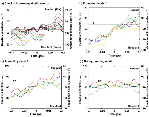
protein vibrational modes alter the reaction by changing the active-site environment such that more reaction trajec-tories cross to the product side. A new theoretical tech-nique has been designed and was used to investigate impact of reaction coupled vibrational modes on the reac-tion mechanism [12]. This technique allows addireac-tion of kinetic energy to a selective vibrational mode and observ-ing the dynamical behavior of the trajectory (see Figure 7). Figure 7(a) shows the change in behavior of trajectories with increasing amount of kinetic energy present in a
pro-Effect of additional kinetic energy in selective protein vibrational modes (a) Increased amount of kinetic energy in a mode cou-pled to the enzyme reaction allows the trajectory to cross the barrier successfully from the reactant side to the product side
Figure 7
Effect of additional kinetic energy in selective protein vibrational modes (a) Increased amount of kinetic energy in a mode cou-pled to the enzyme reaction allows the trajectory to cross the barrier successfully from the reactant side to the product side. The solid black curve represents the native trajectory (no additional kinetic energy), and δ indicates the fraction of system kinetic energy added to the protein vibrational mode. (b)-(d) representative trajectories from simulations with increased kinetic energy in network protein vibrational modes and a non promoting mode. 2% of system kinetic energy was added to protein vibrations mode. Five representative trajectories from each mode are shown in different colors. Not all protein vibra-tional modes show increased barrier recrossing; much less effect on the barrier crossing is seen in a mode not coupled to the reaction [12]. Reprinted with permission from Agarwal et al., J. Am. Chem. Soc. (2005) 127, 15248–15256. Copyright (2005) Amer-ican Chemical Society.
−0.1 −0.05 0 0.05 0.1
Time (ps)
40
80
120
160
Amide bond dihedral angle,
ω
, (
o )
−0.1 −0.05 0 0.05 0.1
Time (ps) 20 60 100 140 Reaction coordinate, ∆ω , ( o ) Reactant (Trans) Product (Cis) TS δ=0.02 δ=0.01 δ=0.005 δ=0.0002 δ=0.001 δ=0.002
−0.1 −0.05 0 0.05 0.1
Time (ps)
60
90
120
150
180 Amide bond dihedral angle,
ω
, (
o )
−0.1 −0.05 0 0.05 0.1
Time (ps) 0 30 60 90 120 Reaction coordinate, ∆ω , ( o ) Reactant Product TS
−0.1 −0.05 0 0.05 0.1
Time (ps) 0 30 60 90 120 Reaction coordinate, ∆ω , ( o )
−0.1 −0.05 0 0.05 0.1
Time (ps)
60
90
120
150
180 Amide bond dihedral angle,
ω , ( o ) Reactant Product TS
−0.1 −0.05 0 0.05 0.1
Time (ps)
60
90
120
150
180 Amide bond dihedral angle,
ω
, (
o )
−0.1 −0.05 0 0.05 0.1
Time (ps) 0 30 60 90 120 Reaction coordinate, ∆ω , ( o ) Reactant Product TS
(a) Effect of increasing kinetic energy (b) Promoting mode 1
tein vibrational mode. The result from further investiga-tions show that the presence of energy in certain reaction coupled promoting modes causes the reaction trajectories to overcome the activation energy barrier quickly and more effectively [see Figure 7(b)–(c)]. Note that not all modes promote the reaction, as indicated by a non-pro-moting mode [that is a mode which is not coupled to the
reaction; see Figure 7(d)]. Also, the theoretical investiga-tions were performed by adding varying amounts of energy to see the effect of these vibrations in a short sim-ulation (picosecond time-scale). The trend indicates that smaller amounts of kinetic energy present in these modes, which is expected to be present in real system, promotes the reaction at longer time-scales (hundreds of microsec-onds). The biophysical role of the discovered network in the enzyme reaction can be understood by observing changes that are introduced in the active-site by reaction promoting vibrations. Detailed analysis indicates that the dynamical behavior of reaction trajectories is correlated with the fluctuations in the enzyme-substrate interactions as a result of increased energy in the protein vibrational mode. Rate-enhancing modes impact the key active-site interactions to make the reaction proceed from reactant side to the product side. An interesting observation, from this analysis, is that the maximum enzyme stabilization occurs close to the TS (consistent with the TS stabilization theory for enzyme catalysis). The role of the reaction pro-moting vibrations could, therefore, be interpreted as inter-nal protein dynamical events that facilitate in the stabilization of the TS.
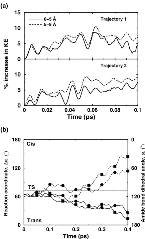
Solvent surrounding the enzyme also plays a role in the enzyme reaction. In many enzyme reactions, hydrolysis of small molecules provides the energy for overcoming the activation energy barrier; however, in other cases the required energy is provided by the thermodynamical fluc-tuations of the solvent. The flucfluc-tuations in the hydration-shell and bulk-solvent surrounding the enzyme are corre-lated with the internal protein motions. Detailed charac-terization of the flexible surface loop regions indicates that the side-chains of several surface residues extend into the solvent and the motion of these residues is coupled to the motion of surrounding solvent molecules. CypA investigations indicated the presence of vibrations (on picosecond) time-scale in several crucial surface residues, which are present in the loop regions showing large dis-placements in reaction promoting vibrational modes. Pre-vious theoretical investigations have also shown that the transition in internal motion of proteins can be driven by the temperature of the solvent [47,48]. In CypA, compu-tational modeling has shown transfer of energy from sol-vent to the external regions of the enzyme. This energy transfer changes the behavior of reaction trajectories, through the network of protein vibrations, to promote catalysis (see Figure 8).
An interesting outcome of detailed characterization of the network of protein vibrations in CypA is the insight into the conservation of protein residues. The genomic analy-sis for sequence conservation reveals that there are several residues which are conserved due to their dynamical role in catalysis. Active-site residues, which are key players in
Effect of additional kinetic energy in first solvation shell of an enzyme
Figure 8
Effect of additional kinetic energy in first solvation shell of an enzyme. (a) Kinetic energy is transferred from the solvent to the protein residues, as indicated by increasing energy in the protein regions (up to 5 Å from protein surface, and between 5 Å and 8 Å from the surface) (b) Two otherwise non-productive regular trajectories (solid lines) become pro-ductive (broken lines) due to transfer of energy from the sol-vent to residues forming parts of the protein vibrations network. The corresponding trajectories are indicated by squares and circles [12]. Reprinted with permission from Agar-wal et al., J. Am. Chem. Soc. (2005) 127, 15248–15256. Copy-right (2005) American Chemical Society.
0 0.1 0.2 0.3 0.4
Time (ps)
0
60
120
180 Amide bond dihedral angle,
ω
, (
o )
0 60 120 180
Reaction coordinate,
∆ω
, (
o )
Trans Cis
TS
(b)
0 5 10 15
0 0.02 0.04 0.06 0.08 0.1
Time (ps) 0
5 10
% increase in KE
Trajectory 1
Trajectory 2 0−5 Å
5−8 Å
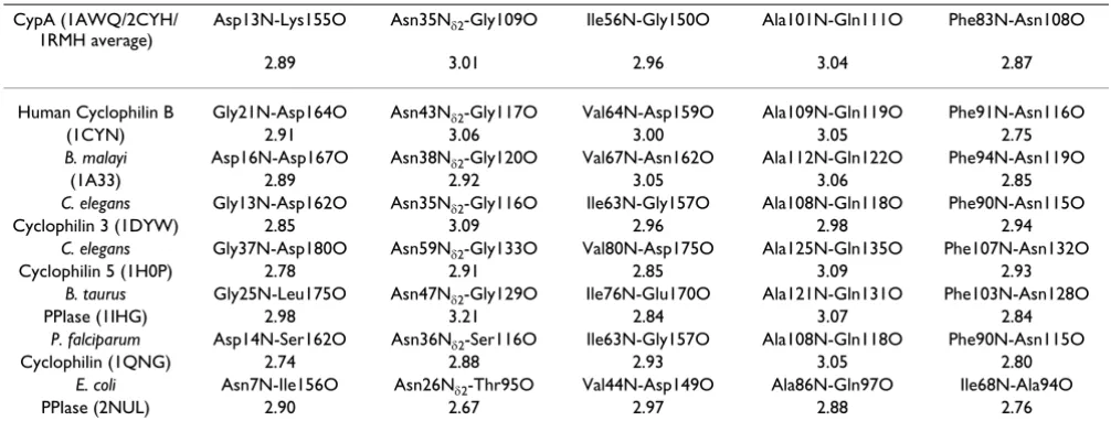
the catalytic step, are often conserved in different species. In addition to the active-site residues (Arg55, Phe60, Asn102 and Ala103), which directly participate in the cat-alytic step, there are several distal residues (including Pro30, Asn35, Phe36, Phe83, Glu86), which are also con-served. Note, that some of these residues are more than 17 Å from the active-site. Results from detailed structural analysis (summarized in Table 1) indicate that these and other distal residues form crucial points and hydrogen bonds in the discovered network and, therefore, are con-served in species ranging from bacteria to human. This finding also has interesting implications on the under-standing of the secondary and tertiary protein structure. The discovered network promoting catalysis may provide some insights into the conservation of protein "folds"; enzymes catalyzing similar reactions often belong to the same fold family, and enzymes catalyzing mechanistically similar reactions belong the same protein super-family [49,50].
Other enzymes: dihydrofolate reductase and liver alcohol dehydrogenase
Experimental and computational investigations have revealed the impact of protein dynamics on catalysis in other enzyme systems. Experimental and computational studies of the enzyme dihydrofolate reductase (DHFR) have indicated a link between protein dynamical events and the substrate turnover step of hydride transfer. X-ray crystallography has demonstrated changes in orientation of surface loops along different sub-states along the
reac-tion pathway [51]. Similarly, the surface loop conforma-tions have been linked to the catalytic step by NMR studies [20]. Theoretical and computational studies using hybrid quantum-mechanical and molecular mechanics (QM/MM) methodology have discovered a network of coupled promoting motions [8,52,53]. Similar to the net-work of protein vibrations in CypA described above, the network in DHFR is also formed by interconnection of residues and crucial interactions ranging from surface regions all the way to active-site. Changes in hydrogen-bonds and crucial interactions along the reaction profile have been observed similar to those present during catal-ysis by CypA. An important discovery by the computa-tional methods was the identification of the residue Ile14 as a dynamical contributor to catalysis. Recently, the importance of this residue in the catalytic step has been confirmed by NMR studies [54]. The presence of this DHFR network has been confirmed by investigations from several research groups [55,56]. Investigations of DHFR have provided evidence that changing the enzyme structure leads to changed dynamics and, therefore, change in function [21,53,57]. Mutation of a single sur-face residue, more than 12 Å away from active site, changes the dynamics and leads to a rate reduction by a factor of 163.
Liver alcohol dehydrogenase (LADH) is another enzyme where dynamical motions of the protein residues have been linked to the catalytic step. Detailed biochemical and computational studies have identified conserved Table 1: Conservation of network hydrogen bonds in cyclophilin structures from various species. 3-dimensional structures were aligned using secondary structure elements and equivalent hydrogen bonds were selected based on sequence and structural
similarities. Hydrogen bond lengths are in given Å and PDB codes are given in parenthesis [10]. Reprinted with permission from Agarwal et al., Biochemistry (2004) 43, 10605–10618. Copyright (2004) American Chemical Society.
CypA (1AWQ/2CYH/ 1RMH average)
Asp13N-Lys155O Asn35Nδ2-Gly109O Ile56N-Gly150O Ala101N-Gln111O Phe83N-Asn108O
2.89 3.01 2.96 3.04 2.87
Human Cyclophilin B Gly21N-Asp164O Asn43Nδ2-Gly117O Val64N-Asp159O Ala109N-Gln119O Phe91N-Asn116O
(1CYN) 2.91 3.06 3.00 3.05 2.75
B. malayi Asp16N-Asp167O Asn38Nδ2-Gly120O Val67N-Asn162O Ala112N-Gln122O Phe94N-Asn119O
(1A33) 2.89 2.92 3.05 3.06 2.85
C. elegans Gly13N-Asp162O Asn35Nδ2-Gly116O Ile63N-Gly157O Ala108N-Gln118O Phe90N-Asn115O Cyclophilin 3 (1DYW) 2.85 3.09 2.96 2.98 2.94
C. elegans Gly37N-Asp180O Asn59Nδ2-Gly133O Val80N-Asp175O Ala125N-Gln135O Phe107N-Asn132O Cyclophilin 5 (1H0P) 2.78 2.91 2.85 3.09 2.93
B. taurus Gly25N-Leu175O Asn47Nδ2-Gly129O Ile76N-Glu170O Ala121N-Gln131O Phe103N-Asn128O
PPIase (1IHG) 2.98 3.21 2.84 3.07 2.84
P. falciparum Asp14N-Ser162O Asn36Nδ2-Ser116O Ile63N-Gly157O Ala108N-Gln118O Phe90N-Asn115O
Cyclophilin (1QNG) 2.74 2.88 2.93 3.05 2.80
E. coli Asn7N-Ile156O Asn26Nδ2-Thr95O Val44N-Asp149O Ala86N-Gln97O Ile68N-Ala94O
active-site residue Val203, whose motion are a key player in altering the active-site chemical environment to pro-mote the reaction [58-63].
Conclusion
In this review, we have presented recent developments that continue to support an emerging integrated view of protein structure, dynamics and function such as enzyme catalysis. The success of microbial cell factories depends on optimal performance of molecular machines inside the cell. Enzymes perform their function with remarkable efficiency, as they increase the reaction rate by many orders of magnitude. Until recently, enzymes (and pro-teins in general) were considered static assemblies; how-ever, recent investigations continue to provide evidence which indicate that enzymes are dynamically active assemblies. Detailed experimental and theoretical/com-putational investigations of enzyme CypA suggest that the internal protein motions are a designed part of the protein structure and contribute to its function of catalyzing pep-tidyl-prolyl cis/trans isomerization. Supporting evidence from other systems (DHFR and LADH) indicates that the interconnection between structure, dynamics and func-tion is present in other enzymes as well.
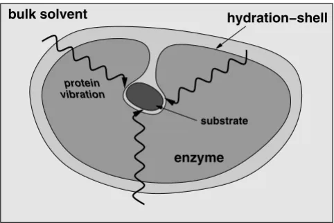
The overall emerging picture of protein dynamics, solvent fluctuations and enzyme function based on recent insights is depicted in Figure 9. Along with structural interactions, internal motions at fast time-scales control
the chemical environment of the active-site favoring the catalytic step to proceed to the product state. The thermo-dynamical fluctuations of the hydration-shell and bulk solvent provide energy to overcome the activation energy barrier (in cases where no other source of energy is avail-able). The flexible surface loop regions of the enzyme show dynamical coupling with the solvent. This dynami-cal coupling allows the transfer of energy from the solvent to the surface regions of the enzyme. This energy is even-tually transferred to the active-site through networks of motions or vibrations. The slower conformational fluctu-ations in the networks (at time-scale of the reaction) alter the enzyme-substrate interactions such that more reaction trajectories cross TS barrier to reach the product state suc-cessfully.
The integrated view is supported by evidence from inves-tigations of many other proteins and enzymes as well [64-66]. Sequence analysis with thermodynamic mapping have indicated long range energetic coupling in proteins [67]; slow conformational fluctuations could possibly be the mechanism of energy transfer over long ranges in pro-tein structure and, therefore, provide insights into under-standing allosteric effects. Simulations have already revealed that energy can be transferred between specific vibrational modes in a protein [68,69]. It is also interest-ing to note that designinterest-ing active-site mimics of the enzymes is difficult and change in enzyme structure far away from the active-site leads to slow or inactive enzymes. The integrated view offers a possible explana-tion, as the distal regions of the enzyme contribute to catalysis through dynamical coupling with the solvent and by transferring the required energy to the active-site. Therefore, this integrated view has wide implications in enzyme chemistry, protein engineering and drug design. Manipulation of enzyme catalyzed reactions may be pos-sible; for example, laser pulse has already been used to ini-tiate an enzyme reaction involving thermally excited protein dynamics (molecular motions on picosecond time-scale) [70]. On the basis of better understanding of enzyme structure, dynamics and function it may be possi-ble to design more efficient enzymes or enzymes with novel functionalities. Further, the understanding of allos-teric and cooperative effects could help in designing better and novel drugs.
List of abbreviations
κ, transmission coefficient
∆G‡, activation energy barrier (energy difference between reactant and the activated state)
CA, capsid protein from HIV-1
CAN, N-terminal of capsid protein A schematic representation of the integrated view of enzyme
structure, dynamics and function
Figure 9
A schematic representation of the integrated view of enzyme structure, dynamics and function. Enzyme structure and internal protein dynamics events play a role in the catalytic step. Conserved residues from the surface to the active-site participate in network of protein motions or vibrations that promotes catalysis. The surface residues are coupled to the thermo-dynamical fluctuations of the solvent, and possibly play a role in transfer of energy from solvent to the protein.
bulk solvent hydration−shell
protein vibration
enzyme
CypA, cyclophilin A
DHFR, dihydrofolate reductase
HIV-1, human immunodeficiency virus type 1
LADH, liver alcohol dehydrogenase
NMR, nuclear magnetic resonance
PPIase, peptidyl-prolyl cis/trans isomerase
TS, transition state
TST, transition state theory
Authors' contributions
PKA drafted and revised the manuscript.
Acknowledgements
PKA would like to thank Dr. Brahma Ghosh for feedback on the manu-script.
References
1. Neet KE: Enzyme catalytic power minireview series. J Biol Chem 1998, 273:25527-25528.
2. Radzicka A, Wolfenden R: A Proficient Enzyme. Science 1995, 267:90-93.
3. Kraut J: How Do Enzymes Work. Science 1988, 242:533-540. 4. Knowles JR: Enzyme Catalysis - Not Different, Just Better.
Nature 1991, 350:121-124.
5. Fischer E: Ber Dtsch Chem Ges 1894, 27:3189.
6. Haldane JBS: Enzymes. London, Longmans, Green; 1930. 7. Cannon WR, Benkovic SJ: Solvation, reorganization energy, and
biological catalysis. J Biol Chem 1998, 273:26257-26260. 8. Agarwal PK, Billeter SR, Rajagopalan PTR, Benkovic SJ,
Hammes-Schiffer S: Network of coupled promoting motions in enzyme catalysis. Proc Natl Acad Sci U S A 2002, 99:2794-2799.
9. Benkovic SJ, Hammes-Schiffer S: A perspective on enzyme catal-ysis. Science 2003, 301:1196-1202.
10. Agarwal PK, Geist A, Gorin A: Protein dynamics and enzymatic catalysis: investigating the peptidyl-prolyl cis-trans isomeri-zation activity of cyclophilin A. Biochemistry 2004, 43:10605-10618.
11. Agarwal PK: Cis/trans isomerization in HIV-1 capsid protein catalyzed by cyclophilin A: insights from computational and theoretical studies. Proteins: Struct Func Bioinform 2004, 56:449-463.
12. Agarwal PK: Role of protein dynamics in reaction rate enhancement by enzymes. J Am Chem Soc 2005, 127:15248-15256.
13. Schramm VL, Shi WX: Atomic motion in enzymatic reaction coordinates. Curr Opin Struct Biol 2001, 11:657-665.
14. Heller WT: Influence of multiple well defined conformations on small-angle scattering of proteins in solution. Acta Crystal-logr D 2005, 61:33-44.
15. Eisenmesser EZ, Bosco DA, Akke M, Kern D: Enzyme dynamics during catalysis. Science 2002, 295:1520-1523.
16. Bosco DA, Eisenmesser EZ, Pochapsky S, Sundquist WI, Kern D: Catalysis of cis/trans isomerization in native HIV-1 capsid by human cyclophilin A. Proc Natl Acad Sci U S A 2002, 99:5247-5252. 17. Wand AJ: Dynamic activation of protein function: A view emerging from NMR spectroscopy. Nat Struct Biol 2001, 8:926-931.
18. Zavodszky P, Kardos J, Svingor A, Petsko GA: Adjustment of con-formational flexibility is a key event in the thermal adapta-tion of proteins. Proc Natl Acad Sci U S A 1998, 95:7406-7411.
19. Zaccai G: Biochemistry - How soft is a protein? A protein dynamics force constant measured by neutron scattering.
Science 2000, 288:1604-1607.
20. Osborne MJ, Schnell J, Benkovic SJ, Dyson HJ, Wright PE: Backbone dynamics in dihydrofolate reductase complexes: Role of loop flexibility in the catalytic mechanism. Biochemistry 2001, 40:9846-9859.
21. Cameron CE, Benkovic SJ: Evidence for a functional role of the dynamics of glycine-121 of Escherichia coli dihydrofolate reductase obtained from kinetic analysis of a site-directed mutant. Biochemistry 1997, 36:15792-15800.
22. Fenimore PW, Frauenfelder H, McMahon BH, Young RD: Bulk Sol-vent and hydration-shell fluctuations, similar to a- and b-fluc-tuations in glasses, control protein motions and functions. P Natl Acad Sci USA 2004, 101:14408-14413.
23. Frauenfelder H, Fenimore PW, McMahon BH: Hydration, slaving and protein function. Biophys Chem 2002, 98:35-48.
24. Handschumacher RE, Harding MW, Rice J, Drugge RJ: Cyclophilin -a Specific Cytosolic Binding-Protein for Cyclosporin-A. Sci-ence 1984, 226:544-547.
25. Takahashi N, Hayano T, Suzuki M: Peptidyl-Prolyl Cis-Trans Iso-merase Is the Cyclosporin-a-Binding Protein Cyclophilin.
Nature 1989, 337:473-475.
26. Gothel SF, Marahiel MA: Peptidyl-prolyl cis-trans isomerases, a superfamily of ubiquitous folding catalysts. Cell Mol Life Sci
1999, 55:423-436.
27. Rovira P, Mascarell L, Truffa-Bachi P: The impact of immunosup-pressive drugs on the analysis of T-cell activation. Curr Med Chem 2000, 7:673-692.
28. Fischer G: Chemical aspects of peptide bond isomerisation.
Chem Soc Rev 2000, 29:119-127.
29. Zhao YD, Ke HM: Crystal structure implies that cyclophilin predominantly catalyzes the trans to cis isomerization. Bio-chemistry 1996, 35:7356-7361.
30. Zhao YD, Ke HM: Mechanistic implication of crystal structures of the cyclophilin-dipeptide complexes. Biochemistry 1996, 35:7362-7368.
31. Vajdos FE, Yoo SH, Houseweart M, Sundquist WI, Hill CP: Crystal structure of cyclophilin A complexed with a binding site pep-tide from the HIV-1 capsid protein. Protein Sci 1997, 6:2297-2307.
32. Page AP, Kumar S, Carlow CKS: Parasite Cyclophilins and Antiparasite Activity of Cyclosporine-A. Parasitology Today
1995, 11:385-388.
33. Chappell LH, Wastling JM: Cyclosporine-a - Antiparasite Drug, Modulator of the Host-Parasite Relationship and Immuno-suppressant. Parasitology 1992, 105:S25-S40.
34. Gamble TR, Vajdos FF, Yoo SH, Worthylake DK, Houseweart M, Sundquist WI, Hill CP: Crystal structure of human cyclophilin A bound to the amino- terminal domain of HIV-1 capsid. Cell
1996, 87:1285-1294.
35. Yoo SH, Myszka DG, Yeh CY, McMurray M, Hill CP, Sundquist WI: Molecular recognition in the HIV-1 capsid/cyclophilin a com-plex. J Mol Biol 1997, 269:780-795.
36. Saphire ACS, Bobardt MD, Gallay PA: trans-complementation rescue of cyclophilin A-deficient viruses reveals that the requirement for cyclophilin A in human immunodeficiency virus type 1 replication is independent of its isomerase activ-ity. J Virol 2002, 76:2255-2262.
37. Howard BR, Vajdos FF, Li S, Sundquist WI, Hill CP: Structural insights into the catalytic mechanism of cyclophilin A. Nat Struct Biol 2003, 10:475-481.
38. Braaten D, Franke EK, Luban J: Cyclophilin A is required for an early step in the life cycle of human immunodeficiency virus type 1 before the initiation of reverse transcription. J Virol
1996, 70:3551-3560.
39. Wiegers K, Krausslich HG: Differential dependence of the infec-tivity of HIV-1 group O isolates on the cellular protein cyclo-philin A. Virology 2002, 294:289-295.
40. Li G, Cui Q: What is so special about Arg 55 in the catalysis of cyclophilin A? insights from hybrid QM/MM simulations. J Am Chem Soc 2003, 125:15028-15038.
Publish with BioMed Central and every scientist can read your work free of charge "BioMed Central will be the most significant development for disseminating the results of biomedical researc h in our lifetime."
Sir Paul Nurse, Cancer Research UK
Your research papers will be:
available free of charge to the entire biomedical community
peer reviewed and published immediately upon acceptance
cited in PubMed and archived on PubMed Central
yours — you keep the copyright
Submit your manuscript here:
http://www.biomedcentral.com/info/publishing_adv.asp
BioMedcentral
42. Torrie GM, Valleau JP: Non-Physical Sampling Distributions in Monte-Carlo Free-Energy Estimation - Umbrella Sampling.
J Comput Phys 1977, 23:187-199.
43. Kumar S, Bouzida D, Swendsen RH, Kollman PA, Rosenberg JM: The Weighted Histogram Analysis Method for Free-Energy Cal-culations on Biomolecules .1. The Method. J Comput Chem
1992, 13:1011-1021.
44. Levy RM, Karplus M, Kushick J, Perahia D: Evaluation of the Con-figurational Entropy for Proteins - Application to Molecular-Dynamics Simulations of an Alpha-Helix. Macromolecules 1984, 17:1370-1374.
45. Eisenmesser EZ, Millet O, Labeikovsky W, Korzhnev DM, Wolf-Watz M, Bosco DA, Skalicky JJ, Kay LE, Kern D: Intrinsic dynamics of an enzyme underlies catalysis. Nature 2005, 438:117-121. 46. Bouvignies G, Bernado P, Meier S, Cho K, Grzesiek S, Bruschweiler
R, Blackledge M: Identification of slow correlated motions in proteins using residual dipolar and hydrogen-bond scalar couplings. P Natl Acad Sci USA 2005, 102:13885-13890.
47. Tournier AL, Xu JC, Smith JC: Translational hydration water dynamics drives the protein glass transition. Biophys J 2003, 85:1871-1875.
48. Tournier AL, Xu JC, Smith JC: Solvent caging of internal motions in myoglobin at low temperatures. Physchemcomm 2003:6-8. 49. Gerlt JA, Babbitt PC: Mechanistically diverse enzyme
super-families: the importance of chemistry in the evolution of catalysis. Curr Opin Chem Biol 1998, 2:607-612.
50. Babbitt PC, Gerlt JA: Understanding enzyme superfamilies -Chemistry as the fundamental determinant in the evolution of new catalytic activities. J Biol Chem 1997, 272:30591-30594. 51. Sawaya MR, Kraut J: Loop and subdomain movements in the
mechanism of Escherichia coli dihydrofolate reductase: Crystallographic evidence. Biochemistry 1997, 36:586-603. 52. Agarwal PK, Billeter SR, Hammes-Schiffer S: Nuclear quantum
effects and enzyme dynamics in dihydrofolate reductase catalysis. J Phys Chem B 2002, 106:3283-3293.
53. Watney JB, Agarwal PK, Hammes-Schiffer S: Effect of mutation on enzyme motion in dihydrofolate reductase. J Am Chem Soc
2003, 125:3745-3750.
54. Schnell JR, Dyson HJ, Wright PE: Effect of cofactor binding and loop conformation on side chain methyl dynamics in dihy-drofolate reductase. Biochemistry 2004, 43:374-383.
55. Garcia-Viloca M, Truhlar DG, Gao JL: Reaction-path energetics and kinetics of the hydride transfer reaction catalyzed by dihydrofolate reductase. Biochemistry 2003, 42:13558-13575. 56. Thorpe IF, Brooks CL: The coupling of structural fluctuations
to hydride transfer in dihydrofolate reductase. Proteins: Struct Func Bioform 2004, 57:444-457.
57. Rajagopalan PTR, Lutz S, Benkovic SJ: Coupling interactions of dis-tal residues enhance dihydrofolate reductase cadis-talysis: Muta-tional effects on hydride transfer rates. Biochemistry 2002, 41:12618-12628.
58. Bahnson BJ, Colby TD, Chin JK, Goldstein BM, Klinman JP: A link between protein structure and enzyme catalyzed hydrogen tunneling. Proc Natl Acad Sci U S A 1997, 94:12797-12802. 59. Colby TD, Bahnson BJ, Chin JK, Klinman JP, Goldstein BM: Active
site modifications in a double mutant of liver alcohol dehy-drogenase: Structural studies of two enzyme-ligand com-plexes. Biochemistry 1998, 37:9295-9304.
60. Agarwal PK, Webb SP, Hammes-Schiffer S: Computational studies of the mechanism for proton and hydride transfer in liver alcohol dehydrogenase. J Am Chem Soc 2000, 122:4803-4812. 61. Webb SP, Agarwal PK, Hammes-Schiffer S: Combining electronic
structure methods with the calculation of hydrogen vibra-tional wavefunctions: Application to hydride transfer in liver alcohol dehydrogenase. J Phys Chem B 2000, 104:8884-8894. 62. Billeter SR, Webb SP, Agarwal PK, Iordanov T, Hammes-Schiffer S:
Hydride transfer in liver alcohol dehydrogenase: Quantum dynamics, kinetic isotope effects, and role of enzyme motion. J Am Chem Soc 2001, 123:11262-11272.
63. Billeter SR, Webb SP, Iordanov T, Agarwal PK, Hammes-Schiffer S: Hybrid approach for including electronic and nuclear quan-tum effects in molecular dynamics simulations of hydrogen transfer reactions in enzymes. J Chem Phys 2001, 114:6925-6936.
64. Hammes GG: Multiple conformational changes in enzyme catalysis. Biochemistry 2002, 41:8221-8228.
65. Kohen A: Kinetic isotope effects as probes for hydrogen tun-neling, coupled motion and dynamics contributions to enzyme catalysis. Prog React Kinet Mec 2003, 28:119-156. 66. Tousignant A, Pelletier JN: Protein motions promote catalysis.
Chem Biol 2004, 11:1037-1042.
67. Lockless SW, Ranganathan R: Evolutionarily conserved pathways of energetic connectivity in protein families. Science 1999, 286:295-299.
68. Moritsugu K, Miyashita O, Kidera A: Vibrational energy transfer in a protein molecule. Phys Rev Lett 2000, 85:3970-3973. 69. Moritsugu K, Miyashita O, Kidera A: Temperature dependence of
vibrational energy transfer in a protein molecule. J Phys Chem B 2003, 107:3309-3317.
![Figure 3Three-dimensional structure of CypAThree-dimensional structure of CypA. Protein secondary structure is represented with cyan arrows (β-sheets) and red helices (α-helices) based on crystal structure from Zhao and Ke (PDB code: 1RMH) [29]](https://thumb-us.123doks.com/thumbv2/123dok_us/9102822.1902979/3.612.57.296.81.452/dimensional-structure-cypathree-dimensional-structure-secondary-represented-structure.webp)
![Figure 6A network of coupled protein vibration promoting catalysis in cyclophilin A [10]A network of coupled protein vibration promoting catalysis in cyclophilin A [10]](https://thumb-us.123doks.com/thumbv2/123dok_us/9102822.1902979/6.612.54.554.87.226/vibration-promoting-catalysis-cyclophilin-vibration-promoting-catalysis-cyclophilin.webp)