R E S E A R C H
Open Access
Pain in experimental autoimmune encephalitis: a
comparative study between different mouse
models
Jianning Lu
1, Martina Kurejova
1, Laura N Wirotanseng
1, Ralf A Linker
2, Rohini Kuner
1and Anke Tappe-Theodor
1*Abstract
Background:Pain can be one of the most severe symptoms associated with multiple sclerosis (MS) and develops with varying levels and time courses. MS-related pain is difficult to treat, since very little is known about the mechanisms underlying its development. Animal models of experimental autoimmune encephalomyelitis (EAE) mimic many aspects of MS and are well-suited to study underlying pathophysiological mechanisms. Yet, to date very little is known about the sensory abnormalities in different EAE models. We therefore aimed to thoroughly characterize pain behavior of the hindpaw in SJL and C57BL/6 mice immunized with PLP139-151peptide or MOG35-55 peptide respectively. Moreover, we studied the activity of pain-related molecules and plasticity-related genes in the spinal cord and investigated functional changes in the peripheral nerves using electrophysiology.
Methods:We analyzed thermal and mechanical sensitivity of the hindpaw in both EAE models during the whole disease course. Qualitative and quantitative immunohistochemical analysis of pain-related molecules and
plasticity-related genes was performed on spinal cord sections at different timepoints during the disease course. Moreover, we investigated functional changes in the peripheral nerves using electrophysiology.
Results:Mice in both EAE models developed thermal hyperalgesia during the chronic phase of the disease. However, whereas SJL mice developed marked mechanical allodynia over the chronic phase of the disease, C57BL/ 6 mice developed only minor mechanical allodynia over the onset and peak phase of the disease. Interestingly, the magnitude of glial changes in the spinal cord was stronger in SJL mice than in C57BL/6 mice and their time course matched the temporal profile of mechanical hypersensitivity.
Conclusions:Diverse EAE models bearing genetic, clinical and histopathological heterogeneity, show different profiles of sensory and pathological changes and thereby enable studying the mechanistic basis and the diversity of changes in pain perception that are associated with distinct types of MS.
Background
Multiple sclerosis (MS) is one of the most common neurological diseases mostly affecting young adults. It is an incurable, chronic inflammatory, progressive neuroin-flammatory and neurodegenerative disease with a still unclear etiology. Among others, pain is one of the critical MS symptoms. While research on pain in MS is per-formed with increasing frequency, the literature remains ambiguous to date. Many studies are based on question-naires and the reports on pain prevalence in MS patients
vary from 29% [1] up to 86% [2]. Some studies report no difference in the frequency of pain in MS patients com-pared to the background population, but report a higher intensity and impact of pain on daily life in MS patients [3]. It has been reported that 32% of patients indicate pain among the most severe symptoms of MS [4], and 12% of various pain syndromes are even classified as the worst symptom of the MS itself [5]. Symptoms of neuro-pathic pain, including mechanical or cold allodynia as well as thermal and mechanical hyperalgesia have been described [6-9]. Chronic pain in MS severely reduces the quality of the patient’s life and therefore deserves detailed analysis. So far, not much is known about the mechan-isms underlying MS-related pain and its treatment * Correspondence:[email protected]
1
Pharmacology Institut, University of Heidelberg, Im Neuenheimer Feld 366, Heidelberg D-69120, Germany
Full list of author information is available at the end of the article
remains difficult. Therefore, there is a major and unmet need for basic research on molecular mechanisms under-lying the development and chronicity of pain in MS.
Various animal models mimicking the disease have been used for decades, the most prevalent being experi-mental autoimmune encephalomyelitis (EAE), which closely resembles MS [10]. The use of diverse immuno-genic peptides against central nervous system (CNS) components in the EAE model enables simulation of di-verse types of MS (for example, relapsing-remitting, pro-gressive, etcetera). A major difference between MS and EAE is that whereas MS is a spontaneous disease, EAE has to be artificially induced using strong immune adju-vants. Only particular combinations of antigen and ro-dent strain can elucidate EAE [11,12], leading to specific disease profiles [11-14]. Moreover, EAE is studied mainly in inbred strains; hence, the genetic heterogeneity which is critical in the MS populations is only reflected when different models of EAE are studied in parallel [11].
Pain hypersensitivity of the hindpaw has been previ-ously reported in mouse EAE models [15-18]. However, a comprehensive temporal analysis and comparison thereof in different models representing different sub-types of MS has been missing so far. In this study, we sought to comprehensively analyze nociceptive sensitiv-ity during the whole disease course in two different EAE mouse models, namely SJL mice immunized with PLP139-151 peptide and C57BL/6 mice immunized with MOG35-55 peptide. Moreover, we performed detailed immunohistochemical analyses to address pathophysio-logical changes that are potentially linked to differences in pain behavior between the two models, and we per-formed electrophysiological measurements on peripheral nerve terminals. Our results showed that distinct EAE models are associated with specific profiles and temporal courses of changes in pain sensitivity as well as particu-lar patterns of neurochemical changes in the spinal cord.
Methods
Animals and induction of experimental autoimmune encephalomyelitis
Female SJL/J mice were purchased from Harlan Labora-tories (Borchen, Germany) and C57BL/6 J mice were pur-chased from Janvier (Le Genest Saint Isle, France). For the induction of EAE, female mice at age eight weeks, received subcutaneous injections in both flanks of either 50 μg MOG35-55 peptide or 100 μg PLP139-151 peptide (synthesized at German Cancer Research Center; DKFZ, Genomics and Proteomics Core Facilities, Peptide Synthe-sis, Heidelberg, Germany) in PBS emulsified in an equal volume of complete Freund's adjuvant (CFA) containing Mycobacterium tuberculosis H37RA (Difco, Detroit, MI, USA) at a final concentration of 0.5 mg/ml under Iso-fluran anesthesia. Control mice were immunized with
ovalbumin (50μg) in PBS/CFA. Two injections of pertus-sis toxin (List Biological Laboratories Inc., Campbell, CA, USA; 200 ng per mouse intraperitoneal) were given on the day of immunization and 48 hours later. Animals were weighed and scored for clinical signs of disease on a daily basis. Disease severity was assessed using a scale ranging from 0 to 10; scores were as follows [19]: 0 = normal; 1 = reduced tone of tail; 2 = limp tail, impaired righting; 3 = absent righting; 4 = gait ataxia; 5 = mild paraparesis of hindlimbs; 6 = moderate paraparesis; 7 = severe paraparesis or paraplegia; 8 = tetraparesis; 9 = moribund; 10 = death. If necessary, food was provided on the cage floor.
Behavioral nociceptive testing
All animal procedures including the EAE protocol under section:`Animals and induction of experimental auto-immune encephalomyelitis´ were conducted with the ap-proval of the ethics commitee by the local governing body (Regierungspräsidium Karlsruhe, Germany). All behavioral measurements were done in awake, unrestrained, age-matched female mice. All tests were performed in an ap-propriate quiet room between 10 am and 4 pm.
Analysis of paw withdrawal latency in response to an infrared beam (which generates a heat ramp) was done as described in earlier publications [20,21] (for example, Plantar test apparatus, Hargreaves' Method, Ugo Basile Inc.). Mechanical sensitivity was tested in the same co-hort of animals via manual application of calibrated von Frey hair filaments (0.04 g to 1.4 g) to the plantar sur-face of the hindpaw as described for earlier studies [20]. The hindpaw withdrawal latency upon heat stimulation using the plantar test apparatus and the hindpaw re-sponse to von Frey hair stimulation was assessed every second to third day, alternately.
Locomotion and exploratory activity
General activity and novelty-induced explorative behavior was measured by using an open field chamber (44 x 44 cm; Ugo Basile, Comerio, Italy) under normal lighting conditions. A video tracking software (ANY-Maze, Ugo Basile, Italy) was used to monitor the mice over ten min-utes. The following parameters were analyzed: distance travelled (horizontal activity), speed and immobility time.
Afferent recordings in skin-nerve preparation
attached and mounted in an organ bathinside-upto ex-pose the dermis. The preparation was perfused with an oxygen-saturated modified synthetic interstitial fluid so-lution containing (in mM) 123 NaCl, 3.5 KCl, 0.7 MgSO4, 1.5 NaH2PO3, 1.7 NaH2PO4, 2.0 CaCl2, 9.5 so-dium gluconate, 5.5 glucose, 7.5 sucrose, and 10 HEPES at a temperature of 32 ± 1°C and pH 7.4 ± 0.05. Fine fila-ments were teased from the desheathed nerve, placed in separate chamber, and placed on a recording electrode.
Nerve fibers were classified according to their conduc-tion velocities, von Frey thresholds, and firing properties. Electrical stimulation of the nerve fiber was employed to calculate conduction velocities of individual nerve fibers. Fibers which conducted <1 m/s, fibers conducting be-tween 1 to 10 m/s, and the fibers conducting with the velocity >10 m/s were considered to be unmyelinated C-fibers, myelinated Aδ-fibers and thickly myelinated low threshold mechanoceptors (RA and SA), respectively. The threshold for each unit was tested using calibrated von Frey filaments; the thinnest filament that elicited three action potentials in the time of approximately 2 seconds of pressing the filament on the units was taken as a threshold.
Once the receptive field was identified using the glass rod, a computer-controlled linear stepping motor (Nanomotor Kleindiek Nanotechnik, Reutlingen, Ger-many) was used to apply standardized mechanical stim-uli. Each fiber was tested with a series of displacement mechanical stimuli ranging from 6 to 384 μm for both control and EAE animals. Electrophysiological data were collected with a Powerlab 4.0 system (ADInstruments, Spechbach, Germany) and analyzed off-line with the spike histogram extension of the software.
Immunohistochemistry
Mice were perfused with 0.1 M phosphate buffer saline and 4% paraformaldehyde (PFA). Spinal cords were
isolated and post-fixed for up to 16 hours in 4% PFA. Free-floating vibratome sections (50 μm) were processed for immunofluorescence protocol. Sections were incu-bated for 30 minutes at 80°C in prewarmed 10 mM so-dium citrate buffer (pH 8) for antigen retrieval [22] and processed according to standard immunofluorescence protocol. The following antibodies were used: rabbit poly-clonal anti-CGRP (Product ID : 24112; 1:200; ImmunoStar Inc., Hudson, WI, USA), Streptavidin-conjugated Isolectin B4 (1:100; Vector laboratories, Burlingame, CA, USA), rabbit polyclonal Iba-1 (Product ID : 019–19741; 1:500; Wako, Richmond, VA, USA), mouse polyclonal anti-GFAP (Product ID : 73–240; 1:200; NeuroMab, Antibodies Incorporated, Davis, CA, USA), mouse monoclonal NeuN (Product ID : MAB377; 1:200; Millipore, Billerica, MA, USA), rabbit polyclonal anti-Fox3 (Product ID : MCA-1B7; 1:500; EnCor Biotechnology, Gainsville, FL, USA).
Illustrations and densitometry
Fluorescence images were obtained using a laser scan-ning confocal microscope (Leica TCS AOBS, Bensheim, Germany). For quantitative measurement of microglia and astrocytes, images were obtained in a confocal series over a thickness of 50 μm using the same laser intensity in all images. The fluorescence signal intensity in per unit area was measured densitometrically using NIH ImageJ software (National Institutes of Health, Bethesda, Maryland, USA) Data were averaged from four areas per section and two sections per mouse in groups of at least four animals in three independent experiments.
Statistics
If not indicated differently, all data are presented as mean ± standard error of the mean (S.E.M.). For com-parisons of multiple groups, analysis of variance (ANOVA) for random measures was performed followed by post-hoc Bonferroni’s test, and for the comparison of
EAE weight control weight EAE score control score
SJL - PLP139 -151 C57BL/6 - MOG35 -55
We
ig
h
t
(g
)
days after immunization
10 20 30 40 50
0 1 2 3 4 5 6 7 9 8 10
0 5 10 15 20 25 30
EA
E s
c
o
re
days after immunization
-1 10 20 30 40
0 1 2 3 4 5 6 7 9 8 10
1
-1
0 5 10 15 20 25 30
E
A
E
sco
re
We
ig
h
t
(g
)
A
B
pre 2 4 7 9 11 141822242729374244 pre 2 7 9 11 15 172125303642444951 time after immunization (days) time after immunization (days)
re s p ons e laten c y (s ec ) re s p ons e laten c y (s ec ) Hargraves test 0 10 20 30 40 50 re s pons e fr eque nc y (% )
pre 3 8 10 12 15 1719212325283036384143
time after immunization (days) von Frey 0.07 g
0 10 20 30 40 50 re sp o n se fr e q u e n cy (% )
pre 3 8 10 12 15 1719212325283036384143
time after immunization (days) von Frey 0.4 g
0 20 40 60 80 100 re s pons e fr eque nc y (% )
pre 3 8 10 12 15 1719212325283036384143
time after immunization (days) von Frey 1.0 g
0 10 20 30 40 50 re s p ons e fr eque nc y (% )
pre 3 8 10 14 16 1822242831353739434550
time after immunization (days) von Frey 0.07 g
60 70 80 0 10 20 30 40 50 re sp o n se fr e q u e n cy (% )
von Frey 0.4 g
60 70 80
pre 3 8 10 14 16 1822242831353739434550
time after immunization (days)
pre 3 8 10 14 16 1822242831353739434550
time after immunization (days) 0 20 40 60 80 100 0 20 40 60 80 100
von Frey 1.0 g Hargraves test * † † † peak onset peak onset peak onset peak onset
B
A
D
C
F
E
H
G
0 2 4 6 8 10 12 0 2 4 6 8 10 12 † † † † † † † † † † * * * * † † * * * † † peak † † * * * † † † † † † † † † † † * * * onset peak peak onset onset peak peak onset * * * * * * * † † † † † † † † † † † † † † † † † † † † † † † † † * † † * † † † † † † † † † † † † † † † † † † † † † † † † †control SJL PLP139-151 control C57 MOG35-55
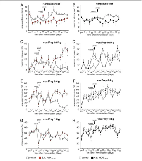
Figure 2Analysis of nociceptive sensitivity in SJL mice immunized with PLP139-151peptide, C57 mice immunized with MOG35-55
two groups Student’st-Test was used to determine sta-tistically significant differences. A value of P<0.05 was considered to be statistically significant.
Results
Disease progression, pain and locomotion
We actively immunized female mice from the SJL and C57BL/6 strains with either the PLP139-151peptide or the MOG35-55peptide (referred to henceforth as SJL-EAE or C57-EAE mice, respectively). Control mice underwent the same immunization protocol using ovalbumin. SJL-EAE mice showed a typical relapsing-remitting disease pattern, whereas C57-EAE mice developed chronic EAE. After immunization, SJL-EAE mice displayed the first signs of disease onset with tail weakness on day 10 and reached a peak in motor deficit functions at day 12 (Figure 1A), whereas C57-EAE mice showed the first symptoms at day 11 and a maximal disease score at day 17 (Figure 1B). As usually seen, EAE mice lost 1 to 2 g of body weight immediately preceding the onset of the dis-ease (Figure 1). The degree of the EAE in the chronic phase was comparable over both models, as indicated by a similar disease score (Figure 1).
In addition to monitoring clinical disease symptoms on a daily basis over 44 days (SJL-EAE mice) or 52 days (C57-EAE mice), we investigated nociceptive thresholds in response to heat and mechanical stimuli. We found that the response latency towards heat stimuli dropped significantly in SJL-EAE and C57-EAE mice following immunization as compared to basal response latencies (Figure 2A,B). Mice in both EAE models developed sig-nificant thermal hyperalgesia in the chronic phase of the disease (Figure 2A,B; Table 1). Thus, the time course of thermal hyperalgesia was not different across the two models.
We applied mechanical pressure via von Frey hair filaments (0.04 g to 1.4 g force) to the plantar surface of the hindpaws. The application of low magnitude of forces (von Frey filaments of forces between 0.04 g to 0.07 g), which do not normally evoke nociceptive with-drawal in control mice, elicited withwith-drawal in SJL-EAE mice in the chronic phase of the disease starting from day 36 onwards and lasting over the whole period of in-vestigation (data with 0.07 g force are shown in Figure 2C). The same stimulus also elicited withdrawal behavior in C57-EAE mice but in a different temporal time frame: in the onset and peak phase of the disease (Figure 2D). The application of more intense forces to the plantar surface of the paw (von Frey hair filaments between 0.16 g to 0.6 g), that normally evoke mild nociceptive withdrawal in control mice, resulted in a significant increase in withdrawal response frequency in SJL-EAE mice in the chronic phase of the disease, start-ing from day 28 after immunization and continustart-ing
over the whole observation period (data with 0.4 g force are shown in Figure 2E), whereas the withdrawal behavior of C57-EAE mice did not differ from control mice (Figure 2F). Moreover, we found that mechanical allodynia correlated with the clinical scores. SJL-EAE mice with higher clinical scores (score 5 to 6) showed a more pronounced mechanical allodynia than EAE mice with moderate symptoms (score 3 to 4) (Figure 3). Interestingly, the paw withdrawal response frequency towards the application of von Frey filaments of stron-ger force (1 g or 1.4 g) was comparable between either SJL-EAE mice and control mice (data with 1.0 g force are shown in Figure 2G) or C57-EAE mice and controls (Figure 2H). This shows that SJL-EAE mice develop nociceptive mechanical allodynia in the chronic phase of the disease. The differences in the behavioral pheno-types are summarized in Table 1.
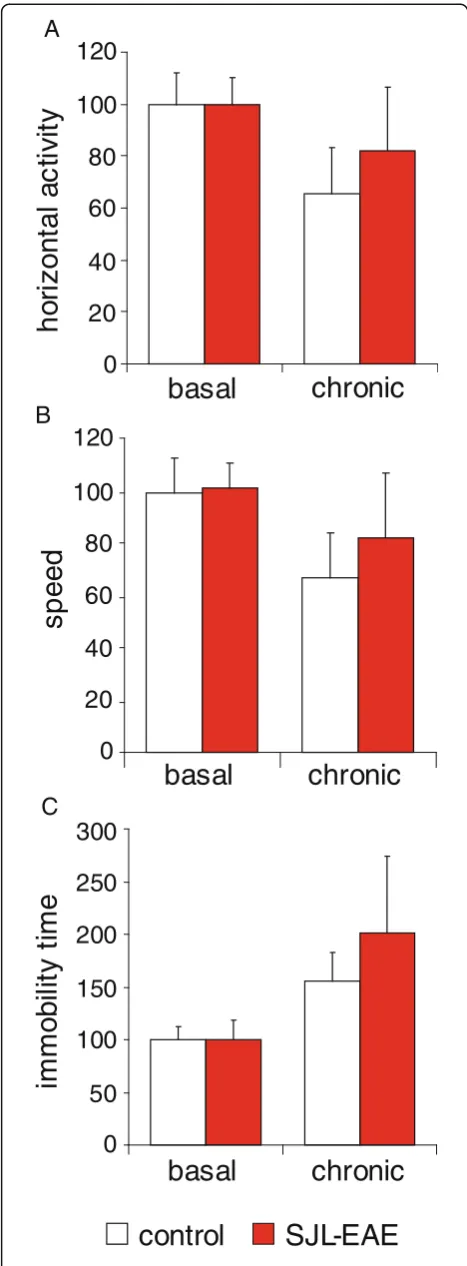
Intrigued by the marked mechanical hypersensitivity in the chronic phase of EAE in SJL mice, we questioned whether their locomotor activity would be altered. Using the open field test apparatus SJL-EAE mice did not dem-onstrate any difference in horizontal activity when com-pared to either the control mice or to their basal behavior before the induction of EAE (Figure 4A). Add-itional parameters, as movement speed (Figure 4B) or immobility time (Figure 4C) were not different between EAE and control animals in the chronic phase of the dis-ease or as compared to basal behavior.
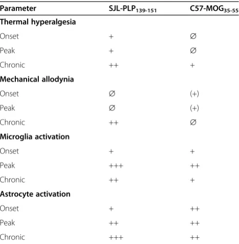
Table 1 Summary and overview of the main
characteristics of SJL PLP139-151 peptide immunized mice and C57 MOG35-55 peptide immunized mice
Parameter SJL-PLP139-151 C57-MOG35-55
Thermal hyperalgesia
Onset + ∅
Peak + ∅
Chronic ++ +
Mechanical allodynia
Onset ∅ (+)
Peak ∅ (+)
Chronic ++ ∅
Microglia activation
Onset + +
Peak +++ ++
Chronic ++ +
Astrocyte activation
Onset + ++
Peak ++ ++
Chronic +++ ++
Thus, SJL-EAE mice did not reveal aberrant behavioral changes associated with EAE despite the presence of nociceptive hypersensitivity to sensory stimuli.
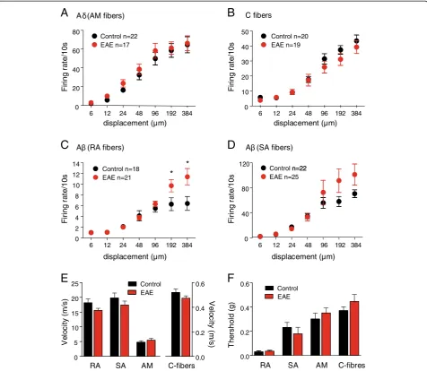
Electrophysiological analyses of peripheral nerve activity In order to characterize the firing properties of peripheral afferents in the chronic phase of the disease, the skin nerve preparation of the saphenous nerve was employed on eight SJL-EAE mice and seven control mice in the chronic phase of the disease (day 35 to 45) (Figure 5). Fir-ing properties of four different fiber types innervatFir-ing the hindpaw were investigated in response to graded mechan-ical stimuli, namely mechanosensitive C-fiber nociceptors, Aδ mechanonociceptors, SA, and RA low-threshold Aβ mechanoceptors, which were identified on the basis of stimulation as well as conduction and firing properties. Stimulus–response functions of C-fibers and Aδ mechan-onociceptors from control and SJL-EAE mice demon-strated no significant changes in the responsiveness to mechanical stimulation (Figure 5A, 5B). Low-threshold SA and RA Aβfibers isolated from the SJL-EAE animals showed a slight or even statistically significant increase in responses to higher stimulus intensities. Additionally RA and SA low-threshold Aβ fibers and non-myelinated C-fibers (Figure 5E) showed a slight decrease in conduction velocity. There were no changes in mechanical thresholds of different afferent fibers (Figure 5F). So, the functional
properties of the nerve fibers in the chronic phase of the EAE are unaltered and unlikely to contribute to the sen-sory abnormalities.
Immunohistochemistry on the spinal cord
We investigated lumbar spinal cord section of SJL-EAE mice and control immunized mice at different time points during EAE for the expression of different pain-or EAE-related markers. Because not only white matter abnormalities but also grey matter abnormalities are a basic phenomenon in EAE, we investigated the expres-sion of various key marker proteins at 2 to 3 days after immunization (‘pre’time point), at disease onset, at peak and in the chronic phase of the disease (day 35 to 45 after EAE induction).
We found a downregulation of NeuN expression throughout the whole spinal cord at disease onset and in the peak phase and an almost complete recovery of NeuN immunogenicity in the chronic phase as compared to con-trol mice (Figure 6A). Recently, NeuN has been identified as the Fox-3 gene product [23]. Therefore, we performed co-labeling of anti-NeuN with anti-Fox-3 antibody. Inter-estingly, we did not find any difference in Fox-3 expres-sion during the time course of the EAE (Figure 6B), indicating no alteration in the amount of neuronal cells during the time course of the EAE. The loss of NeuN immunoreactivity might be accompanied with specific changes in the EAE disease that lead to a change in NeuN antigenicity, as has been reported in other conditions [24,25].
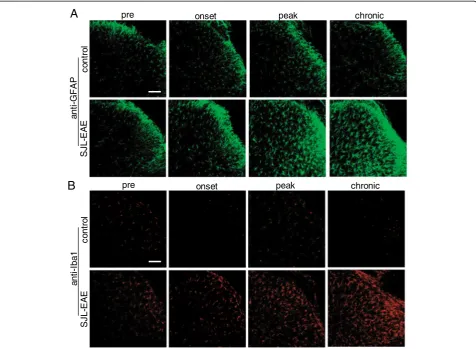
Additionally we analyzed the patterning of the neuro-peptide calcitonin gene-regulated neuro-peptide (CGRP) and the nonpeptidergic isolectin B4 (IB4). Although there was no difference in the density of CGRP-immunoreactive fibers in the spinal dorsal horn in SJL-EAE mice or control mice during the time course of the EAE (Figure 7A), we observed an increase in IB4-positive signals throughout the whole spinal cord at the onset of the disease (Figure 7B). We registered maximal increase in IB4 ex-pression at the peak stage of the disease, which decreased in the chronic phase (Figure 7B). Because IB4 selectively binds activated microglia cells [26], our results indicate a strong activation of microglia in SJL-EAE mice at disease onset and at peak phase of the disease. Co-labeling studies with anti-GFAP, a marker for astrocytes and anti-Iba1, a marker for microglia cells, confirmed the expression of IB4 specifically in microglia.
As glia cells play an important role in EAE we investi-gated the time course of astrocyte and microglia activity in the spinal cord of SJL-EAE and control mice. Immu-nohistochemistry with anti-GFAP antibody showed an increase in GFAP-positive cells at disease onset in the spinal dorsal horn (Figure 8A). The number of GFAP positive cells further increased in the peak and chronic
control
SJL-EAE score 3-4 SJL-EAE score 5-6 0
20 40 60 80
re
s
pon
se
f
re
quen
c
y
(%
)
0.04 0.07 0.16 0.4 0.6 1.0 von Frey force (g)
*
†
† *
*
* * Day 43
Figure 3Comparison of response frequency to von Frey hair filament stimulation in the chronic phase of the EAE (day 43).
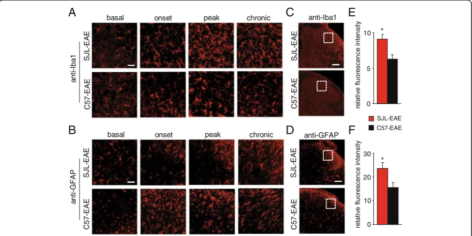
phase of the disease, and cells became activated as seen by their morphological changes (Figure 8A). Similarly, using the microglia specific anti-Iba1 antibody, we saw an induction of microglia cells at disease onset and in the chronic phase of the disease and activation of microglia, which was evident by morphological changes (Figure 8B). Because microglia and astrocyte activation plays an important role in pain, we compared the time course of microglia and astrocyte activation in SJL-EAE and C57-EAE animals in more detail. Interestingly, we found a comparable activation of microglia as shown with anti-Iba1 antibody in the dorsal horn of the spinal cord dur-ing the onset phase in SJL-EAE and C57-EAE mice (Figure 9A), but to a lesser extent in C57-EAE mice as compared to SJL-EAE mice in the peak phase as well as in the chronic phase of the disease (Figure 9A).
To quantify the amount of microglia cells in the chronic phase of the disease, we measured the fluores-cence intensity in lamina I and II of the spinal dorsal horn and found a significantly higher fluorescence inten-sity for Iba1 in SJL-EAE mice as compared to C57-EAE mice (see Figure 9C for example, Figure 9E for quantifi-cation). Additionally we compared the expression profile of astrocytes by using an anti-GFAP antibody. We found a stronger activation of astrocytes in C57-EAE as com-pared to SJL-EAE mice in the onset phase of the disease (Figure 9B). Interestingly, there was an accumulation of GFAP-positive cells in the superficial spinal dorsal horn of SJL-EAE mice in the chronic phase of the disease as compared to C57-EAE mice (Figure 9B). Quantification of the GFAP fluorescence intensity in the spinal dorsal horn revealed a significantly stronger activation of astro-cytes in SJL-EAE mice as compared to C57-EAE mice in the chronic phase of the disease (see Figure 9D for ex-ample, Figure 9F for quantification).
The differences of microglia and astrocyte activation in the spinal dorsal horn between the two EAE models are summarized in Table 1.
Discussion
Clinically significant pain is a severe and debilitating symptom associated with MS, however, to date we are far beyond understanding the mechanisms underlying MS-related pain. Animal models mimicking diverse
h
o
ri
z
ont
al a
c
ti
vit
y
0
20
40
60
80
100
120
basal
chronic
A
SJL-EAE
control
s
p
eed
immo
b
ili
ty
ti
me
0
20
40
60
80
100
120
basal
chronic
B
0
100
50
150
200
250
300
basal
chronic
C
Figure 4Behavioral and motor task analysis of SJL-PLP139-151
aspects of the disease have been used for decades to study pathological features of the disease and more re-cently to investigate behavioral changes with respect to pain hypersensitivity.
Chronic pain symptoms in MS are very complex and diverse and could even be indirectly related to MS (reviewed in [27,28]). Pain symptoms, the number of pain sites, and their severity vary among the patients and are often unrelated to the duration of MS [29]. Pain has been reported at the onset of the disease [4] or even
as an initial symptom of MS [30]. Pain syndromes are described as increasing with the age of patients and the disease progression [2,4,31], but in most MS studies chronic pain was found to have no significant correl-ation to age, disease durcorrel-ation or disease course [29,32-37]. Taking this into account, the use of animal models to study MS-related chronic pain syndromes is very lim-ited. We aimed to investigate the sensory properties of the hindpaws as readout for hyperalgesia and allodynia, which constitute one component of MS-related pain
displacement (μm) displacement (μm)
0 2 4 6 8 10 12 14
Control n=18 EAE n=21
*
6 12 24 48 96 192 384
A (RA fibers)
0 40 80 120
6 12 24 48 96 192 384
(SA fibers)
Control n=22 EAE n=25
n=22 0
20 40 60 80
6 12 24 48 96 192 384
A (AM fibers)
Control n=22 EAE n=17
0 10 20 30 40 50
6 12 24 48 96 192 384
C fibers
Control n=20 EAE n=19
displacement (μm) displacement (μm)
Firi
n
g
ra
te
/1
0
s
F
ir
ing
ra
te
/1
0s
Firi
n
g
ra
te
/1
0
s
F
ir
ing
ra
te
/1
0s
0.0 0.2 0.4 0.6
RA SA AM C-fibres
Control EAE
Th
e
rs
h
o
ld
(g
)
A
F
D
C
B
*
V
e
lo
city
(
m
/s
) V
e
lo
city
(
m
/s
)
0 5 10 15 20 25
0.0 0.2 0.4 0.6
RA SA AM C-fibers
Control EAE
E
A
Figure 5Electrophysiological analysis of excitability of peripheral afferent fibers in the skin nerve preparation in SJL-PLP139-151
peptide immunized mice and control mice.Shown are electrophysiological recordings of firing rates evoked by application of increasing 10 seconds lasting pressure via a nanomotor (expressed in terms of displacement) from (A) Aδ-type of mechanonociceptors, (B)
Here, we provide a thorough investigation of nocicep-tive sensitivity of the hindpaw in two different mouse EAE models over a complete time course of the disease. Additionally, we provide substantiated underlying mechanistical analysis with detailed immunohistochem-ical data. We found that SJL mice immunized with PLP139-151 peptide and C57 mice immunized with MOG35-55 peptide clearly showed thermal hyperalgesia, whereas only SJL-EAE mice developed marked mechan-ical allodynia in the chronic phase of the disease. C57-EAE mice developed mechanical allodynia exclusively towards very low-intensity stimuli during disease onset and peak phase. Our findings are in line with a study from Aicheret al.[15] who showed thermal hyperalgesia in SJL-PLP139-151 EAE mice in the chronic phase of the disease [15]; however, this was found on the tail and forepaw of the mice. Additionally Olechowskiet al. [16] and Rodrigues et al. [17] reported hindpaw mechanical allodynia and hypernociception before and around the onset phase of EAE in C57-MOG35-55mice [16,17]. Our findings are supported from these studies and clearly demonstrate differences in the sensory properties be-tween the two commonly used EAE models. The use of
the same behavioral tests over a long-lasting investiga-tion period under similar condiinvestiga-tions enabled us to dir-ectly compare the sensory profile of both EAE models.
Pain in MS patients is very diverse and one EAE model cannot mirror the heterogeneity of the disease [11] research perspective should therefore be focused to-wards the understanding that one EAE -pain model is not sufficient to study MS-related pain. Moreover, de-pending on the immunization peptides used and their representation in peripheral nervous system [38], periph-eral pain may also add to the mechanism of increased pain in neuroinflammation, especially in models of auto-immune neuritis [39,40].
We found a strong activation of glia cells in the spinal dorsal horn in SJL-EAE and C57-EAE mice. This glia activation occured to a different magnitude and over a different time course in both models, that matched the temporal profile of nociceptive hypersen-sitivity. It is known that microglia and astrocytes are critical players in the effector phase of EAE and MS [41,42] because there is a marked activation of glia cells in both the spinal cord and brain over the course of the disease [43,44]. We hypothesize that the time
pr
e
ons
e
t
pe
a
k
c
h
ro
nic
control SJL -EAE
A
anti -NeuNanti -NeuN
B
anti -Fox3 overlay
pr
e
ons
e
t
pe
a
k
c
h
ro
nic
Figure 6Expression of NeuN and Fox3 in lumbar spinal cord in SJL-PLP139-151peptide immunized mice and control mice over the time
course and extent of microglia and astrocyte activation in SJL-EAE mice as compared to C57-EAE mice and the subsequent release of diverse signaling molecules constitute the marked differences in the development and maintenance of chronic pain. This theory is sup-ported from a study of Olechowski et al. [16], suggest-ing inflammation and reactive gliosis as key mediators
of allodynia in C57-MOG35-55 EAE mice [16].
Activated glia cells not only undergo phenotypic changes, which are characterized by altered morph-ology, but also release a large variety of different sig-naling molecules, including inflammatory cytokines and chemokines [45-50], which are strongly implicated in pain facilitation [51-55].
There is a large variety of molecules and mediators, and thus, diverse signaling scenarios are possible.
B
C
D
anti-GFAP IB4 overlay anti-Iba1 IB4 overlay pre
co
n
tr
o
l
onset peak chronic
A
SJ
L
-EA
E
an
ti
-C
G
R
P
IB4
pre onset peak chronic
co
n
tr
o
l
SJ
L
-EA
E
Temporally regulated key signaling mediators that pos-sibly account for the development and maintenance of chronic pain in EAE include regulated glial factors such as those that comprise the chemokine monocyte chemo-attractant protein-1 (MCP-1), which is released from glia cells and can attract various cell types involved in in-flammation and also pain. Previous studies have demon-strated the expression of MCP-1 in the CNS of patients with MS [56-58] or EAE mice [59]. Additionally, the MCP-1 receptor CCR2 has been shown to be critical for the induction of EAE [60]. Accumulating evidence indi-cates that MCP-1 plays a critical role in chronic pain fa-cilitation via CCR2 receptors [61-64]. Spinal MCP-1 can lead to neuropathic pain behavior [65,66] and induces to the phosphorylation of the mitogen-activated protein kinase (MAPK) extracellular regulated kinase (ERK) [65] in the spinal cord. In addition, Shin et al. [67] found a
significant increase of different MAPK (phosphorylated ERK, c-jun N-terminal kinase (JNK) and p38) in the rat spinal cord at the peak stage of EAE [67]. The activation of ERK is known to play an important role in central sensitization [68], and JNK has been shown to be per-sistently activated in spinal cord astrocytes after nerve injury [69,70]. Moreover MCP-1 has been shown to amplify excitatory glutamatergic currents [65] and inhi-bits GABA-induced currents [71]. Thus, MCP-1 is strongly involved in mechanisms of chronic pain.
Another example is matrix metalloproteinases (MMPs), which are known to be largely implicated in MS and EAE progression [72,73]. A variety of MMPs are upregulated in the spinal cord of EAE mice, among which are MMP-2, MMP-7, MMP-8 and MMP-9 [74-76]. Dong et al. [77] recently reported concordant ele-vated expression of MMP-2 and MMP-9 to a different
co
n
tr
o
l
SJ
L
-EA
E
pre onset peak chronic
A
B
co
n
tr
o
l
SJ
L
-EA
E
an
ti
-G
F
A
P
an
ti
-I
b
a
1
pre onset peak chronic
Figure 8Astrocyte and microglia expression in the spinal dorsal horn of control immunized mice and SJL-PLP139-151peptide
extent in different EAE models [77]. Moreover, MMP-9 plays an important role in neuropathic pain conditions [78,79] as well as in MS [80-83]. Additionally, the ad-ministration of MMP inhibitors or genetical ablation of MMPs reduces the disease severity in different EAE murine models [84-87].
To further support our theory, another mechanistical possibility might be via proinflammatory cytokines (for example, IL-1beta, IL-6 and TNFalpha), which have been shown to lead to the phosphorylation of CREB [79]. CREB is essential for the maintenance of long-term plas-ticity in dorsal horn neurons [79] and thereby plays an essential role in pain sensitization [79,88-90]. Kimet al. suggests that increased phosphorylation of CREB in sen-sory neurons in the dorsal horns might be involved in the generation of neuropathic pain in EAE [91]. Taken together, there are various signaling pathways arising from activated glia cells which may thereby contribute to pain in EAE and possibly also to MS.
Given that neuro-immune interactions play a critical role in other pain states and given that peripheral im-mune function is also changed in MS patients [7] it is possible that peripheral neuro-immune interactions con-tribute to MS-induced pain. In order to clarify potential
changes in the peripheral nervous system in SJL-EAE mice, we investigated the electrophysiological properties of peripheral afferent fibers in EAE mice using the skin nerve preparation. EAE is known to cause central de-myelination, but there is weak evidence for a peripheral component to the disease [92,93]. In case of a peripheral demyelination one would expect a decrease in velocity of the signal transduction of myelinated Aβ and Aδ fibers. Pender et al. observed an impaired response to noxious mechanical stimuli potentially associated with a demyelination-induced conduction block in the small diameter myelinated afferent (Aδ) fibers in the dorsal root ganglia (DRGs) of rabbits or rats with EAE [94-96]. We observed a slight decrease in conduction velocity in myelinated Aβ mechanonociceptors but the observed changes in the peripheral afferents are very mild, indicat-ing only minor peripheral contribution to the disease phenotype which might arise from a different mechan-ism than possible peripheral demyelination processes.
In summary we show clear differences in pain behavior between different EAE mouse models, which may reflect the heterogeneity in human MS. Moreover the observed differences in glia cell activation most likely contribute to the different pain behavior. This study suggests that
C
5
7
-EAE
SJ
L
-EA
E
basal onset peak chronic
C57-E
A
E
SJ
L
-EA
E
re
la
ti
v
e
f
lu
o
re
sce
n
c
e
in
te
n
si
ty
E
SJL-EAE 10
5
0
C57-E
A
E
SJ
L
-EA
E
basal onset peak chronic
C
5
7
-EAE
SJ
L
-EA
E
anti-Iba1
an
ti
-I
b
a
1
C
A
an
ti
-G
F
A
P
C57-EAE
30
20
10
0
* *
F
D
B
re
la
ti
v
e
f
luo
re
s
c
e
nc
e
int
e
n
s
it
y
anti-GFAP
Figure 9Analysis and quantification of astrocyte and microglia activation in SJL-PLP139-151peptide immunized mice and
microglia and astrocytes represent a good target to in-vestigate pain mechanisms in different EAE mouse mod-els. Future studies would be necessary to elucidate differences in downstream signaling cascades in the dif-ferent EAE models.
Conclusions
In summary we show clear differences in pain behavior between different EAE mouse models, which may reflect the heterogeneity in human MS. Moreover the observed differences in glia cell activation most likely contribute to the different pain behavior. This study suggests that microglia and astrocytes represent a good target to in-vestigate pain mechanisms in different EAE mouse mod-els. Future studies would be necessary to elucidate differences in downstream signaling cascades in the dif-ferent EAE models.
Abbreviations
CFA: Complete Freund’s adjuvant; CGRP: Calcitonin gene-regulated peptide; CNS: Central nervous system; DRG: Dorsal root ganglia; EAE: Experimental autoimmune encephalomyelitis; ERK: Extracellular regulated kinase; IB4: Isolectin B4; JNK: c-jun N-terminal kinase; MAPK: Mitogen-activated protein kinase; MMPs: Matrix metalloproteinases; MS: Multiple sclerosis; PFA: Paraformaldehyde; RA: Rapidly adapting; SA: Slowly adapting.
Competing interests
The authors have no conflicts of interest.
Authors’contributions
JL carried out behavioral and histological experiments and analyzed results. MK performed skin-nerve electrophysiological experiments, analyzed data and provided figure. RIW carried out open field behavioral experiments and analyzed data. RAL and RK provided general support and participated in the design of the study. RAL helped to improve the manuscript. ATT conceived, designed, and coordinated the study and wrote the manuscript. All authors read and approved the final manuscript.
Acknowledgements
The authors are grateful to Silvia Seubert for expert technical assistance and Isabella Peruga for her help in the early phase of the project. J.L was supported by the HBIGS-program of the University of Heidelberg and A.T.T. was supported by an Olympia Morata Award from the Medical Faculty Heidelberg of University Heidelberg.
Author details
1Pharmacology Institut, University of Heidelberg, Im Neuenheimer Feld 366, Heidelberg D-69120, Germany.2Department of Neurology,
Universitätsklinikum Erlangen, Schwabachanlage 6, Erlangen D-91054, Germany.
Received: 10 June 2012 Accepted: 18 September 2012 Published: 6 October 2012
References
1. Clifford DB, Trotter JL:Pain in multiple sclerosis.Arch Neurol1984,
41:1270–1272.
2. Stenager E, Knudsen L, Jensen K:Acute and chronic pain syndromes in
multiple sclerosis. A 5-year follow-up study.Ital J Neurol Sci1995,
16:629–632.
3. Svendsen KB, Jensen TS, Overvad K, Hansen HJ, Koch-Henriksen N, Bach FW:
Pain in patients with multiple sclerosis: a population-based study.Arch
Neurol2003,60:1089–1094.
4. Stenager E, Knudsen L, Jensen K:Acute and chronic pain syndromes in
multiple sclerosis.Acta Neurol Scand1991,84:197–200.
5. Pöllmann W, Feneberg W, Erasmus LP:Pain in multiple sclerosis–a still underestimated problem. The 1 year prevalence of pain syndromes, significance and quality of care of multiple sclerosis inpatients.
Nervenarzt2004,75:135–140.
6. Hadjimichael O, Kerns RD, Rizzo MA, Cutter G, Vollmer T:Persistent pain
and uncomfortable sensations in persons with multiple sclerosis.Pain
2007,127:35–41.
7. Kenner M, Menon U, Elliott DG:Multiple sclerosis as a painful disease.Int
Rev Neurobiol2007,79:303–321.
8. Osterberg A, Boivie J, Thuomas KA:Central pain in multiple sclerosis–
prevalence and clinical characteristics.Eur J Pain2005,9:531–542.
9. Svendsen KB, Jensen TS, Hansen HJ, Bach FW:Sensory function and quality
of life in patients with multiple sclerosis and pain.Pain2005,114:473–481.
10. Wekerle H, Kojima K, Lannes-Vieira J, Lassmann H, Linington C:Animal
models.Ann Neurol1994,36(Suppl):S47–53.
11. Gold R, Linington C, Lassmann H:Understanding pathogenesis and therapy of multiple sclerosis via animal models: 70 years of merits and culprits in experimental autoimmune encephalomyelitis research.
Brain2006,129:1953–1971.
12. Kuerten S, Angelov DN:Comparing the CNS morphology and immunobiology of different EAE models in C57BL/6 mice - a step towards understanding the complexity of multiple sclerosis.
Ann Anat2008,190:1–15.
13. Berger T, Weerth S, Kojima K, Linington C, Wekerle H, Lassmann H: Experimental autoimmune encephalomyelitis: the antigen specificity of T lymphocytes determines the topography of lesions in the central and
peripheral nervous system.Lab Invest1997,76:355–364.
14. Schmidt S:Candidate autoantigens in multiple sclerosis.
Mult Scler1999,5:147–160.
15. Aicher SA, Silverman MB, Winkler CW, Bebo BF Jr:Hyperalgesia in an
animal model of multiple sclerosis.Pain2004,110:560–570.
16. Olechowski CJ, Truong JJ, Kerr BJ:Neuropathic pain behaviours in a chronic-relapsing model of experimental autoimmune encephalomyelitis
(EAE).Pain2009,141:156–164.
17. Rodrigues DH, Sachs D, Teixeira AL:Mechanical hypernociception in
experimental autoimmune encephalomyelitis.Arq Neuropsiquiatr2009,
67:78–81.
18. Lisi L, Navarra P, Cirocchi R, Sharp A, Stigliano E, Feinstein DL, Dello Russo C: Rapamycin reduces clinical signs and neuropathic pain in a chronic
model of experimental autoimmune encephalomyelitis.J Neuroimmunol
2012,243:43–51.
19. Linker RA, Maurer M, Gaupp S, Martini R, Holtmann B, Giess R, Rieckmann P, Lassmann H, Toyka KV, Sendtner M, Gold R:CNTF is a major protective factor in demyelinating CNS disease: a neurotrophic cytokine as
modulator in neuroinflammation.Nat Med2002,8:620–624.
20. Stösser S, Agarwal N, Tappe-Theodor A, Yanagisawa M, Kuner R: Dissecting the functional significance of endothelin A receptors in peripheral nociceptors in vivo via conditional gene deletion. Pain2010,148:206–214.
21. Tappe-Theodor A, Constantin CE, Tegeder I, Lechner SG, Langeslag M, Lepcynzsky P, Wirotanseng RI, Kurejova M, Agarwal N, Nagy G,et al:Galpha (q/11) signaling tonically modulates nociceptor function and contributes
to activity-dependent sensitization.Pain2012,153:184–196.
22. Jiao Y, Sun Z, Lee T, Fusco FR, Kimble TD, Meade CA, Cuthbertson S, Reiner A:A simple and sensitive antigen retrieval method for
free-floating and slide-mounted tissue sections.J Neurosci Methods1999,
93:149–162.
23. Kim KK, Adelstein RS, Kawamoto S:Identification of neuronal nuclei (NeuN) as Fox-3, a new member of the Fox-1 gene family of splicing
factors.J Biol Chem2009,284:31052–31061.
24. Portiansky EL, Barbeito CG, Gimeno EJ, Zuccolilli GO, Goya RG:Loss of
NeuN immunoreactivity in rat spinal cord neurons during aging.Exp
Neurol2006,202:519–521.
25. Unal-Cevik I, Kilinc M, Gursoy-Ozdemir Y, Gurer G, Dalkara T:Loss of NeuN immunoreactivity after cerebral ischemia does not indicate neuronal cell
loss: a cautionary note.Brain Res2004,1015:169–174.
26. Streit WJ, Kreutzberg GW:Lectin binding by resting and reactive
microglia.J Neurocytol1987,16:249–260.
27. O'Connor AB, Schwid SR, Herrmann DN, Markman JD, Dworkin RH:Pain associated with multiple sclerosis: systematic review and proposed
28. Pöllmann W, Feneberg W:Current management of pain associated with
multiple sclerosis.CNS Drugs2008,22:291–324.
29. Archibald CJ, McGrath PJ, Ritvo PG, Fisk JD, Bhan V, Maxner CE, Murray TJ: Pain prevalence, severity and impact in a clinic sample of multiple
sclerosis patients.Pain1994,58:89–93.
30. Kalia LV, O'Connor PW:Severity of chronic pain and its relationship to
quality of life in multiple sclerosis.Mult Scler2005,11:322–327.
31. Solaro C, Brichetto G, Amato MP, Cocco E, Colombo B, D'Aleo G, Gasperini C, Ghezzi A, Martinelli V, Milanese C,et al:The prevalence of pain in
multiple sclerosis: a multicenter cross-sectional study.Neurology2004,
63:919–921.
32. Ehde DM, Osborne TL, Hanley MA, Jensen MP, Kraft GH:The scope and
nature of pain in persons with multiple sclerosis.Mult Scler2006,
12:629–638.
33. Grasso MG, Clemenzi A, Tonini A, Pace L, Casillo P, Cuccaro A, Pompa A, Troisi E:Pain in multiple sclerosis: a clinical and instrumental approach.
Mult Scler2008,14:506–513.
34. Hirsh AT, Turner AP, Ehde DM, Haselkorn JK:Prevalence and impact of pain in multiple sclerosis: physical and psychologic contributors.
Arch Phys Med Rehabil2009,90:646–651.
35. Michalski D, Liebig S, Thomae E, Hinz A, Bergh FT:Pain in patients with multiple sclerosis: a complex assessment including quantitative and qualitative measurements provides for a disease-related biopsychosocial
pain model.J Pain Res2011,4:219–225.
36. Moulin DE, Foley KM, Ebers GC:Pain syndromes in multiple sclerosis.
Neurology1988,38:1830–1834.
37. Osborne TL, Jensen MP, Ehde DM, Hanley MA, Kraft G:Psychosocial factors associated with pain intensity, pain-related interference, and
psychological functioning in persons with multiple sclerosis and pain. Pain2007,127:52–62.
38. Garbay B, Heape AM, Sargueil F, Cassagne C:Myelin synthesis in the
peripheral nervous system.Prog Neurobiol2000,61:267–304.
39. Liu H, Shiryaev SA, Chernov AV, Kim Y, Shubayev I, Remacle AG, Baranovskaya S, Golubkov VS, Strongin AY, Shubayev VI:Immunodominant fragments of myelin basic protein initiate T cell-dependent pain.
J Neuroinflammation2012,9:119.
40. Moalem-Taylor G, Allbutt HN, Iordanova MD, Tracey DJ:Pain hypersensitivity in rats with experimental autoimmune neuritis, an animal model of human inflammatory demyelinating neuropathy.
Brain Behav Immun2007,21:699–710.
41. D'Amelio FE, Smith ME, Eng LF:Sequence of tissue responses in the early stages of experimental allergic encephalomyelitis (EAE):
immunohistochemical, light microscopic, and ultrastructural
observations in the spinal cord.Glia1990,3:229–240.
42. Gehrmann J, Gold R, Linington C, Lannes-Vieira J, Wekerle H, Kreutzberg
GW:Microglial involvement in experimental autoimmune inflammation
of the central and peripheral nervous system.Glia1993,7:50–59.
43. Gray E, Thomas TL, Betmouni S, Scolding N, Love S:Elevated activity and microglial expression of myeloperoxidase in demyelinated cerebral
cortex in multiple sclerosis.Brain Pathol2008,18:86–95.
44. Petzold A, Eikelenboom MJ, Gveric D, Keir G, Chapman M, Lazeron RH, Cuzner ML, Polman CH, Uitdehaag BM, Thompson EJ, Giovannoni G: Markers for different glial cell responses in multiple sclerosis: clinical and
pathological correlations.Brain2002,125:1462–1473.
45. Benveniste EN:Role of macrophages/microglia in multiple sclerosis and
experimental allergic encephalomyelitis.J Mol Med (Berl)1997,75:165–173.
46. Gonzalez-Scarano F, Baltuch G:Microglia as mediators of inflammatory
and degenerative diseases.Annu Rev Neurosci1999,22:219–240.
47. Kim SU, de Vellis J:Microglia in health and disease.J Neurosci Res2005,
81:302–313.
48. Milligan ED, O'Connor KA, Nguyen KT, Armstrong CB, Twining C, Gaykema RP, Holguin A, Martin D, Maier SF, Watkins LR:Intrathecal HIV-1 envelope glycoprotein gp120 induces enhanced pain states
mediated by spinal cord proinflammatory cytokines.J Neurosci2001,
21:2808–2819.
49. Ozenci V, Kouwenhoven M, Link H:Cytokines in multiple sclerosis:
methodological aspects and pathogenic implications.Mult Scler2002,
8:396–404.
50. Szczucinski A, Losy J:Chemokines and chemokine receptors in multiple
sclerosis.Potential targets for new therapies. Acta Neurol Scand2007,
115:137–146.
51. Ji RR, Suter MR:p38 MAPK, microglial signaling, and neuropathic pain.
Mol Pain2007,3:33.
52. Milligan ED, Twining C, Chacur M, Biedenkapp J, O'Connor K, Poole S, Tracey K, Martin D, Maier SF, Watkins LR:Spinal glia and proinflammatory
cytokines mediate mirror-image neuropathic pain in rats.J Neurosci2003,
23:1026–1040.
53. Milligan ED, Watkins LR:Pathological and protective roles of glia in
chronic pain.Nat Rev Neurosci2009,10:23–36.
54. Watkins LR, Maier SF:Beyond neurons: evidence that immune and glial
cells contribute to pathological pain states.Physiol Rev2002,82:981–1011.
55. Watkins LR, Milligan ED, Maier SF:Glial proinflammatory cytokines
mediate exaggerated pain states: implications for clinical pain.Adv Exp
Med Biol2003,521:1–21.
56. McManus C, Berman JW, Brett FM, Staunton H, Farrell M, Brosnan CF: MCP-1, MCP-2 and MCP-3 expression in multiple sclerosis lesions: an
immunohistochemical and in situ hybridization study.J Neuroimmunol
1998,86:20–29.
57. Simpson JE, Newcombe J, Cuzner ML, Woodroofe MN:Expression of monocyte chemoattractant protein-1 and other beta-chemokines by resident glia and inflammatory cells in multiple sclerosis lesions.
J Neuroimmunol1998,84:238–249.
58. Van Der Voorn P, Tekstra J, Beelen RH, Tensen CP, Van Der Valk P, De Groot
CJ:Expression of MCP-1 by reactive astrocytes in demyelinating multiple
sclerosis lesions.Am J Pathol1999,154:45–51.
59. Fischer FR, Santambrogio L, Luo Y, Berman MA, Hancock WW, Dorf ME: Modulation of experimental autoimmune encephalomyelitis: effect of altered peptide ligand on chemokine and chemokine receptor
expression.J Neuroimmunol2000,110:195–208.
60. Fife BT, Huffnagle GB, Kuziel WA, Karpus WJ:CC chemokine receptor 2 is critical for induction of experimental autoimmune encephalomyelitis.
J Exp Med2000,192:899–905.
61. Abbadie C, Lindia JA, Cumiskey AM, Peterson LB, Mudgett JS, Bayne EK, DeMartino JA, MacIntyre DE, Forrest MJ:Impaired neuropathic pain
responses in mice lacking the chemokine receptor CCR2.Proc Natl Acad
Sci U S A2003,100:7947–7952.
62. Dansereau MA, Gosselin RD, Pohl M, Pommier B, Mechighel P, Mauborgne A, Rostene W, Kitabgi P, Beaudet N, Sarret P, Melik-Parsadaniantz S:Spinal CCL2 pronociceptive action is no longer effective in CCR2 receptor
antagonist-treated rats.J Neurochem2008,106:757–769.
63. Menetski J, Mistry S, Lu M, Mudgett JS, Ransohoff RM, Demartino JA, Macintyre DE, Abbadie C:Mice overexpressing chemokine ligand 2 (CCL2)
in astrocytes display enhanced nociceptive responses.Neuroscience2007,
149:706–714.
64. Tanaka T, Minami M, Nakagawa T, Satoh M:Enhanced production of monocyte chemoattractant protein-1 in the dorsal root ganglia in a rat model of neuropathic pain: possible involvement in the development of
neuropathic pain.Neurosci Res2004,48:463–469.
65. Gao YJ, Zhang L, Samad OA, Suter MR, Yasuhiko K, Xu ZZ, Park JY, Lind AL, Ma Q, Ji RR:JNK-induced MCP-1 production in spinal cord astrocytes
contributes to central sensitization and neuropathic pain.J Neurosci2009,
29:4096–4108.
66. Thacker MA, Clark AK, Bishop T, Grist J, Yip PK, Moon LD, Thompson SW, Marchand F, McMahon SB:CCL2 is a key mediator of microglia activation
in neuropathic pain states.Eur J Pain2009,13:263–272.
67. Shin BA, Yoo HG, Kim HS, Kim MH, Hwang YS, Chay KO, Lee KY, Ahn BW, Jung YD:P38 MAPK pathway is involved in the urokinase plasminogen
activator expression in human gastric SNU-638 cells.Oncol Rep2003,
10:1467–1471.
68. Ji RR, Baba H, Brenner GJ, Woolf CJ:Nociceptive-specific activation of ERK
in spinal neurons contributes to pain hypersensitivity.Nat Neurosci1999,
2:1114–1119.
69. Ma W, Quirion R:Partial sciatic nerve ligation induces increase in the phosphorylation of extracellular signal-regulated kinase (ERK) and c-Jun N-terminal kinase (JNK) in astrocytes in the lumbar spinal dorsal horn
and the gracile nucleus.Pain2002,99:175–184.
70. Zhuang ZY, Wen YR, Zhang DR, Borsello T, Bonny C, Strichartz GR, Decosterd I, Ji RR:A peptide c-Jun N-terminal kinase (JNK) inhibitor blocks mechanical allodynia after spinal nerve ligation: respective roles of JNK activation in primary sensory neurons and spinal astrocytes for
neuropathic pain development and maintenance.J Neurosci2006,
71. Gosselin RD, Varela C, Banisadr G, Mechighel P, Rostene W, Kitabgi P, Melik-Parsadaniantz S:Constitutive expression of CCR2 chemokine receptor and inhibition by MCP-1/CCL2 of GABA-induced currents in spinal cord
neurones.J Neurochem2005,95:1023–1034.
72. Rosenberg GA:Matrix metalloproteinases in neuroinflammation.Glia 2002,39:279–291.
73. Yong VW, Power C, Forsyth P, Edwards DR:Metalloproteinases in biology
and pathology of the nervous system.Nat Rev Neurosci2001,2:502–511.
74. Kieseier BC, Clements JM, Pischel HB, Wells GM, Miller K, Gearing AJ, Hartung HP:Matrix metalloproteinases MMP-9 and MMP-7 are expressed in experimental autoimmune neuritis and the Guillain-Barre syndrome.
Ann Neurol1998,43:427–434.
75. Nygardas PT, Hinkkanen AE:Up-regulation of MMP-8 and MMP-9 activity in the BALB/c mouse spinal cord correlates with the severity of
experimental autoimmune encephalomyelitis.Clin Exp Immunol2002,
128:245–254.
76. Toft-Hansen H, Nuttall RK, Edwards DR, Owens T:Key metalloproteinases are expressed by specific cell types in experimental autoimmune
encephalomyelitis.J Immunol2004,173:5209–5218.
77. Dong M, Liu R, Guo L, Li C, Tan G:Pathological findings in rats with
experimental allergic encephalomyelitis.Apmis2008,116:972–984.
78. Ji RR, Xu ZZ, Wang X, Lo EH:Matrix metalloprotease regulation of
neuropathic pain.Trends Pharmacol Sci2009,30:336–340.
79. Kawasaki Y, Xu ZZ, Wang X, Park JY, Zhuang ZY, Tan PH, Gao YJ, Roy K, Corfas G, Lo EH, Ji RR:Distinct roles of matrix metalloproteases in the
early- and late-phase development of neuropathic pain.Nat Med2008,
14:331–336.
80. Gijbels K, Masure S, Carton H, Opdenakker G:Gelatinase in the cerebrospinal fluid of patients with multiple sclerosis and other
inflammatory neurological disorders.J Neuroimmunol1992,41:29–34.
81. Lee MA, Palace J, Stabler G, Ford J, Gearing A, Miller K:Serum gelatinase B,
TIMP-1 and TIMP-2 levels in multiple sclerosis.A longitudinal clinical and
MRI study. Brain1999,122(Pt 2):191–197.
82. Leppert D, Ford J, Stabler G, Grygar C, Lienert C, Huber S, Miller KM, Hauser SL, Kappos L:Matrix metalloproteinase-9 (gelatinase B) is selectively elevated in CSF during relapses and stable phases of multiple sclerosis.
Brain1998,121(Pt 12):2327–2334.
83. Lichtinghagen R, Seifert T, Kracke A, Marckmann S, Wurster U, Heidenreich F: Expression of matrix metalloproteinase-9 and its inhibitors in
mononuclear blood cells of patients with multiple sclerosis.
J Neuroimmunol1999,99:19–26.
84. Brundula V, Rewcastle NB, Metz LM, Bernard CC, Yong VW:Targeting leukocyte MMPs and transmigration: minocycline as a potential therapy
for multiple sclerosis.Brain2002,125:1297–1308.
85. Folgueras AR, Fueyo A, Garcia-Suarez O, Cox J, Astudillo A, Tortorella P, Campestre C, Gutierrez-Fernandez A, Fanjul-Fernandez M, Pennington CJ,et
al:Collagenase-2 deficiency or inhibition impairs experimental
autoimmune encephalomyelitis in mice.J Biol Chem2008,
283:9465–9474.
86. Giuliani F, Metz LM, Wilson T, Fan Y, Bar-Or A, Yong VW:Additive effect of the combination of glatiramer acetate and minocycline in a model of
MS.J Neuroimmunol2005,158:213–221.
87. Opdenakker G, Nelissen I, Van Damme J:Functional roles and therapeutic
targeting of gelatinase B and chemokines in multiple sclerosis.Lancet
Neurol2003,2:747–756.
88. Sommer C, Kress M:Recent findings on how proinflammatory cytokines cause pain: peripheral mechanisms in inflammatory and neuropathic
hyperalgesia.Neurosci Lett2004,361:184–187.
89. Sorkin LS, Xiao WH, Wagner R, Myers RR:Tumour necrosis factor-alpha induces ectopic activity in nociceptive primary afferent fibres.
Neuroscience1997,81:255–262.
90. Woolf CJ, Allchorne A, Safieh-Garabedian B, Poole S:Cytokines, nerve growth factor and inflammatory hyperalgesia: the contribution of
tumour necrosis factor alpha.Br J Pharmacol1997,121:417–424.
91. Kim H, Moon C, Ahn M, Lee Y, Kim S, Matsumoto Y, Koh CS, Kim MD, Shin T: Increased phosphorylation of cyclic AMP response element-binding protein in the spinal cord of Lewis rats with experimental autoimmune
encephalomyelitis.Brain Res2007,1162:113–120.
92. Misawa S, Kuwabara S, Mori M, Hayakawa S, Sawai S, Hattori T:Peripheral
nerve demyelination in multiple sclerosis.Clin Neurophysiol2008,
119:1829–1833.
93. Sarova-Pinhas I, Achiron A, Gilad R, Lampl Y:Peripheral neuropathy in
multiple sclerosis: a clinical and electrophysiologic study.Acta Neurol
Scand1995,91:234–238.
94. Pender MP, Sears TA:Involvement of the dorsal root ganglion in acute experimental allergic encephalomyelitis in the Lewis rat. A histological
and electrophysiological study.J Neurol Sci1986,72:231–242.
95. Pender MP, Sears TA:Vulnerability of the dorsal root ganglion in
experimental allergic encephalomyelitis.Clin Exp Neurol1985,21:211–223.
96. Pender MP, Sears TA:The pathophysiology of acute experimental allergic
encephalomyelitis in the rabbit.Brain1984,107(Pt 3):699–726.
doi:10.1186/1742-2094-9-233
Cite this article as:Luet al.:Pain in experimental autoimmune encephalitis: a comparative study between different mouse models.
Journal of Neuroinflammation20129:233.
Submit your next manuscript to BioMed Central and take full advantage of:
• Convenient online submission
• Thorough peer review
• No space constraints or color figure charges
• Immediate publication on acceptance
• Inclusion in PubMed, CAS, Scopus and Google Scholar
• Research which is freely available for redistribution