I J H S
IJHS 2018;4(1):10-14
ijhs.shmu.ac.ir doi: 10.22100/ijhs.v4i1.376
Original Article
International Journal of HealthStudies Dear Author,
Please check your proof carefully and mark all corrections at the appropriate place in the proof (e.g.,by using on-screen annotation in the PDF file) or compile them in a separate list. To ensure fast publication of your paper please return your corrections within 48 hours.
Any queries or remarks that have arisen during the processing of your manuscript are listed below and highlighted by flags in the proof. Click on the ‘Q’ link to go to the location in the proof
Location in
article Please insert your reply or correction at the corresponding line in the proofQuery/remark: click on the Q link to go
Q1
Q2
Q3
Q4
Please check names and affiliations. Also add departments and schools of all authors.
There is not any arrow in the figure 1!
Please insert acknowledgement.
Please check references.
I J H S
Original Article
International Journal of Health Studies
The Correlation between CD44 and ABCG2 Expression and Pathological Grades of
Bladder Cancer
Alireza Palangi1, Samira Bayat2, Nasser Shakhssalim3, Mahmood Parvin4, Abdolamir Allameh1*
1 Dept. of clinical biochemistry, School of Medical Sciences, TarbiatModarres University, Tehran, Iran. 2
Dept. of Biochemistry, School of Science, Payam Noor University Tehran unit, Tehran, Iran.
3 Urology and Nephrology Research Center, ShahidLabbafinejad Medical center, ShahidBeheshti University of Medical Sciences, Tehran, Iran. 4
Dept. of pathology, ShahidLabbafinejad hospital, ShahidBeheshti University of Medical Sciences, Tehran, Iran.
Received: 5 November 2018 Accepted: 11 November 2018
Abstract
Background: The human bladder cancer progression is accompanied
by the growth of side-populations of cancer stem cells (CSCs). The expression of ABCG2 and CD44, as the CSC markers, may be associated with different pathological grades of bladder cancer. This study was designed to identify the changes occurring in ABCG2 and CD44 in different types of bladder tumors at various grades.
Methods: The sample included 67 patients with bladder cancer (63
males; 4 females) with a diagnosis of transitional cell carcinoma (urothelial cancer) using cystoscopy. Based on the tumor pathological grade, we divided the tumor biopsies into two low-grade (N=20) and high-grade (N=21) groups. The tumor samples along with 26 normal-looking bladder tissues were analyzed by techniques like immunohistochemistry (IHC) and total RNA extraction for qPCR.
Results: Based on the results obtained by IHC analysis of ABCG2 and
CD44 protein in bladder low- and high-grade tumors, these CSC markers showed significant elevation in malignant tissues in comparison to the normal bladder tissues. The scoring of ABCG2 expression in normal, low-grade and high-grade tissues was 28, 120, and 140 respectively. The CD44 scores in the normal bladder, low-grade and high-low-grade tissues were found to be 0.6, 11.5, and 29.0, respectively. The IHC data showed inconsistency with the qPCR data, suggesting an overexpression of ABCG2 (4-6.5 folds) and CD44 (15-22 folds) in the low and high-grade tumors in comparison to the normal bladder tissue.
Conclusions: Finding a good relationship between ABCG2 and CD44
markers and different grades of bladder cancer demonstrated that these markers can be seen as potential and predictive indicators of bladder malignancy.
Keywords: ABCG2, CD44, Bladder cancer, Tumor grade, Cancer stem cell.
*Corresponding to: A Allameh, Email: [email protected] Please cite this paper as: Palangi A, Bayat S, Shakhssalim N, Parvin M, Allameh A. The correlation between cd44 and abcg2 expression and pathological grades of bladder cancer. Int J Health Stud 2018;4(1):10-14.
I
ntroduction
Bladder cancer is one the most common urothelial cancers, which are a group of carcinomas involving the bladder, uterus, and renal pelvis tissues.1 According to previous data, the incidence rate of bladder carcinoma is higher in men than in women with a 3:1 ratio.2 Until 2015, bladder urothelial carcinoma has already affected about 3.4 million people that 430000 new cases are annually added to this figure around the world.3,4
Several urine markers have been identified so far to diagnose bladder cancer. However, only a few of are used in practice to detect and screen the bladder urothelial carcinomas, due to varied and low negative and positive predictive value rate of these tests.5,6For example, the utility and use of urine markers such as urovysion (FISH), bladder tumor antigen (BTA), and nuclear matrix protein 22 (NMP22) in the initial diagnosis of bladder cancer is in dispute.7,8
Some elevated levels of inflammatory markers are also found in patients diagnosed as new cases of upper tract urothelial carcinoma (UTUR) such as serum C reactive protein (CRP). However, the specificity of this test is also evaluated low in case of this type of cancer.9
The major current diagnostic procedure in diagnosis of bladder cancer10 includes cystoscopy associated with tissue biopsies. The pathologic grade of the tumor11 appears to be as one of the main prognostic factors in the bladder carcinoma. The histologic grade provides considerable prognostic information; especially to predict the disease progression.12All deaths caused by bladder cancers occurs almost among the patients with high-grade tumors.13
Few molecular and biochemical markers such as hyaluronic acid and p53 introduced for prognosis and grading of bladder cancers are not enough sensitive and specific to replace the histopathologic studies of bladder tissue biopsies.14,15Although known and used as the best method for bladder cancer diagnosis and grading, the general belief is that numerous errors may occur in histopathologic examinations.10
The side population of the stem cells developing to the human bladder cancer is correlated with grade, stage, recurrence, and progression-free survival.14The ATP binding cassette (ABC) family of transporter proteins sub family G2 (ABCG2)/the breast cancer resistance protein 1 (BCRP1) mediates this side population phenotype, which is demonstrated to be the main mediator of the phenotype.16
Accordingly, we assessed the expression of ABCG2 as well as CD44 as cancer stem cell markers in human bladder cancer tissues in association with the tumor pathological grade in this study aimed at finding more about the diagnostic value of these markers in the prediction of human bladder cancers.
Materials and Methods
Palangi et al
collecting the samples. We selected 67 patients (63 males and 4 females). These patients had referred to the hospital who were diagnosed with bladder cancer following primary examination and checking the clinical symptoms. Before any therapeutic measures, a surgeon made the decision to do the surgery. Also, each patient signed a written informed consent before the procedure of sample collection.
The exclusion criteria were having diabetes mellitus, lipid metabolism disorders, and hematologic disorders.
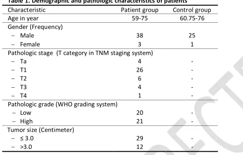
Three groups were considered in this study, including 26 subjects with a normal bladder as the control group, 20 patients with a low-grade urothelial bladder carcinoma as the low-grade group, and 21 patients with a high-grade urothelial bladder carcinoma as the high-grade group. All groups were matched regarding the factors of age and gender (P-value: 0.916 and 0.999, respectively). A summary of the demographic and pathologic information of patients is given in Table 1.
Table 1. Demographic and pathologic characteristics of patients
Control group Patient group
Characteristic
60.75-76 59-75
Age in year Gender (Frequency) 25 38 − Male 1 3 − Female
Pathologic stage (T category in TNM staging system)
-4 − Ta -26 − T1 -6 − T2 -4 − T3 -1 − T4
Pathologic grade (WHO grading system)
-20 − Low -21 − High
Tumor size (Centimeter)
-29
− ≤ 3.0
-12
− >3.0
A pathologist assessed the slides under a light microscope in terms of histopathologic parameters, diagnosis and grading of tumors after the procedures of tissue processing, paraffin embedding, tissue fixation, slide preparation and haematoxylin and eosin (H & E) staining were performed based on the standard.17
The sections with a size of 2 μm were prepared from the paraffin-embedded bladder tissues. Their paraffin was removed and they were then rehydrated. By submerging the sections into 10 mM citrate buffer (pH: 6.0) for 3 minutes at 97˚C, followed by 30 minutes of incubation at 60˚C, the tissue antigens were extracted. The processed of endogenous peroxidase activity blocking and non-specific binding inhibition were done using hydrogen peroxide block reagent and the protein block reagent by employing the expose rabbit specific HRP/DAB detection IHC kit (Abcam, UK) according to the manufacturer`s instructions.
The rabbit monoclonal anti CD44 antibody (ab157107, Abcam, UK) and rabbit monoclonal anti ABCG2 antibody (B7185, Sigma-Aldrich, USA) were diluted by 1:1000 and 1:50 ratios, respectively, which were then incubated with the sections for 1 hour based on the manufacturer`s recommendations. Following rinsed for three times with PBS,
we added the goat anti rabbit HRP conjugate (expose rabbit specific HRP/DAB detection IHC kit, Abcam, UK). After 15 minutes of incubation, they were rinsed again (4 times). We then submerged the tissue sections with DAB chromogene for 10 minutes and counter stained them with haematoxylin and finally cover-slipped them.
We replaced the ABCG2 and CD44 primary antibodies with PBS in the IHC procedure to assess the presence of background staining. An expert pathologist unaware of the patients’ information and qPCR results carefully examined the cell membrane expression of ABCG2 and CD44 at the bladder urothelium.
We calculated the scores of IHC results by multiplying the percentage of positive stained cells by the intensity of staining. The average percentage of positive cells in 5 high-power microscopic fields (40x) was obtained to calculate the percentage of positive stained cells. The slides with positive stained cells lower than 10% were scored as negative. The intensity of staining was graded as 0 for no staining, 1 for weak, 2 for moderate and 3 for strong.
According to the kit package insert, the using hybrid-R extraction kit (GeneAll Biotechnology, South Korea) was used to extract the mRNA from the bladder tissue biopsies. Using spectrophotometry (NanoDrop 2000 C Thermo Scientific, USA) and gel agarose electrophoresis, we assessed the quality and quantity of the extracted mRNA based on a standard procedure. We converted 2 μg of mRNA to complimentary DNA (cDNA) by the RevertAid First Strand cDNA Synthesis Kit (Thermo Fisher scientific, USA) according to the manufacturer`s instructions. Using the primer 3.0 online software version 4.0 (http://bioinfo.Ut.Ee/primer3-0.4.0/), we designed the specific primers for ABCG2 and CD44. The primers’ specificity was assessed using the blast analysis technique (http://bioinfo.Ut.Ee/primer3-0.4.0/).
The primer sequences were as the following:
ABCG2
Forward: 5`GTGGCCTTGGCTTGTATGAT3`
Reverse: 5`AACAATTGCTGCTGTGCAAC3`
CD44
Forward: 5`GGCAAGAAACCTGGGATTGG3`
Reverse: 5`CGTGGTGTGGTTGAAATGGT3`
Using the Norm-finder Excel-based software,18 the GAPDH was selected from beta Actin, HGPRT and GAPDH genes as the reference gene. The specific primers of GAPDH were designed and blasted as described previously. The GAPDH primer sequences were as below:
Forward: 5`CTGACTTCAACAGCGACACC3`
Reverse: 5`GTGGTCCAGGGGTCTTACTC3`
Plus 2x Master Mix Green (Ampliqon, Denmark) with SYBR green method according the following program: 15 minutes at 95˚C activation phase followed by 40 cycles of 60-second denaturation at 95˚C, 30 seconds of annealing at 60˚C, and finally, 60 seconds of extension at 72˚C per cycle.
All the statistical analyses were made using the Graphpad Prism software, Ver. 7.0. We also used the descriptive statistics to analyze the patients’ characteristics. The Kruskal-Wallis test was employed to examine the relationship between the control, low-grade, and high grade groups. In addition, the Chi Square test was applied to assess the correlation between the ABCG2 and CD44 gene expression and the bladder cancer.
We represented all data as mean±standard error of the mean (SEM), and the P-values lower than 0.05 were considered statistically significant.
Results
In this study, we compared the expression of CD44 and ABCG2 in the bladder tissues obtained from patients, which were divided based on histopathological grade of the tumor specified by the H&E staining technique.
As seen in Figure 1, we compared the low-grade and high-grade urothelial carcinomas with the normal tissue. The presence of cytological and architectural disorders is the key for the differential diagnosis of normal, low-grade and high-grade neoplastic urothelium cancers, which includes the variability in size and color of the urothelial cells and the loss of polarity in the urothelium.20
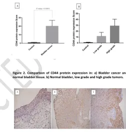
The mean score for the low-grade carcinoma was obtained as 11.33±8.31 in examining the immunohistochemistry scoring in the case of CD44 protein accommodation in the bladder tissue preparation, while this score obtained as 29.00±13.21 in case of the high-grade carcinoma and 0.62±0.26 for the normal bladder tissues (Figure 2).
The carcinoma samples clearly showed relatively higher immunohistochemistry scores for the CD44 with a significant difference in comparison with the CD44 IHC score in the low-grade and high-low-grade samples (Figure 3).
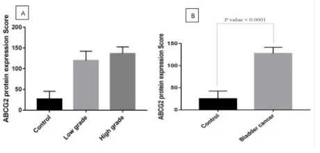
The study of ABCG2 protein expression in the bladder tissue samples also revealed a relatively higher expression of this protein in the high-grade tumors (score: 136.4±14.6) compared to the values estimated in the low-grade tumors (score: 119.6±21.8) (Figure 4).
Moreover, the malignant tissues showed higher levels of ABCG2 accumulation in comparison with the normal tissues (with a score: 26.79±21.61) (Figure 5).
The results of the CD44 and ABCG2 protein expression (IHC analysis) were inconsistent with the qPCR findings of CD44 and ABCG2 expression at the mRNA levels in the normal urothelium, low-grade and high-grade carcinoma samples. Based on the qPCR findings, we found a significant overexpression of the CD44 in the bladder samples from low-grade and high-low-grade carcinoma tissues in comparison with the
Figure 1. Representative histologic photographs of H&E-stained, a) Normal bladder tissue, b) Low grade bladder tumor tissue, c) High grade bladder tumor tissue (400X magnitude). Urothelial cells layer indicated by arrows.
Figure 2. Comparison of CD44 protein expression in: a) Bladder cancer and normal bladder tissue. b) Normal bladder, low grade and high grade tumors.
Figure 3. Representative slides of IHC staining with CD44 antibodies, a) Normal bladder tissue. b) Low grade bladder tumor tissue. c) High grade bladder tumor tissue (400X magnitude). CD44 positive cells are colored brown and negative cells are blue.
Figure 4. Representative slides of IHC staining with ABCG2 antibodies, a) Normal bladder tissue. b) Low grade bladder tumor tissue. c) High grade bladder tumor tissue (400X magnitude). CD44 positive cells are colored brown and negative cells are blue.
Palangi et al
Figure 5. Comparison of ABCG2 protein expression in: a) normal bladder, low grade and high grade tumors. b) Bladder cancer and normal bladder tissue.
Figure 6. Comparison of relative mRNA expression of a: CD44 in normal bladder, low grade and high grade tumors. b) ABCG2 in normal bladder, low grade and high grade tumors.
normal samples. The increased CD44 expression was significantly higher in the low-grade and high-grade tumors (P-value<0.0001) equal to 14.31±4.36 and 21.55±4.31, respectively, compared to the values estimated in the case of normal tissue (Figure 6A). Similarly, a significant elevated level of expression of the ABCG2 specific mRNA in the carcinoma samples was seen compared to the normal bladder (Figure 6B). Increase rates found in the ABCG2 expression in the case of low-grade and high-grade tumors were approximately as 4.27±3.44 and 6.51±3.49 increase, respectively.
Discussion
No sensitive and specific biochemical or molecular marker has been provided to distinguish different pathological grades of bladder cancers despite numerous researches conducted to understand the molecular pathogenesis of bladder cancers. The developed genetic and epigenetic assays for molecules such as growth factors and their receptors21, 22, 23 have not become practical yet in the clinical studies.
However, the emergence of markers related to the cancer stem cells (CSCs) is promising for the diagnosis of bladder cancer. In this research, we found a relationship between the tumor grade in the human urothelial carcinoma and the upregulation of ABCG2 and CD44 as CSC markers in bladder
tumors. Based on the patients’ demographic information a major part of the bladder cancer patients (92%) are men (38 men and 3 women). The patients were classified as T1 in terms of TNM staging system (26 out of 41) (Table 1). However, according to the WHO grading system, the low-grade urothelial carcinomas accounted for approximately 50% of the tumors (low-grade: N=20; high-grade: N=21).
A ~ 4 fold and a ~ 6.5 fold increase were found in the ABCG2 specific mRNA in the low-grade and high-grade tumors by comparing the gene expression in the low- grade and high-grade samples (Figure 6B). The relatively low levels of ABCG2 expression in the normal urothelial tissues (control) can conforms the specificity of this marker to the malignancy condition.
The qPCR data on the ABCG2 gene expression was consistent with the IHC pattern, which shows the differences in the ABCG2 protein accumulation within the tumor tissues. Comparing the ABCG2 protein expression in the tumor tissues revealed a weak staining in the normal urothelium (~25% of the urothelial cells), a moderate staining in ~50% of the neoplastic cells in the low-grade urothelial carcinoma, and a moderate staining in ~75% of the neoplastic cells with a high-grade carcinoma, which were inconsistent with the qPCR data.
We saw a similar pattern of changes in the CD44 gene expression in the urothelium samples diagnosed as normal, low-grade and high-grade carcinomas (Figure 6A). Comparing the CD44 expression at mRNA levels revealed a ~18 fold increase in the CD44 mRNA in the malignant tissues relative to the normal matched tissues. In addition, a significant difference was found in the CD44 expression between the low-grade and high-grade tumors.
Similarly, cases with tongue carcinoma have been also reported with overexpression or rather the co-expression of ABCG2 and CD44 genes. Demonstrated by this study, there is a relationship between CD44v6 or ABCG2 positivity in the neoadjuvant chemotherapy (NAC-treated patients) and the tumor local recurrence. This indicates that the local recurrence in the NAC-treated cases is associated with the activity of cancer stem-like cells.24Further description has been provided regarding the prognostic value of ABCG2 and CD44 in patients with triple negative breast cancer.25According to Wang et al. (2017), ABCG2 can serve as a prognostic marker for the overall survival in patients26 based on the analysis of overall survival in patients with clear renal cell carcinomas. Hepburn et al. (2012) also emphasized the role of ABCG2 as a cancer stem cell (CSC) marker in cancers through introducing the ABCG2 as the major mediator of side population of CSCs in the non-muscular invasive bladder cancer (NMIBC). Accordingly, this marker seems to be a proper therapeutic target for the NMIBC.27Studies conducted by Xiang et al. (2012) further supported this result by suggesting the relationship between ABCG2 expression and histologic grade of the breast invasive ductal carcinoma biopsies. Accordingly, ABCG2 was introduced as a potential prognostic biomarker to predict clinical progression and the response to chemotherapy.28
study on the bladder samples. The presence of a similar relationship in other malignancies such as glioma29 suggests that the CSC marker can be used for the diagnosis of different cancers.
The IHC analysis on the bladder carcinoma biopsies in relation to CD44 as a CSC marker represented this marker can be further trusted in the high-grade bladder carcinoma biopsies.30The detection condition of IHC probably depends on different factors since the detection of CD44 expression by this technique appeared lower in the high-grade tumors compared to the low-grade urothelial carcinoma tissue biopsies.31However, the qPCR is capable of differentiating the CD44 expression the in control and low-grade tumors due to its advantages (Figure 6A).
In conclusion, based on a case-control study on clinical samples, the co-expression of ABCG2 and CD44 together seems to be a promising marker for the diagnosis and grading of urothelial carcinoma. Nevertheless, more studies with larger population are required to identify clinical uses of ABCG2 and CD44 in the diagnosis and grading of bladder carcinomas.
Acknowledgement
?.
Conflict of Interest
The authors declared that they have no conflict of interest.
References
1
.
Hurwitz M, Spiess PE, Garcia JA, Pisters LL. Urothelial and kidney cancers. Oncology 2016;32(12).2. Antoni S, Ferlay J, Soerjomataram I, Znaor A, Jemal A, Bray F. Bladder cancer incidence and mortality: a global overview and recent trends. Eur Urol 2017;71(1):96-108.doi:10.1016/j.eururo.2016.06.010
3. McGuire S. World cancer report 2014. Geneva, Switzerland: World Health Organization, international agency for research on cancer, WHO Press, 2015. Adv Nutr 2016;7(2):418-9.doi:10.3945/an.116.012211
4. Vos T, Allen C, Arora M, Barber RM, Bhutta ZA, Brown A, et al. Global, regional, and national incidence, prevalence, and years lived with disability for 310 diseases and injuries, 1990–2015: a systematic analysis for the Global Burden of Disease Study 2015. Lancet 2016;388(10053):1545-602.doi:10.1016/S0140-6736(16)31678-6
5. D’Costa JJ, Goldsmith JC, Wilson JS, Bryan RT, Ward DG. A systematic review of the diagnostic and prognostic value of urinary protein biomarkers in urothelial bladder cancer. Bladder Cancer 2016;2(3):301-17. doi:10.3233/BLC-160054
6. Miyake M, Goodison S, Rizwani W, Ross S, Bart Grossman H, Rosser CJ. Urinary BTA: indicator of bladder cancer or of hematuria. World J Urol 2012;30(6):869-73.doi:10.1007/s00345-012-0935-9
7. Halling KC, King W, Sokolova IA, Karnes RJ, Meyer RG, Powell EL, et al. A comparison of BTA stat, hemoglobin dipstick, telomerase and Vysis UroVysion assays for the detection of urothelial carcinoma in urine. J Urol 2002;167(5):2001-6.
8. Xylinas E, Kluth LA, Rieken M, Karakiewicz PI, Lotan Y, Shariat SF. Urine markers for detection and surveillance of bladder cancer. Urol Oncol 2014;32(3):222-9.doi:10.1016/j.urolonc.2013.06.001
9. Beig-Mohammadi H, Masoudian N, Tabibi A, Allameh A. Diagnostic value of a combined C-reactive protein and haptoglobin tests in new cases of upper tract urothelial carcinoma. J Bas Res Med Sci 2016;3(4):28-33.
10. Tanagho EA, McAninch JW. Smith's general urology. 15th ed. Lange Medical Books/McGraw-Hill, Health Professions Division, 2000; 2008. 868 p.
11. Kirkali Z, Chan T, Manoharan M, Algaba F, Busch C, Cheng L, et al. Bladder cancer: epidemiology, staging and grading, and diagnosis. Urology 2005;66(6 Suppl 1):4-34.doi:10.1016/j.urology.2005.07.062
12. Sylvester RJ, van der Meijden AP, Oosterlinck W, Witjes JA, Bouffioux C, Denis L, et al. Predicting recurrence and progression in individual patients with stage Ta T1 bladder cancer using EORTC risk tables: a combined analysis of 2596 patients from seven EORTC trials. Eur Urol 2006;49(3):466-5.
doi:10.1016/j.eururo.2005.12.031
13. Herr HW. Tumor progression and survival of patients with high grade, noninvasive papillary (TaG3) bladder tumors: 15-year outcome. J Urol 2000;163(1):60-1.
14. Esrig D, Spruck CH 3rd, Nichols PW, Chaiwun B, Steven K, Groshen S, et al. P53 nuclear protein accumulation correlates with mutations in the p53 gene, tumor grade, and stage in bladder cancer. Am J Pathol 1993;143(5):1389-97. 15. Lokeshwar VB, Öbek C, Pham HT, Wei D, Young MJ, Duncan RC, et al. Urinary hyaluronic acid and hyaluronidase: markers for bladder cancer detection and evaluation of grade. J Urol 2000;163(1):348-56.
16. She JJ, Zhang PG, Wang ZM, Gan WM, Che XM. Identification of side population cells from bladder cancer cells by DyeCycle Violet staining. Cancer Biol Ther 2008;7(10):1663-8.
17. Feldman AT, Wolfe D. Tissue processing and hematoxylin and eosin staining. Methods Mol Biol 2014;1180:31-43.doi:10.1007/978-1-4939-1050-2_3
18. Bonefeld BE, Elfving B, Wegener G. Reference genes for normalization: a study of rat brain tissue. Synapse 2008;62(4):302-9.doi:10.1002/syn.20496
19. Taylor S, Wakem M, Dijkman G, Alsarraj M, Nguyen M. A practical approach to RT-qPCR-publishing data that conform to the MIQE guidelines. Methods 2010;50(4):S1-5.doi:10.1016/j.ymeth.2010.01.005
20. Hoda SA, D’alfonso TM. Sternberg’s Diagnostic Surgical Pathology.
21. Knowles MA, Hurst CD. Molecular biology of bladder cancer: new insights into pathogenesis and clinical diversity. Nat Rev Cancer 2015;15(1):25-41.doi:10.1038/nrc3817
22. Hallinan N, Finn S, Cuffe S, Rafee S, O’Byrne K, Gately K. Targeting the fibroblast growth factor receptor family in cancer. Cancer Treat Rev 2016;46:51-62.doi:10.1016/j.ctrv.2016.03.015
23. Khan FM, Marquardt S, Gupta SK, Knoll S, Schmitz U, Spitschak A, et al. Unraveling a tumor type-specific regulatory core underlying E2F1-mediated epithelial-mesenchymal transition to predict receptor protein signatures. Nat Commun 2017;8(1):198.doi:10.1038/s41467-017-00268-2
24. Yanamoto S, Yamada S, Takahashi H, Naruse T, Matsushita Y, Ikeda H, et al. Expression of the cancer stem cell markers CD44v6 and ABCG2 in tongue cancer: effect of neoadjuvant chemotherapy on local recurrence. Int J Oncol 2014;44(4):1153-62.doi:10.3892/ijo.2014.2289
25. Collina F, Di Bonito M, Li Bergolis V, De Laurentiis M, Vitagliano C, Cerrone M, et al. Prognostic value of cancer stem cells markers in triple-negative breast cancer. Biomed Res Int 2015;2015:158682.doi:10.1155/2015/158682
26. Wang H, Luo F, Zhu Z, Xu Z, Huang X, Ma R, et al. ABCG2 is a potential prognostic marker of overall survival in patients with clear cell renal cell carcinoma. BMC cancer 2017;17(1):222.doi:10.1186/s12885-017-3224-6
27. Hepburn AC, Veeratterapillay R, Williamson SC, El-Sherif A, Sahay N, Thomas HD, et al. Side population in human non-muscle invasive bladder cancer enriches for cancer stem cells that are maintained by MAPK signalling. PloS one 2012;7(11):e50690.doi:10.1371/journal.pone.0050690
28. Xiang L, Liu ZH, Huan Q, Su P, Du GJ, Wang Y, et al. Hypoxia-inducible factor-2a is associated with ABCG2 expression, histology-grade and Ki67 expression in breast invasive ductal carcinoma. Diagn Pathol 2012;7(1):32.doi:10.1186/1746-1596-7-32
29. Jin Y, Bin ZQ, Qiang H, Liang C, Hua C, Jun D, et al. ABCG2 is related with the grade of glioma and resistance to mitoxantone, a chemotherapeutic drug for glioma. J Cancer Res Clin Oncol 2009;135(10):1369-76. doi:10.1007/s00432-009-0578-4
30. Wasfy RE, El-Guindy DM. CD133 and CD44 as cancer stem cell markers in bladder carcinoma: an immunohistochemical study. Egyptian Journal of Pathology 2017;37(1):204-8.doi:10.1097/01.XEJ.0000520912.41715.09
31. Desai S, Lim SD, Jimenez RE, Chun T, Keane TE, McKenney JK, et al. Relationship of cytokeratin 20 and CD44 protein expression with WHO/ISUP grade in pTa and pT1 papillary urothelial neoplasia. Mod Pathol 2000;13(12):1315-23.doi:10.1038/modpathol.3880241