COMPUTATIONAL BINDING STUDIES FOR
HOST-GUEST INTERACTION OF MOSCS
________________________________________________________________________________________
Dr. Anila Ishaque
1, Dr. Muhammad Waqas Rana
2, Dr. Muhammad Bilal Ikram
31 Fatima Jinnah Medical University, Lahore, Pakistan
2
Demonstrator at AJK Medical College, Muzaffarabad Azad Kashmir, Pakistan
3 Sharif Medical & Dental College, Lahore, Pakistan
Abstract: Through simple models,host-guest interactions of biologically relevant moleculesnon-covalently binding to the building
blocks (serving as simplified models) of metal-organic super containers (MOSCs) were determined using ab initio density
functional theory and molecular dynamics analysis of both host and guest molecules. Binding sites were chosen by qualitatively
analyzing electrostaticpotentialmaps.Multiplehost-guestorientationswereexplored,todeterminepreferred binding locations on the
MOSC. Dipole-dipole interactions are primarily responsible for the mutual attraction or repulsion of these molecules.
Keywords: metal-organic super container, host-guest, binding study
Introduction
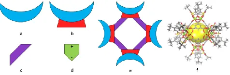
Metal-organic super containers (MOSCs)6represent a new
class of biomimetic container
molecules.MOSCsallowforvariedgeometricplacementsofthe“b uildingblocks”whichpermits a highly modular design. Three
types of such building blocksinclude(a)
sulfonylcalix[4]arenes7(SC) shown in Figure 1(a), (b)
tetranuclear complexes (TNCs),shown in Figure 1(b), which
consist of a SC bound to four M2+ ions furnished by four
carboxylategroups,and aμ4
oxygen,8and(c)variouscarboxylatelinkersshowninFigure1(c).
An example of the complete MOSC is represented in form of a
schematic diagram, in Figure 1(e) and in form of atomistic
model, in Figure 1(f). Each super container possesses multiple
internal and external cavities and has variable geometry
depending upon the selection of the carboxylate building
blocks. It is in part because of their tunable character that
MOSCs can be utilized in a wide range of applications,
including drug delivery and biomedical sensing.
Figure 1 Structural details of MOSC building blocks
Cartoon diagram of the „building blocks‟ of a MOSC. a)
sulfonylcalix[4]arene, b) tetranuclear
complex,c)carboxylatelinkages,d)aspirin.Notethataspirinisa
polarmolecule,asindicatedby a plus and minus sign at
opposite ends of the shape. e) Cartoon diagram depicting a
simplified MOSC structure to aid the reader in visualizing
the super container molecule, and f) Exampleofa molecular
structure of a MOSC.6aThe yellow sphere aids in guiding
the reader‟s eyes in visualizing the MOSC‟s
three-dimensional shape.
Another class of materials closely related to the
MOSCs are metal organic frameworks
(MOFs).MOFsareafamilyoftwo-dimensional(2D)orthree-dimensional(3D)infinitecrystalline lattices formed from the
or metal ion clusters.9Commonly, the organic molecules,
known as linkers, are of a semi-rigid or rigid construction
and form links between the metalionsorion clusters.10This
design works in the same manner as scaffolds or similar
frameworks, hence the name. Unfortunately, the
exceedingly large number of atoms in an asymmetric unit of
a typical MOF makes computational studies of these
systems rather challenging.11This problem is present in
MOSC systems as well. To reduce computational expense,
we model properties of the chemically representative
fragments of a MOSC system.
As is often done when studying MOFs, we model
only parts of the system.12Considering the size of these
MOSCs can approach the threshold of computational
capabilities (at 200-800 atoms), we applied a „divide and conquer‟ strategy and theoretically investigated the building
blocksforthesesuper-containers.Incomputationallyanalyzingthesemolecules,weho
petobetter understand their binding capabilities and
selectivity to guest molecules, in addition to fully
understanding the characteristics of the bindingsites.
Experimentally, these MOSCs were found to
selectively and reversibly bind cationic guests, such as
methylene blue dye.6b, 6cUtilizing ratiometric results from
UV-Vis titrations of varying amounts of host and guest
molecules revealed that SC and TNC bound two molecules
of aspirin each. A MOSC, constructed from
tert-butyl-sulfonylcalix[4]arene, 1,4-benzene-dicarboxylate linkers
and cobalt(II), was found to bind more than thirty molecules
of aspirin.6cIt was this fifteen-fold increase in aspirin
molecule adsorption that led to this study. In order to
simplify our model, we have removed the tertbutyl groups
and have assumed the conformation of SC is a rigid
cup-shaped molecule,13which matches the shape of the TNC
molecule, as TNC is held in the cup shape by the
coordination of the metal ions with carboxylate groups,
sulfonyl oxygens, andμ4-oxygen.
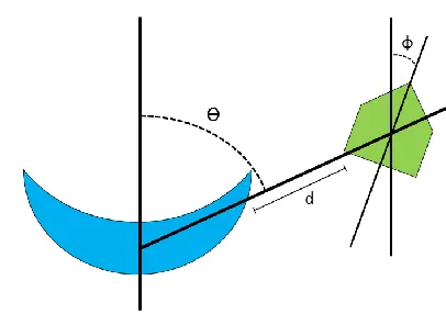
We explore the binding pattern of hosts (SC, TNC)
and guest, as a function of 1) binding site, 2) relative
orientation (of their dipoles), and 3) distance between them
(Figure 2).
Figure 2 Diagram of mutual orientation of host and guest
Schematic diagram depicting the mutual spatial
configurations of host (blue moon) and guest (green
pentagon) can be approximately parameterized by three
elements: 1) angle ϴ = direction,
fromwhichtheguestapproacheshost(specifiesthebindingsite),
2)angleϕ=determinesmutual orientation of electrostatic
dipoles of host and guest, and 3) distance d = between host
andguest
Methods
X-ray crystallographic data6awas used to obtain
structural information for the cobalt- containing TNC,
instead of running optimization, which gives us an exact
correlation between
experimentalandcomputationalwork.Atomicstructuredetermi
nesinitialpositionsofeachion,
⃗𝑅⃗⃗I, in the model. The presence of transition metal ions
provides a challenge for force field molecular mechanics
approaches. Some existing force fields do not account for
electronic
polarizationoftheenvironment,aneffectthatcansignificantlyre
duceelectrostaticinteractionsof partial atomic charges.14The
force fields that do account for electronic polarization are
too time- consuming for our project. Density-functional
theory (DFT) was chosen because it offers the best
balancebetweennecessarycomputationaltimeandaccuracy.Th
𝑖
equation, which is the Schrödinger equation of a
fictitious system of non-interacting particles (typically
electrons) that generate the same density as any given
system of interacting particles.
Here the first term corresponds to kinetic energy T and
uses symbol of gradient ∇= ( 𝜕 , 𝜕 , 𝜕 ).
Insolvingequation(1)onefindsasetofone-electronorbitals𝜑𝐾𝑆(𝑟), andtheirenergies𝜀𝑖.The orbitals are
combined with orbital occupation function 𝑓𝑖for constructing
the total density of electrons
ρ(r) = ∑fiφ KS∗
(r)φKS(r)
i i i≤HO
(2)
The total density determines the potential
which is defined as the functional derivative of the total
energy 𝐸𝑡𝑜𝑡[𝜌] minus kinetic energy 𝑇 in respect to
variation of the total density 𝜌(𝑟 ) and includes interactions
of electrons with ions, and three electron iterations:
Coulomb, correlation, and exchange. Rectangular brackets
symbolize a functional. Equations (1)-(3) are solved in the
iterative, self-consistent manner. Electrostatic potential
introduced in Eq. (3) illustrates interaction of a valence
electron with background charge of electrons and ions.
This electrostatic potential is a byproduct computed in DFT
and several other methods.15
Theelectronicdensityofstates(DOS)describesthenu
mberofstatesperintervalofenergy. We use DOS in order to
characterize electronic structure of the studied models. DOS
wasdefined as
where the Dirac delta function was approximated by
finite width Gaussian function and εiare the energies of a
given KS orbital (calculated using DFT), and the index i
runs over all orbitals calculated.
ESP maps are a three-dimensional representation
of the charge distribution to visualize
regionsofamoleculewithvariablecharge.Singlepointenergycal
culationswererunonstructures after geometry optimization.
Binding energies of the host and guest were calculated using
the equation:
Each molecule‟s ground-state energy was determined
separately. Computed values of binding energy are
compared to thermal quantum 𝑘𝐵𝑇 and to the energy of
hydrogen bond in water.
Results and Discussion
We used Gaussian 09 software to find an electronic
structure with density functional theory.16We used this
software to solve self consistent DFT equations.DFT with
B97 dexchange- correlation functional and 6-311G(d) and
LanL2DZ basis sets (for light elements and transition
metals, respectively) in Gaussian software provides spatial
electron density distributions for the ground state and for
Kohn-Sham orbitals. Gauss View 5 was used as an interface
for model- building and visualization of
results.17Determination of the energy of the molecular
systems was completed using the B97d functional, which
includes a dispersion correction.18The B3LYP functional
was used to generate electrostatic potential maps. n(ε) = ∑δ(ε − εi)
i
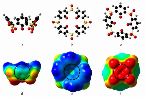
Figure 3 Sulfonylcalixarene models and ESP maps
Ball and stick models of sulfonylcalix[4]arene a) side view,
b) top view, c) bottom view. Atom colors: black=carbon,
red=oxygen, yellow=sulfur, grey=hydrogen. Electrostatic
potential maps of sulfonylcalix[4]arene e) side view, f) top
view, g) bottom view. Blue regions designate
relativepositive charge, transitioning into negative,
indicated by red. The dotted black circles identifythe
binding locations we have chosen.
Electrostatic Potential Maps
Understanding the variable electron density of the hosts
and guests was essential in determining possible binding
sites. In order to do so, electrostatic potential (ESP) maps
were generated, according to Eq. (3).15From these maps,
we have identified the top (ϴ=0˚), side (ϴ=90˚), and bottom
(ϴ=180˚) of each host molecule as candidate sites (Figures
3, 4) due to eitherpositive or negative regions existing on the
surface of the molecule.19In SC, positive regions exist on
the top of the molecule, with negative regions on the
bottom. TNC shares these features, only to a greater extent,
as the metals now draw even more electron density away
from the top side of the molecule. ESP map of aspirin,
shown in Figure 5 allows selection of the mutual orientation
of host and guest.
Figure 4 TNC models and ESP maps
Ball and stick models of the tetranuclear complex a) side
view, b) top view, c) bottom view. Atom colors:
black=carbon, red=oxygen, yellow=sulfur, blue=cobalt,
grey=hydrogen. Electrostatic potential maps of the
tetranuclear complex e) side view, f) top view, g) bottom
view. Blue regions designate relative positive charge,
transitioning into negative, indicated by red. The dotted
black circles identify the binding locations we have chosen.
a) Ball and stick model of aspirin side view. Atom
colors:
black=carbon,
red=oxygen,
grey=hydrogen. b) Electrostatic potential map of
aspirin, side view. Blue regions designate relative
positive charge, transitioning into negative,
indicated by red.
Binding Studies
Each guest molecule was placed near the host, at d= 3, 5,
7 Angstroms from the host molecule.20We expect that a
host-guest complex which displays stabilization will have a
negative binding energy according to Eq. (5).21 In addition,
the guest, i.e. aspirin, has been placed in thedirection of the
host at two orientations: positive and negative dipole attacks
attack,the benzene ring is closes tto thehost molecule,where
as in the negative dipole attack the oxygen molecules are
nearest to the host. These two positions were chosen
because both the host and guest molecules have partially
positive and partially negative regions.
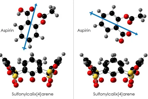
Figure 6 Two orientations of host and guest
Examples of various mutual orientations of host and guest
molecules. Right: Scheme displaying the negative dipole
attack orientation of the aspirin molecule in relation to the
host, sulfonylcalix[4]arene. Note that a true „head-on‟
orientation was not used due to the steric hindrance of the
methane group on the aspirin molecule. Left: Scheme
displaying the positive dipole attack orientation of the
aspirin molecule in relation to the host,
sulfonylcalix[4]arene.
Tables 1 and 2 summarize the calculated binding
energies. Binding energies exhibit clear dependence upon
mutual orientation parameters ϴ, ϕ, and d. Binding energies
decrease with
increasinghost-guestdistance,withtwoexceptions:1)SC,positivedipoleattackf
romthetop,and
2)TNC,positivedipoleattackfromthetop.Weconsiderthepo
ssibilitythatasthepositivedipole of the aspirin approaches the
top of the hosts (which are positively charged at that binding
site), the guest experiences repulsion. Once the aspirin is
displaced, there is a slight attraction betweenthe molecules.
The decrement of E(bind)(d) with distance is attributed
to severalinteractionmechanisms: Coulombic, 1,
dipole-dipole 1, etc.22Both host molecules, SC and TNC,provide𝑑,
𝑑3favorable adsorption, but do so with an opposite
dependence on angle. SC and TNC both prefer a negative
dipole attack from the top and a positive dipole attack from
the bottom. Some areas do
notparticipateinbinding,indicatedbyapositivevalueinthetable,
which corresponds to repulsion of species.
Table 1 Sulfonylcalixarene binding energies
Aspirin to sulfonylcalix[4]arene binding energies in eV as a function of structural parameters ϕ, ϴ, and d.
We have also inspected the influence of the host-guest binding
on electronics structure.We have compared the bandgap
values, εbandgap = εLUMO − εHOMO, for the host, guest, and
combined host and guest systems,as reported inTablesS1,S2.In
both cases,the guest is aspirin.For
provided in
Figures S1-S2
. Examples of frontier
orbitals for host-guest complexes are provided in
Figures S3-S4
.
We have found an interesting correlation between adsorption binding energy and bandgap energy. Specifically, the host-guest pairs, which have larger values of binding
energy, provide a
strongerchangeofbandgapuponbinding.Thecorrelationsofbin dingandgappromptapossibility to monitor the binding of host and guest by spectralprobes.
Conclusions
Our calculations show that through simple models,
(e.g. aspirin) non-covalently binding to the building blocks
of MOSCs can be determined. ESP maps allow us to predict
possible binding sites on host molecules. Specifically, we
were testing the hypothesis that negatively charged sites of
the host preferentially bind with positively charged sites of
guest, which accounts for the experimentally observed
binding selectivity.6cIn addition, the strongest adsorption on
both sulfonylcalix[4]arene and the tetranuclear complex is
observed for two configurations: ϕ=ϴ=180˚, d=3 Å, and
ϕ=180˚, ϴ=0˚, d=3 Å.
Adsorption energies of aspirin to TNC have low
values -0.066 eV < E(bind) < -0.052 eV, corresponding to
thermal quantum, KT~0.027 eV. Such weak binding can be
broken by thermalfluctuations. The upper limit of strong
hydrogen bond energy is generally considered to be about
1.8 eV. Nearly all bonding at 3 Å of the SC occurs in
this range, with the only exception being ϕ=ϴ=0˚, d=3 Å.
This indicates that the binding of the aspirin to the SC is
strong, but still able to be rinsed away, as is anticipated for
such non-covalent host-guest interactions. Additionally, a
dipole dipol einter action is occurring here due to the
permanent dipole momentt hat eachho stand the guest
maintain.Consider ing the strength of the dipole-dipole
interactions,dispersion forces are less important.
Ongoing theoretical studies examine the building
blocks and the MOSC as a whole,
includingstructure,stabilityandbindingenergiesoftheMOSCh
ostwithvariousguestmolecules. The adsorption mechanism
of guest molecules to the various binding sites of the MOSC
will be explored by abinitio molecular
dynamics.23Additionalexperimentalstudies,withtheguidance
of
thesecomputationalstudies,willbeperformed,regardingguests
electivitytooptimizethebinding ability of the MOSCs. Once
a thorough understanding of the binding capability of the
MOSCs is complete, new guest molecules could be
identified and the MOSCs could be modified in order to
incorporatepreviouslynon-bindingguests.TheversatilityoftheMOSCsopensthemuptoawi
de range of applications, such as drug delivery vehicles and
perhaps water treatmentfilters.
References
1. Evans, W. E.; Relling, M. V., Nature 2004, 429
(6990), 464-468.
2. Noteboom, C.; Qureshi, S., Health and Technology
2014, 1 (4), 59-73.
3. Peer, D.; Karp, J. M.; Hong, S.; FaroKhzad, O. C.;
Margalit, R.; Langer, R., Nat. Nanotechnol. 2007,2
(12), 751-760.
4. Kannan, N.; Vakeesan, D., Renew. Sust. Energ. Rev.
2016, 62, 1092-1105.
5. Peng, R.; Zhao, D.; Dimitrijevic, N. M.; Rajh, T.;
Koodali, R. T., The Journal of Physical Chemistry
C2011, 116 (1), 1605-1613.
6. 6. (a) Dai, F.-R.; Wang, Z., J. Am. Chem. Soc.
2012, 134 (19), 8002-8005; (b) Dai, F.-R.; Becht,
D.C.;
7. Wang, Z., Chem. Commun. 2014, 50, 5385-5387; (c)
Dai, F.-R.; Sambasivam, U.; Hammerstrom, A. J.;
Wang, Z., J. Am. Chem. Soc. 2014, 136, DOI:
10.1021/ja502839b.
8. Morohashi, N.; Narumi, F.; Iki, N.; Hattori, T.;
Miyano, S., Chem Rev 2006, 106 (12), 5291-5316.
9. Kajiwara, T.; Kobashi, T.; Shinagawa, R.; Ito, T.;
Takaishi, S.; Yamashita, M.; Iki, N., Eur J Inorg
Chem2006, (9), 1765-1770.
10. Kitagawa, S.; Kitaura, R.; Noro, S. i., Angewandte
Chemie International Edition 2004, 43 (18), 2334-
2375.
11. Eddaoudi, M.; Moler, D. B.; Li, H.; Chen, B.;
Reineke, T. M.; O'keeffe, M.; Yaghi, O. M.,
Accounts Chem. Res. 2001, 34 (4), 319-330.
12. Yaghi, O. M.; O'Keeffe, M.; Ockwig, N. W.; Chae,
H. K.; Eddaoudi, M.; Kim, J., Nature 2003,
423(6941), 705-714.
13. Fischer, M.; Gomes, J. R. B.; Jorge, M., Molecular
Simulation 2014, 40 (7-9), 537-556.
14. Gutsche, C. D., 2nd ed.; The Royal Society of
15. Johnson, B. G.; Gill, P. M.; Pople, J. A.; Fox, D. J.,
Chemical physics letters 1993, 206 (1), 239-246.
16. Dennington, R.; Keith, T.; Millam, J., Semichem
Inc., Shawnee Mission KS 2009.
17. rimme, S., Journal of computational chemistry 2006,
27 (15), 1787-1799.
18. Naray-Szabo, G.; Ferenczy, G. G., Chemical reviews
1995, 95 (4), 829-847.
19. Brunold, T. C.; Conrad, K. S.; Liptak, M. D.; Park,
K., Coord. Chem. Rev. 2009, 253 (5+6), 779-794.
20. Peter, L. M., Journal of Physical Chemistry Letters
2011, 2 (15), 1861-1867.
21. Mora-Sero, I.; Bisquert, J., Journal of Physical
Chemistry Letters 2010, 1 (20), 3046-3052.
22. Hodes, G., Journal of Physical Chemistry C 2008,