Available Online At www.ijpret.
INTERNATIONAL JOURNAL OF PURE AND
APPLIED RESEARCH IN ENGINEERING AND
IMAGE ENHANCEMENT
PRABHJOT KAUR, SATWANT KAUR, PRAJNA VASUDEV,
1. RIET, Phagwara.
2. Assistant Professor RIET, Phagwara
Accepted Date: 16/11/2012 Publish Date: 01/12/2012 Keywords Contrast Stretching, Histogram Equalization, Gamma Correction Corresponding Author Er. Rishma Chawla
Assistant Professor RIET, Phagwara.
IJPRET-QR CODE
Available Online At www.ijpret.com
INTERNATIONAL JOURNAL OF PURE AND
APPLIED RESEARCH IN ENGINEERING AND
TECHNOLOGY
A PATH FOR HORIZING YOUR INNOVATIVE WORK
IMAGE ENHANCEMENT USING SPATIAL TECHNIQUES
PRABHJOT KAUR, SATWANT KAUR, PRAJNA VASUDEV, RISHMA CHAWLA
Assistant Professor RIET, Phagwara.
Abstract
Image Enhancement is a process to improve the visual appearance of original image. It results in more pleasing image for human and machine analysis for some specific applications such as forensics field, biology field, X
enhancement is used as a preprocessing step in digital image processing in some specific applications. Image enhancement can be done in two domains. Spatial domain and Frequency domain. Spatial domain techniques are those which directly operate on pixels in an image and frequency domain techniques operate on Fourier transform of an image. Image acquired through devices suffer from low contrast and noise. Low contrast images can be due to the poor illumination, lack of timing range in the imaging sensor, or due to the wrong setting of the lens. Low contrast affects the image visual quality. These problems are removed by image enhancement
techniques. This paper focuses on different image
enhancement techniques in spatial domain
INTERNATIONAL JOURNAL OF PURE AND
APPLIED RESEARCH IN ENGINEERING AND
A PATH FOR HORIZING YOUR INNOVATIVE WORK
TECHNIQUES
PRABHJOT KAUR, SATWANT KAUR, PRAJNA VASUDEV,
Image Enhancement is a process to improve the visual It results in more pleasing image analysis for some specific applications field, biology field, X-ray etc. Image enhancement is used as a preprocessing step in digital image processing in some specific applications. Image enhancement omain and Frequency . Spatial domain techniques are those which directly operate on pixels in an image and frequency domain techniques operate on Fourier transform of an image. Image acquired through devices suffer from low contrast and noise. ntrast images can be due to the poor illumination, lack of timing range in the imaging sensor, or due to the wrong setting of the lens. Low contrast affects the image visual quality. These problems are removed by image enhancement
cuses on different image
enhancement techniques in spatial domain
Research Article ISSN: 2319-507X Rishma Chawla, IJPRET, 2012; Volume 1(4): 147-153 IJPRET
Available Online At www.ijpret.com INTRODUCTION
Image enhancement is a method of
modifying the appearance of acquired image
through some device in a better way, so that
resultant image is better than original image
in looks1. Image enhancement is used as a
preprocessing step in digital image
processing before the image segmentation
step. The main aim of image enhancement is
to make the original image better so that the
enhanced image can be used as input in
further digital image processing steps. By
using this method, image can be enhanced in
various ways like, we can improve the
contrast, remove the noise etc. image
enhancement is used in various fields. For
example medical, remote sensing, biology,
automated vision inspection, military.
Image enhancement can be broadly
categorized into two domains that are as
follow:
Spatial Domain-spatial domain techniques
are those which directly operate on pixels in
an image. Pixel values of the input image are
manipulated to get the output image. Spatial
domain methods are denoted by the
expression2.
G(x, y)=T[F(x, y)] (1)
Where G(x, y) is the output image, T is the
Operation performed on F(x, y), F(x, y) is
original image that undergoes enhancement
process. Grey level transformation function
can be written as
S = T(r) (2)
Where r and s are intensity of pixels of f(x, y)
and g(x, y).
Frequency Domain-frequency domain
techniques operate on Fourier transform of
an image. The image is first transferred into
frequency domain. It means that, the Fourier
Transform of the image is computed first. All
the enhancement operations are performed
on the Fourier transform of the image and
then the Inverse Fourier transform is
performed to get the resultant image In the
frequency domain the image enhancement
can be done as follows[3]:
G (u, v) =H (u, v) F (u, v) (3)
Where G (u, v) is enhanced image, F (u, v) is
input image and H (u, v) is transfer function.
But here we will discuss only some of the
Available Online At www.ijpret. Spatial Domain techniques
Contrast stretching
Contrast is the difference of the brightness
between objects and their backgrounds .In
the digital image processing, contrast
enhancements can be done by the contrast
stretching enhancement technique .contrast
stretching is a point processing
enhancement technique in which we stretch
the range of intensity value of the pixels in
an image. This technique improves the
brightness difference uniformly across the
dynamic range of the image as a result of
this low contrast images look clear and
sharp4. The Contrast set to stretching is also
called as normalization. Before the
stretching can be performed it is necessary
to specify the upper and lower pixel value
limits over which the image is to be
normalized. Often these limits will just be
the minimum and maximum pixel values
that the image type concerned allows.
and b be the lower and upper limits
respectively for example: 8
images have 0 as the lower limit a
the upper limit. The simplest sort of
normalization then scans the image to find
Available Online At www.ijpret.com
Contrast is the difference of the brightness
between objects and their backgrounds .In
the digital image processing, contrast
enhancements can be done by the contrast
ching enhancement technique .contrast
stretching is a point processing
enhancement technique in which we stretch
the range of intensity value of the pixels in
an image. This technique improves the
brightness difference uniformly across the
the image as a result of
this low contrast images look clear and
. The Contrast set to stretching is also
called as normalization. Before the
stretching can be performed it is necessary
to specify the upper and lower pixel value
he image is to be
these limits will just be
the minimum and maximum pixel values
that the image type concerned allows. Let a
and b be the lower and upper limits
respectively for example: 8-bit gray level
images have 0 as the lower limit and 255 as
simplest sort of
normalization then scans the image to find
the lowest and highest pixel
present in the image
for each pixel, the original value
to output value s using the
Figure 1 showing the result of contrast
stretching
Histogram Equalization
highest pixel values currently
.call this c and d .Then
for each pixel, the original value r is mapped
using the function4:
(4)
the result of contrast
Research Article ISSN: 2319-507X Rishma Chawla, IJPRET, 2012; Volume 1(4): 147-153 IJPRET
Available Online At www.ijpret.com
An image histogram is a graphical
representation of the tonal distribution in a
digital image. It plots the number of pixels
for each tonal value. The horizontal axis of
the graph represents the tonal variation,
while the vertical axis represents the number
of pixel in the particular area. The left side of
the horizontal axis represents the black and
dark areas, the middle represent medium
grey and right hand side represent light and
pure white areas.
Histogram equalization is a non-linear
contrast enhancement method for modifying
the dynamic range and contrast of the image
by altering that image so that the histogram
of the resultant image is equalized to
become a constant. When an image
histogram is equalized, all pixel values of the
image are redistributed so there are
approximately an equal number of pixels to
each of the user specified output grayscale
classes[5].The Histogram of digital image
with the intensity levels in the range [0,L-1]
is a discrete function[5]
H (rk) = nk (5)
Where rk is the intensity value, nk is the
number of pixels in the image with intensity
rk and H (rk) is the histogram of the digital
image with Gray Level rk
Histograms are frequently normalized by the
total number of pixels in the image.
Assuming a M × N image, a normalized
histogram.
P(rk) = nk/MN K=0,1,2,3……….L-1
(6)
is related to probability of occurrence of rk in
the image.
Where p(rk) gives an estimate of the
probability of occurrence of gray level rk..The
Sum of all components of a normalized
Available Online At www.ijpret.com
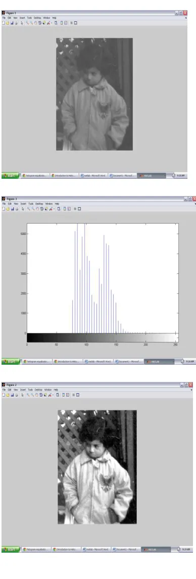
Figure 2 showing the effect of histogram
equalization
Gamma correction
Gamma correction operation is used for
brightness adjustment .Brightness for the
darker pixels are increased but it is almost
the same for the bright pixels6. Gamma
correction is also known as power law
transformation. Power-law transformations
have the basic form1.
s = crγ (7)
Where c and γ are positive constants.
For the various values of gamma different
Research Article ISSN: 2319-507X Rishma Chawla, IJPRET, 2012; Volume 1(4): 147-153 IJPRET
Available Online At www.ijpret.com Applications of image enhancement
• In forensics field, to improve the evidence
photographs and videos that is not very
clear and unidentifiable.
•In biology field, to analyze the forms and structure of animals and plants.
•In automated visual inspection of manufactured goods to inspect for their
missing parts.
•In x-ray imaging, ultrasonic and magnetic resonance imaging that is quite Often seen
in daily life.
CONCLUSION
Image enhancement techniques provide
different approaches for modifying the
appearance of an image. The choice of
enhancement techniques depends upon
various factors such as image content,
viewing conditions, characteristics by
observer and particular application. Like,
Image Negation is used for highlighting the
white details embedded in dark regions and
has its uses in medical imaging. Contrast
Stretching techniques are applied by
stretching the grey levels of an
image. Histogram equalization is used by
maximize the image contrast by applying a
gray level transform which tries to flatten
the resulting histogram. Gamma correction
improves the brightness of the acquired
image.
ACKNOWLEDGEMENT
We express our gratitude to the Ramgharia
institute of Engineering and Technology,
Phagwara, for giving us the opportunity to
work on the Review Paper during our final
year of Masters of technology in Computer
Science Engineering.
Our special thanks to Er. Rishma Chawla,
HOD (Department Of Computer Science) as
our guide, for their invaluable guidance
throughout our work and endeavor period
has provided us with the requisite
motivation to complete our Review paper
Available Online At www.ijpret.com REFERENCE
1. Raman Maini and Himanshu Agrawal, a
Comprehensive Review of Image
Enhancement Techniques, 2010: 8-13.
2. Gaba Gurjot Singh, Paramdeep Singh and
Gurpreet Singh, Implementation of Image
Enhancement Techniques, IOSRJECE, 2012;
1(2): 20-23.
3. HK sawant and Mahentra Deore, A
Comprehensive Review of Image
enchancement technique, 1(2): 39-44.
4. Suneetha and T Venkateswarlu,
Enhancement Techniques for Gray scale
Images in Spatial Domain”, International
Journal of Emerging Technology anpd
Advanced Engineering, 2012: 13-20.
5. Komal Vij and Yaduvir Singh,
Enhancement of Images Using Histogram
Processing Techniques”, int. J. Comp. Tech.
Appl. 2(2):309-313,
6. VV Starovoitov, DI Samal and DV Briliuk,
Image Enhancement for Face Recognition,
International Conference on Ionics, 2003,