MEMBRANE DEFORMATION AND LIPID SIGNALING: FUNCTIONS OF SRGAP FAMILY PROTEINS AND PI(4,5)P2
Jaeda Coutinho-Budd
A dissertation submitted to the faculty of the University of North Carolina at Chapel Hill in partial fulfillment of the requirements for the degree of Doctor of Philosophy in the
Curriculum in Neurobiology
Chapel Hill 2012
2012
ABSTRACT
JAEDA COUTINHO-BUDD: Membrane Deformation and Lipid Signaling: Functions of srGAP Family Proteins and PI(4,5)P2
(Under the direction of Franck Polleux, Ph.D., and Mark Zylka, Ph.D.)
The plasma membrane plays a structural and functional role in the life of a cell. Not only does it aid in encapsulating the intracellular contents to separate one cell from the next, but it also serves as an achor for the actin cytoskeleton scaffold, as well as a home base for lipids that serve as messengers in a number of downstream signaling pathways. Given the importance of these aspects in cellular regulation, variations in the plasma membrane could lead to vast consequences in cellular function. This work explores plasma membrane alterations in two ways: 1) investigating membrane deformation by the slit-robo GTPase Activating Protein (srGAP) proteins of the Bin/Amphiphysin/Rvs (BAR) superfamily, and 2) reducing the levels of phosphatidylinositol (4-5)-bisphosphate (PI(4,5)P2) using chemical
dimerization. The work presented in this thesis demonstrates that srGAP2 can induce neurite outgrowth and branching, and inhibit migration of cortical pyramidal neurons, through the ability of its N-terminal F-BAR domain to induce filopodia-like protrusions. srGAP2 is more potent at inducing protrusions than srGAP1 or srGAP3 in non-neuronal cells, an activity mimicked by their respective F-BAR domains. This work also explores the ways in which the F-BARs of srGAP proteins vary in their regulation of membrane dynamics.
To Graham and the rest of the Coutinho and the Budd families for your never-ending
ACKNOWLEDGEMENTS
TABLE OF CONTENTS
LIST OF FIGURES...xi
LIST OF ABBREVIATIONS... xiv
CHAPTER 1: General Introduction...1
1.1 Cortical development ...1
1.2 Neuronal morphology and neurite outgrowth ...2
1.3 BAR SUPERFAMILY: Proteins that coordinate membrane deformation with actin cytoskeleton dynamics...4
1.3.1 Bin/Amphyphysin/Rvs domain-containing proteins, the founding Subfamily...4
1.3.2 F-BAR domains, the elongated BAR domain...5
1.3.3 Inverse BAR domains form filopodia-like protrusions...6
1.4 srGAP family of F-BAR proteins...7
1.5 PI(4,5)P2-mediated interactions between the plasma membrane and actin cytoskeleton ... 10
1.6 PI(4,5)P2 is a critical regulator of cellular function ... 12
1.6.1 PI(4,5)P2 aids in subcellular protein localization ... 14
1.6.2 PI(4,5)P2 acts as a second messenger... 14
1.6.3 PI(4,5)P2 regulates ion channel function... 15
1.6.3.i PI(4,5)P2 regulation of TRPV1 ... 16
1.7 Rapamycin-induced depletion of PI(4,5)P2... 18
1.8 Nociceptive neuronal subsets of the dorsal root ganglia ... 19
neighboring actin cytoskeleton affect cortical neuronal
development?... 21
1.9.2 Are the F-BAR domains of srGAP1, srGAP2, and srGAP3 ... 21
1.9.3 Do alterations in the lipid composition of the plasma membrane regulate cellular function in vivo?... 22
1.10 Figures and legends... 23
CHAPTER 2: The F-BAR domain of srGAP2 induces membrane protrusions required for neuronal migration and morphogenesis... 30
2.1 INTRODUCTION ... 30
2.2 RESULTS... 32
2.2.1 Expression of srGAP2 in the developing cortex... 32
2.2.2 Full-Length srGAP2 and its F-BAR domain induce filopodia formation... 32
2.2.3 The F-BAR domain of srGAP2 deforms membrane like an I-BAR domain... 34
2.2.4 srGAP2 regulates neurite formation and branching through the ability of Its F-BAR domain to form filopodia ... 35
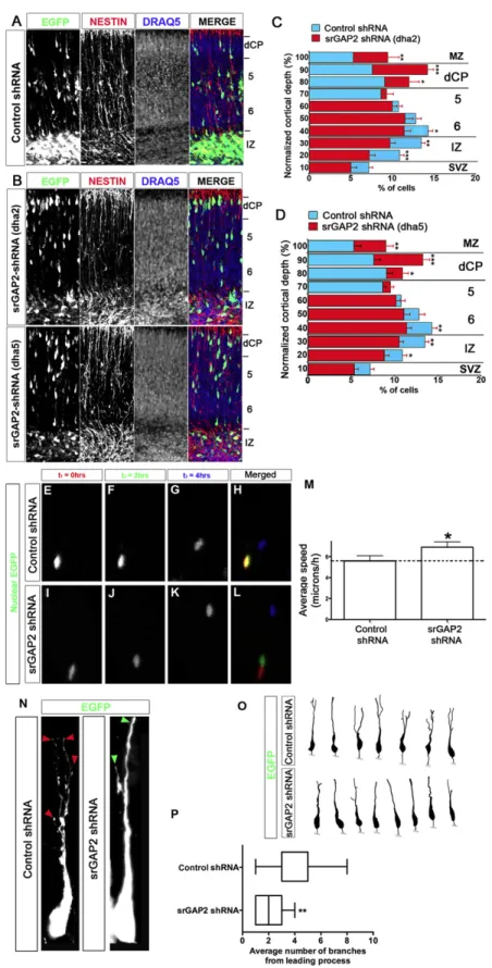
2.2.5 Reduction of srGAP2 expression promotes neuronal migration ... 37
2.2.6 The F-BAR domain is necessary and sufficient for srGAP2-mediated inhibition of radial migration... 38
2.2.7 srGAP2 inhibits migration by increasing leading process dynamics and branching ... 38
2.2.8 srGAP2 partially requires its RhoGAP and SH3 domains to inhibit migration ... 40
2.3 DISCUSSION ... 43
2.3.1 srGAP2 is a novel F-BAR domain-containing protein ... 43
2.3.2 A role for srGAP2 during neuronal development ... 44
2.3.3 Regulation of srGAP2: GAP and SH3 domains ... 45
2.4.1 Animals ... 46
2.4.2 Protein purification ... 46
2.4.3 In vitro GAP assay ... 47
2.4.4 Liposome preparation, liposome tubulation assays, and electron microscopy ... 47
2.4.5 Ex vivo cortical electroporation and primary cortical neuron cultures ... 47
2.5 SUPPLEMENTAL EXPERIMENTAL PROCEDURES ... 48
2.5.1 Sequence alignments... 48
2.5.2 shRNA design and validation ... 48
2.5.3 Constructs... 49
2.5.4 COS7 cell culture, transfections, staining and filopodia measurements ... 50
2.5.5 Ex vivo cortical electroporation and primary cultures ... 51
2.5.6 Time lapse confocal microscopy of cortical sections... 51
2.5.7 Dissociated cortical neuron culture... 52
2.5.8 Quantification of neuron migration and neurite branching... 52
2.5.9 In situ hybridization ... 53
2.5.10 Liposome preparation... 53
2.5.11 Electron microscopy ... 53
2.6 FIGURES AND LEGENDS ... 54
CHAPTER 3: The F-BAR domains from srGAP1, srGAP2, and srGAP3 differentially regulate membrane deformation ... 85
3.1 INTRODUCTION ... 85
3.2 RESULTS... 89
3.2.1. The srGAP family of proteins, through their respective F-BAR domains, exhibit different abilities to induce filopodia in non-neuronal cells ... 89
3.2.3 The F-BAR domains of different srGAP proteins localize
to distinct regions of filopodia ... 90
3.2.4 Molecular dynamics of the F-BAR Domains of srGAP1-3... 91
3.2.5 Lipid specificity varies between the F-BARs of srGAP proteins ... 93
3.2.6 F-BAR(1) constrains cellular protrusions in cortical neurons, whereas F-BAR(2) and F-BAR(3) induce protrusions ... 95
3.3 DISCUSSION ... 96
3.4 MATERIALS AND METHODS ... 101
3.4.1 Plasmid constructs and sequence alignments ... 101
3.4.2 Cell culture ... 101
3.4.3 Live cell imaging... 103
3.4.4 Biochemistry ... 104
3.5 FIGURE LEGENDS... 106
CHAPTER 4: Rapamycin-induced depletion of PI(4,5)P2 is effective in cell lines, but not in dorsal root ganglia neurons in vitro or in vivo... 117
4.1 INTRODUCTION ... 117
4.2 RESULTS... 119
4.2.1 Rapamycin-induced dimerization is an effective tool for studying biological processes in cell lines ... 119
4.2.2 Construction of two mouse lines to study rapamycin- induced PI(4,5)P2 depletion in thermal sensitivity in vivo... 120
4.2.3 Injection of rapamycin does not induce translocaion of Venus-FKBP-Inp54p from the cytoplasm to the plasma membrane in DRG neurons... 121
4.2.4 Expression of Inp54p in CGRP+ neurons leads to basal depletion of PI(4,5)P2 In cultured DRG neurons ... 122
4.2.5 Expression of Inp54p in CGRP+ neurons does not affect behavior in vivo ... 124
4.4 MATERIALS AND METHODS ... 128
4.4.1 DNA plasmid constructs ... 128
4.4.2 Cell culture and live-imaging ... 128
4.4.3 Generation of FRBPLF-CFP and Venus-FKBP-Inp54p Knockin Mice... 129
4.4.4 Neuronal dissociation and imaging... 130
4.4.5 Immunohistochemistry ... 131
4.4.6 Drug administration ... 132
4.4.7 Behavior... 132
4.5 FIGURES AND LEGENDS ... 133
CHAPTER 5: DISCUSSION ... 139
5.1 Summary of Findings ... 139
5.2 srGAP2 in neuronal morphology... 141
5.2.1 Regulation of srGAP2 autoinhibition... 142
5.3 Functional Differences Between F-BARs of the srGAP protein family ... 144
5.4 Challenges to rapamycin-inducible PI(4,5)P2 depletion in vivo... 146
5.4.1 Future directions with the current design ... 146
5.4.1i Behavioral assessent Rosa-FRBPLF/CGRP-Inp54p double heterozygous mice... 146
5.4.1ii Stabilization of the FRBPLF domain might be necessary for sufficient PI(4,5)P2 depletion in vivo... 148
5.4.1iii The magic ratio problem ... 149
5.4.1iv Rapamycin-Induced Depletion of PI(4,5)P2 with Inp54p Might Not Work in Neurons ... 150
5.4.2 Alternative design and approach to rapamycin-inducible PI(4,5)P2 depletion in vivo ... 151
LIST OF FIGURES
Figure 1.1 Cortical neuron development in vivo and in vitro... 23
Figure 1.2 BAR superfamily proteins ... 24
Figure 1.3 PI(4,5)P2-mediated actin regulation through actin-related proteins ... 25
Figure 1.4 PI(4,5)P2 metabolism... 26
Figure 1.5 PI(4,5)P2 signaling and ion channel regulation... 27
Figure 1.6 Schematic of rapamycin-induced depletion of PI(4,5)P2 from the plasma membrane... 28
Figure 1.7 Subtypes of nociceptive neurons in the DRG... 29
Figure 2.1 srGAP2 is expressed in neuronal progenitors and postmitotic neurons and localizes to sites of membrane protrusion ... 54
Figure 2.2 F-BAR-induced filopodia required F-Actin for their dynamic formation but not for their structural maintenance... 56
Figure 2.3 Knockdown of srGAP2 in cortical neurons reduces axonal and dendritic branching ... 58
Figure 2.4 srGAP2 promotes filopodia formation and neurite outgrowth in an F-BAR-dependent manner... 60
Figure 2.5 Knockdown of srGAP2 promotes neuronal migration and reduces LP branching ... 62
Figure 2.6 srGAP2-mediated inhibition of migration requires F-BAR-mediated membrane deformation ... 64
Figure 2.7 Model for srGAP2-regulated membrane protrusion in neuronal migration ... 66
Figure 2.S1 srGAP2 induces filopodia formation in a F-BAR-dependent manner in COS7 cells... 67
Figure 2.S2 Expression of the F-BAR domain of srGAP2 in COS7 cells does not inhibit endocytosis ... 69
Figure 2.S3 Structural alignment of the predicted F-BAR domain of srGAP2... 70
Figure 2.S4 Localization of srGAP2-EGFP, F-BAR-EGFP and F-BARΔ49-EGFP fusion proteins in COS7 cells and stage 1 E15 cortical neurons ... 72
Figure 2.S6 Control FBP17 F-BAR tubulates liposome... 74
Figure 2.S7 Neurites induced by different srGAP2 constructs contain microtubules ... 75
Figure 2.S8 Definition of layers in slices following dorsal electroporation and organotypic culture ... 76
Figure 2.S9 Expression of srGAP2 in post mitotic neurons inhibits radial migration ... 77
Figure 2.S10 2.2 kD NeuroD promoter drives gene expression in non-radial glial intermediate progenitors 24 hours after electroporation... 78
Figure 2.S11 The RhoGAP domain of srGAP2 is specific for Rac1 ... 79
Figure 2.S12 The GAP and SH3 domains participate in srGAP2’s ability to promote filopodia formation in neurons ... 80
Figure 2.S13 srGAP2 expressing cells accumulate in stage 2 ... 82
Figure 2.S14 The GAP and SH3 domains participate in srGAP2’s ability to inhibit migration ... 83
Figure 3.1 srGAP2 induces significantly more filopodia than srGAP1 or srGAP3... 106
Figure 3.2 srGAP proteins interact through their F-BAR domains... 107
Figure 3.3 Synergy between F-BAR domains towards filopodia induction ... 108
Figure 3.4 The three F-BAR domains of srGAP proteins differ in their subcellular molecular dynamics ... 109
Figure 3.5 F-BAR(2) binds multiple negatively-charged phospholipids ... 110
Figure 3.6 F-BAR domains of srGAP proteins differ in their ability to induce filopodia in cortical neurons ... 111
Figure 3.7 Real-time imaging of membrane and F-Actin dynamics induced by F-BAR domains in cortical neurons... 112
Figure 3.S1 Conservation and alignments of the srGAP family of proteins ... 113
Figure 3.S2 The F-Actin network is disrupted after Cytochalasin-D treatment ... 114
Figure 3.S3 Dose-dependent binding of F-BAR(2) to various phosphoinositides ... 115
Figure 4.1 Rapamycin-induced translocation effectively allows for PI(4,5)P2
reduction in HEK293 cells... 133 Figure 4.2 Both knockin mouse lines express the correct protein in the
appropriate DRG neurons... 134 Figure 4.3 Rapamycin does not induce translocation in double heterozygotic
mice in vivo... 135 Figure 4.4 PI(4,5)P2 is depleted in CGRP-Inp54p knockin neurons ... 136
Figure 4.5 PI(4,5)P2 add-back does not rescue deficits in calcium signaling in
CGRP-Inp54p neurons, most likely due to poor cell heath... 137 Figure 4.6 Expression of Venus-FKBP-Inp54p in heat-sensing CGRP neurons
LIST OF ABBREVIATIONS E – embryonic day (post-conception)
RGC – radial glial cells CP - cortical plate
GTP – guanine triphosphate GDP – guanine diphosphate
GEF – guanine nucleotide exchange factors GAP – GTPase activating proteins
BAR – Bin/Amphiphysin/Rvs
N-BAR – N-terminal Amphiphathic helix BAR FCH – Fer/Fes CIP4 homology
F-BAR – FCH BAR domain I-BAR – inverse BAR
PI(4,5)P2 – phosphatidylinositol 4,5-bisphosphate
CC – coiled coil
SH3 – Src Homology 3
FBP17 – forming binding protein 17
toca-1 – transducer of cdc42 actin assembly 1 WASP – Wiscott aldrich syndrome protein
IRSp53 – insulin receptor tyrosine kinase substrate p53 GFP – green fluorescent protein
CFP – cyan fluorescent protein RFP – red fluorescent protein
t1/2 – half time of fluorescence recovery
PI – phosphoinositide PLC – phospholipase C DAG – diacylglycerol
TRPV1 – transient receptor potential vanilloid 1 GPCR – G protein coupled receptor
FKBP12 – FK506 binding protein 12 FRB – FKBP rapamycin binding
mTOR – mammalian target of rapamycin
Inp54p – Inositol polyphosphate 5-phosphatase 4 rapalog – rapamycin analog
C20-MaRap – carbon 20 methylallylrapamycin DRG – dorsal root ganglia
CHAPTER 1 General Introduction
1.1 Cortical development
Formation of functional neuronal circuits involves the coordinated migration of neurons to their final location, the subsequent generation of a single axon and multiple dendrites and finally the formatin of functional synaptic contacts(Barnes and Polleux, 2009). To achieve these complex steps, neurons undergo substantial changes in
morphology, involving both cytoskeletal and membrane remodeling (Luo, 2002; Noctor et al., 2004; Oshima et al., 2007). The developing cerebral cortex is a prime example of the intricacy and dynamics of these morphological changes.
regulated in well-defined steps (Fig. 1.1A). Cortical neurons, born through asymmetric divisions of radial glial cells (RGC), transition through a multipolar morphology
characterized by short, immature neurites that protrude from the cell body to dynamically sense the surrounding environment. These neurons then attach to a RGC, which serves as a scaffold for their migration. At this stage, neurons are characterized by the presence of a single, polarized leading process, and a trailing process at the rear. Recent evidence shows that the trailing process of radially migrating pyramidal neurons extends rapidly to become the axon (Ayala et al., 2007; Luo, 2002). Upon reaching their final destination at the cortical plate (CP), the leading process becomes the apical dendrite and will undergo tremendous branching, and formation of dendritic spines during early postnatal
development (Barnes and Polleux, 2009).
1.2 Neuronal morphology and neurite outgrowth
morphogenesis rely on the ability to regulate actin and microtubule dynamics(Ayala et al., 2007; Gupta et al., 2002).
The basis of neurite initiation, outgrowth, and branching is rooted in the ability of the cytoskeleton to undergo dynamic changes. Small Rho GTPases, such as RhoA, Rac1, and Cdc42, play an important role in modulating the cytoskeletal transformations that take place during neuronal morphogenesis (Threadgill et al., 1997). These small GTPases switch between an activated state when bound to guanine triphosphate (GTP), and inactivated state when bound to guanine diphosphate (GDP). The GTP-GDP cycle is regulated by and Guanine nucleotide exchange factors (GEFs) to activate the proteins, and GTPase
activating proteins (GAPs) to cause inactivation. The classic view of cytoskeletal
modulation by these three major GTPases is that Rac1 leads to lamellopodial protrusions through the formation of branched actin networks, Cdc42 regulates filopodia formation by the formation of filamentus actin bundles, and RhoA induces actin depolymerization (Hall, 1994). All three of these GTPases have been shown to be important regulators in
cytoskeletal rearrangement during neurite outgrowth (Threadgill et al., 1997; Ng and Luo, 2004).
further demonstrated the dependence of neurite outgrowth on filopodia using neurons from mmeevv mice that lack all three murine actin anti-capper proteins: mammalian enabled (Mena), vasodilator stimulated phosphoprotein (VASP), and Ena-VASP like (EVL). Ena/VASP are potent inducers of filopodia, and their triple knockout in cortical neurons resulted in loss of filopodia and failed neurite initiation, both in vitro and in vivo.
Interestingly, loss of Ena/VASP proteins also resulted in cortical lamination defects (Bear et al., 2002; Goh et al, 2002; Kwiatkowski et al., 2007), suggesting a complex functional relationship between filopodia formation, neurite initiation, and neuronal migration.
1.3 BAR SUPERFAMILY: Proteins that coordinate membrane deformation with actin cytoskeleton dynamics
An emerging field is providing novel insights into a family of proteins that directly bind and deform cellular membranes. In addition to inducing membrane curvature,
members of the Bin/Amphyphysin/Rvs (BAR) superfamily of proteins link the membrane to the actin cytoskeleton, either directly (Yamagishi et al., 2004; Lee et al., 2007) or indirectly (Tsujita et al., 2006; Suetsugu et al., 2006; Scita et al., 2008), to become multifaceted regulators of cell morphology and function. However, this interaction is not necessary for cellular membrane deformation, as it has also been shown that membrane deformation induced by these proteins can preclude the emergence of F-actin bundles (Yang et al., 2009).
1.3.1 Bin/Amphyphysin/Rvs domain-containing proteins, the founding subfamily
The six alpha-helices, making up the BAR domain homodimer, form a banana-shaped structure with positive amino acids located on the concave surface, allowing the BAR domain to directly bind negatively-charged, PI(4,5)P2-containing membrane via electrostatic interactions (Fig. 1.3D). These dimeric proteins have been shown to oligomerize to invaginate membrane into the cell to create tubular networks (Itoh et al., 2005; Shimada et al., 2007). A specialized subset of the BAR domain, the N-BAR domain, contains an N-terminal amphipathic alpha-helix that directly inserts into the membrane, increasing membrane curvature (Itoh et al., 2006). Many of these BAR and N-BAR proteins, such as amphiphysin and endophilin, have been implicated in membrane deformation relating to synaptic vesicle formation (Di Paolo et al., 2002; Schuske et al., 2003).
1.3.2 F-BAR domains, the elongated BAR domain
(Shimada et al., 2007) and cryo-electron microscopy (cryo-EM; Frost et al., 2008). These studies reveal that individual F-BAR dimers can bind end-to-end to form long ‘string-like’ oligomers, which also display a lateral binding interface to form a ‘wall-like’ structure reminiscent of the structure adopted by septins (reviewed in Kinoshita, 2006). Like the BAR domain proteins, F-BAR domain proteins, Syndapins and FBP17, have been implicated in endocytosis of synaptic vesicles (Qualmann et al., 1999; Koch et al., 2011; Rodal et al., 2008; Wu et al., 2010). Most of these F-BAR proteins have been found to be important in presynaptic vesicle recycling; however, formin binding protein 17
(FBP17/Toca-2), one of the canonical members of this BAR domain subclass, has been shown to play a role in formation of synaptic spines. Wakita et al. (2011) demonstrated that shRNA-mediated knockdown of endogenous FBP17 results in reduced spine density in cultured hippocampal rat neurons. Interestingly, a very similar protein, cdc42-interacting protein 4 (CIP4/Toca-3), has been shown to inhibit neurite formation by inducing
lamellipodial protrusions (Saengsawang et al., 2012). Both of these proteins are related to the transducer of cdc42-dependent actin assembly 1 (Toca-1), which has been shown to suppress neurite outgrowth in PC12 cells (Kakimoto et al., 2006), presumably through its membrane-invaginating activity. These conclusions were later complicated when it was shown that Toca-1 induces neurites in N1E115 neuroblastoma cells through its ability to induce filopodia by complex formation with N-WASP (Bu et al., 2009). This Toca-1-induced filopodia formation was blocked with inhibitors of endocytosis, suggesting a complex link between filopodia and endocytosis.
1.3.3 Inverse BAR domains form filopodia-like protrusions
(Millard et al., 2005), hence the name “Inverse-BAR.” Accordingly, in contrast to the canonical BAR and F-BAR domains, I-BAR domains induce filopodia-like membrane protrusions in vivo and in vitro (Millard et al., 2005; Matilla et al., 2007; Saarikangas et al., 2008; Saarikangas et al., 2009), and have actually been shown to inhibit endocytosis (Quinones et al., 2010). The exact structural mechanism underlying I-BAR-induced
protrusive activity is currently unknown, but thought to differ from the oligomerization-based tubulation characterizing F-BAR domains (Saarikangas et al., 2009). Although these I-BAR domain-containing proteins have been long thought of as inducers of membrane protrusion, a relatively new I-BAR domain protein, Pinkbar, has been shown to induce the formation of planar membrane structures (Pykäläinen et al., 2011). It is likely that the functions of many of the BAR superfamily proteins will differ from their original functionalities.
1.4 srGAP family of F-BAR proteins
While canonical F-BAR domains of certain family members such as Toca-1, FBP17, and CIP4 are known to invaginate membrane (Itoh and Camilli, 2006), there are known F-BAR proteins that induce filopodia-like membrane protrusions. Proteins such as GAS7 and PSTPIP2 (MAYP) were shown to induce filopodia formation prior to their characterization as F-BAR domain-containing proteins (Chitu et al., 2005; She et al., 2002). The function of approximately 25 predicted F-BAR proteins in the human genome remain to be identified. One such poorly characterized family of F-BAR domain-containing proteins is the slit-robo GTPase Activating Proteins (srGAPs).
inactivate small GTPases by increasing their intrinsically slow rate of GTP hydrolysis (Schutes and Der, 2006). srGAP1 specifically binds and inactivates Cdc42 and RhoA (Wong et al., 2001), while srGAP3 is specific for Rac1 (Soderling et al., 2002). The orthologue to mammalian srGAP proteins in C. elegans, SRGP-1, has been shown to regulate cell-cell junctions and engulfment of apoptotic cells through its regulation of Rac1 (Zaidel-Bar et al. 2011; Neukomm et al., 2011). SH3 domains are polyproline-binding motifs that mediate protein-protein interactions. In addition to Robo, the SH3 domain of srGAP2 has been shown to bind to actin-related proteins such as Wiscott-Aldrich Syndrome protein (WASP), WASP interacting protein (WIP), Diaphanous homologous protein 1 (mDia1) (Linkermann et al., 2009), formin-like 1 (FMNL-1, Mason et al., 2011), and Palladin (Okada et al., 2011). The SH3 domain of srGAP3 has been shown to bind the Wasp family
member, WAVE-1 (Soderling et al., 2002) and lamellipodin (Endris et al., 2011) to regulate Rac-dependent cellular protrusions.
In Chapter 2, we propose a model of autoinhibition for srGAP2, and hypothesize that the SH3 domain of srGAP2 is the key to unlocking its filopodia-inducing activity. It is possible that SH3 binding to one or more of these proteins releases the autoinhibition, allowing the F-BAR domains to dimerize, and induce protrusions. Subsequent work involving the crystal structure of Syndapin I (Rao et al., 2010) demonstrates that the SH3 domain of Syndapin I directly binds its F-BAR domain, leading to autoinhibition.
Furthermore, this autoinhibition can be released by binding of the SH3 domain to dynamin 1. Alternatively, Guo et al. (2010) found that protein arginine methyltransferase 5 (PRMT5) binds srGAP2 in its N-terminus, and methylates arginine 927 in the C-terminus of srGAP2. The authors show that the methylation mutant, srGAP2R927A, fails to undergo dimerization,
Like many RhoGAP proteins, the srGAP family members have also been shown to regulate cell morphology and migration (Yang et al., 2006; Soderling et al., 2002; Soderling et al., 2007; Vogt et al., 2007; Endris et al., 2011; Carlson et al., 2011; Zaidel-Bar et al., 2011). Recently, the expression pattern of srGAP1, 2, and 3 was examined throughout the developing central nervous system, although the spatial and temporal patterns differ between the three (Bacon et al., 2009). srGAP2 and srGAP3 are present in the cerebral cortex throughout late embryonic development, suggesting a similar role for these proteins in regulation of morphology and migration of cortical neurons. srGAP1 expression emerges postnatally, suggesting that it has an alternative role in neuronal function. Recent work has revealed the presence of srGAP1 and srGAP3 in large diameter neurons of adult DRG, with almost complete absence of srGAP2 (Chen et al., 2012). Interestingly, only srGAP3 increased in expression after spaired nerve injury, further suggesting separate functions for the three srGAP proteins.
However, until recently (including work in chapters 2 and 3), little was known about the F-BAR domains of these proteins. Work from the Polleux lab (Guerrier et al, 2009; Chapter 2) was the first to identify the F-BAR of srGAP2 as an inducer of outward
membrane protrusions. The formation of these filopodia-like protrusions led to decreased cortical migration, and increased neurite formation in dissociated cortical neurons.
Subsequently, srGAP3 has also been shown to regulate neuronal protrusions, specifically spine density and shape (Carlson et al., 2011). Loss of srGAP3 reduces the density of spines in hippocampal and cortical neurons, as well as counter-intuitively reducing the number of mature, mushroom-shaped spines. Interestingly, heterozygous loss of srGAP3 increases the number of thin spines. Recent work from the Polleux lab has found similar results regarding the regulation of spine shape with srGAP2. More interestingly, this work shows that a human-specific, duplicated portion of the srGAP2 F-BAR domain (srGAP2p12)
(Charrier at al., In Press); however, in this work both loss of srGAP2 and inhibition by srGAP2p12 lead to increased spine density. These neuronal results for srGAP2 and
srGAP3 are particularly exciting given that both have been implicated in forms of brain-related disorders. Loss of srGAP3 is known to play a role in 3p deletion syndrome (Endris et al., 2002), a form of mental retardation, and recent work has revealed a role for srGAP2 in early infantile epileptic encephalopathy (Saitsu et al., 2011). Although these proteins are quite similar, there are subtle differences in their function, which are explored more
thoroughly in Chapter 3; however, it is currently unclear as to whether these differences arise from their interactions with the actin cytoskeleton, the plasma membrane, or both.
1.5 PI(4,5)P2-mediated interactions between the plasma membrane and actin cytoskeleton
The BAR superfamily proteins are known to bind electrostatically to negatively-charged lipids, such as phosphatidylinositol 4,5-bisphosphate (PI(4,5)P2) and
phosphatidylserine (PS), embedded in the plasma membrane (Takei et al., 1998; Itoh and De Camilli, 2006). Some of these family members not only bind to negatively-charged phospholipids, but have been shown to cluster PI(4,5)P2 specifically, such as the I-BAR of
IRSp53 (Saarikangas et al., 2009). Although IRSp53 can interact with the actin cytoskeleton both directly (Yamagishi et al., 2006) and indirectly (Scita et al., 2008), PI(4,5)P2 also has been found to interact with actin filaments to modulate cytoskeletal
activity through a variety of other actin-associated proteins, such as cofilin (Ojala et al., 2001), vinculin (Huttelmaier et al., 1998), talin (Martel et al., 2001), gelsonin (Yu et al., 1992; Azuma et al., 2000), neuronal Wiskott-Aldrich syndrome protein (N-WASP; Miki et al., 1996), (Fig 1.3).
High concentrations of PI(4,5)P2 often lead to actin polymerization, whereas
its actin-severing activity, thereby blocking actin depolymerization (Ojala et al., 2001). PI(4,5)P2 also inhibits gelsolin, a capping protein with actin-severing activity. PI(4,5)P first
interferes with gelsolin’s ability to bind actin, thereby uncapping actin filaments, and secondly inhibits its actin-severing activity (Yu et al., 1992). Binding to PI(4,5)P2 is able to
relieve the autoinhibition of many of these actin-interacting proteins, such as N-WASP, talin, and vinculin. Talin links membrane-bound adhesion proteins like β-1 integrins to the actin cytoskeletal in a PI(4,5)P2-dependent manner (Martel et al., 2001). The same
regulation allows talin to bind vinculin (Gilmore and Burridge, 1996), which also links proteins to actin, as well as binds VASP proteins (Huttelmaier et al. 1998). Upon
electrostatic binding to PI(4,5)P2, N-WASP intermolecular binding is inhibited, opening up
the protein and revealing other protein-binding domains (Miki et al., 1996); N-WASP can also be activated by the binding of cdc42 to its cdc42/rac-interactive binding (CRIB) domain (Symons et al., 1996). Once open, the verprolin-homology domain/cofilin-homology
domain/and acidic domain (VCA) binds a G-actin monomer and actin-related protein 2/3 (ARP2/3) to induce actin nucleation and the formation of branched actin networks.
Activated N-WASP can also bind and insert profilin-actin complexes into actin polymers to enhance actin polymerization. The proline-rich region binds SH3-containing proteins, such as the F-BAR and I-BAR containing proteins Toca-1 (Takano et al., 2008) and IRSp53 (Lim et al., 2008), respectively. Both of these proteins also interact directly with small Rho GTPases, adding more complexity to the relationship between PI(4,5)P2 and
actin-interacting proteins. It is likely that many of the other SH3-containing BAR superfamily proteins could interact with N-WASP as well.
Additionally, one of the major kinase classes responsible for the production of PI(4,5)P2, PIP5KI, interacts strongly with multiple regulators of the actin cytoskeleton (Fig
et al., 2004), Rho (Chong et al., 1994), Rac (Halstead et al., 2010), and Arf6 (Honda et al., 1999). Cdc42 has been shown biochemically to stimulate PIP5K-induced production of PI(4,5)P2 (Weernink et al., 2004); however, to my knowledge no functional analysis of
cdc42 and PIP5K has been performed, suggesting a possible indirect activation through another GTPase. PIP5KIα binds Rho in a GTP-dependent manner, and complexes with Rho-dependent seronine-threonine kinase (ROCK) to activating downstream pathways to induce actin stress fiber formation (Yamamoto et al., 2001). Rac1 has been shown to recruit PIP5KIβ to the plasma membrane and induce neurite retraction in N1E115
neuroblastoma cells and cerebellar granule neurons (Halstead et al., 2001). The authors identified the rac1-binding residue in PIP5KIβ, and mutated the corresponding, highly-conserved glutamate residue in PIPK5Iα and PIP5KIγ. All three wildtype isoforms induced neurite retraction in N1E115 cells, however, the glutamate mutations abolished both neurite retraction and membrane-localization in all three isoforms. Another important GTPase is Arf6, which has been shown to recruit PIP5Ks to the plasma membrane and lead to
membrane ruffles, actin comet formation, as well as regulate vesicle trafficking (reviewed in Funakoshi et al., 2011).
Clearly PI(4,5)P2 plays an important role in regulating morphological changes due to
membrane deformation and rearrangement of the actin cytoskeleton. Additionally, PI(4,5)P2
acts as a guidepost for other non-cytoskeletal-related proteins, serves as a second messenger in intracellular signaling pathways, and operates as modulator of ion channel activity. PI(4,5)P2 is paramount in cellular biology.
1.6 PI(4,5)P2 is a critical regulator of cellular function
PI(4,5)P2 is the most abundant phosphoinositide (PI) present in the plasma
species that interconvert through the local positioning of phosphates around the inositol ring of the lipid headgroup. PI(4,5)P2 can be synthesized through addition of a phosphate to
position 5 of the inositol ring of phosphatidylinositol 4-phosphate (PI(4)P) or the position 4 of phosphatidylinositol 5-phosphate (PI(5)P), or the removal of the phosphate at position 3 of phosphatidylinositol 3,4,5-triphosphate (PI(3,4,5)P3). PI(4,5)P2 can also be
downregulated by the removal of a phosphate from either positions 4 or 5. Phosphoinositide anabolism and catabolism are regulated by lipid kinases and
phosphatases, respectively, which are specific for each phosphate location in the inositol ring (Fig. 1.4) (reviewed in Liu and Bankaitis, 2010).
Conversion between PI species is carefully regulated by these kinases and phosphatases in order to maintain the appropriate phospholipid balance necessary for proper cellular function. For example, loss of PIP5Kγ, a kinase that converts PI(4)P to PI(4,5)P2, results in neural tube closure defects (Wang et al., 2007), defects in synaptic
transmission through inhibited endocytosis, and eventually perinatal death (Di Paolo et al., 2004). Interestingly, mice lacking synaptojanin, a 5-phosphatase domain-containing protein that reduces PI(4,5)P2 to PI(4)P, also die just after birth (Kim et al, 2002).
Synaptojanin knockout mice have an overabundance of PI(4,5)P2, leading to poor synaptic
transmission due to an accumulation of clathrin coated vesicles in their presynaptic
terminals. In contrast, overexpression of synaptojanin, due to trisomy of the synaptojanin 1 gene, has been linked to the cognitive impairments associated with Down’s syndrome (Voronov et al., 2008). Loss of a similar protein, the Oculocerebrorenal Lowe syndrome (OCRL) 5-phosphatase, also leads to mental retardation, among other abnormalities such as insufficient kidney absorption and hypotonia (Lowe et al., 1952).
These diseases arising from altered PI(4,5)P2 regulation suggest that PI(4,5)P2 is a
settings, the yeast 5-phosphatase, Inositol polyphosphate 5-phosphatase (Inp54p) is often used given its simple domain structure. Inp54p lacks the additional protein domains
commonly found in other 5-phosphatase family members (Stefan et al., 2002), resulting in a constitutively-active 5-phosphatase domain (Várnai et al., 2006; Nebl et al., 2000).
Removal of the C-terminal 13 amino acids disrupts the tether to endoplasmic reticulum, resulting in a cytoplasmic 5-phosphatase that is more amenable to manipulation for experimental reduction in PI(4,5)P2 (Wiradjaja et al., 2000).
1.6.1 PI(4,5)P2 aids in subcellular protein localization
Many proteins have been identified that contain domains or motifs to specifically target proteins to PI(4,5)P2 (Heo et al., 2006). This can be visualized using one of these
protein domains, the pleckstrin homology (PH) domain of phospholipase C ∂1 (PLC∂1), which has become a biosensor for PI(4,5)P2 abundance at the plasma membrane. When
high levels of PI(4,5)P2 are present, fluorescently-tagged versions of this PH domain can be
seen at the plasma membrane. When PI(4,5)P2 levels are low, the domain is found in the
cytoplasm. These PI(4,5)P2-binding domains often occur in cytoplasmic proteins, which are
translocated to the plasma membrane upon interaction with PI(4,5)P2; therefore, the
amount of available PI(4,5)P2 at the membrane regulates the amount of translocation, and
ultimately the extent of protein activity at the plasma membrane. Consequently, fluctuations in the amount of PI(4,5)P2 at the plasma membrane create differential effects in a number
of cellular responses due to PI(4,5)P2-binding, with implications in vivo ranging from
phototransduction (Huang et al., 2004), to bipolar disorder (Soares et al., 2000; Soares et al., 2001), to regulation of pain sensitivity (Sowa et al., 2010).
1.6.2 PI(4,5)P2 acts as a second messenger
In addition to its role in regulating the actin cytoskeleton and protein binding, PI(4,5)P2 can be cleaved by phospholipase C (PLC) into diacylglycerol (DAG) and inositol
1.5A) (Berridge et al., 1983). DAG remains at the plasma membrane to activate protein kinase C (PKC), and IP3 travels to the endoplasmic reticulum to bind IP3 receptors and
stimulate the release of intracellular calcium. Multiple isoforms of PLC exist, and are
activated by different stimuli. PLCβ-induced cleavage of PI(4,5)P2 is predominantly evoked
by the activation of Gαq-coupled G-protein coupled receptors (GPCRs), whereas receptor
tyrosine kinase (RTK) activation by nerve growth factor (NGF) leads to PLCγ activity (Rebecchi and Pentyala, 2000). Both of these subtypes respond to high levels of intracellular calcium (Ca2+), but the third major class, PLC∂, is the most sensitive to fluctuations in Ca2+ levels (Allen et al., 1997). Regardless of the route of activation, all
three of these isoforms have the ability to hydrolyze PI(4,5)P2 and activate downstream
signaling.
1.6.3 PI(4,5)P2 regulates ion channel function
The levels of PI(4,5)P2 not only regulate protein localization and intracellular
signaling cascades, but they can directly affect the function and activity of ion channels. The first reported case of PI(4,5)P2-dependent ion channel modulation came from
Hilgemann and Ball (1996). Using an inside-out membrane patch, the authors found that a sodium-calcium (Na+-Ca2+) exchanger and ATP-inhibited potassium (K+) channel currents
decayed over time in these isolated membrane patches; however, currents could be
restored with the addition of PI(4,5)P2 or components that facilitated in PI(4,5)P2 assembly.
Rescue of channel current was blocked with the addition of PLC. In other words, these channels require a basal level of PI(4,5)P2 in order to function (Fig. 1.5B). These findings
have since been corroborated with a variety of other Na+, K+, Ca2+, and non-selective cation
channels (Suh and Hill, 2005).
PI-binding regions of these ion channels have been identified based on
tranduction channels (Brauchi et al, 2007; Prescott and Julius, 2003; Ufret-Vincenty et al., 2011). These mutations might not be direct PI(4,5)P2 binding sites; they might instead alter
the conformation of the protein so that it can no longer interact with PI(4,5)P2. Suh and
Hille (2008) put forth two models of PI recognition: 1) electrostatic binding of clustered basic residues merely attract the protein to the negatively-charged lipids, and 2) the arrangement of basic charges that come together after protein folding create a precise PI binding pocket. Both of these models could explain PI specificity based on either the spacing of the basic amino acids along the protein sequence, or the arrangement of the basic residues in this binding pocket. However, without a crystal structure, it is difficult to determine the mechanism of interaction. Crystal structures of membrane-bound proteins are difficult to determine, however, one group has recently crystallized the inward rectifying K+ channel, Kir2.2, in the presence of short-chain PI(4,5)P
2, and shown a direct interaction
of the ion channel and PI(4,5)P2 (Fig. 1.5B). Hansen et al. (2011) found that one PI(4,5)P2
binds each of the four protein subunits of Kir2.2, between the transmembrane and cytoplasmic domains, inducing a conformational change in the channel that results in increased channel activity. It is possible that a similar mechanism exists for the PI(4,5)P2
regulation of other ion channels as well. 1.6.3.i PI(4,5)P2 regulation of TRPV1
One non-selective cation channel that has been shown to interact with PI(4,5)P2 is
the sensory transduction channel, transient receptor potential vanilloid 1 (TRPV1). TRPV1 responds to acid (H+), chemical ligands such as capsaicin, and noxious thermal stimuli
above 42º C, and TRPV1-null mice show sensory deficits to each of these stimuli (Caterina et al., 2000). The role of PI(4,5)P2 in TRPV1 activation has been more controversial over
the years. The first studies to report its PI(4,5)P2 dependence suggested that TRPV1 was
inhibited by PI(4,5)P2 (Chuang et al., 2001; Prescott and Julius, 2003); however, it has
2007). In the presence of low concentrations of capsaicin (10-100 nM) or subtle changes in temperature, PI(4,5)P2 depletion increases TRPV1 currents, and production of PI(4,5)P2
inhibits these currents. The authors also found that this inhibitory effect is most likely not due to PI(4,5)P2 directly, as similar results were not seen with excised membrane patches,
suggesting this inhibition is due to interaction between PI(4,5)P2 with other cellular proteins.
More recently, however, Klein et al. (2008) reported that depletion of PI(4,5)P2 inhibited
TRPV1 activity at both high and low concentrations. Moreover, they report that while PI(4,5)P2 is not the only PI to activate TRPV1, it is the most physiologically relevant player
in both membrane patches and intact cells.
Recently, PI(4,5)P2 depletion downstream of the G-coupled Adenosine receptor,
A1R, has been shown to reduce thermal sensitivity through TRPV1, and chronic
sensitization through pronociceptive GPCRs in mice in vivo (Sowa et al., 2010). The authors found that Prostatic Acid Phosphatase (PAP), an ectonucleotidase that reduces adenosine monophosphate (AMP) to adenosine, reduces PI(4,5)P2 levels through A1R
activation. PAP knockout mice have increased levels of endogenous PI(4,5)P2, and the
authors have previously shown these mice have increased sensitivity to nociceptive stimuli (Zylka et al., 2008). Intrathecal injection of secretory PAP (S-PAP) reduces PI(4,5)P2
levels, and inhibits both thermal sensation through TRPV1, and thermal and mechanical sensitization due to inflammation and nerve injury in wildtype animals. In contrast, injection of PI(4,5)P2 intensifies thermal hypersensitivity and mechanical allodynia with nerve injury
models and with pronociceptive stimulation. While many of the previously discussed reports have shown that PI(4,5)P2 is an important modulator of TRPV1, this work
demonstrates that PI(4,5)P2 is necessary for pain sensitization in vivo. Moreover, the
authors demonstrated a role in both acute and chronic pain models; however, manipulation of PI(4,5)P2 in this work was still indirect. A specific genetic reduction in vivo would be a
1.7 Rapamycin-induced depletion of PI(4,5)P2
A variety of methods have been used to reduce PI(4,5)P2 signaling, such as
activation of PLC, application of PI 4-kinase inhibitors, overexpression of PI(4,5)P2-binding
proteins or antibodies, or the expression of PI 5-phosphatases. While these methods are effective, they can have off-target effects, do not always allow for temporal control of manipulation, and are difficult to implement in vivo. Recently, two groups simultaneously developed a temporally-controlled method of PI(4,5)P2 depletion using a
chemically-induced dimerization system (Várnai et al., 2006; Suh et al., 2006).
Protein function often relies on the proximity of its effector. In other words, even an active protein is ineffective if it can’t reach its target. Nature has developed ways of
targeting proteins to their location of interest, either with specific codes like nuclear localization signals embedded within their sequences (Dingwall et al., 1982), or
electrostatic binding as discussed for BAR domains and other PI(4,5)P2-binding proteins.
Some proteins have the ability to bind with multiple proteins, but only do so in the presence of certain chemicals. For instance, the immunosuppressant FK506 Binding Protein 12 (FKBP12) binds multiple drugs, two of which are FK506 and rapamycin. When FKBP12 is bound to FK506, it inactivates the phosphatase calcineurin; however, in the presence of rapamycin, FKBP forms a complex with the mammalian target of rapamycin (mTOR) through its FBKP rapamycin binding (FRB) domain (Crabtree and Schreiber, 1996). These FKBP binding interactions subsequently initiate two dinstinct signaling pathways.
at the plasma membrane, and an FKBP-fused 5-phosphatase in the cytoplasm. Upon addition of rapamycin, the FKBP-phosphatase fusion translocates to the plasma membrane due to dimerization with the FRB domain. The enhanced proximity of the lipid
5-phosphatase to its target in the plasma membrane allows for the rapid reduction of PI(4,5)P2 to PI(4)P (Fig. 1.6). Overexpression of constitutively active 5-phosphatases
causes the loss of membrane-cytoskeletal adhesion, leading to increased membrane blebbing (Raucher et al., 2000), and long-term depletion of PI(4,5)P2 can lead to activation
of the apoptotic cell death pathway (Azuma et al., 2000; Mejillano et al., 2001). This inducible model of PI(4,5)P2 depletion has the advantage over constitutive expression of
5-phosphatases because it allows for temporal control, and reduces the risk of complications due to overexpression side-effects.
Currently, there are no known methods to deplete PI(4,5)P2in vivo. Given that the
FRB and FKBP components of this system are genetically encoded, it is possible to implement this inducible system in vivo with the addition of rapamycin. Natural rapamycin will interact with the endogenous mTOR pathway; however, it is possible to utilize mutated FRB domains that interact with rapamycin analogs (rapalogs; Stankunas et al., 2003; Bayle et al., 2006). These rapalogs do not interact with endogenous mTOR, but still function to induce dimerization between mutated FRB and FKBP. One FRB mutation that seemed promising for use in vivo given its interaction with the rapalog, C20-marap, consists of three amino acid mutations: K2095P, T2098L, and W2101F (Bayle et al., 2006). In order to acutely deplete PI(4,5)P2 to determine its affects on thermal sensitization in vivo, this
system needs to be targeted to heat-sensing neurons.
1.8 Nociceptive neuronal subsets of the dorsal root ganglia
twelve subtypes of nociceptive, or pain-sensing neurons (Neacsu and Flonta, 2006), that can be broken down into two main groups: A∂ and C fibers (reviewed in Julius and
Bausbam, 2001). A∂ fibers are lightly myelinated neurons of medium diameter that respond to mechanical, thermal, and chemical stimuli, with a thermal threshold of ~53°C (type I) and ~43°C (type II). C fibers are small diameter, unmyelinated neurons that sense mechanical, thermal, and chemical stimuli, in addition to innocuous temperature and itch. The thermal threshold of C fibers is approximately 43°C. C fibers can be further broken down into two neuronal subtypes based on their molecular markers and stimuli-evoked response: peptidergic and nonpeptidergic (Cavanaugh et al., 2009). Peptidergic neurons express substance P (SP), the nerve growth factor (NGF) receptor TrkA, as well as calcitonin gene related protein (CGRP), while peptidergic neurons are marked by proteins such as PAP and Mrgprd (Fig. 1.7). Cavanaugh et al. (2009) showed that acute ablation of TRPV1-expressing neurons using the TRPV1 ligand, capsaicin, eliminated heat sensitivity within the parameters of the behavioral tests. In contrast, inducible ablation of non-peptidergic neurons using diphtheria toxin (DTX) application to mice expressing the human diphtheria toxin receptor in the Mrgprd locus causes a significant reduction in mechanical sensitivity. As demonstrated by Cavanaugh et al. (2009), these subtypes of nociceptive DRG neurons can be genetically targeted, and the effectiveness of targeting can be verified molecularly or by specific behavioral responses. TRPV1 is expressed in a portion of A∂, peptidergic, and non-peptidergic nociceptive subtypes; however, 50% of CGRP-expressing neurons express TRPV1, making these neurons adequate genetic targets for the manipulation of heat-sensing neurons in vivo.
1.9 Overview and rational of aims explored in this thesis
next, but it also serves as an achor for the actin cytoskeleton scaffold, as well as a home base for lipids that serve as messengers in a number of downstream signaling pathways. Given the importance of these aspects in cellular regulation, variations in the plasma membrane could lead to vast consequences in cellular function. The overall aim of my dissertation was to study how perturbations to the plasma membrane affect cellular responses.
1.9.1. Do changes in plasma membrane curvature and its neighboring actin cytoskeleton affect cortical neuronal development? Immense morphological changes take place during neuronal development. As neurons mature, the cell must extend multiple neurite that eventually become the axon and dendrites. In order to construct these
neuronal processes, the outer membrane and internal components must be constructed, rearranged, and added at a relatively rapid pace compared to a cell at rest; therefore, it is possible that membrane-deforming proteins play a role in the changes occurring during this period of rapid outgrowth. srGAP2 is a member of the BAR superfamily of membrane-deforming proteins; however, it has internal domains that facilitate interaction with both the plasma membrane, the actin cytoskeleton, and other cellular proteins. Previously
characterized F-BAR domains had only been shown to regulate membrane invagination and endocytosis. In Chapter 2, I present work demonstrating that the membrane-deforming F-BAR domain of srGAP2 is sufficient to induce filopodia-like protrusions, and subsequently regulate neuronal development in vitro and in vivo.
1.9.2 Are the F-BAR domains of srGAP1, srGAP2, and srGAP3 functionally
different developmental timepoints (Bacon et al., 2009) and the original paper suggesting that all three srGAP proteins bound the Robo receptor (Wong et al., 2001). In chapter 3, I demonstrate that the three F-BAR domains of the srGAP proteins are not functionally identical. The BAR of srGAP2 (BAR(2)) is a more potent filopodia inducer than F-BAR(1) or F-BAR(3) in non-neuronal cells; however, F-BAR(3) functions more similarly to F-BAR(2) in cortical neurons. F-BAR(1) seems to function to restrict
membrane-deformation and protrusions. Moreover, this work demonstrates that the F-BARs of srGAP proteins can interact to more intricately regulate membrane deformation and cellular morphology.
1.9.3 Do alterations in the lipid composition of the plasma membrane regulate cellular function in vivo? PI(4,5)P2, the most prominent phosphoinositide isoform in the
plasma membrane, interacts directly with many members of the BAR superfamily of
proteins, as well as the actin cytoskeleton and other actin-related proteins. In addition to its cytoskeletal scaffolding role, PI(4,5)P2 acts as a second messenger in an abundance of
cellular signaling pathways. Additionally, PI(4,5)P2 regulates the activity of many ion
channels, such as TRPV1, a key player in thermosensation of noxious temperatures. As discussed in chapter 4, we sought to reduce PI(4,5)P2 in heat-sensing neurons in vivo
using the rapamycin-induced dimerization system in order to reduce pain sensitization and chronic pain. Although we successfully adapted and expressed the rapamycin-induced PI(4,5)P2 depletion system in vivo, we were unable to induce translocation. Chapter 4
illuminates some of the challenges and obstacles that arose along the way.
1.9 Figures and legends
Figure 1.1. Cortical neuron development in vivo and in vitro
Figure 1.2. BAR superfamily proteins
(A-C) Domain organization of members of the Bin/Amphiphysin/Rvs (BAR) domain-containing superfamily of proteins. A) The founding subgroup, the BAR-containing proteins. B) Select members of the F-BAR domain-containing subgroup. F-BARs are elongated FCH-BAR domain that share similar sequence and helical domain secondary structure with BAR domains. C) Members of the inverse BAR (I-BAR) subfamily. In addition to BAR domains, many domains are common amongs the BAR domain-containing members, such as RhoGAP domains and SH3 domains. Many of these common additional domains aid in cytoskeletal rearrangement or protein-protein binding. Membrane-binding domains: BAR, F-BAR, I-BAR, and pleckstrin homology (PH). Small Rho GTPase binding domains: RhoGAP and HR1, and Cdc42/Rac1 Interactive Binding (CRIB). Domains that aid in protein-protein interaction: src homology 3 (SH3), WW (2 highly-conserved tryptophan residues), Wiscott-Aldrich syndrome homology 2 (WH2), and WH2-like (WH2-l). D) Crystalized dimers of F-BAR and BAR/N-BAR dimers are banana-shaped structures with positive amino acids (+) located on their concave surfaces. Inverse BAR (I-BAR) dimers are less curved, with phospholipid-binding positive amino acids on their convex surface. F-BAR and F-BAR/N-F-BAR domains oligomerize and bind membrane via electrostatic charge from the concave surface. These end to end oligomers form in a corkscrew fashion, invaginating membrane into the cell. Conversely, I-BAR domains protrude membrane, inducing filopodia-like structures. The mechanism for I-BAR-induced protrusions is currently unknown. (Fig 1.2D is modified from Scita et al., 2008, with permission from
Figure 1.3. PI(4,5)P2-mediated actin regulation through actin-related proteins
Small GTPases activate and mediate localization of the PI(4,5)P2 producing kinase family
PIP5Ks. High levels of PI(4,5)P2 lead to actin polymerization through activation of actin
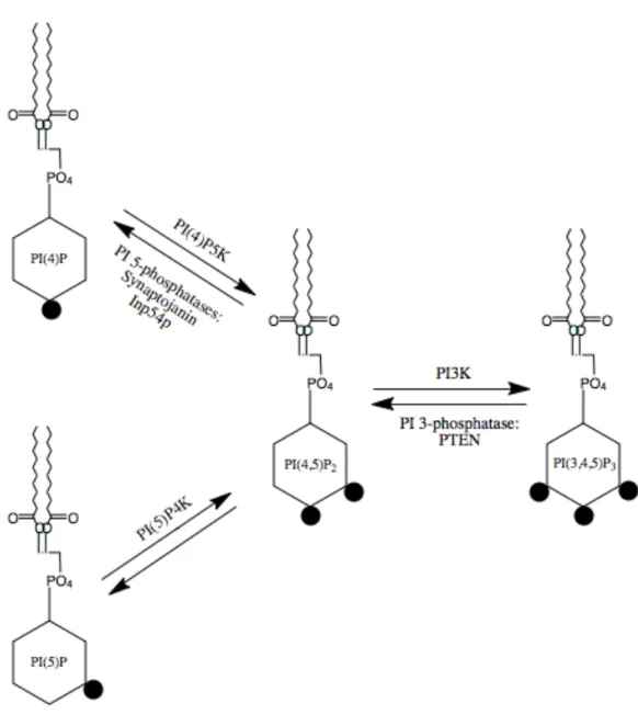
Figure 1.4. PI(4,5)P2 metabolism
Interconversion between phosphoinositide species occurs with the addition and removal of phosphates (black circles) to certain positions of the inositol ring headgroup of the lipid by specific kinases and phosphatases, respectively. PI(4)P5K adds a phosphate group to the 5 position of PI(4)P to create PI(4,5)P2, which can be reversed by PI 5-phosphatases such
as the mammalian Synaptojanin or the yeast Inp54p. PI(4,5)P2 can also be formed by the
addition of a phosphate to the 4th position of the inositol ring by PI(5)P4K. There is currently no identified enzyme to reduce PI(4,5)P2 to PI(5)P; however, bacterial IpgD can
cause this reduction, resulting in decreased membrane-cytoskeletal adhesion. Formation of PI(4,5)P2 can also occur in the opposite direction, from the removal of the 3-position
phosphate of PI(3,4,5)P3. Likewise, PI(4,5)P2 can be converted to PI(3,4,5)P3 with the
Figure 1.5. PI(4,5)P2 signaling and ion channel regulation
A-B) PI(4,5)P2 (signified by lipid with black ball) is cleaved by phospholipase C (PLC) into
inositol triphosphate (IP3) and DAG. DAG remains at the membrane to activate protein
kinase C (PKC), and IP3 goes on to stimulate the IP3 receptor (IP3R) in the endoplasmic
reticulum (seafoam green structure) and release intracellular calcium stores. B) When PI(4,5)P2 is abundant at the plasma membrane, ion channels like TRPV1 (red channel)
allow influx of ions. This channel current is inhibited by the reduction of PI(4,5)P2 due to
PLC activity. PLC activation can occur through G-protein (green α and βγ subunits) activation due to activation of a G-protein coupled receptor (GPCR, blue channel) by a ligand (yellow circle) like adenosine, or through Ca2+ binding. C) The proposed mechanism
for PI(4,5)P2 ion channel regulation is due to conformational changes that occur between
PI(4,5)P2-bound and –unbound states. The inward rectifying potassium channel, Kir2.2,
was crystallized in the presence of PI(4,5)P2. Each of the 4 channel subunits bound to one
molecule of PI(4,5)P2, with the hydrophobic acyl chains binding to the transmembrane
domain and the phophorylated head group binding to cytoplasmic domain of the subunit. The conformational change induced by PI(4,5)P2 binding is proposed to open the channel
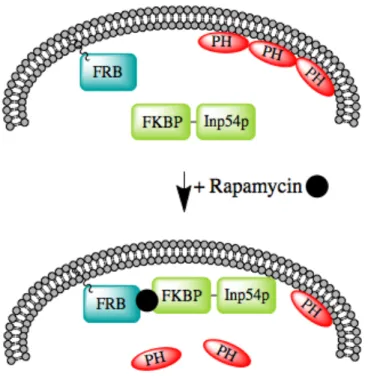
Figure 1.6. Schematic of rapamycin-induced depletion of PI(4,5)P2 from the plasma membrane
Prior to rapamycin treatment, the FRB domain of mTOR is tagged to the plasma membrane with the membrane-targetting motif of GAP43, and the FKBP-Inp54p fusion protein is cytoplasmically localized. The PH domain of PLC∂1 acts as a biosensor, and binds PI(4,5)P2 in the plasma membrane. After rapamycin treatment, the FKBP domain
dimerizes with the FRB domain, translocating the FKBP-Inp54p fusion protein to the plasma membrane, where the Inp54p phosphatase reduces PI(4,5)P2 to PI(4)P, releasing
Figure 1.7. Subtypes of nociceptive neurons in the DRG
CHAPTER 2
The F-BAR Domain of srGAP2 Induces Membrane Protrusions Required for Neuronal Migration and Morphogenesis1
2.1 INTRODUCTION
During brain development, neural progenitor proliferation, neuronal migration, and differentiation require considerable changes in cell shape involving coordinated cytoskeletal and membrane remodeling (Ayala et al., 2007; Luo, 2002). Neuronal migration involves the coordinated extension and adhesion of the leading process (LP) along the radial glial scaffold with the forward translocation of the nucleus, which requires regulation of
centrosome and microtubule dynamics by proteins such as Lis1, Doublecortin, and Nudel among others (Ayala et al., 2007; Higginbotham and Gleeson, 2007; Lambert de Rouvroit and Goffinet, 2001). However, little is known about the molecular mechanisms underlying membrane dynamics during neuronal migration and morphogenesis.
The basis of neurite initiation, outgrowth, and branching is rooted in the ability of the actin and microtubule cytoskeleton to undergo dynamic changes (Gupton and Gertler, 2007; Luo, 2002; Mattila and Lappalainen, 2008). Filopodia have been shown to play a role in neurite initiation (Dent et al., 2007; Kwiatkowski et al., 2007), growth cone dynamics (Burnette et al., 2007; Gallo and Letourneau, 2004), neurite outgrowth (Luo, 2002), and
1
Sabrice Guerrier, Jaeda Coutinho-Budd, Takayuki Sassa, Aurelie Gresset, Nicole Vincent Jordan, Keng Chen, Wei-lin Jin, Adam Frost, and Franck Polleux (2009) srGAP2 regulates neuronal
migration and morphogenesis through the ability of its F-BAR domain to induce membrane protrusions. Cell 138, 990-1004.
branching (Dent et al., 2004; Gallo and Letourneau, 1998). Downregulation of the actin anti-cappers ENA/VASP proteins, which are potent inducers of filopodia, resulted in failed neurite initiation and also in defects in cortical lamination (Kwiatkowski et al., 2007), suggesting a functional relationship between filopodia formation, neurite initiation, and neuronal migration.
Classically, filopodia formation is thought to be primarily dependent on proteins that regulate actin polymerization at the barbed end of actin filaments and proteins bundling F-actin (Gupton and Gertler, 2007). Interestingly, the BAR superfamily member IRSp53 has been shown to induce filopodia through membrane deformation independently of its F-actin bundling activity (Lim et al., 2008; Mattila et al., 2007; Saarikangas et al., 2009). The BAR domain superfamily contains three main groups: (1) the Bin/Amphiphysin/Rvs (BAR) domain subfamily (Itoh and De Camilli, 2006), (2) the Fes-Cip4 homology BAR (also called F-BAR or EFC) domain subfamily (Itoh et al., 2005; Tsujita et al., 2006; reviewed in Frost et al., 2009), and (3) the I-BAR subfamily (reviewed in Scita et al., 2008). Structural analysis of three F-BAR domains demonstrated that these domains are elongated homodimers characterized by a shallow curvature formed by the antiparallel interaction of two α-helical coiled coils (Henne et al., 2007; Shimada et al., 2007). In addition to sharing the general fold and quaternary organization of the BAR domain superfamily, F-BAR domains share functional properties with ‘‘classical’’ BAR domains, most notably the ability to bind and deform membranes in vitro and in living cells (Frost et al., 2008; Itoh et al., 2005; Kakimoto et al., 2006; Shimada et al., 2007). However, to date, the in vivo functions of F-BAR
domain-containing proteins are largely unknown (Frost et al., 2009).
I-BAR domains. Our results highlight the functional importance of proteins directly regulating membrane deformation for proper neuronal migration and axon-dendrite morphogenesis.
2.2 RESULTS
2.2.1 Expression of srGAP2 in the developing cortex
To begin our study of the role of srGAP2 in cortical development, we first examined its pattern of expression. srGAP1–3 have recently been reported to be expressed
throughout the cortex during and after radial migration (Bacon et al., 2009; Mattar et al., 2004; Yao et al., 2008). Our analysis confirmed that srGAP2 mRNA is expressed
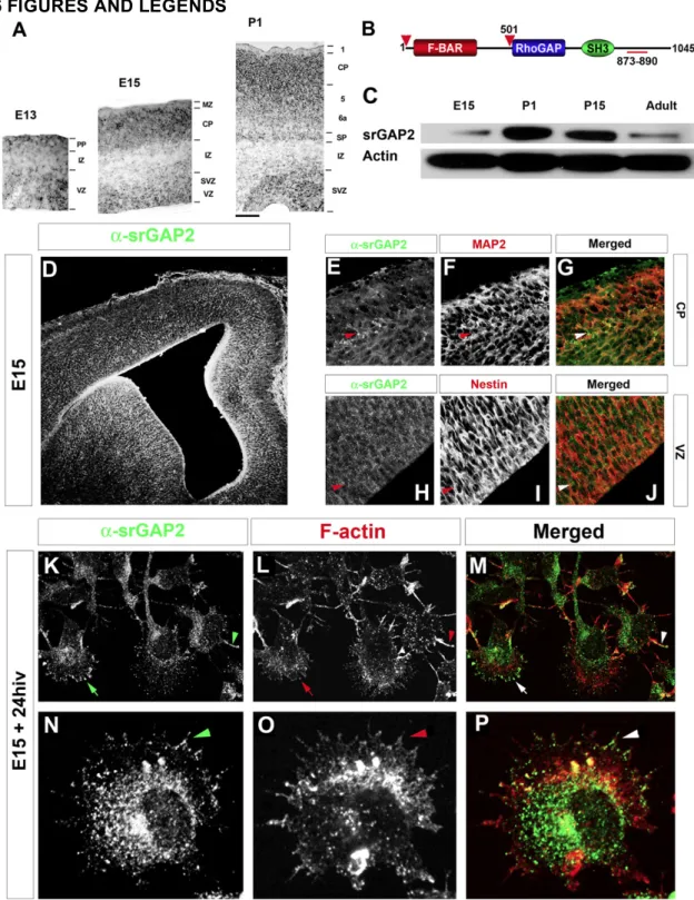
throughout the developing cortex and is found both in proliferative zones (ventricular zone [VZ] and subventricular zone [SVZ]) at embryonic day 13 (E13) and E15 and in postmitotic regions (cortical plate [CP]) at E15 and postnatal day 1 (P1) (Figure 1A). In order to determine the pattern of srGAP2 protein expression, we used a polyclonal antibody raised against the C terminus of srGAP2 (Figures 1B and 1C; Yao et al., 2008). srGAP2 protein is expressed throughout cortical development culminating at P1 corresponding to the peak of neuronal migration in the cortex. Its expression is maintained at P15 and reduced, but still present, in adult cortex (Figure 1C).
Immunofluorescent staining for srGAP2 shows that it is ubiquitously expressed in the cortical wall (Figure 1D) being found both in Nestin-positive neuronal progenitors in the VZ (Figures 1H–1J) and MAP2-positive postmitotic neurons in the CP (Figures 1E–1G). At the subcellular level, endogenous srGAP2 is found at the cell periphery (Figures 1K– 1M, arrows) and was often localized along F-actin-rich filopodia-like protrusions (arrowhead in Figures 1K–1P) in acutely dissociated E15 cortical neurons.
2.2.2 Full-length srGAP2 and its F-BAR domain induce filopodia formation
Tsujita et al., 2006). Surprisingly, expression of srGAP2 did not induce any membrane invaginations, but instead induced filopodia formation (see Figures S1D–S1Fand S1P available online). This effect requires its F-BAR domain since deletion of the F-BAR domain (srGAP2ΔF-BAR-EGFP) does not induce filopodia formation in COS7 cells (Figures S1G–S1I
and S1P).
Interestingly, unlike the F-BAR domains of FBP17 and CIP4 (Itoh et al., 2005), expression of the F-BAR domain of srGAP2 did not inhibit endocytosis, as assessed using Alexa546-Transferrin uptake assay (Figure S2). Furthermore, expression of the isolated F-BAR domain fused to EGFP induced filopodia formation similar to full-length srGAP2 (Figures S1J, S1K, and S1P). Of note, the F-BAR domain is a potent membrane-targeting motif (Figure S1J). These data suggest that the F-BAR domain of srGAP2 is necessary and sufficient for membrane localization and the induction of filopodia-like membrane protrusions.
In order to distinguish the membrane-targeting function of the F-BAR domain from its membrane deformation activity, we identified a small truncation of the last C-terminal 49 amino acids (F-BARΔ49) (Figure S3A and Supplemental Experimental Procedures for details). Expression of F-BARΔ49-EGFP results in significant membrane targeting (Figure
domain (amino acids 1–358) in mammalian cells or bacteria (data not shown). Furthermore, as shown for other F-BAR domains (Frost et al., 2008; Itoh et al., 2005; Kakimoto et al., 2006; Shimada et al., 2007), srGAP2 forms a stable dimer in solution as assessed by light scattering assays (Figure S3C), and deletion of the Fes-Cip4 homology (FCH) domain (green box in Figure S3A), which represents a significant portion of the dimerization interface, abolishes the ability of srGAP2 to induce filopodia in COS7 cells (data not shown). Altogether, these data suggest that all eight predicted α-helices are likely to be required for formation of the functional F-BAR domain of srGAP2.
2.2.3 The F-BAR domain of srGAP2 deforms membrane like an I-BAR domain The ability of srGAP2 or its F-BAR domain to induce filopodia in COS7 cells is reminiscent of the activity of the structurally related I-BAR domain-containing proteins (Mattila et al., 2007; Millard et al., 2007; Saarikangas et al., 2009; Scita et al., 2008). Interestingly, F-actin depolymerization prevents the dynamics and formation of new
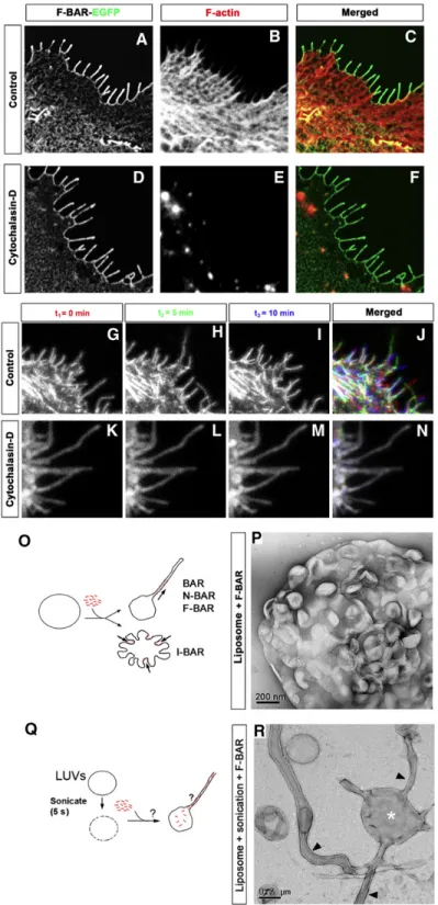
filopodia, but does not affect the maintenance of pre-existing filopodia induced by the I-BAR domains of IRSp53 or MIM (Mattila et al., 2007). We found the same results for the F-BAR domain of srGAP2 (Figures 2A–2C), while cells treated with cytochalasin D were depleted of F-actin. Strikingly, this treatment had no effect on membrane localization of the F-BAR domain or on the maintenance of filopodia-like protrusions (Figures 2D–2F). F-BAR-induced filopodia were highly dynamic in COS7 cells (Figures 2G–2J and Movie S1). Treatment with cytochalasin D significantly impaired the extension and retraction of F-BAR-induced filopodia (Figures 2K–2N and Movie S2), suggesting that F-actin is required for the dynamics of these protrusions.
F-BAR domain of srGAP2 induced an inward dimpling or ‘‘scalloping’’ of the liposome surface (Figures 2O and 2P), which is reminiscent of the activity of I-BAR domains in the same conditions (Suetsugu et al., 2006), suggesting that the F-BAR domain of srGAP2 can induce ‘‘inverse’’ membrane tubulation.
These results suggested the possibility that if the purified F-BAR domain of srGAP2 could be exposed to the inside surface of liposomes, then protrusive tubules would form (Figure 2Q). To test this hypothesis, mixtures of the F-BAR domain with intact, large unilamellar vesicles (LUVs) were briefly sonicated, which presumably resulted in transient pore formation in liposomes and introduction of the recombinant F-BAR inside LUVs. Following a wash, liposomes were fixed, negatively stained, and imaged using transmission electron microscopy. As predicted by the I-BAR model, this resulted in numerous long tubular extensions emerging from LUVs (Figure 2R), which is in stark contrast with control sonicated liposomes not incubated with recombinant protein (Figure S6A). Consistent with the dimensions of tubules induced by other members of the F-BAR and I-BAR families (Frost et al., 2008; Mattila et al., 2007), the srGAP2 F-BAR-induced tubules were 83 nm ± 15 nm (average ± SD, n = 38) in diameter. Importantly, at higher magnification, the tubules observed by negative staining electron microscopy after sonication do not have an obvious protein coat surrounding the liposomes (Figure 2R). This is in contrast with tubules induced by other F-BAR and BAR domains that coat the outer surface of the tubule (Figure S6B; Frost et al., 2008; Shimada et al., 2007). Together, these results suggest that unlike previously characterized F-BAR domains, the F-BAR domain of srGAP2 functions as an I-BAR domain (Mattila et al., 2007; Suetsugu et al., 2006).
2.2.4 srGAP2 regulates neurite formation and branching through the ability of its F-BAR domain to form filopodia