Overview: Lines of Communication
The cone snail kills prey with venom that disables
neurons
Neurons are nerve cells that transfer information within the body
Neurons use two types of signals to communicate:
Interpreting signals in the nervous system involves sorting a complex set of paths and connections
Concept 37.1: Neuron structure and organization
reflect function in information transfer
The neuron is a cell type that exemplifies the close fit of form and function that often arises over the
Neuron Structure and Function
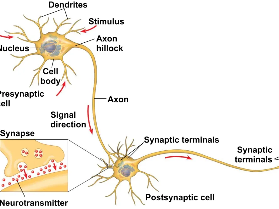
Most of a neuron’s organelles are in the cell body
Most neurons have dendrites, highly branched extensions that receive signals from other neurons
The single axon, a much longer extension, transmits signals to other cells
The cone-shaped base of an axon, where signals are generated, is called the axon hillock
The branched ends of axons transmit signals to other cells at a junction called the synapse
At most synapses, chemical messengers called
Neurons of vertebrates and most invertebrates require supporting cells called glial cells
Figure 37.3
Cell bodies of
neurons
Glia
Introduction to Information Processing
Nervous systems process information in three stages
Sensory input Integration
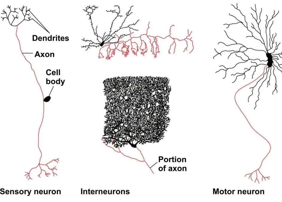
Sensory neurons transmit information from eyes and other sensors that detect external stimuli or internal conditions
This information is sent to the brain or ganglia, where
interneurons integrate the information
Neurons that extend out of the processing centers
trigger muscle or gland activity
In many animals, neurons that carry out integration are organized in a central nervous system (CNS)
The neurons that carry information into and out
of the CNS form the peripheral nervous system
(PNS)
Figure 37.5
Dendrites
Axon
Cell body
Portion of axon
Interneurons
Concept 37.2: Ion pumps and ion channels
establish the resting potential of a neuron
The inside of a cell is negatively charged relative to the outside
This difference is a source of potential energy, termed membrane potential
The resting potential is the membrane potential of a neuron not sending signals
Formation of the Resting Potential
K and Na play an essential role in forming the
resting potential
In most neurons, the concentration of K is highest
inside the cell, while the concentration of Na is
highest outside the cell
Sodium-potassium pumps use the energy of ATP
to maintain these K and Na gradients across the
plasma membrane
Figure 37.6
OUTSIDE OF CELL
INSIDE OF CELL Key
Na
K
Sodium-potassium pump
Potassium channel
The opening of ion channels in the plasma
membrane converts the chemical potential energy of the ion gradients to electrical potential energy
Ion channels are selectively permeable, allowing only certain ions to pass through
A resting neuron has many open potassium
channels, allowing K to flow out
Modeling the Resting Potential
Resting potential can be modeled by an artificial membrane that separates two chambers
The concentration of KCl is higher in the inner chamber and lower in the outer chamber
K diffuses down its gradient to the outer chamber
Negative charge (Cl−) builds up in the inner chamber
Figure 37.7
Inner
chamber Outerchamber
140 mM
KCI
5 mM
KCI
−90 mV Inner
chamber
Outer chamber
15 mM
NaCI 150 mNaCIM
62 mV
Cl− Cl− Potassium channel Artificial membrane
K Na
Sodium channel
(b) Membrane selectively permeable to Na
(a) Membrane selectively permeable to K
EK 62 mV log 140 m5 mMM −90 mV ENa 62 mV log150 mM 62 mV
In a resting neuron, the currents of K and Na are
Concept 37.3: Action potentials are the signals
conducted by axons
Researchers can record the changes in membrane potential when a neuron responds to a stimulus
Figure 37.9
Ions
Change in membrane potential (voltage)
(b) Gate open: Ions flow through channel. (a) Gate closed: No ions
flow across membrane. Ion
When gated K channels open, K diffuses out,
making the inside of the cell more negative
This is hyperpolarization, an increase in magnitude of the membrane potential
Figure 37.10
(a) Graded hyperpolarizations produced by two stimuli that increase membrane permeability to K
(b) Graded depolarizations produced by two stimuli that increase membrane permeability to Na
(c) Action potential triggered by a depolarization that reaches the threshold
Resting potential
Time (msec) 0 1 2 3 4 5 6 Threshold −100 −50 0 50 M e m b ra n e p o te n ti al ( m V ) Action potential Strong depolarizing stimulus
Resting potential
Opening other types of ion channels triggers a
depolarization, a reduction in the magnitude of the membrane potential
For example, depolarization occurs if gated Na
Graded potentials are changes in polarization
where the magnitude of the change varies with the strength of the stimulus
Graded potentials decay with distance from the source
If a depolarization shifts the membrane potential sufficiently, it results in a massive change in
membrane voltage, called an action potential
Action potentials have a constant magnitude and transmit signals over long distances
They arise because some ion channels are voltage
Action potentials occur whenever a depolarization increases the membrane potential to a particular value, called the threshold
Generation of Action Potentials:
A Closer Look
An action potential can be considered as a series of stages
At resting potential
1. Most voltage-gated sodium (Na) channels are
closed; most of the voltage-gated potassium (K)
channels are also closed
Animation: Action Potential
1 Figure 37.11 Key Na K Action potential Threshold Resting potential Time −100 −50 0 50 M e m b ra n e p o te n ti al (m V )
Rising phase of the action potential
Depolarization
Falling phase of the action potential
Resting state Undershoot Sodium channel Potassium channel Inactivation loop OUTSIDE OF CELL
INSIDE OF CELL
When stimulus depolarizes the membrane
2. Some gated Na+ channels open first and Na flows into the cell
3. During the rising phase, the threshold is crossed, and the membrane potential increases
4. During the falling phase, voltage-gated Na channels
become inactivated; voltage-gated K channels
5. During the undershoot, membrane permeability to K
During the refractory period after an action potential, a second action potential cannot be initiated
The refractory period is a result of a temporary inactivation of the Na channels
For most neurons, the interval between the start of an action potential and the end of the refractory
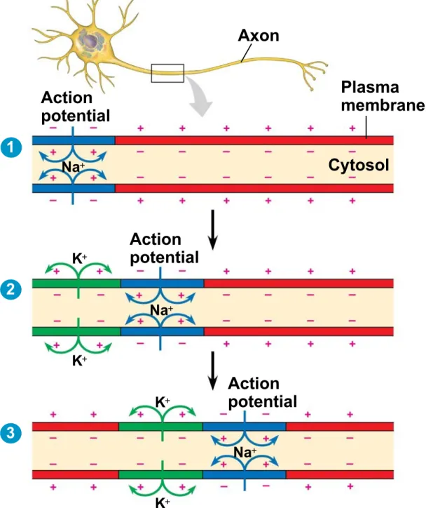
Conduction of Action Potentials
At the site where the action potential is initiated (usually the axon hillock), an electrical current depolarizes the neighboring region of the axon membrane
Action potentials travel only toward the synaptic
terminals
Inactivated Na channels behind the zone of
Evolutionary Adaptations of Axon Structure
The speed of an action potential increases with the axon’s diameter
In vertebrates, axons are insulated by a myelin
sheath, which enables fast conduction of action potentials
Myelin sheaths are produced by glia—
oligodendrocytes in the CNS and Schwann cells
Figure 37.13
Axon Myelin
sheath
Schwann cell
Nodes of
Ranvier Nucleus of
Schwann cell Schwann cell
Node of Ranvier
Layers of myelin Axon
Action potentials are formed only at nodes of
Ranvier, gaps in the myelin sheath where voltage-gated Na channels are found
Action potentials in myelinated axons jump between the nodes of Ranvier in a process called saltatory conduction
A selective advantage of myelination is space
Figure 37.14
Cell body
Schwann cell
Depolarized region (node of Ranvier)
Concept 37.4: Neurons communicate with
other cells at synapses
At electrical synapses, the electrical current flows from one neuron to another
The presynaptic neuron synthesizes and packages the neurotransmitter in synaptic vesicles located in the synaptic terminal
The arrival of the action potential causes the release of the neurotransmitter
The neurotransmitter diffuses across the synaptic
cleft and is received by the postsynaptic cell
Animation: Synapse
Figure 37.15
Presynaptic cell Postsynaptic cell
Axon Synaptic vesicle
containing neurotransmitter Synaptic
cleft
Postsynaptic membrane
Ca2
K
Na
Ligand-gated ion channels Voltage-gated
Ca2 channel
Presynaptic membrane
1
2
Generation of Postsynaptic Potentials
Direct synaptic transmission involves binding of neurotransmitters to ligand-gated ion channels
in the postsynaptic cell
Postsynaptic potentials fall into two categories
Excitatory postsynaptic potentials (EPSPs) are
depolarizations that bring the membrane potential toward threshold
Inhibitory postsynaptic potentials (IPSPs) are
The duration of postsynaptic potential is limited by rapidly clearing neurotransmitter molecules from the synaptic cleft
Some neurotransmitters are recaptured into
presynaptic neurons to be repackaged into synaptic vesicles
Some are recaptured into glia to be used as fuel
or recycled to neurons
Others are removed by simple diffusion or hydrolysis
Summation of Postsynaptic Potentials
The cell body of one postsynaptic neuron may receive inputs from hundreds or thousands of synaptic terminals
Neurotransmitters
Signaling at a synapse brings about a response that depends on both the neurotransmitter from the presynaptic cell and the receptor on the
postsynaptic cell
A single neurotransmitter may have more than a dozen different receptors
Acetylcholine
Acetylcholine is vital for functions involving muscle stimulation, memory formation, and learning
Vertebrates have two major classes of acetylcholine receptor, one that is ligand gated and one that is
The best understood function of the ligand-gated ion channel is in the vertebrate neuromuscular junction
When acetylcholine released by motor neurons
binds to this receptor, the ion channel opens and an EPSP is generated
A number of toxins disrupt neurotransmission by acetylcholine
These include the nerve gas sarin and a bacterial toxin that causes botulism
Neuropeptides
Several neuropeptides, relatively short chains of amino acids, also function as neurotransmitters
Neuropeptides include substance P and
endorphins, which both affect our perception of pain
Opiates bind to the same receptors as endorphins
Gases
Gases such as nitric oxide (NO) and carbon monoxide (CO) are local regulators in the PNS
Unlike most neurotransmitters, these are not stored