INTEGRATED APPROACHES TO IDENTIFY AND PREDICT
PHARMACOKINETIC-BASED DIETARY SUBSTANCE-DRUG INTERACTIONS
Christina S. Won
A dissertation submitted to the faculty of the University of North Carolina at Chapel Hill in partial fulfillment of the requirements for the degree of Doctor of Philosophy in Pharmaceutical Sciences in the Eshelman School of Pharmacy (Division of Pharmacotherapy and Experimental Therapeutics).
Chapel Hill 2012
Approved by:
iii ABSTRACT
CHRISTINA S. WON: Integrated Approaches to Identify and Predict Pharmacokinetic-Based Dietary Substance-Drug Interactions
(Under the direction of Mary F. Paine)
The large variation in bioactive ingredient composition inherent to natural products, including dietary substances, can confound the design and interpretation of natural product-drug interaction studies. The purpose of this dissertation was to address this overlooked issue by developing a framework to evaluate pharmacokinetic-based dietary substance-drug interactions such that ultimately, firm clinical recommendations can be made. Fruit juices represent a diverse market of popular foods containing phytochemicals that can inhibit drug metabolizing enzymes and transporters in the intestine. The potential increase or decrease in systemic drug exposure could lead to adverse effects or therapeutic failure, respectively. A multi-experimental approach utilizing in vitro
iv
v
vi
ACKNOWLEDGEMENTS
I would to express my deepest gratitude to the six members of my dissertation advisory committee for their encouragement, generosity, and wise counsel. Their enthusiasm for science and passion for their work are qualities which I admire and hope to maintain throughout my career as a scientist. I would like to acknowledge my advisor Mary F. Paine for her unwavering support, guidance, and patience. Her dedication to professional and personal excellence is inspiring.
I would like to thank past and present members of the Paine lab, particularly Garrett Ainslie, Scott Brantley, Cathrine Denton, Richa Dua, Vanessa González-Pérez, Brandon Gufford, J. P. Jones III, and Kristina Wolf (especially Kristina!) for their scientific assistance and sense of humor. I am grateful to Arlene S. Bridges of the ADME Mass Spectrometry Center for her scientific and emotional support. I would also like to thank past and present members of the Brouwer and Thakker labs for their scientific advice and friendship. Many thanks to Kathleen Köck and Chester Costales for listening and advising.
I am very grateful for the collaborations which made this dissertation work possible. I am thankful to Paul A. Dawson and Tian Lan at Wake Forest University School of Medicine for sharing their knowledge of and providing materials for the in vitro OATP experiments in Chapter 2. I am much obliged to Nicholas H. Oberlies and Karen VanderMolen at the University of North Carolina at Greensboro for enlightening me on the ways of the natural product chemistry world, as well as providing juice extracts for the in vitro OATP studies in Chapters 2 and 3. I am grateful to the great staff and cooperative healthy volunteers of the Clinical and Translational Research Center.
vii
I am very appreciative of past and present administrative staff, especially Amber Allen, Arlo Brown, and Kathy Maboll (especially Kathy!).
I would like to thank the developers of WinNonlin®, Simcyp®, and GastroPlus™. I feel lucky and privileged to learn these software programs.
I would like to thank the University for awarding me a Graduate School Merit Assistantship (2007-2008) and the National Institutes of Health for funding my education and research.
I would like to express my eternal appreciation and love to my family and friends, who kindly helped me to survive the graduate school experience by listening, comforting, advising, and sending care packages. Collectively, they were the unofficial seventh member of my committee. A million thanks to my mother for being her tenacious, bold self and to my father for being his rational, calm self. I cannot say enough about my awesome little sister who amazes and inspires me every day with her intelligence, empathy, compassion, and ardent dedication to caring for people. Her votes of confidence always give me a boost when I need it most. My wonderful friends and colleagues have helped me through all kinds of tough times and I will be forever grateful for their emotional support. I will not list all their names for fear of omission but they know who they are!
I am indebted to my late mentor George N. Sideris who was, and still is, a great influence in my life. I am also indebted to his lovely partner Anna Condoulis.
Finally, I would like to close by sharing one of many motivational quotes which decorated my lab desk and apartment:
“A smooth sea never made a skillful mariner, neither do uninterrupted prosperity and success
qualify men for usefulness and happiness. The storms of adversity, like those of the ocean, rouse the faculties, and excite the invention, prudence, skill, and fortitude of the voyager.”
viii
TABLE OF CONTENTS
LIST OF TABLES...x-xi LIST OF FIGURES...xii-xiii LIST OF ABBREVIATIONS...xiv-xix CHAPTERS
1 INTRODUCTION
PROLOGUE...1 PART 1: INFLUENCE OF DIETARY SUBSTANCES ON INTESTINAL DRUG
METABOLISM AND TRANSPORT...5 PART 2: MECHANISMS UNDERLYING FOOD-DRUG INTERACTIONS:
INHIBITION OF INTESTINAL METABOLISM AND TRANSPORT...51
2 A MODIFIED GRAPEFRUIT JUICE ELIMINATES FURANOCOUMARINS AND POLYMETHOXYFLAVONES AS CANDIDATE MEDIATORS OF THE
FEXOFENADINE-GRAPEFRUIT JUICE INTERACTION IN HEALTHY VOLUNTEERS...109
3 BIOACTIVITY-GUIDED FRACTIONATION OF GRAPEFRUIT
(Citrus paradisi Macfad.) JUICE AND EVALUATION OF
REPRESENTATIVE COMPONENTS AS INHIBITORS OF AN INTESTINAL
ORGANIC ANION-TRANSPORTING POLYPEPTIDE...137
4 EVALUATION OF SELECT GRAPEFRUIT JUICE CONSTITUENTS AS MARKER
COMPOUNDS PREDICTIVE OF THE GRAPEFRUIT JUICE EFFECT………..166
5 CONCLUSIONS...203 EPILOGUE...211
APPENDICES
A PILOT STUDY EVALUATING POMEGRANATE JUICE AS AN
INHIBITOR OF CYP3A ACTIVITY IN HEALTHY VOLUNTEERS...217
B INHIBITION OF AN INTESTINAL ORGANIC ANION-TRANSPORTING POLYPEPTIDE BY ORANGE (Citrus sinensis (L.) Osbeck), APPLE
ix
C BIOACTIVITY-GUIDED FRACTIONATION OF ORANGE
(Citrus sinensis (L.) Osbeck), APPLE (Malus domestica Borkh),
AND POMEGRANATE (Punica granatum L.) JUICES TO IDENTIFY
INHIBITORS OF ORGANIC ANION-TRANSPORTING POLYPEPTIDE 2B1……...………227 D EVALUATION OF ATENOLOL AND CIPROFLOXACIN AS
SUBSTRATES AND INHIBITORS OF ORGANIC ANION-TRANSPORTING
POLYPEPTIDE 2B1………..233
E INTERDAY VARIABILITY OF ESTRONE 3-SULFATE TRANSPORT IN TRANSFECTED MDCKII CELLS EXPRESSING ORGANIC
x
LIST OF TABLES CHAPTER 1
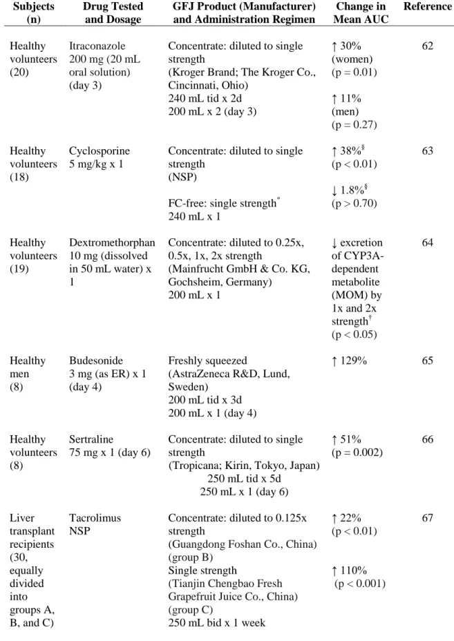
Table 1.1 Controlled clinical drug-grapefruit juice (GFJ) interaction studies
reported since 2008………..………...33-34
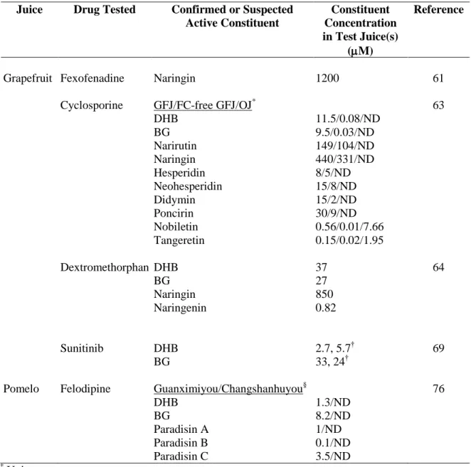
Table 1.2 Clinical drug-citrus juice interaction studies since 2006 in which candidate
causative ingredients in the test juice were quantified……..………...……..35
Table 1.3 Summary of recent controlled clinical studies involving CYP3A-mediated
citrus juice-drug interactions………..………...84-85
Table 1.4 Potential explanations for lack of concordance between in vitro and in vivo dietary
substance-drug interaction studies……..………...……….86
Table 1.5 Questions to consider when reviewing clinical dietary substance-drug
interaction studies………..………...……..87
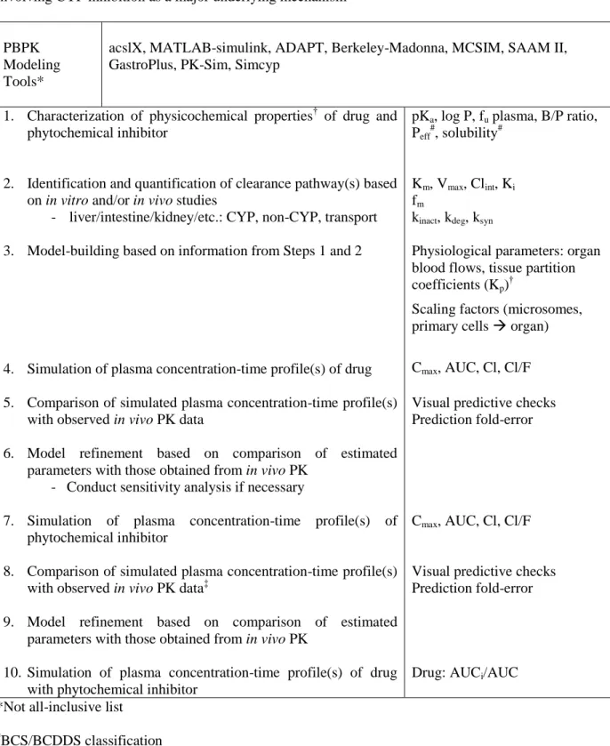
Table 1.6 General framework for quantitative prediction of dietary substance-drug interactions involving CYP inhibition as a major underlying mechanism………..…………..88
CHAPTER 2
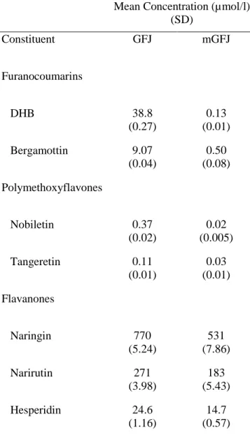
Table 2.1 Concentrations of representative furanocoumarins, polymethoxyflavones, and
flavanones in grapefruit juice (GFJ) and modified GFJ (mGFJ).………...…...124
Table 2.2 Pharmacokinetics of fexofenadine (120 mg) in 18 healthy volunteers after administration with 240 mL of water, grapefruit juice (GFJ), or
modified GFJ (mGFJ) …………..………...125
CHAPTER 3
Table 3.1 IC50 and concentration of representative flavanones, furanocoumarins, and
polymethoxyflavones in grapefruit juice (GFJ)………...…160
CHAPTER 4
Table 4.1 Summary of eligible clinical interaction studies ………...181
Table 4.2 Input data for substrate files in Simcyp®.………..…………...….182
xi
Table 4.4 Comparison of observed and predicted systemic midazolam exposure…….……..…….186
Table 4.5 Comparison of predicted and observed systemic felodipine exposure………...187
CHAPTER 5
Table 5.1 Minimum recommended requirements for conducting in vitro, clinical,
xii
LIST OF FIGURES CHAPTER 1
Figure 1.1 Timeline of key observations in drug-grapefruit juice (GFJ)
interaction research...………32
Figure 1.2 Schematic representation of enterocytes.………83
CHAPTER 2
Figure 2.1 Inhibitory effects of grapefruit juice extracts (GFJ, mGFJ) on estrone 3-sulfate (E1S) uptake in COS-1 and HEK293T/17 cells overexpressing human OATP1A2 or
OATP2B1………128
Figure 2.2 Inhibition of fexofenadine uptake by grapefruit juice extracts (GFJ, mGFJ) in
OATP1A2-transfected cells.………..………..129
Figure 2.3 Fexofenadine uptake in OATP2B1-transfected cells.………...……...…130
Figure 2.4 Geometric mean plasma fexofenadine concentration-time profile following
coadministration with 240 ml of water, GFJ, or mGFJ for 18 healthy volunteers...131
Figure 2.5 Fexofenadine AUC changes in 18 healthy volunteers administered fexofenadine
with 240 ml water, GFJ, and mGFJ...………..132
CHAPTER 3
Figure 3.1 Separation scheme of grapefruit juice extracts ………...152
Figure 3.2 Bioactivity-guided fractionation of grapefruit juice and effects of juice extracts
and fractions on OATP2B1-mediated uptake of estrone 3-sulfate in vitro……….153
Figure 3.3 HPLC chromatogram of subfraction m3; UV absorption spectra of DHB and m3; furanocoumarin moiety and structure of DHB; NMR spectrum of
m3 and DHB; and HRMS of m3 and DHB………..………...………155-156
Figure 3.4 Structures of constituents from representative classes of compounds
in grapefruit juice………157
Figure 3.5 IC50 curves for grapefruit juice constituents representative of three
xiii CHAPTER 4
Figure 4.1 Structures of midazolam, felodipine, 6’,7’-dihydroxybergamottin, and
bergamottin…...189
Figure 4.2 Observed midazolam plasma concentration-time profile following administration of water and GFJ and simulated midazolam plasma concentration-time profiles following administration of DHB and
DHB plus BG………..190
Figure 4.3 Observed felodipine plasma concentration-time profile following administration of water or orange juice control and GFJ and simulated
felodipine plasma concentration-time profile following administration of DHB
and DHB plus BG………....192-193
Figure 4.4 Comparison of observed and simulated mean AUC ratios of selected clinical
GFJ-drug interaction studies.….……..………195
CHAPTER 5
Figure 5.1 Considerations for the evaluation, interpretation, and conduct of natural
xiv
LIST OF ABBREVIATIONS (AUCm/AUCp)last metabolite/parent AUClast ratio
+BG 6’,7’-dihydroxybergamottin plus bergamottin 1’-OH MDZ 1’-hydroxymidazolam
1X single strength 2X double strength AA African American
ACAT advanced compartmental absorption & transit ADAM advanced dissolution, absorption & metabolism ADME absorption, distribution, metabolism, excretion ANOVA analysis of variance
APAP acetaminophen Aq aqueous
AUC0-∞ area under the curve from zero to infinite time
AUClast area under the curve from zero to the last measured concentration B/P blood to plasma partition ratio
BCA bicinchoninic acid
BCRP breast cancer resistance protein BG bergamottin
bid two times daily
BLQ below limit of quantification BSP bromosulfophthalein
xv CI confidence interval
Cl/F apparent oral clearance Clast last measured concentration Clint, intrinsic clearance
Cmax maximum concentration
COS-1 CV-1 (simian) origin SV40 virus
CTRC Clinical and Translational Research Center CV coefficient of variation
CYP cytochrome P450 d day(s)
DDIs drug-drug interactions DHB 6’,7’-dihydroxybergamottin
DMEM Dulbecco’s modified Eagle’s medium DMSO dimethyl sulfoxide
E maximum effect E1S estrone 3-sulfate
EDTA ethylenediaminetetraacetic acid EGCG epigallocatechin gallate
ELSD evaporative light scattering detector Eo baseline effect
ER extended release
fa fraction available to be absorbed from dosage form FC furanocoumarin
FDA Food and Drug Administration
fu,gut unbound fraction in enterocytes
xvi
fu,plasma unbound fraction in plasma
g gram
GCRC General Clinical Research Center GFJ grapefruit juice
GI gastrointestinal h hour
HBSS Hank's balanced salt solution HEK human embryonic kidney HeLa cervical cancer cells
HEPES 4-(2-hydroxyethyl)-1-piperazineethanesulfonic acid HPLC high pressure liquid chromatography
HRMS high resolution mass spectrometry IC50 half maximal inhibitory concentration Imax maximum inhibitory effect
INR International Normalized Ratio ka first-order absorption rate constant
Kapp concentration of mechanism-based inhibitor associated with half maximal inactivation rate (KI)
KCl potassium chloride kg kilogram
KH2PO4 monopotassium phosphate Ki inhibition constant
kinact maximal inactivation rate constant
Km substrate concentration at which reaction rate is half of Vmax L liter
l liter
xvii M men
MDCKII Madin-Darby canine kidney type II
MDR1 gene encoding for P-glycoprotein MDZ midazolam
MES 2-(N-morpholino)ethanesulfonic acid hydrate mg milligram
mGFJ modified grapefruit juice MgSO4 magnesium sulfate min minute
ml milliliter mL milliliter mM millimolar mol mole
MOM 3-methoxymorphinan
MRP multi-drug resistance-associated protein
m/z mass to charge ratio NaCl sodium chloride NaOH sodium hydroxide NC not calculated ND not detected NM not measured
nmol/l nanomoles per liter NMR nuclear magnetic resonance NR not reported
xviii OATPs organic anion transporting polypeptides OJ orange juice
Org organic
OST α/β organic solute transporter alpha/beta
PBPK physiologically-based pharmacokinetic PBS phosphate-buffered saline
PD pharmacodynamics
PDA photodiode array detector P-gp P-glycoprotein
PK pharmacokinetics
pKa acid dissociation constant
p-nitrophenylacetate PNPA POM pomegranate juice qd daily
rCYP, recombinant CYP s second
SD standard deviation SDS sodium dodecyl sulfate SE standard error
SLCO gene encoding for organic anion transporting polypeptides SS statistically significant
SULTs sulfotransferases t½ terminal half-life tid three times daily
xix UTI urinary tract infection
UV ultraviolet VER verapamil Vmax maximum rate
Vmax, maximum rate of metabolite formation
Vss volume of distribution at steady state w week
W women y year
γ Hill coefficient
λz terminal elimination rate constant μg microgram
μg/mL micrograms per milliliter μM micromolar
Part 1 has been published in Current Drug Metabolism (2010;11(9):778-92) and is presented in the style of that journal. Copyright 2010 by Bentham Science Publishers Ltd. Reprinted with permission from Bentham Science Publishers.
Part 2 of this chapter has been published in Pharmacology & Therapeutics (2012; 136(2):186-201) and is presented in the style of that journal. Copyright 2012 by Elsevier B.V. Reprinted with permission from Elsevier.
CHAPTER 1 INTRODUCTION
PROLOGUE
The diet is an underappreciated modifiable environmental factor that influences human health status. Pharmaceutical agents have long overshadowed dietary interventions as primary modifiers of health outcomes and disease amelioration. Multi-drug regimens have become commonplace and are expected to continue and escalate, predisposing millions of people to adverse drug reactions, as well as drug-drug interactions. The public’s burgeoning interest in holistic, complementary, and alternative medicine has led to the adoption of additional practices to augment the effects of their drug therapies. Certain plant-derived chemicals (i.e., phytochemicals) consumed through foods and dietary supplements, including herbal preparations, have become viable therapeutic options used in conjunction with prescribed, as well as over-the-counter, medications. This growing shift in the practice of health care and maintenance may put patients at risk for additional types of drug interactions.
Globalization has enhanced the opportunities for people to be exposed to a variety of exotic phytochemicals, as well as diverse medical practices, perceived to be beneficial to health. Innovative agricultural engineering and food fortification also have led to a rise in ‘superfoods’ and/or ‘functional foods.’ Coupled with the dangers of polypharmacy, it is inevitable that these worlds will
2
adverse events or therapeutic failure. Various food products have the potential to perturb the absorption, distribution, metabolism, and excretion of medications. The underlying mechanisms can be physicochemical (e.g., chelation), biochemical (e.g., inhibition of drug metabolizing enzymes and/or transporters), or physiologically-based (e.g., altered blood flow to intestine).
Plant-derived beverages, including fruit juices, are ubiquitous and represent a diverse market of food products. Aside from purported health benefits, fruit juices contain constituents that have been shown to inhibit drug metabolizing enzymes (e.g., cytochrome P450s) and transport proteins (e.g., organic anion transporting polypeptides) in the intestine, leading to altered PK of the victim drug. The clinical impact of such beverage-drug interactions is discussed in the following two parts. A review of the literature demonstrates challenges in designing, conducting, and interpreting clinical studies to assess dietary substance-drug interactions. Numerous in vitro and in vivo methods, similar to those used to bring a new chemical entity to market, are available to screen for drug interaction potential. However, results can be confounded by the between-brand variability in bioactive ingredient composition. Large variations, even within the same lot of the same brand, can contribute to varying magnitudes of effect on multiple substrates both in vitro and in vivo. A disregard for the phytochemical type and content can lead to lack of replication, as well as discrepancies between in
vitro predictions and in vivo observations.
3
consumption. A multi-experimental approach, which included a clinical study in healthy volunteers,
in vitro studies with transporter-expressing cells, bioactivity-guided fractionation/isolation, and PK
modeling and simulation, was utilized as outlined in the following specific aims:
Aim 1: In vitro-in vivo approach to elucidate the molecular and cellular factors mediating dietary substance-drug interactions
Evaluate the contribution of specific grapefruit juice (GFJ) components to the inhibition of intestinal organic anion transporting polypeptide (OATP) activity and consequent oral absorption of the transporter substrate by GFJ.
Hypothesis: A food-grade GFJ devoid of certain classes of compounds (modified GFJ) can be used
as a tool to identify or eliminate the contribution of specific classes of compounds to enteric OATP
inhibition and reduction of oral absorption.
1a. Evaluate the OATP inhibitory effect of GFJ and modified GFJ extracts in vitro using OATP1A2- and OATP2B1-transfected cell lines and estrone 3-sulfate and fexofenadine as substrates. 1b. Compare the magnitude of effect of GFJ and modified GFJ to water on the systemic exposure to
the OATP substrate fexofenadine in healthy volunteers.
Aim 2: In vitro approach to identify the active ingredients responsible for the targeted bioactivity of dietary substances
Identify and characterize enteric OATP inhibitors in GFJ.
Hypothesis: A bioactivity-guided fractionation approach can be used to isolate enteric OATP
inhibitors using an established stably transfected cell system and an OATP probe substrate.
4
2b. Identify potent OATP inhibitory fractions by evaluating the effects of generated GFJ fractions on OATP activity using Madin-Darby Canine Kidney II (MDCK II) cells stably transfected with OATP2B1 and estrone 3-sulfate as the probe substrate.
2c. Determine the IC50 of individual GFJ components towards OATP2B1 using MDCKII cells stably transfected with OATP2B1 and estrone 3-sulfate as the probe substrate.
Aim 3: In silico approach to model in vivo drug disposition based on in vitro and human-derived parameters/data
Evaluate select CYP3A4 inhibitors in GFJ as candidate marker substances predictive of the effect of GFJ on CYP3A4 substrate PK behavior.
Hypothesis: In vitro data encompassing absorption, distribution, elimination, and physicochemical
properties of substrates and GFJ inhibitors can be integrated into a PBPK model to determine the
predictive nature of marker GFJ ingredients and to predict the magnitude and variability of
GFJ-drug interactions in humans.
3a. Develop a whole-body human PBPK interaction model for select CYP3A4 substrates (midazolam, felodipine) and CYP3A4 inhibitors in GFJ (6’,7’-dihydroxybergamottin, bergamottin) using in vitro and in vivo data from the literature and commercially available software programs.
5
PART 1: INFLUENCE OF DIETARY SUBSTANCES ON INTESTINAL DRUG METABOLISM AND TRANSPORT OVERVIEW
6 INTRODUCTION
Interpatient differences in response to therapeutic agents represent one of the most challenging complications in clinical practice. Such complications can delay, even prevent, optimal treatment outcomes, which can negatively impact quality of life and health care costs. Genetic, pathophysiologic, and environmental factors all contribute to variation in drug response, which is in part due to large interindividual differences in processing xenobiotics via absorption, distribution, and elimination. Significant resources continue to be invested in delineating genetic factors associated with variation in drug disposition, and in turn drug response, with the promise of “personalized medicine” [1-3]. Comparatively less attention has been directed toward non-genetic factors, which
are equally important in determining drug response [4], and whose contribution increases with age [5]. Because ingestion of dietary substances, as foods or supplements, undoubtedly constitutes the largest portion of environmental exposure to xenobiotics, evaluation of the influence of dietary substances on drug disposition is prudent to improving the understanding of interindividual differences in response to therapeutic agents.
Dietary substances perhaps have the greatest impact on drug disposition processes in the intestine, as most drugs and dietary substances enter the body by the oral route and are absorbed subsequently by enterocytes. Like hepatocytes, enterocytes express myriad metabolizing enzymes and transport proteins that influence, at least in part, the extent of systemic drug exposure [6, 7]. The clinical significance of the intestine as a contributor to drug disposition and as a site for drug-drug interactions (DDIs) is widely recognized. Incorporation of intestinal biochemical processes in DDI prediction models is the topic of several recent reviews and original research articles [8-15].
7
17]. Evaluation of drug interaction liability of new drug candidates is strictly defined [18, 19], whereas that for foods and supplements is not. Consequently, robust guidelines on the evaluation of potential drug-dietary substance interactions are essentially non-existent. Lack of guidance in this area has led to a multitude of studies that often are difficult to compare, inconclusive, and fail to meet strict definitions required to make informed clinical and regulatory decisions. The current review focuses on new findings and developments over the last two years in drug-dietary substance interaction research and addresses concerns regarding interpretation of associated studies.
DRUG-DIETARY SUBSTANCE INTERACTIONS
A drug-dietary substance interaction is defined as the result of a physical, chemical, physiologic, or pathophysiologic relationship between a drug and a nutrient(s) present in a food, nutritional supplement, or food in general [20]. Such an interaction manifests clinically as compromised nutritional status due to addition of a drug or altered pharmacokinetics (PK) and/or pharmacodynamics (PD) of a drug or dietary substance. Like drugs, dietary substances can act as objects or precipitants [21], the latter of which can increase systemic drug exposure, augmenting the risk of adverse events and toxicity, or decrease systemic drug exposure, leading to therapeutic failure. These interactions are challenging to assess because, unlike most drug products, dietary substances are mixtures, composed of multiple, and usually unknown, bioactive ingredients. A mechanistic understanding of the varied effects of dietary substances on drug disposition would form a basis for optimizing pharmacotherapy by minimizing potential unwanted effects.
Clinical Considerations
8
Management of these relatively unexplored interactions is a challenge in clinical practice. The clinician must identify short- and long-term consequences, determine the need for dosing and/or timing adjustments for the drug(s), and consider alternative treatment approaches [23]. Understanding underlying mechanisms of the interaction and causative bioactive compounds will facilitate making the most appropriate decision. However, prospective and systematic investigations on mechanisms and outcomes of many interactions are insufficient or lacking altogether. Clinical interaction studies often do not support in vitro observations [24]. These in vitro-in vivo discordances raise questions about how research is conducted and interpreted. Taken together, practical approaches in the management of these interactions are difficult to formulate. Development of common practice guidelines to provide a consistent and comprehensive recommendation on avoiding or assessing drug-dietary substance interactions can be achieved only by designing and conducting robust clinical studies.
Dietary Substances as Precipitants of Altered Drug Exposure and Response
Dietary substances as precipitants can alter drug absorption, distribution, and/or elimination
9
delay gastric emptying or change gastric pH, causing reduced absorption of some drugs, including penicillins and proton pump inhibitors [30]. Vitamin K-rich foods, such as dark green leafy vegetables, are examples of dietary substances that interfere with co-factor function [31]. These foods should be consumed cautiously with the anticoagulant, warfarin, as they can interfere with vitamin K metabolism and increase risk of bleeding or clot formation [32]. Fruit juices are examples of dietary substances that modify drug metabolism/transport and are the focus of this review. Other examples of drug-dietary substance interactions are discussed comprehensively in several sources [33-36].
MECHANISMS OF ALTERED SYSTEMIC DRUG EXPOSURE VIA INHIBITION OF INTESTINAL BIOCHEMICAL PROCESSES
The gastrointestinal tract is exposed continuously to a variety of xenobiotics, the majority of which are components of the diet. Fruit juices are touted frequently as healthy foods due to high antioxidant content, which is believed to slow onset of disease and aging [37]. These ubiquitous products are ready-made, easily obtained, and affordable. They have become highly recommended supplements to routinely prescribed and over-the-counter drugs and/or as monotherapy for prevention, treatment, and maintenance of common diseases (e.g., hypercholesterolemia, hypertension, and diabetes mellitus). The prevalence of these chronic conditions, and associated use of medications and fruit juices, is expected to rise [38-40].
10 Cytochrome P450 3A
Cytochrome P450 (CYP) enzymes constitute the major catalysts of phase I drug biotransformation [42]. The CYP3A subfamily, consisting primarily of CYP3A4 and CYP3A5, is the most abundantly expressed in the intestine, representing, on average, approximately 80% of total immunoquantified CYP protein [43]. CYP3A is believed to be involved in the oxidative metabolism of over 50% of pharmaceutical agents [44]. Some fruit juices have been shown to inhibit enteric CYP3A, leading to clinical consequences [45]. Although several in vitro observations have translated to the clinic, generalizations about the effect of fruit juices on the metabolism of CYP3A substrates should be avoided since the effect may be substrate-dependent.
Grapefruit Juice. The grapefruit (Citrus paradisi), particularly as juice, is one of the most
extensively studied dietary substances shown to interact with an array of medications [46]. Grapefruit juice (GFJ) can enhance systemic drug exposure, by up to 1400%, by inhibiting CYP3A-mediated pre-systemic (first-pass) metabolism in the intestine [47]. Inhibition is localized largely in the gut, as reflected by a lack of effect on the PK of an intravenously administered substrate and on the elimination half-life of an orally administered substrate [48]. The increase in systemic drug exposure can be sufficiently large to produce untoward effects, including muscle pain with some HMG CoA reductase inhibitors (statins) and severe hypotension with some calcium channel antagonists [49]. Accordingly, the package insert of more than 40 drugs, encompassing a range of therapeutic classes, carries a warning to avoid concomitant GFJ intake.
The serendipitous observation of a PK/PD interaction between GFJ and the anti-hypertensive agent, felodipine [50], spurred numerous investigations of various drug-GFJ interactions. Modes of intestinal CYP3A inhibition by GFJ include reversible and mechanism-based [51], as well as destruction of the protein [52]. A number of causal ingredients were examined over a span of 15 years before a class of compounds, furanocoumarins, was established as a major mediator of the ‘GFJ effect’ in human subjects [53]. The discovery of the GFJ effect and subsequent investigations
11
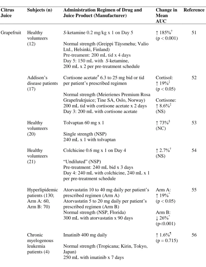
importance of other dietary substances as modifiers of drug performance. Figure 1.1 highlights other key observations since the discovery of the felodipine-GFJ interaction in 1989 [54]. Table 1.1 summarizes the design and major results of recent healthy volunteer and patient studies reporting modest to significant PK interactions with medications that are CYP3A substrates.
Pomelo Juice. The pomelo (Citrus maxima), or pummelo, is a large citrus fruit native to
Asia and is consumed typically as the fresh fruit. The grapefruit is believed to be an accidental hybrid of the pomelo and sweet orange (Citrus sinensis) [72]. Accordingly, it is reasonable to expect furanocoumarins are present in pomelos. Indeed, juice prepared from some species of pomelo has been reported to contain furanocoumarins in concentrations comparable to those in GFJ [73]. Clinical interactions with tacrolimus [74] and cyclosporine [75] via enteric CYP3A inhibition, albeit modest, have been reported. A clinical study of pomelo juice evaluated the extent of inhibition based on the species of pomelo [76]. Freshly prepared juices from two varieties of fruit (‘Guanximiyou’ and ‘Changshanhuyou’) were given, on separate occasions, with felodipine (10 mg) to 12 healthy
volunteers. Each juice was measured for furanocoumarin content and tested for inhibition of CYP3A activity (testosterone 6β-hydroxylation) in human liver microsomes prior to clinical testing; at 2.5%
juice (v/v), extents of inhibition were ~30% (Guanximiyou) and <5% (Changshanhuyou), relative to control. The more potent juice increased both mean area under the curve (AUC) and maximum concentration (Cmax) of felodipine, by ~40% (p<0.05), whereas the less potent juice increased these values by ~15% (NS) relative to water. Heart rate also was measured to determine effects on felodipine PD. Neither juice altered mean heart rate significantly. Unlike the tacrolimus and cyclosporine studies, the felodipine study acknowledged and attempted to account for PK variability with respect to furanocoumarin composition in the juice (see DISCORDANCE BETWEEN IN
VITRO AND CLINICAL STUDIES).
12
and pomelos contain furanocoumarins, an increase in systemic sildenafil exposure (relative to water) was expected. However, the juice significantly decreased mean AUC and Cmax of sildenafil. The authors speculated the mechanism was either a physicochemical interaction between sildenafil and components of the juice or inhibition of an intestinal uptake process (see Uptake Transport Proteins). Unlike the aforementioned study with felodipine, furanocoumarins were not measured in the juice.
Cranberry Juice. The cranberry (Vaccinium macrocarpon) has long been considered a
health food, touted for beneficial effects on diverse ailments [78]. More than 150 individual compounds have been identified [79]. As a rich source of phytochemicals, cranberries have shown anti-atherosclerotic and anti-proliferative properties, which may be protective in cardiovascular disease and certain cancers [80]. Cranberry juice (CBJ) continues to maintain popularity, largely as prophylaxis and treatment for urinary tract infections (UTIs) [81]. An in vivo study in rats given CBJ and the CYP3A/Cyp3a substrate, nifedipine, indicated that the juice inhibited enteric Cyp3a activity to an extent comparable to that by GFJ [82]. A subsequent clinical study involving 12 healthy volunteers given cyclosporine and a single 240-mL glass of CBJ indicated no interaction [83]. However, use of cyclosporine as a CYP3A probe was not ideal since cyclosporine also is a substrate for the efflux transporter, P-glycoprotein (P-gp) (see Efflux Transport Proteins), and whether or not CBJ modulates intestinal P-gp activity is not known.
Two clinical trials using midazolam as a CYP3A/non-P-gp probe showed conflicting results. The first study involved 10 healthy volunteers given a thrice daily regimen of a commercially available CBJ concentrate (diluted 1:4) for 10 days and a single dose of midazolam (0.5 mg on day 5) [84]. Relative to water, a change in midazolam PK was not detected. The second study involved 16 healthy volunteers given a single dose of midazolam (5 mg) and three 240-mL glasses of ‘double strength’ CBJ [85]. Prior to the clinical study, five brands of juice were tested in vitro to identify a
13
the second study was that in vitro bioactivity-guided fractionation was utilized to isolate and identify candidate CYP3A inhibitors. The clinical test juice, a concentrate, was fractionated to generate hexane-, chloroform-, butanol-, and aqueous-soluble fractions. The hexane- and chloroform-soluble fractions (50 μg/mL) inhibited CYP3A activity (midazolam 1’-hydroxylation) in human intestinal
microsomes by ~80 and 60%, respectively, suggesting the CYP3A inhibitors resided in these more lipophilic fractions. The juice was purified further until three triterpenes were isolated (maslinic acid, corosolic acid, ursolic acid) as candidate causative ingredients, with IC50 values ≤10 μM) [86]. The discrepancy between the two clinical studies may be explained by the difference in concentration of bioactive components. Only one brand was tested in the first study [84], and various components (anthocyanins, flavonols, hydroxycinnamic acids, hydroxybenzoic acids, and catechins) were measured. The most abundant was the flavonol, rutin, but CYP3A inhibition potency was not evaluated in vitro. Recognizing the substantial variability of bioactive components in natural products, the second study [85] began with in vitro testing to inform selection of the most appropriate brand for clinical testing and to generate candidate enteric CYP3A inhibitors for further investigation.
Although not intestinal CYP3A metabolism-based, the presumed warfarin-CBJ interaction
14
which was equivalent to 57 g of cranberry fruit daily and was more than triple the UTI prophylaxis “dose” recommendation [88]. A review published in 2010 discussed studies to date (including the
aforementioned sole finding) and concluded that moderate consumption of CBJ does not affect anticoagulation and that inclusion of precautionary warnings in warfarin product labeling should be re-examined [89]. Nevertheless, warfarin labeling continues to advise patients to avoid taking cranberry juice or cranberry products [90]. A more thorough understanding of the CBJ product in question is necessary to ascertain whether or not CBJ can enhance systemic exposure to clinically relevant CYP2C9, as well as CYP3A, substrates in humans.
Pomegranate Juice. The pomegranate (Punica granatum) and associated by-products is one
of the most popular superfoods on the market. Like CBJ, pomegranate juice is a complex mixture of polyphenolic compounds with high antioxidant potency [91]. Human in vitro and rat in vivo studies suggested that pomegranate juice can inhibit enteric CYP3A/Cyp3a activity (carbamazepine expoxidation) [92]. However, a subsequent clinical study involving 13 healthy men given 240 mL of pomegranate juice and a single oral dose of midazolam (6 mg) suggested minimal interaction, despite inhibition of CYP3A activity (triazolam hydroxylation) in human liver microsomes [93]. Likewise, another study involving 12 healthy subjects given a single dose of simvastatin (40 mg) after treatment with a different brand of pomegranate juice (300 mL three times daily for three days) reported no interaction [94]. Generalizations about the enteric CYP3A inhibition potential of pomegranate juice are cautioned, as minimal to no information was provided about the test juices and their composition, precluding between-study comparisons. In addition, neither clinical study provided a sample size justification.
15
rhabdomyolysis possibly due to an interaction with pomegranate juice [98]. This observation has yet to be investigated experimentally.
Esterase
Ester prodrugs are designed commonly to increase drug absorption [99]. Upon ester bond cleavage through hydrolysis or oxidation, active drug is released. Major esterases that hydrolyze prodrugs include carboxylesterase, acetylcholinesterase, butyrylcholinesterase, paraoxonase, and arylesterase [100]. Esterases are localized in multiple tissues, particularly blood, liver, and intestine [101]. Esterase inhibition could lead to increased stability of the ester in the lumen and enterocytes, resulting in higher absorption of the ester and higher exposure to active metabolite via rapid hydrolysis in plasma.
Grapefruit Juice. Enalapril is a prodrug that is metabolized primarily by carboxylesterase to
enalaprilat, an angiotensin converting enzyme inhibitor [102]. Lovastatin, indicated for hypercholesterolemia, is a prodrug that is hydrolyzed to the active acid by carboxylesterase, as well as oxidized to several inactive metabolites by CYP3A/Cyp3a; hydrolysis is considered the major metabolic pathway [103]. Lovastatin also has been suggested to be a weak substrate for P-gp [104]. Effects on the apical-to-basolateral (absorptive) permeability and/or metabolism of enalapril and lovastatin by GFJ (diluted 1:3 from frozen concentrate) were evaluated in a human intestine-derived cell line (Caco-2) and human intestinal and liver S9 fractions [105]. Relative to 0% (v/v) juice (buffer), the permeability of enalapril (5 μM) in Caco-2 cells increased significantly, by 30-133%, over the range of juice concentrations tested (6.25-50%). Cellular accumulation of enalapril at 1 h increased by 39-87%, while that of enalaprilat decreased by 12-32%. Enalapril hydrolysis in both S9 fractions was inhibited by <20% up to 40% juice. The permeability of lovastatin (5 μM) increased in
16
hydrolysis was reduced by ~50% in human intestinal S9 fractions up to 40% juice. Collectively, these in vitro observations suggested that GFJ inhibited enteric esterase activity, leading to increased prodrug stability.
When GFJ concentrate (diluted 1:3) was administered orally to rats, before intravenous administration of enalapril or lovastatin (2 mg/kg), clearance and half-life of both prodrugs were unchanged relative to water, indicating that GFJ had no effect on hepatic esterase/Cyp3a activity [105]. After oral administration of enalapril (10 mg/kg) with water or GFJ concentrate (diluted 1:3, 1:2, and undiluted), mean AUC of enalaprilat was increased, by 65, 70, and 16%, respectively, relative to water; prodrug was not measured. The decreased exposure at the higher strength was attributed to binding of drug to GFJ pulp. These results were consistent with observations with the esterase inhibitor, bis-p-nitrophenylphosphate. After oral administration of lovastatin (10 mg/kg) with water or GFJ concentrate (diluted 1:3, 1:2, and undiluted), mean AUC of lovastatin acid was increased by 279, 157, and 170%, respectively, relative to water; prodrug was not measured. Since lovastatin is a substrate for CYP3A/Cyp3a, the contribution of esterase inhibition was differentiated by measuring Cyp3a- and esterase-mediated metabolites in portal vein-cannulated rats pre-treated with GFJ (diluted 1:3). Both Cyp3a and esterase inhibition by GFJ led to similar increases in exposure to lovastatin and the active acid, as well as unchanged CYP3A-mediated metabolites, suggesting equal contribution by Cyp3a and esterase to the interaction. Taken together, these in vivo
observations were consistent with enteric esterase inhibition by GFJ, leading to increased prodrug stability in enterocytes and higher exposure to active metabolite via hydrolysis in plasma.
17
(20 µM) in Caco-2 cells was increased with kaempferol and naringenin (each at 250 μM) by 80% and ~200%, respectively, whereas that of lovastatin (20 µM) was increased by ~65% with both flavonoids. Intracellular concentrations of enalaprilat and lovastatin acid decreased by ~60% and 46-70%, respectively, consistent with inhibition of esterase activity. Oral administration of enalapril and lovastatin (both at 10 mg/kg) with naringenin (10 mg/kg) to rats increased active metabolite AUCs significantly, by 38% and 288%, respectively, relative to water. Similarly, oral administration with kaempferol (10 mg/kg) increased metabolite AUCs by 109 and 246%, respectively. Finally, in portal vein-cannulated rats, kaempferol (10 mg/kg) increased portal plasma exposure to lovastatin and lovastatin acid by 154% and 113%, respectively. Collectively, these observations suggested some flavonoids as potential candidate enteric esterase inhibitors in GFJ. However, more studies are needed to determine the clinical utility, as well as other causative ingredients, of this new type of drug-GFJ interaction.
Sulfotransferase
Conjugative enzymes generally increase hydrophilicity, facilitating elimination of endogenous substrates and xenobiotics [107]. Sulfotransferases (SULTs) catalyze the conjugation of 3’-phosphoadenosine 5’-phosphosulfate with a number of endogenous low molecular weight
compounds (e.g., steroids, catecholamines) and xenobiotics [108]. Three human SULT subfamilies have been identified, with at least 13 distinct members distributed in liver, brain, intestine, lung, kidney, and other tissues [109]. Some fruit juices have been shown to inhibit two members of the SULT1 family in vitro: SULT1A1 and SULT1A3, the latter of which is expressed only in extrahepatic tissues, including the intestine.
Grapefruit Juice. SULT1A1 and SULT1A3 inactivate 2-adrenergic agonists in the liver and
intestine, respectively [110]. The bronchodilators albuterol and terbutaline undergo extensive
18
and SULT1A3, respectively [112]. GFJ, at a concentration of 10% (v/v), inhibited SULT1A1 and
SULT1A3 by >90% and 50%, respectively, relative to control. Specific juice components also were tested and included naringin, naringenin, quercetin, bergamottin, and 6’,7’-dihydroxybergamottin. Quercetin was the most potent, inhibiting by >90% (SULT1A1) and 50% (SULTA3), at a concentration of 10 μM.
Orange Juice. Orange juice was tested in the same manner as GFJ in the aforementioned in vitro study [112]. As observed with GFJ, orange juice (10%, v/v) inhibited both SULTs, by >95%
(SULT1A1) and 20% (SULT1A3). The orange juice components, tangeretin and nobiletin (both at 10 μM), were the most potent single components, inhibiting SULT1A1 almost completely and
SULT1A3 by ~20%. As with GFJ, whether or not these observations translate to the clinic merits further investigation.
Pomegranate Juice. The effect of pomegranate juice on sulfoconjugation was evaluated in
Caco-2 cells [113]. The extent of inhibition of 1-naphthol sulfation by pomegranate juice was both concentration- and cell culture time-dependent. At the highest concentration tested (5%, v/v), the juice had no effect on SULT1A1 and SULT1A3 expression for up to 24 hours. Punicalagin, the most abundant polyphenol in pomegranate juice, was isolated and shown to inhibit sulfoconjugation in the cells, with an IC50 of 45 M. Clinical significance of these in vitro observations has not been reported.
Efflux Transport Proteins
19
Grapefruit Juice. Whether or not GFJ modulates intestinal P-gp activity remains
controversial [110]. One reason for the inconsistency is use of P-gp substrates that also are CYP3A substrates [116]. The contribution by P-gp and CYP3A is difficult to distinguish. Cyclosporine, a commonly used immunosuppressant with a narrow therapeutic window, is one such dual CYP3A4/P-gp substrate shown to interact with GFJ [117]. The increase in cyclosporine AUC ranges from 20 to 60%, relative to water or orange juice [47]. To assess whether furanocoumarins mediate the cyclosporine-GFJ interaction, a randomized crossover study involving 18 healthy volunteers compared the effects of GFJ, a “furanocoumarin-free” GFJ (prepared from the GFJ), and orange juice
(control) on oral cyclosporine PK [63]. Median dose-corrected cyclosporine AUC with GFJ was significantly higher (by ~38%) than that with orange juice. In contrast, relative to orange juice, furanocoumarin-free GFJ had no consistent effect, with a median concentration-time profile that was indistinguishable from that with orange juice. Complementary in vitro studies with Caco-2 cell monolayers showed that, relative to vehicle, diluted extracts derived from GFJ and orange juice, as well as two purified furanocoumarins (bergamottin and 6’,7’-dihydroxybergamottin), partially increased cyclosporine apical-to-basolateral translocation, whereas the furanocoumarin-free GFJ extract had no effect. These observations supported furanocoumarins as candidate P-gp inhibitors in GFJ. Furanocoumarins were concluded to mediate, at least partially, the cyclosporine-GFJ interaction
in vivo through inhibition of enteric CYP3A and possibly enteric P-gp.
Recent studies in rats evaluated potential interactions with the anti-gout agent colchicine and antiemetic domperidone, both of which are dual CYP3A/P-gp substrates. The effect of a GFJ concentrate on colchicine intestinal permeability was evaluated in Caco-2 cell monolayers and in the
in situ rat intestinal perfusion model [118]. With Caco-2 cells, at the highest concentration of GFJ
tested (10%, v/v), colchicine apical-to-basolateral translocation was increased by 75%, and
basolateral-to-apical translocation was decreased by 45%, relative to control (transport buffer). In
addition, GFJ (10%, v/v) increased colchicine ileal and jejunal permeability by 2- and 1.5-fold,
20
P-gp by GFJ. The effect of a commercially available GFJ extract was evaluated on domperidone exposure in rats [119]. Domperidone (10 mg/kg) was administered orally, two hours after GFJ
extract (2 mL/kg). The sum of partial AUCs (0-0.25 h, 0-2 h, 4-8 h) with GFJ extract was 16%
greater than that with water, albeit the difference was not significant (p>0.05). As with cyclosporine, interpretation of the underlying mechanism of the colchicine- and domperidone-GFJ interactions is confounded by the dual CYP3A/P-gp nature of these substrates. However, unlike cyclosporine, clinical relevance has not been established.
The inconclusive results of studies utilizing dual CYP3A/P-gp substrates could be resolved using P-gp substrates that undergo negligible metabolism. Although GFJ has been shown to inhibit translocation of such substrates in vitro, observations have not translated to the clinic. For example, GFJ had a negligible effect on systemic exposure to digoxin, as evidenced by a <10% increase in mean AUC0-24 relative to water [120]. Other minimally metabolized substrates, including the antihistamine fexofenadine and the -blockers talinolol and celiprolol, also have been tested. Unexpectedly, healthy volunteer studies showed a significant decrease in mean AUC of these three drugs when taken with GFJ, by 13-63% relative to water [59, 121, 122], prompting investigations of this newly identified mechanism underlying drug-fruit juice interactions (see Uptake Transport Proteins).
Orange Juice. Pravastatin, which undergoes minimal metabolism, is a substrate for P-gp and
21
interaction. Naringin and some polymethoxyflavones have been shown to inhibit P-gp and MRP2 in
vitro and may represent candidate causative ingredients [125, 126].
Uptake Transport Proteins
Organic anion transporting polypeptides (OATPs) are transmembrane transporters that facilitate uptake of a number of endogenous compounds and drugs [127]. These transporters are gaining attention as important determinants of drug disposition [128]. The human OATP family consists of 11 members, with OATP1A2, OATP1B1, OATP1B3 and OATP2B1 as the most characterized [129]. Of these, OATP1A2 and OATP2B1 have been reported to be expressed on apical membranes of enterocytes [130].
Grapefruit Juice. The initial clinical study examining effects of fruit juices, including GFJ,
22
consequent potential for reduced efficacy. This relatively new type of mechanism is discussed in detail in two recent reviews [139, 140].
OATP1A2 and the flavonoid, naringin, have been proposed as the major transporter and causative ingredient involved in the interaction between GFJ and fexofenadine, as well as talinolol.
In vitro studies with OATP-transfected human epithelial cervical cancer (HeLa) cells supported a role
for OATP1A2 in uptake of both drugs [59, 61, 141]. One in vitro study assessed the uptake activity of several OATPs, and showed OATP1A2 as the only enteric OATP capable of taking up fexofenadine [133]. A clinical study investigating the impact of GFJ on intestinal transporter expression showed no difference in OATP1A2 protein (and P-gp) expression between GFJ and water in duodenal biopsies obtained from healthy volunteers, suggesting GFJ may not destroy transport proteins via mechanism-based inhibition [133]. That is, the mechanism of inhibition of enteric transporter activity by GFJ may differ from that of enteric CYP3A activity. Unlike OATP2B1-transfected HeLa cells, a separate study with OATP2B1-OATP2B1-transfected human embryonic kidney (HEK) 293 cells demonstrated fexofenadine as a substrate for OATP2B1 [142]. GFJ and components (including naringin) have been shown to inhibit uptake of the OATP substrate estrone-3-sulfate in OATP2B1-transfected HEK293 cells, by up to 80% [134], but additional studies are needed to determine whether GFJ/components inhibit OATP2B1-mediated uptake of fexofenadine, as well as to clarify differences in fexofenadine uptake between transfected cell lines.
23
A recent in vitro study involving the leukotriene receptor antagonist, montelukast, and a clinical trial involving the renin-inhibiting antihypertensive agent, aliskiren, added two potential drugs to the growing list of enteric OATP-mediated drug-GFJ interactions [143, 71]. In vitro studies with Caco-2 cells and OATP2B1-transfected Madin-Darby canine kidney cells demonstrated that montelukast undergoes carrier-mediated transport by OATP2B1. GFJ at 5% and 10% (v/v), and orange juice at 10%, reduced montelukast permeability significantly (p<0.05), by ~30% relative to control (buffer). Clinical relevance of these interactions has not been examined. Aliskiren is a substrate for OATP2B1, as well as CYP3A and P-gp. Healthy volunteers (n = 11) were administered single-strength GFJ (200 mL) three times daily for 5 days, and aliskiren (150 mg) was given on the third day. Relative to water, GFJ significantly reduced mean aliskiren AUC and Cmax, by 61% and 81%, respectively. Mean elimination half-life remained essentially unchanged. Net inhibition of enteric OATP-mediated uptake by GFJ could account for the reduced exposure. Other potential mechanisms included a physicochemical interaction between GFJ and aliskiren or an alteration of physiologic conditions in the gut by GFJ. Follow-up in vitro and clinical studies are needed to clarify the role of OATP in the aliskiren-GFJ interaction, as well as effect on PD outcomes.
Orange Juice. Orange juice contains trace amounts of furanocoumarins and has minimal
enteric CYP3A inhibitory effect [126, 144]. As such, orange juice has been used as a control juice, rather than water, in some clinical studies. However, orange juice has been shown to reduce systemic exposure, significantly (by 22-83%), to fexofenadine [59], atenolol [145], and celiprolol [146]. Decreased mean AUC (up to 38%) also has been observed for the fluoroquinolones ciprofloxacin [147] and levofloxacin [148] with calcium-fortified and non-fortified orange juice. Any or all of these interactions could involve inhibition of enteric OATP by orange juice, as fexofenadine, levofloxacin, and celiprolol, have been shown to be substrates for OATP in vitro [59, 149, 150].
24
However, hesperidin produced only 60% maximum inhibition at the highest tested concentration (100 µM). A study in rats duodenally administered celiprolol (5 mg/kg) and orange juice or aqueous solution of hesperidin (208 µg/mL or 340 µM, the same concentration as that in the orange juice) showed a mean AUC decrease of 75% and 78%, respectively, relative to water. The AUC in the hesperidin group was not significantly different than that of the orange juice group, suggesting hesperidin contributes to the celiprolol-orange juice interaction. Studies with other OATP substrates would clarify the in vivo significance of hesperidin.
Pomelo Juice. The previously mentioned clinical study of six healthy male volunteers showed a
significant decrease (by ~60%) in mean AUC and Cmax of sildenafil after ingestion of a 240-mL glass of fresh-squeezed pomelo juice [77] (see Cytochrome P450 3A). One possible explanation was inhibition of intestinal uptake (e.g., by OATP) of sildenafil by the juice. No follow-up studies examining sildenafil as an OATP substrate have been reported.
Apple Juice. The effect of apple juice on fexofenadine uptake also was evaluated in the initial
fexofenadine-GFJ interaction study [59]. The OATP-mediated uptake of [14C]fexofenadine was
examined in the presence and absence of increasing concentrations (0-5%, v/v) of apple juice in OATP1A2-transfected HeLa cells. The highest concentration of juice inhibited activity by >85% relative to water. Clinical study results also were significant, as apple juice decreased mean AUC of
fexofenadine by ~70% compared to water. To the authors’ knowledge, no follow-up in vitro and
clinical studies involving apple juice per se have been published. However, a recent in vitro study
investigated the effect of three flavonoids (apigenin, kaempferol, quercetin) on OATP activity in
OATP1A2- and OATP2B1-transfected HEK293 cells using fexofenadine and bromosulfophthalein as
substrates [152]. Quercetin, known to be present in apples (Malus x domestica) and apple juice [153], inhibited OATP1A2-mediated fexofenadine uptake, with an IC50 of 13 μM. Quercetin also inhibited
OATP1A2- and OATP2B1-mediated bromosulfophthalein uptake, with Ki values of 22 and 8.7 μM,
respectively. Further studies are needed to determine if quercetin is a major causative ingredient in
25
DISCORDANCE BETWEEN IN VITRO AND CLINICAL STUDIES
Although a number of fruit juices have been shown to inhibit several intestinal CYPs and transporters in vitro, some of the interactions have not translated to the clinic. Fruit juices clearly inhibit intestinal metabolism and transport, but the magnitude of change in Cmax and/or AUC often is insignificant, unpredictable, and highly variable, which cannot be explained adequately. These in
vitro-in vivo discordances may be due to a lack of sufficient information to determine a positive
interaction. A deficiency common to most drug-fruit juice interaction studies is a limited or non-existent chemical description of the juice. Although several reasons account for discrepancies between in vitro and clinical studies [24, 154], one explanation is that the concentration of inhibitors in the juice might not be sufficient to inhibit metabolism/transport in vivo. The sources and complexity of a plant’s chemical constituents are underappreciated. Concentrations of bioactive compounds in a natural food product vary depending on ecological conditions, manufacturing process, storage conditions, and a host of other environmental factors [155]. Thus, testing a random juice product in vitro and in vivo without understanding the chemical makeup provides no basis for comparison between studies. One of the most fundamental solutions to establishing meaningful physiological dose-response relationships for dietary substances is to characterize the product prior to use.
Few in vivo drug-fruit juice interaction studies reported concentrations of bioactive
constituents in the clinical test juice. Since the establishment of furanocoumarins as unequivocal mediators of enteric CYP3A-based interactions in 2006 [53], only a handful of clinical studies involving CYP3A substrates reported furanocoumarin content in the test juice (Table 1.2). Furanocoumarins have been studied to the extent that they can be considered ‘marker’ compounds,
26
can be made. Indeed, 6′7′-dihydroxybergamottin was used in a recent PK modeling study investigating the impact of CYP3A-based inhibition on drug disposition [157]. In addition to grapefruit and related citrus juices, furanocoumarins are present in substantial amounts in umbelliferous vegetables (e.g., parsnips, celery, parsley) and are not destroyed by normal cooking procedures [158]. Furanocoumarins also are present in Kampo extract medicines, which originated in Japan and are used widely in Asia [159]. A similar strategy could be applied to predict likelihood and magnitude of interactions between these foods/natural medicines and traditional medications.
APPLICATIONS: NEW TWISTS ON OLD INTERACTIONS
Mechanisms and causative ingredients underlying enteric CYP3A inhibition by GFJ have been studied for more than two decades [160]. The information gained has been used by different groups to their advantage. For example, the potential for GFJ to increase systemic drug concentrations, and consequent PD effect, of certain opiates is a widely discussed topic among recreational users in online forums [161, 162]. The scientific community also has attempted to exploit enteric CYP3A inhibition for pharmacoeconomic and therapeutic purposes. Intentional manipulation of enteric CYP3A by GFJ and individual components is of particular interest in the cancer treatment and organ transplantation areas [137, 163-166]. For example, inhibition of enteric CYP3A by GFJ could improve oral bioavailability of some agents without GFJ exerting additional adverse effects. In addition, the cost and side effect severity of these multi-drug and toxic regimens could be reduced through dose and/or dosing frequency reduction by coadministration with GFJ, possibly improving adherence. However, several drugs in these therapeutic classes have a narrow therapeutic range and require close monitoring. Without thorough characterization of the juice product, these practices are at best ineffective, and at worst, place the patient at risk for unfavorable outcomes.
An in vivo study of wild-type and humanized CYP3A4 transgenic mice orally administered
27
[163]. Results illustrated the potential of BAS 100 to “boost” systemic drug exposure, and decrease associated variability, of erlotinib in cancer patients. The erlotinib study is one of the first attempts to test the strategy of deliberate inhibition of intestinal first-pass metabolism.
A clinical trial investigating the effect of GFJ on sirolimus PK in advanced solid tumor patients is ongoing [167]. Initial results showed no effect, possibly due to insufficient furanocoumarin content in the GFJ product selected [168]. This approach is unsettling because GFJ was chosen as a ‘boosting’ agent for a narrow therapeutic index drug but was not characterized before administration. A more ‘potent’ GFJ containing inhibitory concentrations of furanocoumarins (not
reported) given subsequently to the subjects significantly increased plasma concentrations of sirolimus, by up to 400% relative to water [164]. A daily glass of GFJ (240 mL) was projected to lower sirolimus costs by 50% [169]. The same investigators have suggested ‘GFJ boosting’ to reduce dose and cost for the tyrosine kinase inhibitor, lapatinib [165]. One 250-mg lapatinib tablet, accompanied by food and/or GFJ, was speculated to increase systemic exposure comparable to that of five 250-mg tablets on an empty stomach, resulting in a total cost savings of 80%.
28
relationship between the causative ingredients in GFJ (e.g., furanocoumarins) and tacrolimus, as well as the large interindividual variability in response, making therapeutic outcomes virtually unpredictable.
It seems tempting to take advantage of the effects of a natural product like GFJ to boost systemic drug exposure and decrease inter-/intraindividual variability in PK, and ideally PD, outcomes. The dose and dosing schedule of certain drugs could be reduced to lower drug costs and improve patient compliance. However, further research is required on the mechanisms of action, causative ingredients, and PK-PD relationship with respect to individual juice components and the drug of interest. Given the possibility of using GFJ and/or individual constituents as a ‘drug-sparing agent,’ a standardized approach to investigating interaction potential is imperative. Early evaluation
of CYP and transporter inhibition properties of new chemical entities is routine during drug development. A similar approach could be adopted for dietary substances. However, information providing a systematic approach for the study, prediction, and management of drug-dietary substance interactions is lacking. Ideal management approaches would be those developed based on well-designed in vitro studies. Information gained from rigorous in vitro studies, combined with that gained from in silico methods, could optimize clinical study design and clarify the clinical significance of an interaction. Robust PK/PD models could then be used to determine potential risks of long-term inhibition of intestinal metabolism/transport by a given dietary substance on pharmacotherapeutic outcomes.
CONSIDERATIONS FOR IMPROVED RESEARCH PRACTICES
29
more extensive drug-dietary substance interaction studies other than those recommended. However, legislation need not be passed to undertake sound scientific research. Several approaches can be adopted for rigorous evaluation of potential drug-dietary substance interactions. Practices regarding peer review of the drug interaction liability of a dietary substance should be the same as those for a drug, particularly with respect to reproducibility. If the dietary substance is not described in detail, then other investigators will be unable to reproduce one or more facets of the study.
Since dietary substances contain multiple, often unknown, bioactive ingredients that vary in composition between batches and manufacturers, characterization of causative ingredients and mechanisms of action is essential. Identification of components responsible for these interactions can be challenging. Generally, biologic action is not determined by a single active compound. A set of ‘marker’ compounds that can be applied for definitive authentication of the test material would serve
as an indicator of quality and potency. The selected markers should be unique to the selected species and represent health-relevant principles [156]. The identity of constituent(s) should be confirmed initially by in vitro methods that screen for potential interactions. Such experiments provide mechanistic information about inhibitory capacities, as well as specific enzymes and/or transporters involved.
Reporting of the characterization of dietary substances used in clinical trials must be improved. Many clinical studies lack basic information about the test material. Considering the requirements for a drug investigated clinically, the disconnect becomes obvious. A substance derived from a “natural source” does not imply that the rigors of reproducibility should be abandoned. At