TRAUMATIC DENTAL INJURIES IN PRESCHOOL-AGE CHILDREN: PREVALENCE AND RISK FACTORS
Catherine D. Born
A thesis submitted to the faculty at the University of North Carolina at Chapel Hill in partial fulfillment of the requirements for the degree of Master of Science in the School of Dentistry
(Orthodontics).
Chapel Hill 2018
Approved by:
Kimon Divaris
Tate H. Jackson
ABSTRACT
Catherine D. Born: Traumatic Dental Injuries in Preschool-Age Children: Prevalence and Risk Factors
(Under the direction of Kimon Divaris)
Purpose: This study examined the prevalence, socio-demographic correlates, and clinical predictors of traumatic dental injuries (TDIs) in the primary dentition among a community-based
sample of preschool-age children.
Methods: The sample comprised 1,546 preschool-age children [mean age 49 (range: 24-71) months] in Early Head Start and Head Start programs in North Carolina, enrolled in the
Zero-Out Early Childhood Caries (ZOE) study. Information on socio-demographic, extraoral,
and intraoral characteristics was collected and analyzed with bivariate and multivariate methods,
including logistic regression modeling and marginal effects estimation.
Results: The prevalence of dental trauma was 47% and 8% of TDI cases were “severe”, defined as pulp exposure, tooth displacement, discolored or necrotic tooth, or tooth loss. In
bivariate analyses, overjet and lip incompetence were significantly associated with TDI, whereas
age, body mass index, and canine occlusion showed weaker positive associations. Overjet
remained positively associated with severe trauma in multivariate analysis: OR=1.3 (95% CI:
1.2-1.5), corresponding to an absolute 1.0% (95% CI: 0.6-1.5) increase in the likelihood of
Conclusions: Overjet is a strong risk factor for TDIs in the primary dentition.
Incorporating and operationalizing this information may help traumatic dental injury prevention
ACKNOWLEDGEMENTS
Thank you to my mentor, Kimon Divaris, and my committee members, Dr. Jackson and
Dr. Koroluk, for their expertise and guidance. Thank you to all of the personnel and clinicians
involved in the ZOE 1.0, ZOE-pilot, and ZOE 2.0 studies, the participating families, and Early
Head Start and Head Start staff who have made it possible to conduct numerous studies to further
our understanding of dental disease across the state of North Carolina. Thank you to Tom Pahel
and John Cantrell for data treatment, migration, and organization, without which this project
would not be possible. Thank you to the International Association for Dental Research for the
graciously awarded research grants. Thank you to my husband Andrew and my family for their
TABLE OF CONTENTS
LIST OF TABLES ... viii
LIST OF FIGURES ... ix
LIST OF ABBREVIATIONS ...x
LIST OF SYMBOLS ... xi
REVIEW OF THE LITERATURE ...1
Prevalence of traumatic dental injuries ...1
Consequences of TDIs ...2
Etiology of TDIs ...3
Classification of TDIs ...6
Orthodontic treatment and TDIs ...7
Importance of risk factor information and risk indices ...10
Preliminary studies ...11
Conclusions ...12
TRAUMATIC DENTAL INJURIES IN PRESCHOOL-AGE CHILDREN:
PREVALENCE AND RISK RACTORS ...17
Introduction ...17
Materials and methods ...18
Study population ...18
Data collection ...19
Analytical approach ...21
Results ...22
Discussion ...23
Conclusions ...27
Tables ...28
Figures...34
LIST OF TABLES
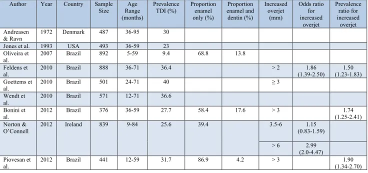
Table 1 – Prevalence of traumatic dental injury in the primary dentition
and classification for increased overjet: summary of past studies ...28
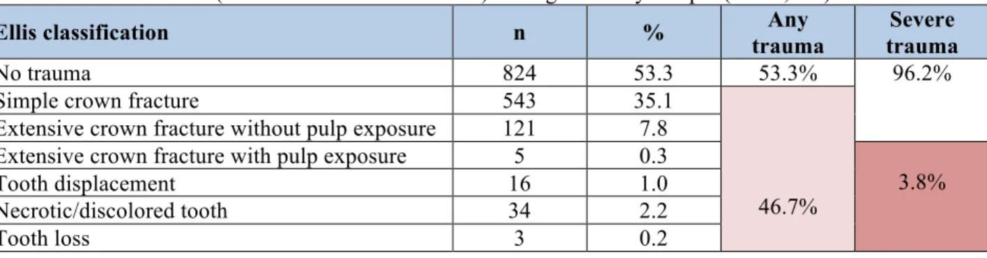
Table 2 – Dental trauma (modified Ellis classification) among the study sample ...29
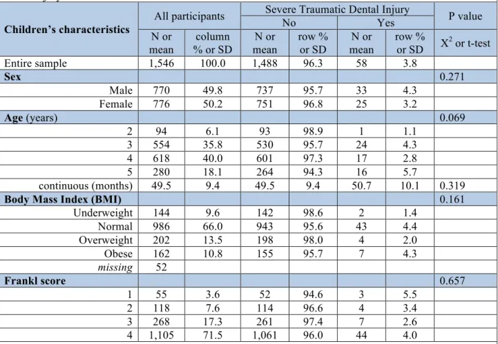
Table 3 – Descriptive information of participating children ...30
Table 4 – Intraoral and extraoral characteristics of participating children ...31
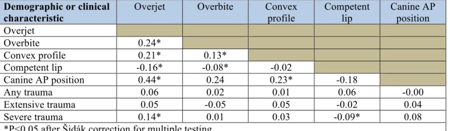
Table 5 – Pairwise correlation coefficients between clinical variables and trauma ...32
LIST OF FIGURES
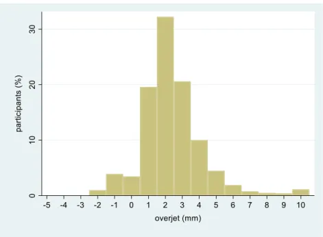
Figure 1 – Distribution of overjet values in the study sample ...34
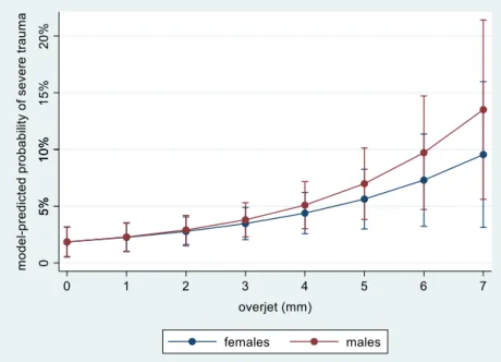
Figure 2 – Final logistic regression model-predicted probabilities and
95% confidence intervals of severe trauma, for males and females, according to overjet ...35
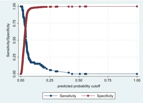
Figure 3 – Final logistic regression model sensitivity and specificity ...36
LIST OF ABBREVIATIONS
AAP American Academy of Pediatrics
AAPD American Academy of Pediatric Dentistry
ADHD Attention-deficit/hyperactivity disorder
BMI Body Mass Index
CI Confidence Interval
ECOHIS Early Childhood Oral Health Impact Scale
EHS Early Head Start
HS Head Start
ICD International Classification of Disease
ICDAS International Caries Detection and Assessment System
NC North Carolina
OHRQoL Oral health-related quality of life
QoL Quality of life
RCT Randomized Controlled Trial
SES Socioeconomic status
TDI Traumatic dental injury
WHO World Health Organization
LIST OF SYMBOLS
© Copyright Symbol
A REVIEW OF THE LITERATURE
Prevalence of traumatic dental injuries
Traumatic dental injuries (TDIs) are one of the most common oral conditions experienced
by children, along with dental caries, infectious diseases, and hereditary conditions.1 With the
decline in caries prevalence and severity in recent years, dental trauma and its sequelae are
receiving an increasing amount of attention. Sports injuries, violence, and traffic accidents are
frequently cited as important contributors in the etiology of dental trauma.2 Epidemiologic data
from two large national surveys in the United States indicate that one in six adolescents and one
in four adults have experienced a traumatic dental injury.3,4 A 2006 study found similar results in
North Carolina (NC), with 25% of individuals between six and 50 years old having a history of
dental trauma to one or more permanent anterior teeth, and approximately half of adolescents
having suffered dental trauma by the time they graduate from high school.5 The evidence also
supports gender and age predilections: boys suffer from an injury to the permanent dentition
twice as often as girls do, and children between the ages of eight and 10 years old are the most
likely to sustain an injury to a permanent tooth.6
Although much has been reported on dental trauma in the permanent dentition, less is
known about TDIs in the primary dentition. A 12-year literature review on traumatic dental
injuries revealed that up to 36% of children experience dental trauma in their primary dentition,
with the greatest incidence occurring between two and three years of age, when motor
that approximately one-third of toddlers, infants, and children experience a traumatic dental
injury to their primary dentition.9 However, most injuries to the dentition in these investigations
were minor and were limited to enamel-only fractures, which require little to no intervention.
Consequences of TDIs
The consequences of TDIs extend well beyond the traditional clinical implications.
Traumatic episodes can confer clinical and quality of life impacts to children and their families
when there is severe injury to oral and dental structures. A recent systematic review explored the
effects of TDIs in the primary dentition on oral health-related quality of life (OHRQoL) of
preschool-age children and their families.10 OHRQoL was measured using the Early Childhood
Oral Health Impact Scale (ECOHIS), the only instrument to date that is validated to measure oral
health impacts among preschool-age children and their families.11 In the cross-sectional studies
included in the systematic review, TDIs were found to have a statistically significant negative
impact on OHRQoL, as measured by the overall ECOHIS score, with children experiencing TDI
having a “24% greater chance of experiencing a negative impact on OHRQoL”.10 The domains
of the ECOHIS that were most affected by TDIs in the sample of preschool-age children were
within the domain of “child impacts” and included function—difficulty drinking hot or cold
beverages, difficulty eating some foods—and child self-image/social interaction—avoided
smiling or laughing, avoided talking.10,11 Although OHRQoL impacts from TDIs decrease over
time, negative effects are still seen 12 or more months after the injury.12 In sum, Borges and
colleagues emphasize the importance of prevention, early diagnosis, and treatment of TDIs in
order to prevent or reduce the impacts on OHRQoL of preschool-age children and their families
Furthermore, a wide spectrum of social, psychological and financial consequences have
been reported in the literature. According to a population-based Canadian study, trauma to the
maxillary incisors had more of an impact on social well-being than on psychological or
functional parameters among 12- to 14- year-old schoolchildren.13 In another Canadian study,
Nguyen and colleagues14 found that 90% of patients and 86% of parents indicated that some
school and work time were lost due to pediatric dental trauma. In regards to financial impacts,
Borum estimated that the annual cost of treatment for patients at a major trauma center in
Denmark was $600,000-$1,000,000 per year. This included acute trauma care, follow up care,
and subsequent restoration, and translated to between two and five million dollars per million
inhabitants per year, irrespective of age.15,16 The costs at a population-level may appear low;
however, costs for individual cases are high and the lifetime costs may range up to hundreds of
thousands of dollars for avulsion of the permanent maxillary central incisors.16,17 Treatment costs
for emergency and long-term care, as well as time missed from work can be significant for
families of children who have incurred dental trauma. Of note, 50% of children with a history of
multiple TDIs are likely to re-injure a previously traumatized tooth, requiring long-term
follow-up care and increased treatment costs.18 It is evident that the consequences of traumatic dental
injuries are not limited to just physical consequences—some of the psychosocial, clinical, and
financial consequences can extend well beyond childhood, impacting the quality of life of the
child and the family.16
Etiology of TDIs
The high prevalence of TDIs and their negative impact on OHRQoL have motivated
substantial research and scholarship investigating possible etiologic factors. Dental trauma
factors of TDIs can be grouped into three domains, including human behavior, environmental
determinants and oral factors. The “human behavior” domain generally includes risk-taking
behaviors, learning difficulties, executive function disorders, and conditions such as
attention-deficit/hyperactivity disorder (ADHD), which affects up to five percent of children.20 A study
investigating executive function disorders and trauma suggested that there is a link between
certain subscales of executive dysfunctions, specifically impulsivity and emotional control, and
TDIs.21 A recent systematic review summarized the findings of 14 studies investigating the
association between ADHD and TDIs and determined that the prevalence of TDIs in children
with ADHD is approximately one in three children, corroborating evidence from the past 10
years that has linked ADHD to TDIs.20 Moreover, the 1997 Health Survey for England provided
data enabling the study of behavioral risk factors for TDIs. In that study, Lalloo22 found a
statistically significant association between hyperactivity and major injuries to the teeth, jaws,
and face.
“Environmental determinants” include more contextual parameters such as
socioeconomic status (SES), material deprivation, and an unsafe environment. There are two
opposing views on the impact of SES on TDIs: some state that children of lower SES have a
higher risk of dental trauma, whereas others indicate that children of higher SES are more likely
to experience TDIs due to increased access to leisure activities and ability to afford toys, sports
equipment, and other goods.23 Paiva et al.24 found no association between SES markers and
TDIs. Lalloo20, using a nationally-representative sample of 5,913 children from England, found
that low SES, families receiving benefits, and children in single parent households were linked to
increased prevalence of TDIs; however, these associations were not statistically significant. This
cross-sectional study conducted in India found that 57.2% of three to five year-old children
categorized as high SES sustained anterior dental trauma compared to only 42.9% of children of
low SES.25 Another study that examined an environmental determinant of TDIs found that
children in public schools sustained more TDIs (11.4%) than those in private schools (9.5%).24
Studies investigating “oral factors”, including increased overjet with protrusion, lip
incompetence, and other factors, comprise the majority of the literature on etiology of TDIs.
Numerous studies support the association between increased overjet and TDI to maxillary
incisors. Bauss et al.27 conducted a cross-sectional study to examine the influence of increased
overjet and inadequate lip coverage on permanent maxillary incisor trauma among 1,367
individuals with mean age of 14.8 years.27 The investigators found that subjects with increased
overjet with and without adequate lip coverage had higher prevalence of TDIs, compared to
those with normal overjet (< 3 mm) and adequate lip coverage. While the majority of studies on
oral risk factors for TDIs focus on the permanent dentition, similar trends have been noted in the
primary dentition, with certain extraoral and dental occlusion characteristics placing children at
increased risk of sustaining a TDI. Several studies on TDIs in the primary dentition have
reported positive associations between increased overjet and prevalence of maxillary incisor
trauma.28-31 Goettems et al.31 investigated numerous dental characteristics, including overjet,
open bite, overbite, anterior crossbite, crowding, tooth rotations, and canine classification, and
found that TDIs were associated with Class II canine classification, overjet ≥ 3 mm, and overbite
≥ 3 mm. Lip competence is also an important soft tissue characteristic that has been shown to
play a protective role in the prevention of TDIs. Children with increased overjet or anterior open
bite along with inadequate lip coverage had a statistically significant increased prevalence of
adequate lip coverage.32 In the primary dentition, one of the main causes of anterior open bite is
non-nutritive sucking habits.33 Non-nutritive sucking habits, such as thumb and finger sucking or
prolonged pacifier use, can be responsible for other dental characteristics as well. In addition to
anterior open bite, increased overjet, Class II canine and molar relationships, posterior crossbite,
and inadequate lip coverage are frequently associated with a history of non-nutritive sucking.34,35
The TDI etiology triad developed by Glendor is certainly not an all-inclusive list, but
offers a helpful categorization of postulated risk factors for dental trauma. BMI and sex, for
example, do not necessarily fall into one particular category, but are included as risk factors for
TDIs. Soriano et al.26 found a statistically significant correlation between obesity and TDIs
among a sample of 1,046 Brazilian children. In contrast, Martins et al.36 found lower TDI
prevalence (8.7%) among overweight/obese schoolchildren (BMI ≥ 85th percentile) ages seven to
14 years, compared to 13.3% TDI prevalence among schoolchildren who were not categorized as
overweight/obese (15th percentile < BMI < 85th percentile). Evidently, findings linking BMI to
TDIs are inconsistent; examining children’s activity levels stratified by BMI might provide a
better, causal explanation for the postulated association between BMI and TDIs. Additional
factors, that do not necessarily fall into one of these three categories but might also increase the
risk of TDIs, were presence of illness, physical limitations, inappropriate use of teeth, and oral
piercings.23
Classification of TDIs
A multitude of classification systems have been utilized for classification of TDIs, dating
back to G.E. Ellis’ first classification system for dental injuries developed in 1950. Factors such
as anatomy, pathology, etiology, and therapeutic considerations have been taken into
review of the evidence base between 1936 and 2003 identified 54 classification systems in the
literature.2 Thirty-two percent of the articles included in the search utilized Andreasen’s
classification system, followed in prevalence by Ellis’ classification system, which was cited in
14% of the articles. Andreasen’s classification system is remarkably similar to the World Health
Organization (WHO) classification system found in the Application of the International
Classification of Disease (ICD), which defines 19 groups, including groups for injury to the
teeth, gingiva, oral mucosa, and supporting structures.2 Although Ellis’ modified classification
system, developed in 1962, provides an anatomical and numerical basis for classification, it has
been criticized for its subjective interpretation because of the use of terms such as “simple” or
“extensive”,37 whereas Andreasen’s classification system, as well as the WHO classification
system, account for minimal subjective interpretation. Moreover, Ellis’ simplified classification
system groups multiple injuries and does not provide a classification for injury to the alveolar
socket or fractures of the maxilla or mandible. The authors of the 2006 systematic review
concluded the following: “Ellis’ classification system…is the most suitable, once it follows the
hierarchical structure, proposed by the WHO, as regards to the ideal properties of
standardization. However, for epidemiological purposes, some changes may be needed”.2 Having
a consistent diagnostic classification system is important for communication between providers
and for epidemiological reasons in order to accurately report incidence and prevalence within
diagnostic categories.
Orthodontic treatment and TDIs
Early Class II treatment should not be performed on all children presenting with a Class
II Division 1 malocclusion. It is important to weigh the benefits of early orthodontic treatment in
prolonging total treatment time with two phases of orthodontic treatment. The primary indication
is a child with psychosocial issues that are related to his or her dental or facial appearance.
Children who are treated early because they are bullied for their dental or facial appearance
report improved self-concept and happiness. In 2016, Choi et al.38 found that severity of
malocclusion was associated with decreased OHRQoL, providing added support for early
treatment of severe malocclusions due to the psychosocial and quality of life benefits. Another
reason for early treatment is susceptibility to trauma, such as having a Class II skeletal profile or
certain Class II dental characteristics, as these children tend to suffer from more TDIs.
Numerous studies have investigated the role of early and timely intervention of Class II
malocclusions with orthodontic treatment in the reduction of TDIs. Thiruvenkatachari et al.39
conducted a systematic review to examine the effects of early treatment for children with Class II
Division 1 malocclusion. After identifying three randomized controlled trials (RCTs)
investigating early treatment for procumbent incisors, the authors concluded that early treatment
with functional appliances or headgear did not demonstrate any significant differences in
outcomes, with the exception of decreased incidence of trauma to a maxillary incisor, compared
to one-phase treatment in the permanent dentition. The summary of evidence from these three
RCTs included in the systematic review revealed that 10 patients would need to be treated early
with a functional appliance in order to prevent one new episode of incisor trauma. However,
trauma could range from simple craze lines to complete avulsion, as trauma was categorized as a
simple ‘yes’ or ‘no’ in one of the studies included in the systematic review, therefore forcing the
categorization of trauma in all studies as none or any. Trauma can vary from minor craze lines to
more serious luxation or complete avulsion. Simplifying this wide continuum of traumatic dental
clinically relevant because the majority of trauma incidents are limited to enamel-only fracture.
Ultimately, the authors indicated that early treatment should not be performed solely as a risk
reduction strategy to prevent traumatic dental injuries, but that early treatment is a multifactorial
decision, and prevention of new incisor trauma can be part of the risk evaluation process in
deciding whether or not to treat early.39
One of the clinical trials included in the systematic review found that there was no
statistically significant difference between the early treatment groups and the control group, in
which treatment was delayed until the permanent dentition. Furthermore, there was no significant
difference between the four dental age categories examined (<9, 9-9.9, 10-10.9, ≥11),
suggesting that the number of patients that experienced trauma did not increase substantially and
remained fairly stable in the late mixed and early permanent dentition. Therefore, if early
intervention was recommended, it would likely be more beneficial to begin treatment shortly
after the permanent incisors erupt in order to prevent new incisor trauma. Based on this study’s
findings, Koroluk et al.40 caution against using trauma reduction as the sole determinant of early
treatment, but explain that the choice is a complex one that needs to be evaluated on an
individual basis, taking into consideration dental factors as well as non-dental factors, including
social factors, activity level, and risk-taking behaviors.40 Chen et al. also stated that early
orthodontic treatment did not reduce the incidence of incisor injury and that the “cost-benefit
ratio of orthodontic treatment primarily to prevent incisor trauma is unfavorable”.41 Most studies
agree that early orthodontic intervention should be an informed decision, relying on more than
just risk of incisor trauma. Parents and caregivers must be adequately informed of the expected
Importance of risk factor information and risk indices
Risk indices for caries, oral hygiene, periodontal diseases, and malocclusion are routinely
used in dentistry to inform decision-making and treatment planning. Indices help summarize and
quantify clinical and behavioral characteristics, and are considered valuable aids in the context of
personalized care and making between-group comparisons. Indices also facilitate the collection
and presentation of objective, standardized information that can lead to improvements in
diagnosis, communication, planning, and management of dental conditions.
Additionally, risk indices, which can be used by physicians, dentists, parents, and
caregivers, can be used for parent/caregiver education and communication, even if their
objectively-determined performance is low. The American Academy of Pediatrics (AAP)
developed an Oral Health Risk Assessment Tool for pediatricians to use at every well child visit,
beginning at or before six months of age. This tool includes risk factors obtained via history and
clinical evaluation. Caries, inflammation of the gums, ulcers, pattern of tooth eruption,
malocclusion, and evidence of trauma are all components of the clinical exam for the Oral Health
Risk Assessment Tool.42 Importantly, its use can facilitate the establishment of a dental home for
children, provide valuable information for referral to dentists for early intervention if needed,
and educate parents on oral hygiene and prevention strategies for caries and trauma, especially in
high-risk children. Identification of risk indicators can help the stratification of children into risk
groups, which allows the opportunity to provide parents and caregivers with a more complete
picture of oral health, as well as the information needed for proper risk reduction and disease
prevention.
The WHO has also published information regarding prevention and treatment of common
children are playing in safe physical environments and using protective sports equipment in
order to reduce the risk of traumatic dental and facial injuries.1 Providing practical and easily
accessible information to parents and caregivers is key to reducing the incidence of TDIs among
children.
Preliminary studies
We have conducted and reported43 a preliminary analysis of the association between
clinical characteristics and TDI among 345 children who were participants of the first phase of
the Zero-Out Early Childhood Caries (ZOE) study (ZOE 1.0; PI: Gary Rozier). These children
were enrolled in Early Head Start (EHS) centers across NC. Clinical examinations were
conducted by one examiner (a board-certified pediatric dentist) under field conditions. The
participating children had mean age of 38 months (range: 30-52); 36% were Hispanic, 31%
African American, 23% white, and 9% Native American or of other race. The prevalence of
dental trauma was 18%. Sixty-nine percent of these with TDI had enamel-only fractures,
whereas a small proportion (6%) showed evidence of more extensive trauma (extensive crown
fracture without pulp exposure, extensive crown fracture with pulp exposure, tooth displacement,
or necrotic/discolored tooth). Lip incompetence, class II canine relationship, and increased
overjet were associated with significantly (P<0.05) higher prevalence of TDI, whereas obesity
and male sex showed weaker positive associations. Overjet remained strongly associated with
‘any and severe forms of’ TDI in multivariate analyses, with a corresponding 4% (95%
confidence interval: 1-7%) increase in the likelihood of TDI per millimeter of overjet. These
preliminary analyses indicated that the prevalence of TDI among this community-based sample
enamel-only fractures. Increased overjet emerged as a modest risk factor [odds ratio (OR)=1.3 (95% CI:
1.1-1.6)] for TDI in the primary dentition, consistent with previous reports in the literature.
Conclusions
Several etiologic and risk factors for TDIs have been reported in the literature including
socio-demographic, behavioral, environmental, and clinical or oral characteristics. However, data
specific to preschool-age children are scant. We sought to further understand the importance of
oral factors, which are relatively easily and reliably measured by clinical examination. Although
previous studies have investigated the association of oral factors (including additional
characteristics such as sex, BMI, behavior, and more) with TDIs, none has actually incorporated
this information in a clinically useful risk model. Such a tool could be used for risk assessment
and outcome prediction and would be beneficial for family education, screenings, personalized
prevention and risk reduction, and for planning early orthodontic treatment. Studies that examine
community-dwelling or non-clinical samples and TDI are rare, especially among preschool-age
children. Such samples are key to developing valid population-level estimates of the prevalence
of dental trauma, because clinical samples are typically formed by care-seeking individuals who
are more likely to have dental issues. In addition, most studies of TDIs examine the permanent
dentition, and very few have investigated primary dentition TDIs. We focused on the
preschool-age group where traumatic dental injuries peak in incidence, when motor coordination is
developing. Our study sought to address these gaps, via the development of a primary dentition
REFERENCES 1. World Health Organization. Fact sheets. Available at:
http://www.who.int/mediacentre/factsheets/fs318/en/. Accessed 2017-12-27
2. Feliciano KMPC, de França Caldas Jr. A. A systematic review of the diagnostic classifications of traumatic dental injuries. Dental Traumatology 2006;22:71-76.
3. Shulman JD, Peterson J. The association between incisor trauma and occlusal characteristics in individuals 8-50 years of age. Dent Traumatol 2004;20:67-74.
4. Kaste LM, Gift HC, Bhat M, Swango PA. Prevalence of incisor trauma in persons 6 to 50 years of age: United States, 1988-1991. J Dent Res 1996;75:696-705.
5. Goslee MT, Lee JY, Rozier RG, Quinonez RB. The impact of dentoalveolar trauma on oral health-related quality of life in young children and their families. Masters of Public Health Thesis. University of North Carolina-Chapel Hill, Chapel Hill, NC, 2006.
6. Andreasen JO, Andreasen FM. Textbook and Color Atlas of Traumatic Injuries to the Teeth (ed 3). Copenhagen: Munkgaard, Mosby, 1994.
7. Glendor U. Epidemiology of traumatic dental injuries – a 12 year review of the literature. Dent Traumatol 2008;24:603-11.
8. Flores MT. Traumatic injuries in the primary dentition. Dental Traumatology 2002;18(6):287-98.
9. Lam R. Epidemiology and outcomes of traumatic dental injuries: a review of the literature. Australian Dental Journal 2016;61:(1 Suppl):4-20.
10. Borges TS, Vargas-Ferreira F, Kramer PF, Feldens CA. Impact of traumatic dental injuries on oral health-related quality of life of preschool children: A systematic review and meta-analysis. PLoS ONE 2017;12(2):e0172235.
11. Pahel BT, Rozier RG, Slade GD. Parental perceptions of children’s oral health: The Early Childhood Oral Health Impact Scale (ECOHIS). Health Qual Life Outcomes 2007;5:6-15.
12. Berger TD, Kenny DJ, Casas MJ, Barrett EJ, Lawrence HP. Effects of severe dentoalveolar trauma on the quality-of-life of children and parents. Dental Traumatology 2009;25:462-469.
13. Fakhruddin KS, Lawrence HP, Kenny DJ, Locker D. Impact of treated and untreated dental injuries on the quality of life of Ontario schoolchildren. Dent Traumatol 2008; 24:309-313.
15. Borum MK, Andreasen JO. Therapeutic and economic implications of traumatic dental injuries in Denmark: an estimate based on 7549 patients treated at a major trauma centre. Int J Paediatr Dent 2001;11:249-258.
16. Lee JY, Divaris K. Hidden Consequences of Dental Trauma: The Social and Psychological Effects. Pediatric Dentistry 2009;31:96-101.
17. Cohen BD, Cohen SC. Realistic monetary evaluation of dental injuries (a current view). J N Dent Assoc 1998;69:37,59.
18. Glendor U. On dental trauma in children and adolescents: Incidence, risk, treatment, time, and costs. Swed Dent J Suppl 2000;140:1-52.
19. Glendor U. Aetiology and risk factors related to traumatic dental injuries – a review of the literature. Dental Traumatol 2009;25:19-31.
20. Sabuncuoglu O, Irmak OS. The attention-deficit/hyperactivity disorder model for traumatic dental injuries: a critical review and update of the last 10 years. Dent Traumatol 2017;33:71-76.
21. Nyquist J, Phillips C, Stein M, Koroluk L. Exploring the Association Between Executive Function and Incisor Trauma: A Pilot Study. Masters of Oral Biology Thesis. University of North Carolina-Chapel Hill, Chapel Hill, NC, 2016.
22. Lalloo R. Risk factors for major injuries to the face and teeth. Dent Traumatol 2003;19:12-14.
23. Zaleckiene V, Peciuliene V, Brukiene V, Drukteinis S. Traumatic dental injuries: etiology, prevalence and possible outcomes. Stomatologija, Baltic Dental and Maxillofacial Journal 2014;16:7-14.
24. Paiva PCP, Paiva HN, de Oliveira Filho PM, de Souza Côrtes. Prevalence and risk factors associated with traumatic dental injury among 12-year-old schoolchildren in Montes Claros, MG, Brazil. Ciência & Saúde Coletiva 2015;20(4):1225-1233.
25. Chalissery VP, Marwah, N, Jafer M, Chalisserry EP, Bhatt T, Anil S. Prevalence of anterior dental trauma and its associated factors among children aged 3-5 years in Jaipur City, India – A cross sectional study. J Int Soc Prev Community Dent 2016;6:S35-S40.
26. Soriano EP, Caldas AF Jr, De Carvalho MV, Amorim Filho HA. Prevalence and risk factors related to traumatic dental injuries in Brazilian schoolchildren. Dental Traumatology
2007;23:232-240.
28. Feldens CA, Kramer PF, Ferreira SH, Spiguel MH, Marquezan M. Exploring factors
associated with traumatic dental injuries in preschool children: a Poisson regression analysis. Dental Traumatology 2010;26:143-148.
29. Piovesan C, Guedes RS, Casagrande L, Ardenghi TM. Socioeconomic and clinical factors associated with traumatic dental injuries in Brazilian preschool children. Braz Oral Res 2012;26(5):464-70.
30. Norton E, O’Connell AC. Traumatic dental injuries and their association with malocclusion in the primary dentition of Irish children. Dental Traumatology 2012;28:81-86.
31. Goettems ML, Azevedo MS, Correa MB, da Costa CT, Wendt FP, Schuch HS, Bonow MLM, Romano AR, Torriani DD. Dental Trauma Occurrence and Occlusal Characteristics in Brazilian Preschool Children. Pediatric Dentistry 2012;34:104-7.
32. Bonini GC, Bönecker M, Braga MM, Mendes FM. Combined effect of anterior malocclusion and inadequate lip coverage on dental trauma in primary teeth. Dental Traumatology
2012;28:437-440.
33. Ngan P, Fields HW. Open bite: a review of etiology and management. Pediatric Dentistry 1997;19(2):91-98.
34. Kohler L, Holst K. Malocclusion and sucking habits of four-year-old children. Acta Paediat Scand 1973;62:373-9.
35. Adair SM, Milano M, Lorenzo I, Russell C. Effects of current and former pacifier use on the dentition of 24- to 59-month-old children. Pediatric Dentistry 1995;17:437-44.
36. Martins VM, Sousa RV, Rocha ES, Leite RB, Gomes MC, Granville-Garcia AF. Assessment of the association between overweight/obesity and traumatic dental injury among Brazilian schoolchildren. Acta Odontol Latinoam 2014;27:26-32.
37. Pagadala S, Tadikonda DC. An overview of classification of dental trauma. International Archives of Integrated Medicine 2015;2(9):157-164.
38. Choi SH, Kim JS, Cha JY, Hwang CJ. Effect of malocclusion severity on oral health-related quality of life and food intake ability in Korean population. Am J Orthod Dentofacial Orthop 2016;149:384-390.
39. Thiruvenkatachari B, Harrison J, Worthington H, O’Brien K. Early orthodontic treatment for Class II malocclusion reduces chance of incisal trauma: Results of a Cochrane systematic review. American Journal of Orthodontics and Dentofacial Orthopedics 2015;148:47-59.
41. Chen DR, McForray SP, Dolce C, Wheeler TT. Effect of early Class II treatment on the incidence of incisor trauma. Am J Orthod Dentofacial Orthop 2011;140:e155-e160.
42. American Academy of Pediatrics. Children’s Oral Health. Available at: https://www2.aap.org/oralhealth/pact/ch3_intro.cfm. Accessed 2017-12-27.
TRAUMATIC DENTAL INJURIES IN PRESCHOOL-AGE CHILDREN: PREVALENCE AND RISK FACTORS
Introduction
Traumatic dental injuries (TDIs) are relatively common among children.1 It is estimated
that 17-50% of adolescents and adults experience dental trauma to one or more permanent teeth 2-4 and 9-40% of children experience trauma in their primary dentition5,6 (Table 1). The wide range
in reported prevalence of traumatic dental injuries in the primary dentition is likely due to
variation in the studied populations and sample characteristics, study design, as well as injury
diagnosis and classification.7 The clinical consequences of TDIs are obvious and measureable;
however, they extend well beyond the traditional clinical implications and can affect the quality
of life (QoL) of those affected and their families. Negative economic, social, and psychological
impacts due to TDI have been well documented,8-11 highlighting the public health problem posed
by injury to the teeth, face, and jaws.
The high prevalence of TDIs and their negative impact on QoL have motivated research
into possible etiologic factors. It is common ground that dental trauma etiology is multifactorial
and complex. In 2009, Glendor suggested that the three main etiologic factors for TDIs can be
grouped in the domains of “human behavior”, which generally includes risk-taking behaviors,
conditions like attention-deficit/hyperactivity disorder (ADHD), and others; “environmental
determinants”, wherein more contextual parameters such as material deprivation, or an “unsafe”
environment are included; and “oral factors”, including increased overjet with protrusion, lip
all-inclusive list, but offers a helpful categorization of all postulated risk factors for dental trauma.
Additional risk factors that do not necessarily fall into one of these three categories but might
also increase the risk of TDIs are body mass index (BMI), sex, presence of illness, learning
difficulties, physical limitations, inappropriate use of teeth, and oral piercings.13
Although previous studies have investigated the prevalence of TDIs, as well as the
association of oral factors and other characteristics such as sex, BMI, and non-nutritive sucking
habits,14-20 very few studies have examined traumatic dental injury in the primary dentition in the
United States,21 and none has actually incorporated this information in a clinically useful risk
model. Such a tool could be used for risk assessment and would be beneficial for family
education, screenings, personalized prevention, risk reduction, and planning early orthodontic
treatment. Our study sought to address this gap and sought to 1) examine the prevalence of TDIs
in the primary dentition among a community-based cohort of preschool-age children 2)
determine the socio-demographic and clinical predictors of TDIs in this population and 3) use
this information to develop a risk model for TDIs.
Materials and methods Study population
The sample was drawn from the Zero-Out Early Childhood Caries (ZOE) study, a
prospective, population-based investigation among young children and their parents in North
Carolina (NC). The sample comprised three contiguous “waves”: ZOE 1.0 [n=345, conducted
among Early Head Start (EHS) during 2012-13 and preliminary results reported by Born and
colleagues],22 ZOE-pilot [n=353, conducted among Head Start (HS) during 2013-14] and ZOE
the current ZOE 2.0 were undertaken with an identical clinical examination protocol. The study
design and patient selection are described in detail elsewhere.22,23
The participants in ZOE comprise a multi-ethnic cohort of preschool-age children in NC,
with African American and Hispanic children being the most represented racial/ethnic groups,
and between the ages of three and four. Children were from low-income families and were either
enrolled in EHS or HS, or were Medicaid-enrolled controls (in ZOE 1.0).22 Selection of HS
programs and centers in ZOE was based upon a representative sample design (probability
proportional to HS center size) of all HS (total enrollment in 2017 was about 17,000) in NC. To
be included in the study, children had to be enrolled and have undergone a clinical examination
as part of ZOE 1.0, ZOE-pilot, or ZOE 2.0 study waves. Children were excluded from the
present analyses if they were <24 months or >71 months of age, or had key socio-demographic
(e.g., gender) or clinical (e.g., trauma) information missing. After exclusions, the analytical
sample consisted of 1,546 children.
Data collection
The clinical exams in all ZOE phases followed a previously described standardized
protocol23 and were performed in EHS/HS centers during normal school hours. In brief,
examination teams (four across the state, in ZOE 2.0, including seven clinical examiners) used
portable equipment to conduct clinical examinations under field conditions. The examination
was performed in the following sequence: 1. Height and weight were obtained after removing
heavy clothing and shoes; 2. The child was accompanied to the dental chair by the recorder while
BMI and BMI percentile for age and sex were calculated using a tablet application; 3. The
examiner brushed the child's teeth; 4. A clinical examination was done to record tooth-surface
most-affected upper anterior tooth (if more than one), as follows: simple enamel-only fracture,
extensive fracture with dentin and no pulp involvement, traumatic pulp exposure, tooth
displacement, necrotic/discolored tooth, and total tooth loss due to trauma,. The Ellis’ modified
classification system provides an anatomical and numerical basis for classification with a
hierarchical structure that groups various injuries into categories.7,25,26 Additional information
was systematically collected on extraoral (e.g., profile and lip competence) and intraoral (e.g.,
overjet, overbite, molar and canine classification) clinical parameters, as well as behavior using
the Frankl Scale.
Profile was classified as convex, straight, or concave based on the angle between soft
tissue nasion, soft tissue A point, and soft tissue B point. Lips were considered incompetent if the
lips were everted and separated by ≥ 3 mm. To assess intraoral characteristics, children were
asked to bite down on their back teeth; frequently, children were instructed to say “cheese” or
swallow to aid in assessment of occlusion. The examiner then made an effort to guide the child
into centric relation.23 Overjet was measured using a periodontal probe from the incisal edge of
the most anteriorly placed maxillary central incisor to the labial portion of the most lingually
placed mandibular central incisor. Overbite was assessed as the amount of vertical overlap of the
maxillary central incisors over the mandibular central incisors and was reported as a percentage
of the total height of the mandibular incisor. Both right and left molar and canine relationships
were reported and were categorized as Class I, Class II, or Class III. Children with an
edge-to-edge molar relationship were classified as Class II. Similarly, children did not have to be a full
cusp Class III in order to be classified as Class III.
The Frankl Scale is a reliable tool used to rate behavior using the following categories as
refusal of treatment, forceful crying, fearfulness, or any other overt evidence of extreme
negativism; 2. Negative—reluctance to accept treatment, uncooperative, some evidence of
negative attitude but not pronounced (sullen, withdrawn); 3. Positive—acceptance of treatment;
cautious behavior at times; willingness to comply with the dentist, at times with reservation, but
patient follows the dentist’s directions cooperatively; 4. Definitely positive—good rapport with
the dentist, interest in the dental procedures, laughter and enjoyment.27 Socio-demographic (e.g.,
race/ethnicity and sex) information was collected from the participating families via a
self-administered parent questionnaire that was digitized using a Teleform® (scan) system.
Analytical approach
Statistical analysis. Data were initially analyzed using descriptive methods and
univariate statistics (e.g., mean, standard deviation, median, range). Bivariate tests of association
between severe TDI prevalence (binary definition: no trauma, simple crown fracture, extensive
crown fracture without pulp involvement versus extensive fracture with pulp involvement, tooth
displacement, necrotic discolored tooth, or total tooth loss due to trauma) included X2 tests,
Fisher’s exact tests, ANOVA, or t-tests, and pairwise correlations using a conventional P<0.05
statistical significance threshold. A Šidák correction was applied to account for multiple-testing
in pairwise correlations. All analyses were done with Stata (StataCorp LLC, College Station,
TX) version 15.1.
Development of a TDI risk model. Because severe TDI was not a common occurrence (< 20%), the use of logistic (versus log-binomial) regression for multivariate modeling was
justified. Selection of covariates for inclusion in the final multivariate model departed from a
backward variable elimination criterion using a Likelihood Ratio X2 test (P<0.20) comparing the
fit of ‘full’ versus ‘reduced’ models. To facilitate interpretation and determination of clinical
relevance we estimated marginal predictive effects (i.e., changes in the predicted probability of
having severe TDI adjusting for all other model covariates) and 95% confidence intervals (CI).
We based our inference on adjusted marginal effect estimation (model-predicted probabilities)
and 95% CIs. We examined the predictive properties of the final model via conventional
classification metrics (e.g., sensitivity and specificity), proportion of subjects correctly classified,
and Receiver Operating Characteristic (ROC) area under the curve (AUC).
Results
The study population included 1,546 preschool-age children [mean age 49 (range: 24-71)
months] in EHS and HS programs in NC, enrolled in the ZOE study. Seven hundred-seventy
children (50%) were male, and 776 children (50%) were female. The prevalence of dental trauma
was 47%. Three quarters of TDI cases had enamel-only fractures, whereas a small proportion
(12%) showed evidence of more extensive trauma (dentin involvement or worse). The
prevalence and distribution of dental trauma diagnoses are presented in Table 2.
The socio-demographic, intraoral, and extraoral characteristics of study participants,
overall and stratified by incidence of severe dental injury are presented in Tables 3 and 4. In
bivariate analyses, lip incompetence and overjet (distribution of values shown in Figure 1) were
significantly associated with TDI (P<0.05), whereas age, BMI, and canine occlusion showed
weaker positive associations. The pairwise correlation coefficients between severe trauma and
overjet and lip competence were 0.14 and -0.09 respectively, with P-values less than 0.05 after
The final model for severe trauma (extensive fracture with pulp involvement, tooth
displacement, necrotic discolored tooth, or total tooth loss due to trauma) is presented in Table 6.
The model included terms for children’s age, sex, lip competence, and overjet. Overjet remained
positively associated with severe trauma in multivariate analysis, OR=1.3 (95% CI: 1.2-1.5) per
added millimeter, corresponding to an absolute 1.0% (95% CI: 0.6-1.5) increase in the likelihood
of severe trauma per millimeter of overjet. Figure 2 illustrates the predicted probabilities of
severe trauma for males and females, for overjet values ranging from 0 to 7 mm. Overall, the
model explained a small proportion (approximately 8%) of the observed variance in severe
trauma. As such, it demonstrated weak predictive properties—its sensitivity and specificity in
identifying severe trauma cases was optimized at model-predicted probability of 4% (Figure 3),
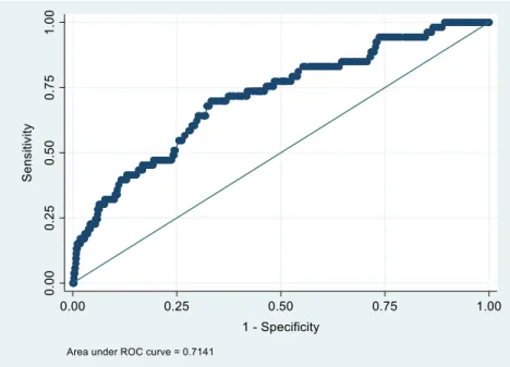
which is virtually identical to the severe trauma prevalence in the sample. The model had 60%
sensitivity and 71% specificity (area under the curve=0.71, Figure 4), resulting in a 7.6% positive
predictive value and 98% negative predictive value.
Discussion
Studies that examine community-dwelling samples and TDI such as this one are rare, as
most studies investigating the prevalence of TDIs have used clinical samples, comprising
care-seeking individuals who are more likely to have dental issues. Furthermore, few studies have
investigated TDIs in the primary dentition, with only one known study in the United States.21
This investigation is one of the largest community-based studies reporting findings on the topic
of TDIs in the primary dentition.
The overall prevalence of TDIs among the sample of 1,546 preschool-age children was
prevalence of TDIs in the primary dentition (Table 1). One potential explanation for the higher
percentage of TDIs reported in this sample is that all children in this study were from
low-income families and were Medicaid-eligible, as participation to EHS/HS is determined by
qualification based on social and economic criteria. Some reports have shown that more children
of lower socioeconomic status receive dental injuries compared to those in higher socioeconomic
groups.28,29
Because such a significant percentage of the injuries sustained were categorized as simple
crown fractures (75%), which have minimal clinical consequence, analyses were focused and
stratified according to severity of the trauma sustained. We considered that ‘severe trauma’
cases, including extensive fracture with pulp involvement, tooth displacement,
necrotic/discolored tooth, and total tooth loss due to trauma, would require immediate
management or intervention, thus were the ones most clinically relevant.
Several factors emerged as being associated with severe TDI; overjet and lip
incompetence showed strong correlations, whereas age, BMI, and canine occlusion showed
weaker positive associations. In the final multivariate logistic regression model, age, sex, lip
incompetence and overjet were retained. This is consistent with numerous reports supporting the
association between increased overjet and risk of TDIs to maxillary incisors in both the
permanent and primary dentitions.6,7,14,16,20,30 Overbite, canine classification, and lip
incompetence have also been linked to higher incidence of TDIs in the primary dentition.6,7,15
Our results did not show a strong link between sex and incidence of severe trauma,
consistent with other studies that suggest that there is no significant difference between sex and
higher percentage of dental trauma in males.7,8 BMI, although not included in our final
multivariate model, was weakly associated with increased incidence of TDI. Other reports
examining postulated links between BMI to TDIs are also inconsistent. Soriano et al. found a
statistically significant correlation between obesity and TDIs among a sample of 1,046 Brazilian
children.18 In contrast, Martins et al. found lower TDI prevalence (9%) among overweight/obese
schoolchildren (BMI ≥ 85th percentile) ages seven to 14 years, compared to 13% TDI prevalence
among schoolchildren who were not categorized as normal weight (15th percentile < BMI <
85th).19 Examining children’s activity levels stratified by BMI might provide a better, causal
explanation for the postulated association between BMI and TDIs.
This study’s findings should be framed by acknowledging its limitations. The ZOE 2.0
clinical examiners were only calibrated on caries diagnosis [weighted kappa ≥ 0.65 for
International Caries Detection and Assessment System (ICDAS) classification] before
conducting research examinations. They were not calibrated on dental trauma detection,
occlusion, overjet, and other intraoral and extraoral parameters. Another potential weakness is
the assumption that tooth loss in certain circumstances was due to trauma instead of caries or
incisal wear. It is not uncommon for children to have significant wear on the primary maxillary
incisors. More severe forms of incisal wear that extend into dentin or expose the pulp are not as
likely to be mistaken as trauma; however, there is less confidence in differentiating enamel-only
incisal wear and enamel-only trauma. Evaluating tooth loss symmetry, caries risk, distribution of
caries lesions, and number of teeth missing in the anterior region all aided in the clinical
examiners’ determination of the reason for tooth loss. Lastly, although the Frankl Scale is a
reliable rating system used frequently in pediatric dentistry to record the observed behavior and
risk-taking behavior and may not be a good indicator of which children are more accident-prone.
Including information on activity level, participation in recreational activities or organized
sports, and other behavioral markers, in a questionnaire could provide helpful information for a
more complete picture of a child’s risk for TDI.
In summary, including terms for behavioral factors, environmental factors, and oral
factors into a risk model should provide parents, caregivers, dentists, and other healthcare
professionals with a more contextual view of TDIs in order to reduce the prevalence of TDIs and
identify those children at heightened risk of TDIs to provide proper education on prevention
strategies. Additional studies, in larger community-based samples including collection of
additional possible predictors of dental trauma, are needed to further understand the interaction
of factors that contribute to TDI in the primary dentition. This added information may enhance
the education and communication opportunities between healthcare providers and caregivers and
improve prevention strategies. Development of a risk assessment index as well as examination of
the validity and generalization of a TDI risk index in external samples and populations is a
logical future application of the current study.
After examination of behavioral, environmental, and oral factors, oral factors and
particularly overjet, proved to be the most significant predictors of TDI in this sample of
preschool-age children. Orthodontic interventions to reduce overjet, although advocated by some
in the mixed dentition, would be focused more on interventions to eliminate non-nutritive
sucking habits if present. Incorporating and operationalizing this information may help traumatic
Conclusions
The following conclusions can be made based on this study’s findings:
1. The prevalence of TDI among this community-based sample of preschool-age
children was 47% and 8% of TDI cases were “severe”, defined as pulp exposure,
tooth displacement, discolored or necrotic tooth, or tooth loss.
2. Overjet and lip incompetence were strong risk factors for TDIs in the primary
dentition.
3. Accounting for age, sex and lip incompetence, we found that each added millimeter
of overjet was associated with 30% increased likelihood of severe dental trauma,
corresponding to an absolute 1% probability increase.
Table 1. Prevalence of traumatic dental injury in the primary dentition and classification for increased overjet; summary of past studies.
Author Year Country Sample Size Age Range (months) Prevalence TDI (%) Proportion enamel only (%) Proportion enamel and dentin (%) Increased overjet (mm) Odds ratio for increased overjet Prevalence ratio for increased overjet Andreasen & Ravn
1972 Denmark 487 36-95 30 Jones et al. 1993 USA 493 36-59 23 Oliveira et
al. 2007 Brazil 892 5-59 9.4 68.8 13.8 Feldens et
al.
2010 Brazil 888 36-71 36.4 > 2 1.86
(1.39-2.50)
1.50 (1.23-1.83) Goettems et
al. 2010 Brazil 501 24-71 40 ≥ 3
Wendt et al.
2010 Brazil 571 12-71 36.6 Bonini et
al.
2012 Brazil 376 36-59 27.7 58.4 17.6 > 3 1.74
(1.25-2.41) Norton &
O’Connell 2012 Ireland 839 9-84 25.6 39.4 3.5-6 (0.83-1.59) 1.15 > 6 2.99
(2.0-4.47) Piovesan et
al.
2012 Brazil 441 12-59 31.7 86.9 4.2 > 3 1.90
(1.34-2.70)
Table 2. Dental trauma (modified Ellis classification) among the study sample (N = 1,546).
Ellis classification n % trauma Any trauma Severe
No trauma 824 53.3 53.3% 96.2%
Simple crown fracture 543 35.1
46.7% Extensive crown fracture without pulp exposure 121 7.8
Extensive crown fracture with pulp exposure 5 0.3
3.8%
Tooth displacement 16 1.0
Necrotic/discolored tooth 34 2.2
Tooth loss 3 0.2
Table 3. Descriptive information of participating children and their association with severe traumatic dental injury.
Children’s characteristics
All participants Severe Traumatic Dental Injury P value
No Yes
N or mean
column % or SD
N or mean row % or SD N or mean row %
or SD X2 or t-test Entire sample 1,546 100.0 1,488 96.3 58 3.8
Sex 0.271
Male 770 49.8 737 95.7 33 4.3 Female 776 50.2 751 96.8 25 3.2
Age (years) 0.069
2 94 6.1 93 98.9 1 1.1
3 554 35.8 530 95.7 24 4.3 4 618 40.0 601 97.3 17 2.8 5 280 18.1 264 94.3 16 5.7
continuous (months) 49.5 9.4 49.5 9.4 50.7 10.1 0.319
Body Mass Index (BMI) 0.161
Underweight 144 9.6 142 98.6 2 1.4 Normal 986 66.0 943 95.6 43 4.4 Overweight 202 13.5 198 98.0 4 2.0 Obese 162 10.8 155 95.7 7 4.3
missing 52
Frankl score 0.657
1 55 3.6 52 94.6 3 5.5
2 118 7.6 114 96.6 4 3.4 3 268 17.3 261 97.4 7 2.6 4 1,105 71.5 1,061 96.0 44 4.0
Severe trauma = extensive fracture with pulp involvement, tooth displacement, necrotic/discolored tooth, total tooth loss due to trauma
Table 4. Intraoral and extraoral characteristics of participating children and their association with severe traumatic dental injury.
Children’s characteristics
All participants Severe Traumatic Dental Injury P value
No Yes
N or mean
column % or SD
N or mean row % or SD N or mean row % or SD
X2, t-test, or Fisher’s
exact* Entire sample 1,546 100.0 1,488 96.3 58 3.8
Overjet <0.005
4 mm or more 271 19.2 250 92.3 21 7.8 <4 mm 1,138 80.8 1,106 97.2 32 2.8
missing 137
continuous (mean, SD) 2.4 1.8 2.3 1.8 3.7 2.3 <0.005
Overbite (%) 0.542
Negative 63 4.5 61 96.8 2 3.2 0 - <25 371 26.6 358 96.5 13 3.5 25 - <50 258 18.5 245 95.0 13 5.0 50 - <75 440 31.6 428 97.3 12 2.7 75 - 100 262 18.8 250 95.4 12 4.6
missing 152
Profile 0.429*
Convex 1,417 92.9 1,362 96.1 55 3.9 Not convex 109 7.1 107 98.2 2 1.8
missing 20
Lip competence <0.005
Competent 1,480 97.1 1,429 96.6 51 3.5 Incompetent 45 3.0 39 86.7 6 13.3
missing 21
Canine occlusion 0.009
At least one canine Class II 261 18.1 243 93.1 18 6.9 Both canines Class I 1,064 73.7 1.030 96.8 34 3.2 At least one canine Class
III (no canines Class II) 118 8.2 116 98.3 2 1.7
missing 103
Severe trauma = extensive fracture with pulp involvement, tooth displacement, necrotic/discolored tooth, total tooth loss due to trauma
Table 5. Pairwise correlation (Pearson) coefficients between clinical variables of interest and trauma among the analytical sample of preschool-age children.
Demographic or clinical characteristic
Overjet Overbite Convex profile
Competent lip
Canine AP position Overjet
Overbite 0.24*
Convex profile 0.21* 0.13*
Competent lip -0.16* -0.08* -0.02
Canine AP position 0.44* 0.24 0.23* -0.18
Any trauma 0.06 0.02 0.01 0.06 -0.00
Extensive trauma 0.05 -0.05 0.05 -0.02 0.04
Severe trauma 0.14* 0.01 0.03 -0.09* 0.08
Table 6. Estimates of association [odds ratio and 95% confidence intervals (CI)] of demographic and clinical characteristics with the prevalence of severe dental trauma and corresponding predictive margins. Demographic or clinical characteristic
Association Predicted marginal effect OR 95% CI Probability
(%)
95% CI
Age (months) 1.02 0.99-1.06 0.1 0.0, 0.2
Sex: male (referent: female) 1.21 0.69-2.12 0.7 -1.3, 2.6 Lip: competent (referent: incompetent) 0.38 0.12-1.27 -0.3 -0.8, 1.0
Figure 1. Distribution of overjet values (mm) in the study sample (N=1,546).
Figure 2. Final multivariable logistic regression model-predicted probabilities and 95% confidence intervals of severe trauma, for males and females, according to overjet (mm).
REFERENCES
1. World Health Organization. Fact sheets. Available at:
http://www.who.int/mediacentre/factsheets/fs318/en/. Accessed 2017-12-27.
2. Shulman JD, Peterson J. The association between incisor trauma and occlusal characteristics in individuals 8-50 years of age. Dent Traumatol 2004;20:67-74.
3. Kaste LM, Gift HC, Bhat M, Swango PA. Prevalence of incisor trauma in persons 6 to 50 years of age: United States, 1988-1991. J Dent Res 1996;75:696-705.
4. Goslee MT, Lee JY, Rozier RG, Quinonez RB. The impact of dentoalveolar trauma on oral health-related quality of life in young children and their families. Masters of Public Health Thesis. University of North Carolina-Chapel Hill, Chapel Hill, NC, 2006.
5. Oliveira LB, Marcenes W, Ardenghi TM, Sheiham A, Bonecker M. Traumatic dental injuries and associated factors among Brazilian preschool children. Dental Traumatology
2007;23:76-81.
6. Goettems ML, Azevedo MS, Correa MB, da Costa CT, Wendt FP, Schuch HS, Bonow MLM, Romano AR, Torriani DD. Dental Trauma Occurrence and Occlusal Characteristics in Brazilian Preschool Children. Pediatric Dentistry 2012;34:104-7.
7. Bastone EB, Freer TJ, McNamara JR. Epidemiology of dental trauma: A review of the literature. Australian Dental Journal 2000;45:(1):2-9.
8. Borum MK, Andreasen JO. Therapeutic and economic implications of traumatic dental injuries in Denmark: an estimate based on 7549 patients treated at a major trauma centre. Int J Paediatr Dent 2001;11:249-258.
9. Lee JY, Divaris K. Hidden Consequences of Dental Trauma: The Social and Psychological Effects. Pediatric Dentistry 2009;31:96-101.
10. Fakhruddin KS, Lawrence HP, Kenny DJ, Locker D. Impact of treated and untreated dental injuries on the quality of life of Ontario schoolchildren. Dent Traumatol 2008;24:309-313.
11. Nguyen PM, Kenny DJ, Barrett EJ. Socioeconomic burden of permanent incisor replantation on children and parents. Dent Traumatol 2004;20:123-33.
12. Glendor U. Aetiology and risk factors related to traumatic dental injuries – a review of the literature. Dental Traumatol 2009;25:19-31.
14. Feldens CA, Kramer PF, Ferreira SH, Spiguel MH, Marquezan M. Exploring factors
associated with traumatic dental injuries in preschool children: a Poisson regression analysis. Dental Traumatology 2010;26:143-148.
15. Bonini GC, Bönecker M, Braga MM, Mendes FM. Combined effect of anterior malocclusion and inadequate lip coverage on dental trauma in primary teeth. Dental Traumatology
2012;28:437-440.
16. Piovesan C, Guedes RS, Casagrande L, Ardenghi TM. Socioeconomic and clinical factors associated with traumatic dental injuries in Brazilian preschool children. Braz Oral Res 2012;26(5):464-70.
17. Andreasen JO, Ravn JJ. Epidemiology of traumatic dental injuries to primary and permanent teeth in a Danish population sample. Int J oral Surg 1972;1:235-239.
18. Soriano EP, Caldas AF Jr, De Carvalho MV, Amorim Filho HA. Prevalence and risk factors related to traumatic dental injuries in Brazilian schoolchildren. Dental Traumatology
2007;23:232-240.
19. Martins VM, Sousa RV, Rocha ES, Leite RB, Gomes MC, Granville-Garcia AF. Assessment of the association between overweight/obesity and traumatic dental injury among Brazilian schoolchildren. Acta Odontol Latinoam 2014;27:26-32.
20. Norton E, O’Connell AC. Traumatic dental injuries and their association with malocclusion in the primary dentition of Irish children. Dental Traumatology 2012;28:81-86.
21. Jones ML, Mourino AP, Bowden TA. Evaluation of occlusion, trauma and dental anomalies in African-American children of metropolitan Headstart programs. J Clin Pediatr Dent 1993:18:51-4.
22. Born CD, Divaris K, Hom JM, Rozier RG. Clinical predictors of traumatic dental injuries in preschool children. J Dent Res 2015;94(Spec Iss A):0435 (IADR/AADR).
23. Ginnis J, Ferreira Zandoná AG, Slade GD, Cantrell J, Antonio-Obese ME, Pahel BT, Meyer BD, Divaris K. Measurement of early childhood oral health for research purposes: dental caries experience and developmental defects of the enamel in the primary dentition. Methods Mol Biol. In Press.
24. Ellis RG, Davey EW. Classification and Treatment of Injuries to the Teeth of Children, 5th ed. Chicago;Year Book Medical Publishers,1970.
25. Feliciano KMPC, de França Caldas Jr. A. A systematic review of the diagnostic classifications of traumatic dental injuries. Dental Traumatology 2006; 22:71-76.
27. American Academy of Pediatric Dentistry Clinical Affairs Committee – Behavior Management Subcommittee. Guideline on Behavior Guidance for the Pediatric Dental Patient. Reference Manual 2015;37(6):180-193.
28. Hamilton FA, Hill FJ, Holloway PJ. An investigation of dentoalveolar trauma and its treatment in an adolescent population. Part 1: The prevalence and incidence of injuries and the extent and adequacy of treatment received. Br Dent J 1997;182:91-95.
29. Lalloo R. Risk factors for major injuries to the face and teeth. Dent Traumatol 2003;19:12-14.
30. Bauss Influence of Overjet and Lip Coverage on the Prevalence and Severity of Incisor Trauma. Journal of Orofacial Orthopedics 2008;69:402-10.
31. Bijella MF, Yared FN, Bijella VT, Lopes ES. Occurrence of primary incisor traumatism in Brazilian children: a house-by-house survey. ASDC Journal of Dentistry for Children 1990;57(6):424-427.

![Table 6. Estimates of association [odds ratio and 95% confidence intervals (CI)] of demographic and clinical characteristics with the prevalence of severe dental trauma and corresponding predictive margins](https://thumb-us.123doks.com/thumbv2/123dok_us/8327915.2208535/44.918.113.817.153.288/estimates-association-confidence-demographic-characteristics-prevalence-corresponding-predictive.webp)