POLY(2-OXAZOLINE)S AS NEW DRUG DELIVERY SYSTEMS
Zhijian He
A dissertation submitted to the faculty at the University of North Carolina at Chapel Hill in partial fulfillment of the requirements for the degree of Doctor of Philosophy in the Division of Molecular Pharmaceutics in the UNC Eshelman School of Pharmacy.
Chapel Hill 2015
Approved By
Alexander V. Kabanov Elena V. Batrakova Leaf Huang
© 2015 Zhijian He ALL RIGHTS RESERVED
ABSTRACT
Zhijian He: Poly(2-oxazoline)s As New Drug Delivery Systems (Under the direction of Alexander V. Kabanov)
Taxane drugs, a class of important cancer chemotherapeutics, are often associated with excipient-related hypersensitivity, severe neurotoxicity, peripheral neuropathy, drug
resistance and other limitations that influence the patients’ quality of life and treatment prognosis. In order to overcome these limitations, this dissertation research focuses on the new poly(2-oxazoline)s (POx) polymers as versatile nano-scale drug delivery systems. The synthesis and characterization of several new monomers including benzyl-oxazoline, 2-methyl-2-oxazine, and a cationic monomer is summarized. In addition, the exploration of newly synthesized POx polymers using above monomers and new functionality groups (e.g. clickable initiator) are also briefly discussed.
The potential of a specific doubly amphiphilic POx triblock copolymer (previously termed P2) is further explored as a polymeric micelle system in delivering taxanes
(paclitaxel, the 3rd generation of toxoids, and multiple-drug combinations). The P2/paclitaxel
micelle formulation is characterized by a facile preparation, up to 50 % wt. drug loading, excellent shelf stability, controllable sub-100 nm size, a very high maximum tolerated dose in mice, and a significant enhancement in efficacy when treating three different breast cancer
models. In addition to paclitaxel, a number of 3rd generation toxoids able to avoid common
drug resistance mechanisms, are formulated in P2 micelles. An excellent solubilization of different 3rd generation taxoids is achieved irrespective of drug structures with up to 46 %
wt. drug loading and less than 100 nm micelle size. Furthermore, a selected formulation with the new taxoid SB-T-1214 shows about one to two orders of magnitude more active in vitro than paclitaxel in LCC6-MDR, a multidrug resistant breast cancer cell line. It significantly inhibits the growth of LCC6-MDR orthotropic tumors, outperforming Taxol and Cremophor formulated SB-T-1214. Moreover, a number of chemotherapeutic drugs are simultaneously formulated in P2 micelles. The multi-drug loaded P2 micelles indicate a ratio-dependent synergistic activity against multiple cancer cells in vitro.
This dissertation also involves the study of a new and first cationic oxazoline monomer and corresponding polymer containing side-chain secondary amine groups. The cationic polymer is able to condense plasmid DNA into a sub 100 nm polyplex which is stable upon dilution in saline and thermal challenge. These polyplexes exhibit minimum serum protein binding and very low cytotoxicity in vitro compared to the polyplexes of DNA and commonly used poly(ethylene glycol)105-b-poly(L-lysine)51 (PEG-PLL). The in vitro transfection efficiency
of the polypelxes is also studied in B16 murine melanoma cells as well as RAW264.7 macrophage cells. The cationic polymer represents a comparably safe and promising platform for delivering genes to macrophages and cancer cells.
In summary, POx polymers are versatile and promising platforms for taxane anticancer drugs delivery and gene delivery.
To my parents Fei He and Yulan Sun and my wife, Lin Zheng.
ACKNOWLEDGEMENT
First of all, I would like to express my utmost gratitude and appreciation to my supervisor, Dr. Alexander Kabanov who offered me this great educational opportunity in his lab as a graduate student. In the past few years, he provided me constant support, priceless guidance and extraordinary wisdom throughout my PhD journey. He is a remarkable and exceptional mentor I am so blessed to have. I will be forever thankful to his mentorship, inspiration and sense of humor. I would also like to thank my committee members, Dr. Elena Batrakova, Dr. Leaf Huang, Dr. William Zamboni and Dr. Lisa Carey for their precious guidance and advices throughout the years. They advised me through the obstacles in completion of my research work and reviewed all my progress and dissertation. In addition, I would like to extend my special appreciation to Dr. Rainer Jordan, Dr. Tantiana Bronich, Dr. Robert Luxenhofer, Dr. Yingchao Han, Dr. Iwao Ojima, Dr. Anita Schulz, Dr. Devika Manickam, Dr. Marina Solkosky, Dr. Daria Alakhova, Herdis Bludau and Xiaomeng Wan for their collaboration and as my external advisors.
Moreover, I want to express my gratitude to UNMC School of Pharmacy and UNC Eshelman School of Pharmacy for allowing me pursuing higher education. I also like to acknowledge my colleagues and friends, Dr. Jing Tong, Dr. Yi Zhao, Dr. Xiang Yi, Dr. Jing Gao, Matt Haney, Jubina Bregu, Nazar Filonov, Vivek Mahajan, Hemant Vishsaowa, Soo Kim,
Youngee Seo, Yuling Zhao, Shu Li, Yuhang Jiang, Dongfen Yuan, Dr. Yuanzeng Min, Dr. Zagit Gaymalov, Lei Miao, Dr. Lei Peng, Duhyeong Hwang, and all past and present members of the kabanov Laboratory for their sincere friendship and kindly help throughout my graduate study. Particularly, I will never forget the happiness, frustration, jokes, and parties we had in addition to the inspiring scientific conversations. I would like to acknowledge BRIC imaging core facility and Mouse Phase I Unit for their assistance especially Dr. Hong Yuan, Dr. Zibo Li, David Darr, and Charlene Santos.
I would particularly thank my parents, my wife Lin, my parents-in-law, brother-in-law and my sister for their unconditional support and love all along the journey. I cannot go this far without their understanding, patience, standing by my side, faith and love.
TABLE OF CONTENTS
LIST OF TABLES…...………..………...xi
LIST OF FIGURES……….…...xii
LIST OF ABBREVATIONS AND SYMBOLS………..…...xvi
Chapter I Nanomedicine of Taxane Anticancer Drugs………..…...1
1.1 Summary………..1
1.2 Introduction………2
1.3 Currently approved formulations of taxane……….………..4
1.4 New taxane analogues in clinical investigation………..……..7
1.5 Mechanism of action (MOA) of taxane drug………..…..11
1.6PK and dose regimen of currently approved taxane formulations………….13
1.7 Limitations of taxane drug………..18
1.8 Taxane drug delivery using nanocarriers (nanomedicine)……….……25
1.8.1 Lipsome formulation of taxanes in clinical trials...……..26
1.8.2 Conjugates and dendrimers for taxane delivery………29
1.8.3 Taxane delivery using polymeric micelle……….……33
1.8.4 Other taxane products in clinical trial……….………..39
Chapter II synthesis and characterization of new 2-oxazoline
monomer and Poly(2-oxazoline)s polymer……….…………..51
2.1 Summary………..51
2.2 Introduction………52
2.3 Initiator containing clickable functionality………55
2.4 Sythesis of new monomer 2-Benzyl-2-Oxazoline (BzOx) and BzOx containing polymer……….………58
2.5 Synthesis of new monomer 2-methyl-2-oxazine (MeOz) for gradient polymerization……….………..…..62
2.6 Synthesis of new cationic POx monomer and polymer CPOx……….64
Chapter III A Simple and Non-Smart Micellar Formulation of POx/Paclitaxel With Superior Safety and Efficacy In Vivo……….……….77
3.1 Summary………..77
3.2 Introduction………78
3.3 Materials and Methods……….….81
3.4 Results and Discussion………..………...……….86
Chapter IV Other Applications of P2 POx Polymers (1): Preparation, In Vitro and In Vivo Evaluation of POx Micelles with High Capacity for 3rd Generation Taxoids……….………..111
4.1 Summary………..…….111
4.2 Introduction……….112
4.3 Materials and Methods………115
4.4 Results……….……….………..………...121
Chapter V Other Applications of P2 POx Polymers (2): Synergistic Combinations of Multiple Chemotherapeutic Agents in High
Capacity POx Micelle………..………140
5.1 Summary………140
5.2 Introduction……….141
5.3 Materials and Methods………...144
5.4 Results……….149
5.5 Discussion……….154
Chapter VI: Poly(2-oxazolines) with Pending Cationic Side Chains as a Novel Non-Viral Platform for Gene Delivery With Low Protein Binding……….………173
6.1 Summary………173
6.2 Introduction……….174
6.3 Materials and Methods………..177
6.4 Results……….186
6.5 Discussion……….194
Chapter VII: Summary and Future Experiments……….213
References………217
LIST OF TABLES
Table 1.1 Current approved taxane formulations………...42
Table 1.2 New taxane analogues in clinical investigation…...43
Table 1.3 Liposomal taxanes in clinical investigation……..……….44
Table 1.4 Conjugates and dendrimers for taxanes in clinical investigation………..45
Table 1.5 Polymeric micelles of taxanes in clinical investigation………....46
Table 3.1 Drug concentration, loading efficiency (LE), loading capacity (LC), drug loaded micelle size and PDI of POx/PTX formulations………...92
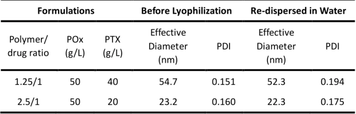
Table 3.2 Size and PDI of two POx/PTX formulations before and after Lyophilization……….93
Table 3.3 Monitoring of animal death and weight in the MTD experiments...94
Table 3.4 Summary of cytotoxicity to liver Hep G2 and kidney LLC-PK1 cells...95
Table 3.5 Clinical chemistry parameters of Balb/c mice receiving POx and POx/PTX……….96
Table 4.1 Data of solubilization experiments………...132
Table 4.2 IC50 values of POx/SB-T-1214 micelles vs. other PTX formulations………….133
Table 5.1 Solution concentration, LE and LC of drugs in POx aqueous dispersions………161
Table 5.2 Comparison of our results with others for drug formulation in LC and DL values………..162
LIST OF FIGURES
Figure 1.1 MOA of taxane drugs (exemplified with PTX)….…...47
Figure 1.2 Mechanism of drug resistance to taxanes….………...48
Figure 1.3 Nanomedicine for taxane drug delivery………..………..49
Figure 1.4 3rd generation of taxanes and POx micelle delivery systems….………....50
Figure 2.1 Schematic demonstration of P2 synthesis………68
Figure 2.2 Azide-Alkyne Huisgen Cycloadition………...69
Figure 2.3 Synthesis of Propargyl-P4 with clickable alkyne moiety………..70
Figure 2.4 Synthesis of BzOx monomer and polymer HEZ007 and HEZ012………71
Figure 2.5 Impact of PTX concentration on the solubilization of PTX in HEZ012 (10 g/L)………72
Figure 2.6 Monomer synthesis of MeOz……….73
Figure 2.7 Synthesis of the new monomer, Boc-MeAmMeOx……….74
Figure 2.8 1H-NMR spectra (d 3-MeOD, 300 K) of the novel monomer 2-(N-methyl,N-Boc-amino)-methyl-2-oxazoline with the assignment of all observed signals………..……..75
Figure 2.9 Block copolymerization of MeOx and Boc-MeAmMeOx using propargyl tosylate as the initiator and 5% K2CO3 as the terminating agent……….……..…..….76
Figure 3.1 Preparation and physicochemical properties………....97
Figure 3.2 SOP for small scale (1-5 mg) micelle production………..98
Figure 3.3 SOP for large scale (200 mg) micelle production………..99
Figure 3.5 Size of POx/PTX 50/40 micellar nanoformulation in water, PBS
or 10mM nacl………..101
Figure 3.6 Zeta-potential of POx/PTX 50/40 micelles in water or 10 mM nacl by zeta sizer………102
Figure 3.7 Characterization of POx polymers………103
Figure 3.8 MTD in nude mice and toxicology profiles……….104
Figure 3.9 In vitro toxicity evaluation……….105
Figure 3.10 Cytotoxicity to LLC-PK1 normal pig kidney epithelial cells………106
Figure 3.11 Cytotoxicity to Hep G2 human liver hepatocellular carcinoma cells……….107
Figure 3.12 Efficacy of POx/PTX 50/40 in A2780 tumors………..108
Figure 3.13 Efficacy of POx/PTX 50/40 in A2780 tumors (treatment starting at later stage, tumor volume of ca. 400 mm3)……….……109
Figure 3.14 Efficacy of POx/PTX 50/40 in LCC6-MDR and T11 tumors...110
Figure 4.1 Preparation of POx/taxoid micellar nanoformulations……….134
Figure 4.2 Physicochemical properties of various POx/SB-T-1214 polymeric micelle formulations……….135
Figure 4.3 In vitro cytotoxicity of various PTX and SB-T-1214 formulation in LCC6-MDR cells, LCC6-WT, and T11 cells………...136
Figure 4.4 Establishment of the safe dose of SB-T-1214 in nude mice………137
Figure 4.5 Efficacy of various drug formulations in LCC6-MDR tumors………..138
xiv Figure 4.6 Efficacy of various drug formulations in T11 OST tumors………139
Figure 5.1 Chemical structure of amphiphilic POx copolymer, P(MeOx40-b-BuOx21-b-MeOx34), and schematic representation of multiple drug loaded micelles……….…….163
Figure 5.2 Chemical structures of anti-cancer drugs………164 Figure 5.3 Size distribution and stability studies of POx micelles loaded
with single drugs……….……….165 Figure 5.4 Size distribution and stability studies of POx micelles
co-loaded with two drugs………….……….166 Figure 5.5 Size distribution and stability studies of POx micelles
co-loaded with three drugs……….……….167 Figure 5.6 CLSM images of MCF-7 cells incubated for 1 h with free
PTX, POx copolymer alone and POx micelles co-loaded with PTX and
17-AAG……….…..168 Figure 5.7 CLSM images of MCF-7 cells incubated for 4 h with free PTX,
POx copolymer alone and POx micelles co-loaded with PTX and 17-AAG………..169 Figure 5.8 Cytotoxicity of single drugs and binary drug combination,
ETO/17-AAG solubilized in POx micelles, in MCF-7 cells and the
corresponding CI vs. Fa plot………....170
Figure 5.9 Cytotoxicity of single drugs and binary drug combinations, BTZ/17-AAG in POx micelles, in MCF-7 cells and the corresponding
log CI vs. Fa plot………...171
Figure 5.10 IC50 values of micellar 17-AAG, BTZ and BTZ/17-AAG
combination in PC3, MDA-MB-231, and HepG2 cancer cells and
the CI vs. Fa plot……….…………172
Figure 6.1 Synthesis of the new monomer, Boc-MeAmMeOx………..198 Figure 6.2 1H-NMR spectra (d
3-MeOD, 300 K) of the novel monomer
2-(N-methyl,N-Boc-amino)-methyl-2-oxazoline with the assignment
of all observed signals………199 Figure 6.3 Block copolymerization of MeOx and Boc-MeAmMeOx
using propargyl tosylate as the initiator and 5% K2CO3 as the terminating
Figure 6.4 1H-NMR spectra (400 MHz, d
3-MeOD) ofBoc-CPOx and
the deprotected CPOx after single and repeated deprotection procedures, along with the respective structures and the peak
assignments……….…201 Figure 6.5 Titration curves for CPOx copolymer and PEG-PLL in 0.1M
aqueous NaCl using 0.1M HCl……….……….………..202 Figure 6.6 pDNA condensation by CPOx assay by ethidium bromide exclusion
and gel retardation assay of pDNA and polyplexes at various N/P ratios…....…………203 Figure 6.7 DLS measurements of the size distributions,particle size and PDI,
And ζ-potential of CPOx/pDNA polyplexes as a function of N/P ratios………..204 Figure 6.8 Representative TEM image……….…..205 Figure 6.9 Size and PDI of CPOx/pDNA polyplexes at various N/P ratios as a
function of storage time………..206 Figure 6.10 Stability of CPOx/pDNA polyplexes upon dilution and heating………207 Figure 6.11 SDS-PAGE and micro BCA assay of adsorbed plasma proteins on
PEG-PLL/pDNA, PLL/pDNA, and CPOx/pDNA polyplexes……….208 Figure 6.12 In vitro cytotoxicity of the polymers and the polyplexes in
RAW264.7 murine macrophages and B16 murine melanoma cells……….…………209 Figure 6.13 Cellular uptake of CPOx/pDNA polyplex with N/P 5 in
RAW264.7 macrophage cells……….……….210 Figure 6.14 Cellular uptake of CPOx/pDNA polyplex with N/P 10 in
RAW264.7 macrophage cells……….……….211 Figure 6.15 In vitro transfection of B16 murine melanoma and
RAW264.7 murine macrophage cells……….………..212
LIST OF ABBREVIATIONS AND SYMBOLS
α-TAS alpha-tocopheryl acid succinate ABC accelerated blood clearance ACN acetonitrile
AFM atomic force microscope
AIDS acquired immune-deficiency syndrome ALD adrenoleukodystrophy
ALP alkaline phosphatase ALT alanine aminotransferase
APTT activated partial thromboplastin time ASCO American Society of Clinical Oncology AUC area under the curve
BBB blood brain barrier
BC breast cancer
BCA bicinchoninic acid BCL-2 B-cell lymphoma 2 BMS Bristol-Myer Squibb Boc tert butyloxycarbonyl BSA bovine serum albumin
BTZ bortezomib
BUN blood urea nitrogen CHK1 checkpoint kinase 1
CI combination index
CLSM confocal laser scanning microscopy Cmax maximum concentration
CMC critical micelle concentration CPOx cationic poly(2-oxazoline)s CrEL Cremophor EL xvi
CTX cabazitaxel
DC drug concentration DHA docosahexaenoic acid DLS dynamic light scattering
DMEM Dulbecco’s Modified Eagle Medium DMF dimethylformamide
DMSO dimethyl sulfoxide
DMPC 1,2-dimyristoyl-sn-glycero-3-phosphocholine DNA deoxyribonucleic acid
DOPA 1,2-dioleoyl-sn-glycero-3-phosphoethanolamine DOPC 1,2-dioleoyl-sn-glycero-3-phosphocholine
DOTAP N-[1-(2,3-Dioleoyloxy)propyl]-N,N,N-trimethylammonium chloride DSPC 1,2-distearoyl-sn-glycero-3-phosphocholine
DTX docetaxel
ECOP Eastern Cooperative Oncology Group ECM extracellular matrix
EGFR epidermal growth factor receptor EPR enhanced permeability and retention EtBr ethidium bromide
ETO etoposide
FLT3 Fms-like tyrosine kinase 3 GC gastric carcinoma
GEM genetically engineered mouse GOG Gynecologic Oncology Group GPC gel permeation chromatography GTP guanosine-5’-triphosphate
h hours
HEPES 4-(2-hydroxyethyl)-1-piperazineethanesulfonic acid H&E hematoxylin & eosin
HPLC high-performance liquid chromatography HRPC hormone refractory prostate cancer HPMA N-(2-hydroxypropyl)methacrylamide IC50 half maximal inhibitory concentration
IIT investigator initiated trial
IP intraperitoneal
IV intravenous
LC loading capacity
LCROP living cationic ring opening polymerization LDH lactate dehydrogenase
LE loading efficiency
LRP1 low-density lipoprotein receptor-related protein 1 MBC metastatic breast cancer
mCRPC metastatic castration-resistant prostate cancer MDR multiple drug resistance
MeOH methanol
mg milligram
min minute
mL milliliter
MOA mechanism of action
mPEG-PLA methoxy poly(ethylene glycol)-b-poly(lactide)
MS mass spectrum
MTD maximum tolerated dose
MTT 3-[4,5-dimethylthiazol-2-yl]-2,5-diphenyl tetrazolium bromide MW molecular weight
ng nanogram
NMR nuclear magnetic resonance NSCLC non-small cell lung cancer
OC ovarian cancer
ORR overall response rate
OST orthotopic syngeneic transplant PBS phosphate buffered saline pBuOx poly(2-butyl-2-oxazoline) pBzOx poly(2-benzyl-2-oxazoline) PD progressive disease PDI polydispersity index PEG poly(ethylene glycol)
PEG-PLA methoxy poly(ethylene glycol)-block-poly(D,L-lactic acid)
PEI polyethylenimine
pEtOx poly(2-ethyl-2-oxazoline) PFS progression free survival P-gp P-glycoprotein
PK pharmacokinetics
PLA poly(D,L-lactic acid)
PLGA poly(lactic-co-glycolic acid)
PLL poly(L-Lysine)
pMeOx poly(2-methyl-2-oxazoline) pNoOx poly(2-nonyl-2-oxazoline) POx poly(2-oxazoline)s
PR partial response
PSMA prostate specific membrane antigen
PT prothrombin time
PTX paclitaxel
q1w every week
q3w every three-weeks
Ref. reference
RES reticuloendothelial system
RT room temperature
SANS small angle neutron scattering
SC subcutaneous
SCCHNC squamous cell carcinoma of head and neck cancer SCID severe combined immune-deficiency
SD stable disease
SEC size exclusion chromatography SEM standard error of the mean siRNA small interfering RNA
SPARC secreted protein acidic and rich in cysteine t1/2 half-life
TAM tumor associated macrophage TEM transmission electron microscopy TFA trifluoroacetic acid
TGA thermogravimetric analysis TGFβ transforming growth factor beta THF tetrahydrofuran
TIC tumor initiating cells
TNBC triple negative breast cancer
μg microgram
μL microliter
USP United States Pharmacopeia
VCR vincristine
Vdss steady state volume of distribution VEGF vascular endothelial growth factor
VRP verapamil
vs. versus
VTE venous thromboembolism
17-AAG 17-allylamino-17-demethoxygeldanamycin
1
CHAPTER I: NANOMEDICINE OF TAXANE ANTICANCER DRUGS 1
1.1 Summary
Taxane anticancer drugs such as paclitaxel, docetaxel, and cabazitaxel, are one class of the most important chemotherapeutics in treating broad range of cancers such as ovarian, breast, non-small cell lung, head and neck, and prostate cancers. However, taxanes are associated with excipient-related hypersensitivity, severe neurotoxicity and peripheral neuropathy, drug resistance and other limitations that influence the patients’ quality of life and treatment prognosis. In order to overcome these limitations, a tremendous research effort is focused on new delivery strategies, especially nanomedicine of taxanes, and have already resulted in clinical trials or approvals. This review emphasizes current approved and clinical investigational taxanes, the updates of taxane mechanism of actions, the limitations of taxanes in clinical use, in particularly, sensory neuropathy and drug resistance; and provides an extensive overview of various clinical stage nanomedicine of taxanes such as liposomes, taxane-conjugates, dendrimers, polymeric micelles (e.g. poly(2-oxazoline)s micelles), and others for delivery of taxane drugs.
2 1.2Introduction
Taxanes are diterpenes originally isolated from the plants of Taxus brevifolia (The bark of Pacific Yews) [1]. It has been more than 50 years since U.S. National Cancer Institute (NCI) discovered first taxane - paclitaxel (PTX) [1] and later Dr. Susan Horwitz and colleagues found it can promote tubulin polymerization thus stabilize the microtubules [2]. The resulting deficiency of microtubule dynamics can arrest cells at G2-M phase during mitosis and ultimately cause apoptosis of rapid proliferating cells [3-5]. Current taxane agents are either natural-origins or semi-synthesized compounds including PTX [1], docetaxel (DTX) [6, 7], cabazitaxel (CTX) [8, 9], and other taxane derivatives [10]. They are widely used in medical oncology and made major impact in treating breast, ovarian, lung, head and neck, and other malignancies [11-14].
Taxanes are generally white crystalline powder with a melting point at approximately 200
oC (PTX, 215-217 oC; DTX, 187-192 oC; CTX, 170-180 oC) [15, 16] and their clinical applications
3
taxanes including DTX and CTX are still considered insoluble [Defined in US Pharmacopeia (USP)] and surfactants (other excipients) are necessary for the clinical formulations [24, 25].
4 1.3Currently approved formulations of taxanes
PTX formulations
A summary of currently approved formulations of taxanes is presented in Table 1.1. The first three formulations are based on classical taxane member, namely PTX. PTX has a high molecular weight (MW) of 854 Da with formula of C47H51NO14, a very low aqueous solubility
(approximately 1 mg/L), and no ionizable groups to form salts to increase solubility through pH adjustment. Although the chemical structure of PTX was identified in 1960’s, it was until 1994 that two groups independently reported total synthesis procedures starting from a baccatin core structure containing the ABCD ring (Table 1.1 PTX structure), followed by addition of the "tail" part to the C13-hydroxyl group [52-54]. Anticancer activity of PTX is mainly due to the side chain, A ring, D oxetane ring and C2 benzoyl group. The C3’ amide acyl group in the C13 chain maintains the activity and the hydroxyl group at C2’ enhances its function [48, 55].
Taxol®, the first injectable dosage form of PTX, was approved by FDA in 1992 exclusively to American company Bristol-Myers Squibb (BMS). It is formulated in 50% CrEL and 50% dehydrated ethanol. The only clinically approved alternative to Taxol® in United States is Abraxane®. It consists of lyophilized cakes of PTX-albumin nanoparticles in each 50 mL vial, containing 100 mg of paclitaxel and nearly 900 mg of human albumin. These novel nanoparticles are 130 nm in size prepared by high-pressure homogenization of PTX with human albumin to form colloidal nanosuspension [56-58].
methoxy-5
poly(ethylene glycol)-b-poly(lactide) (mPEG--PLA) based polymeric micelle formulation of PTX, showing 3 fold increase in maximum tolerated dose (MTD), high levels of distribution of drug in various tissues including tumors, and significantly improved antitumor efficiency in tumor mice models compared to Taxol® [41, 43]. The high MTD of Genexol-PM® without additional toxicity in Phase I clinical trial may showcase a good alternative to Taxol® and Phase II trial is undergoing in United States for treating advanced breast cancer (BC) and non-small cell lung cancer (NSCLC) [40, 41]. Another approved PTX formulation is the liposome of PTX, Lipusu® injectables from Luye Pharma Group and was approved for Chinese market by Chinese FDA [28]. The implementation of liposomes may solve the solubility problems of PTX while avoiding excipient-related side-effects. Lipusu® formulations demonstrated higher tolerated dose, enhanced efficacy and reduced side-effects to the blood pressure, the medulla, peripheral blood and the liver [28, 59].
DTX formulation
6
possibly due to higher binding affinity to microtubules [63]. Another explanation for enhanced efficacy is speculated from in vitro studies in P388 leukemia cells that DTX has 3 times higher intracellular concentration and lower efflux rate than PTX. Current only clinical formulation of DTX is Taxotere® developed by Sanofi. Indications for Taxotere® include metastatic and recurrent breast cancer (MBC), gastric adenocarcinoma (GC), advanced and metastatic NSCLC, squamous cell carcinoma of head and neck cancer (SCCHNC), and hormone refractory prostate cancer (HRPC). One vial of Taxotere® contains single use of 80 mg DTX in polysorbate 80/dehydrated ethanol at 20 mg/mL concentration [6, 61].
CTX formulation
Another example of second-generation taxanes is CTX (previously as XRP-6258), a 7, 10-dimethyloxy derivative of DTX with MW of 854 Da and formula of C45H57NO14. It can also be
7 1.4New taxane analogues in clinical investigation
There are many taxane drugs under clinical investigation. Here we mainly focus on new taxane analogues, which are chemically modified on traditional PTX or DTX. Most of new taxane analogues undergoing clinical trials are listed in Table 1.2. For example, larotaxel (XRP9881) is a promising agent for multidrug resistance (MDR) BC treatment. It is a new semi-synthesized taxoid originated from 10-deaceyl baccatin III and also capable to cross blood-brain barrier (BBB) possibly due to minimal recognition by P-glycoprotein (Pgp) efflux pumps. Phase I and II trials identified dose regimen of larotaxel in solid tumors as 90 mg/m2,
intraveneous (IV) infusion for 1 h every three-weeks (q3w). Using this regimen larotaxel exhibited good efficacy against taxane-pretreated MBC patients while dose-limiting toxicities include neutropenia and neutropenic complications [65, 66].
Milataxel (TL139) is a novel analog to DTX that overcomes Pgp involved resistance. One advantage is that milataxel does not require polysorbate 80 or CrEL to formulate which avoids excipients-related hypersensitivity reactions [67]. A phase II study reported in 2008 for colorectal cancer treatment (35 mg/m2 IV q3w) demonstrated no clinical significance while six
patients suffered neutropenic sepsis and two patients were deceased [68]. Another Phase II study in treating platinum-refractory NSCLC patients showed tolerability of the milataxel at a dose of 35 mg/m2, nevertheless peripheral neuropathy was the most frequent
non-haematological toxicity [69].
8
mg/m2 every week (q1w) for 3 weeks with 1-week rest. Drug induced toxicities included
nausea, vomiting, diarrhoea, fatigue, anorexia, rash, and anaemia while dose limiting toxicity is still grade 3 peripheral neuropathy [72]. Currently, TPI-287 are studied in several Phase II clinical trials (total 17 hits in clinicaltrials.gov website using TPI-287 as a keyword to search) in treating mostly brain malignancies such as glioblastoma, recurrent neuroblastoma and medulloblastoma, and brain metastasis originated from BC, as well as melanoma. 3 trials were completed while 4 were terminated or withdrawn. Up to date there is no more report from Pubmed search. In contrast, clinical data for TL310 is very limited both in Pubmed and clinicaltrials.gov. In a Phase I study, patients with advanced refractory solid tumors were administered orally up to 160 mg/m2 TL310 on days 1, 8 and 15 of a 28-day cycle. One out of
five patients experienced grade 5 neutropenic sepsis, grade 4 neutropenia and grade 4 thrombocytopenia. A partial response (PR) was observed in one GC patient dosed at 80 mg/m2
of TL310 and disease were stable for more than two treatment cycles in six patients (3 cases of oesophageal, 1 melanoma, 1 ovary, 1 cervix cancer) within total 18 patients [71]. Ortataxel (BAY-59-8862; IDN-5109) is a semisynthetic analogue to DTX synthesized originally by Dr. Iwao Ojima group, and was further developed by Indena in collaboration with Bayer. It showed efficacy in preclinical studies in multi-drug resistant tumors and a superior and durable effects on reduction of the tumor microvessel density against HRPC compared to PTX. Phase I and II studies were performed in 1 h IV infusion q3w of 75 mg/m2. A 1 h IV infusion of 45 mg/m2
9
NSCLC. The oral bioavailability were reported as 19 - 31% although no clinical trials were performed on oral administration of the drug to date [74]. Dr. Ojima’s research group also developed a library of third generation taxanes aiming lower toxicity and higher activity against resistance cancer cells than PTX. In section 1.8.3, we will discuss these new taxanes in combination with polymeric micelle delivery systems.
Another oral formulation of new taxane is Tesetaxel (DJ-927). A Phase I study was performed in patients with advanced malignancies using a single oral dose ranging from 1.5 to 40 mg/m2 with a q3w regimen and MTD was identified as 27 mg/m2. However, Tesetaxel
showed modest to low (oral dose 27 mg/m2 q3w) activity in patients with advanced GC and
other cancers, while similar toxicities to current therapies such as neutropenia, sepsis, diarrhoea and peripheral neuropathy were observed [75]. Therefore, Daiichi Pharmaceutical (Daiichi Sankyo) has discontinued development of Tesetaxel (Scrip Daily Online, 7 Nov 2006, S00939534).
Last but not least, BMS developed several taxane analogues investigated clinically including BMS-275183, BMS-184476, and BMS-188797. BMS-275183 is an oral formulation of C4 methyl carbonate analogue of PTX with additional modifications to the side chain and was shown active against PTX resistant tumors in vitro and in vivo. BMS-275183 has oral bioavailability of 24% in human, considerably higher than PTX, and Phase I studies determined oral dose up to 200 mg/m2 q1w was tolerable but twice a week regimen of 100 mg/m2
appeared less toxic in terms of neuropathy, fatigue, diarrhoea and neutropenia. It was shown to be partial responsive for q1w 200 mg/m2 oral administration in 9 patients of 38 total (23.7%)
10
neuroectodermal tumor (1 of 1 patient), cholangio carcinoma (1 of 2 patients) and undifferentiated sarcoma (1 of 1 patient) [76].
BMS-184476 is also a new taxane analogue with Chydroxyl group replaced by a 7-methylthiomethyl ether group compared to PTX. The substitution increases the solubility and potency over PTX and DTX in vitro and in vivo [77]. A Phase I study of BMS-184476 indicated that a 1 h IV infusion at 60 mg/m2 showed no grade 3 or 4 peripheral neuropathy, which is a
recommended dose for Phase II trial [78]. Another Phase I trial also tested BMS-184476 q1w for 3 weeks followed by one week break as a treatment cycle and found neutropenia was the dose-limiting toxicity and later recommended 50 mg/m2 on days 1 and 8 every 21 days. Two
patients died of neutropenia-related complications [79, 80]. A Phase II study for BMS-184476 as second line treatment of NSCLC suggested that the 60 mg/m2 was well tolerable and can
achieve antitumor activity in previously treated patients [81]. No recent report on further Phase II or III trials on this drug since 2005.
The third drug candidate is BMS-188797, a synthetic analogue to PTX with a C4 carbon formed 4-desacetyl-4-methylcarbonate. It has very low solubility (using the same excipient as Taxol®) and approximately twice potent over PTX [78, 82]. The recommended Phase II dose was 50 mg/m2 q1w or 175 mg/m2 q3w IV. The dose-limiting toxicity was febrile neutropenia.
A combination trial of BMS-188797 with cisplatin determined a recommended dose of 110 mg/m2 (1 h infusion, q3w) followed by cisplatin 75 mg/m2 [82-85]. All above three analogues
11 1.5MOA of taxane drugs
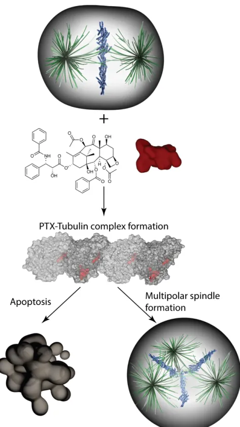
Taxol® was originally as second line treatment for ovarian cancer (OC) in 1980's but soon be applied as first line treatment due to great antitumor responses in nearly 26% of OC patients that intrinsically resistance to platinum class agents [86]. The general antitumor mechanism for taxane drugs is to bind tubulin and interfere tubulin polymerization steps, thus in turn stabilize microtubules and cause cell cycle arrest at G2-M phase and ultimately induce apoptosis [87]. Microtubules are assembled from a 100kDa protein tubulin which is the heterodimer of α-tubulin and β-tubulin proteins (50kDa each). The dimer formation requires binding to a guanosine-5'-triphosphate (GTP) molecule at the nucleotide exchangeable site (E site of β-tubulin) and the non-exchangeable site (N-site of α-tubulin). The binding of taxanes occurs at the E site of β-tubulin heterodimers where GTP-binding sites locate, and therefore disrupt assemble and disassemble dynamics of microtubules [88, 89]. The microtubule dynamics is crucial for chromosome segregation during mitosis. A recent study found that levels of PTX in primary breast tumors are well below the concentration required for retaining cells in mitotic arrest. The authors further identified that cells progressed through mitosis under lower concentrations of PTX can separate their chromosomes on multipolar spindles which led to chromosome missegregation followed by cell death [90] (Fig. 1.1).
12
cycle [63, 91]. Further in vivo evidence demonstrated that DTX has a longer retention time in tumor cells than PTX due to greater uptake, slower efflux and longer terminal elimination half-life in tumor tissue [92, 93]. The major drawbacks of taxane drugs including PTX and DTX are low solubility and drug resistance. For solubility issues, by structural modification, DTX has already shown improved water solubility as several times higher (5.0-6.0 mg/L) than PTX, although it is still considered insoluble [94]. For HRPC treatment, patients often develop resistance to DTX, resulting in therapy failure [95].
13
1.6PK and dose regimen of currently approved taxane formulations
Taxol®
After IV injection of Taxol®, the elimination of PTX follows a rapid and biphasic feature and average distribution and elimination half-life is 0.34 and 5.8 h respectively. Facilitated by CYP (CYP2C8 and 3A4) metabolism and biliary excretion, PTX is rapidly eliminated from circulation. The steady-state volume of distribution (Vdss) is large for PTX due to its high plasma protein binding. Taxol® has a nonlinear PK in which reported cause is the formulation vehicle CrEL. When administering at high dose, the large amount of CrEL in the formulation may alter the hepatic transport function and biliary excretion rates of PTX. Another explanation is that PTX is incorporated in CrEL micelles and lead to altered erythrocyte accumulation [22, 23, 87].
In clinic, Taxol® is administered IV over a period of 3-24 h after dilution to a concentration of 1 mg/mL. The most commonly prescribed dosage regimen is 135 mg/m2 or 175 mg/m2 q3w.
14
to treat pancreatic cancer is 125 mg/m² IV in combination with gemcitabine [98].
Abraxane®
The PK of PTX plasma concentrations after IV administration of Abraxane® showed a biphasic profile that the initial rapid decline indicates drug distribution from the circulation to the peripheral compartment followed by a slower drug elimination phase. The PK data of 260 mg/m2 of Abraxane® over 30 min infusion was compared to the PK of 175 mg/m2 Taxol® IV
over 3 h infusion. Clearance and Vd was 43% and 53% larger for Abraxane® than PTX injection respectively while elimination half-lives were almost the same [56-58]. For MBC, 260 mg/m2
IV over 30 min q3w while for NSCLC recommended dose regimen of Abraxane® is 100 mg/m2
IV over 30 min on days 1, 8, and 15 along with the administration of carboplatin (immediately after Abraxane®) on day 1 of each 21-day cycle. Furthermore, in treating pancreatic adenocarcinoma, dose regimen follows 125 mg/m2 IV over 30-40 min on days 1, 8 and 15 of
each 28-day cycle; as combination therapy regimen, gemcitabine needs to be administered on days 1, 8 and 15 of each 28-day cycle immediately after Abraxane® [99].
Genexol-PM®
PK parameters of Genexol-PM® was reported upon 13 patients receiving 135-390 mg/m2
15
toxicities; however, the AUCinf and Cmax parameters between two groups of patients are not statistically significant different. The phase I trial found major dose limiting toxicity were neuromuscular toxicity and myelosuppression, and identified MTD of Genexol-PM® as 390 mg/m2, relatively higher than Abraxane® at 300 mg/m2. The following phase II trial used a
dose regimen of 230 mg/m2 IV infusion for 3 h along with cisplatin 60 mg/m2 on day 1 through
a 3-week cycle for treating advanced NSCLC patients. If no pre-specified toxicity observed after first treatment cycle, then doses for following cycles were escalated to 300 mg/m2. A
recent report on another phase II trial on NSCLC appears to use Genexol-PM® at 230 mg/m2
on day 1 combined with gemcitabine 1,000 mg/m2 on day 1 and day 8 in a 3-week cycle
regimen [40, 100-102].
Lipusu®
Following IV administration, PK profile of Lipusu® in both rats and dogs indicated a biphasic model that a rapid distribution phase in the first hour was observed followed by a slow elimination phase. The rapid distribution phase is thought due to clearance of liposomal nanoparticles by reticuloendothelial system (RES) out of circulation. Biodistribution results also confirmed that Lipusu® was mainly located at RES organs including liver and spleen following IV bolus injection. For example, spleen has highest drug concentration followed by liver and PTX concentration decreased slowly in liver. In addition, very limited amount of PTX penetrated BBB to the brain and drug concentration fell below the detection limit at 4 h post injection.
The dose regimen for Lipusu® is 175 mg/m2 (q3d or q2w) IV to patients with advanced
16
premedication is recommended including IV administration of methylprednisolone 40 mg at least 30 min prior to Lipusu®, or oral administration of dexamethasone 2.25-3 mg 2 h before Lipusu® [28, 59].
Taxotere®
Usually DTX is administered via IV and PK of DTX follows a three-compartment model in which a rapid initial decline phase (average half-life: 4.5 min) representing the distribution of DTX from the central to peripheral compartments; second phase (half-life of 38.3 minutes) and a relatively slow decline phase (average half-life: 12.2 h) representing efflux of DTX from the peripheral compartments. Excretion of DTX and its metabolites are mainly through renal and biliary following oxidation by metabolism enzyme CYP3A4. Approximately 94% of DTX bound to α1-acid glycoprotein, albumin or lipoproteins in vitro; however, polysorbate 80 as excipients in clinical formulation compete with serum proteins and thus changing PK of DTX by modifying protein binding profile. Further studies identified that unbound DTX is associated with severe hematological toxicity. For oral administration, bioavailability of DTX is very poor mainly because of the existence of Pgp proteins in the apical epithelial of the gastrointestinal tract as an efflux pump. Also due to high levels of Pgp in the BBB, the penetration of DTX into the brain tissue is very limited [91-93, 103-107].
The Taxotere® injectable formulation is a 20 mg/mL DTX solution using polysorbate 80/dehydrated ethanol (50/50, v/v). It requires dilution to final concentration of 0.3-0.74 mg/mL in either saline or 5% dextrose solution. Premedication with oral corticosteroids is also needed for all patients. Taxotere® is usually administered at a dose of 60 to 100 mg/m2 q3w
17
locally advanced or MBC or 75 mg/m2 administered 1 h after doxorubicin 50 mg/m2 and
cyclophosphamide 500 mg/m2 q3w for 6 cycles as an adjuvant therapy for BC. For the rest
indications DTX is normally administered at 75 mg/m2. For instance, treating
chemotherapy-naive NSCLC, it will be followed by cisplatin 75 mg/m2, while in case of platinum therapy failure
DTX is used as a single agent. In HRPC, medication of 5 mg prednisone twice a day continuously is also required. A dose regimen of 75 mg/m2 DTX followed by cisplatin 75 mg/m2 (both on
day 1 only) followed by fluorouracil 750 mg/m2 per day as a 24 h IV (days 1 - 5), starting at the
end of cisplatin infusion is used for GC. Furthermore, two regimens are recommended for SCCHNC: first is the same as GC dose regimen for 4 cycles; second is DTX 75 mg/m2 followed
by cisplatin 100 mg/m2 IV (day 1), followed by fluorouracil 1000 mg/m2 per day as a 24 h IV
(days 1 - 4) for 3 cycles [108].
Jevtana®
The PK profile of CTX also exhibit a triphasic model in which half-life of α, β, and γ phase are 4 min, 2 h, and 95 h respectively and a drug concentration peaked at 1 h following IV infusion. 89 - 92% of CTX bound to serum albumin and lipoproteins and free drug is heavily metabolized by CYP3A4/5 enzymes in the liver and only partially (10 - 20%) by CYP2C8. CTX and metabolites are mainly excreted into feces and urine [95, 109, 110]. For treating HRPC that previously treated with DTX-containing regimen, JEVTANA® is administered at 25 mg/m2 as a 1-h IV infusion q3w in combination of oral prednisone 10 mg daily throughout
18 1.7 Limitations of taxane drugs
Taxol®
General issue with taxane drugs is the low solubility. Excipients are needed to deliver the drug in clinical formulations. Taxol®, the first approved PTX formulation, is characterized by very low drug load (ca. 1% wt. of PTX), and the rest 99% of formulation are excipients, CrEL/ethanol. It is extensively reported that CrEL causes severe allergic, hypersensitivity, anaphylactic reactions, and neurotoxicity such as ganglionopathy, axonopathy and demyelination in both animals and humans, which become dose limiting toxicity for clinical intervention. A standard premedication of corticosteroid (e.g., dexamethasone) and H2 blockers before Taxol® infusion is required to reduce hypersensitivity reactions although 40% of patients still develop minor hypersensitivity reactions. In addition, as discussed in section 1.4, CrEL is also reported to cause nonlinear, unpredictable and undesirable PK profile of PTX [11, 19, 58, 98].
Abraxane®
19
period of infusion time (30 min) without the need of special IV tubing; 3) Approximately 74% increase of response rates in MBC patients; 4) Significantly higher MTD in human; 5) Lower incidence of grade 4 neutropenia [56-58]. Abraxane® was approved by the FDA in 2005 for treating BC and 2012 for NSCLC.
However, in the phase III clinical trial CA-031 in advanced NSCLC patients, weekly albumin-bound PTX plus carboplatin demonstrated only 10% improvement of PFS (p = 0.21) and overall survival (p = 0.27) compared to Taxol® plus carboplatin which were not statistically significant even though albumin-bound PTX had decreased toxicity [113]. In addition, Abraxane® dosing at 260 mg/m2 displayed more predictable linear PK profile, nevertheless the AUC (p = 0.52)
and half-life (p = 0.48) were not significantly improved in comparison with 175 mg/m2 Taxol®.
Last not least, Abraxane® treatment is also associated with peripheral neuropathies which will be discussed separately [114].
Lipusu®
No significant improvement in efficacy was observed in the clinical data as compared to Taxol® but the toxicity associated with Taxol® including anaphylaxis, peripheral neurotoxicity, nausea and vomiting etc. was much lower or rare seen for Lipusu®. Furthermore, intrapleural administration of Lipusu® showed more mild and manageable symptoms of diarrhea, anemia, neutropenia, thrombocytopenia, hepatotoxicity and chest pain than Taxol® [28, 29, 115].
Taxotere®
20
neurologic function following the first IV administration of DTX at 100mg/m2 and total of 34%
of the patients cannot complete the 6 courses therapy in the trial [116, 117]. To minimize toxicity without compromising therapeutic outcome, an alternative lower dose but more frequent schedule was tested. For example, a phase II clinical study of DTX used weekly dose of 25mg/kg IV administration with 12 consecutive weeks of therapy (cumulative dose of 300mg/kg), very mild side-effects (1 of the 6 patients) and enhanced clinical responses (prolonged progression free intervals from 99 to 332days) were observed [117, 118]. Another strategy is to locally administer (e.g. intraperitoneal, IP) Taxotere® which causes longer retention of the drug in the peritoneal cavity in contrast to IV route. Therefore, the same 100 mg/m2 dose of DTX administered IP to eight patients showed no dose-limiting toxicities [119].
DTX-induced neuropathies will be discussed separately in detail.
Jevtana®
21
Peripheral neuropathy, a serious problem for taxanes
Sensory neuropathy is a very common, hard to quantify, but serious toxicity occurred during cancer treatment with taxane-containing regimens including Taxol®, Abraxane® and Taxotere®. Distal symmetrical paresthesias are the typical sign of taxane-related neuropathy which can be partially and gradually reversed taking months to years if stop the taxane treatment. When pre-conditions (e.g. long-standing diabetes mellitus) exist or co-treatment with other known neurotoxic drugs, patients can experience other adverse symptoms including autonomic and motor changes. Distal fingertip paresthesia is a frequent event while neurogenic damage from discogenic root compression is rare, different from cisplatin-induced neuropathies [120, 121]. When receiving taxanes, other tubulin poisons, and platinum drugs, most susceptible neurons are distal ones that transmit pain and touch sensations.
Although mechanism is still not fully understood, the taxane-induced neuropathy may ascribe to following reasons: 1) Taxanes promote microtubule polymerization in the soma of sensory neurons and nerve axons thus interfering with axonal transport process [122-124]; 2) Microglial activation within the spinal cord and macrophage activation in the dorsal root ganglia and peripheral nerve may initiate taxane-induced peripheral neuropathy by signal transduction pathways [125]; 3) Taxane-damaged cells include axons, neurons and Schwann cells.
22
and two different taxanes combined with platinum therapy in ovarian and NSCLC. First, when compared different schedules in the Eastern Cooperative Oncology Group (ECOP) trial, Taxol® 80 mg/m2 weekly seems to have better 5 year survival vs. 175 mg/m2 Taxol® q3w schedules
(89.7% vs. 86.5%; P=0.01). More incidence of grade 2 – 4 sensory neuropathy were observed in weekly regimen; however, grades 3 and 4 sensory neuropathy in weekly of Taxol® is similar to DTX [126]. The data on Abraxane® are inconsistent and difficult to interpret. For example, clinical trials revealed an increased peripheral neuropathy by 260 mg/m2 Abraxane® as
compared to 175 mg/m2 Taxol® and a case of superficial keratopathy uniquely associated with
Abraxane® [114]. A more recent randomized phase III clinical trial of weekly 90 mg/m2 Taxol®
compared to weekly 150 mg/m2 Abraxane® in combination with Bevacizumab as the first-line
therapy for locally recurrent MBC, indicates that Abraxane® offers no benefits to PFS compared to Taxol®, but inducing greater hematologic toxicity and sensory neuropathy. The results were presented by Rugo at the American Society of Clinical Oncology (ASCO) 2012 meeting and is awaiting full publication [127]. Since formulation requires HSA, the cost of Abraxane® is considerably high while the improvement in efficacy remains marginal. Considering the lower cost, equivalent if not better efficacy, pre-manageable hypersensitivity and lower toxicity of peripheral neuropathy, oncologists recommend to give Taxol® rather than Abraxane® to MBC patients as part of first line therapy regimen. On the contrary, Abraxane® was studied in NSCLC trials where carboplatin was paired with Abraxane® 100 mg/m2 q1w vs.
Taxol® 200 mg/m2 q3w. Results showed similar overall survival and PFS but Abraxane®
23
a clinical demand for a formulation of PTX with improved therapeutic outcome and less severe sensory neuropathy and other neurotoxicities.
The Gynecologic Oncology Group (GOG) trials compared combination of Taxol® with carboplatin or cisplatin to treat OC. For example, GOG104 trial found longer Taxol® infusion such as 96 h had less neuropathy while unchanged efficacy compared to 24 h infusion time when combining with cisplatin [129]. Following GOG158 trial demonstrated noninferiority of carboplatin + Taxol® over cisplatin + Taxol® regarding efficacy and neuropathy although carboplatin has reported less neurotoxicity than cisplatin [130]. Moreover, a Phase III trial by SCOTROC group compared carboplatin + Taxol® vs. carboplatin + Taxotere® and identified that patients under Taxol®-containing q3w regimen had significantly higher rates of grades 2-4 neurotoxicity than Taxotere® study arm [131].
In addition, the sensory neuropathy is also reported in Taxotere®, exemplified in a phase II clinical trial that 20 of the 41 patients dosed with cumulative 150-500 mg/m2 Taxotere®
developed mild neuropathy, while 15 patients exhibited moderate or severe predominant sensory neuropathy after the cumulative dose exceeding 600 mg/m2 [132, 133]. The DTX
induced neurotoxic effects also include paraesthesias and numbness in hands and feet with early sign of lacking tendon reflexes and vibratory perception; motor weakness (found in half of the patients) [134]; and predominant proximal muscle weakness [121].
Hematological toxicity for taxanes
24
bone marrow, 2) the renal impairment with a secondary deficiency to generate erythropoietin, or 3) stress-induced erythrocyte death (eryptosis). In a phase II clinical study, the most serious side effects of hematologic toxicity induced by Taxol® include grade IV granulocytopenia in 20 cases (48%), grade III-IV anaemia in 13 cases (31%) and grade III/IV thrombocytopenia in 14 cases (33%) [135].
MDR, another problem for Taxanes
25
such as the cytochrome P450 family CYP1B1 and CYP3A4 in liver cells [140, 141]. 1.8Taxane drug delivery using nanocarriers (nanomedicine)
26
at tumor sites (e.g. Folate receptor) [150-155]. Last but not least, nanomedicine allows the co-delivery of multiple anticancer drugs simultaneously in order to achieve synergistic anticancer effects [50, 156, 157]. In this section, we provide an extensive overview of various nanomedicine of taxanes such as liposomes, taxane-polymer conjugates, dendrimers and polymeric micelles that are mostly under clinical investigation (Fig. 1.3 a, b, c).
1.8.1 Lipsomal formulation of taxanes in clinical trials
Liposomes, first discovered by Bangham and colleagues, became popular vehicles to deliver numerous drugs [158]. They are lipid bilayer vesicles with interior aqueous compartment. The bilayer is normally made of various amphiphilic phospholipids and capable of carrying hydrophobic drugs while the aqueous cavity can house hydrophilic drugs. Several liposome drugs are already reached clinic while more liposomal formulations are in clinical investigation. The size and surface charge of liposomes can be tailored by extruding liposomes through designated pore-size membranes and by adjusting recipe of phospholipids respectively [143]. In addition, PEGylated liposomes showed extended circulation time due to evading RES system. Targeting groups or other functions can be realized at the terminal of PEG. Previously we have discussed Lipusu® as the first approved liposomal PTX commercialized in China and it is undergoing clinical trials in US (clinicaltrials.gov). Here we will focus on the taxane liposomal formulations as single agent currently in clinical trials (Table 1.3) [159] (Pubmed and clinicaltrials.gov.).
1,2-dimyristoyl-sn-27
glycero-3-phosphocholine (DMPC), 1,2-distearoyl-sn-glycero-3-phosphocholine (DSPC), cardiolipin, alpha-tocopheryl acid succinate (α-TAS) and cholesterol. The average size of produced liposomes is approximately 150 nm [30, 160]. The MTD, PK, dose regimen and toxicity as well as efficacy of LEP-ETU were determined in a Phase I trial on 25 patients with advanced solid tumors (breast, colon, and ovarian). The patients were treated at 135 - 375 mg/m2 dose over 90 min IV infusion q3w for 6 cycles. The 375 mg/m2 dose of LEP-ETU was
associated with febrile neutropenia and peripheral neuropathy and MTD was identified as 325 mg/m2 while most common side effects observed were fatigue, nausea, anemia, hypoesthesia,
and neutropenia. Antitumor efficacy was evidenced in 3 PR and 11 stable disease (SD) for the 25 patients [161, 162], enabling LEP-ETU into the phase II clinical trial to treat MBC. A multicenter, open-label trial performed in 35 India MBC patients at the dose of 275 mg/m2
q3w found: 15 patients had PR, 1 patient had complete response, and 10 patients had SD while 9 patients had progression of the disease [163]. Currently NeoPharm is planning the Phase III trial.
28
to treat pancreatic cancer [169, 170]. A recent phase II trial in advanced triple-negative breast cancer (TNBC) patients indicated a benefit PFS rate at week 16 was 59.1%] when combining EndoTAG®-1 (22 mg/m2) and Taxol® (70 mg/m2) weekly over single drug treatment (PFS rate
= 34.2% on EndoTAG®-1 or 48.0% on Taxol®) [171]. The phase I/II study in non-resectable therapy-refractory SCCHNC patients also demonstrated the safety of EndoTAG®-1 but efficacy needs to be further evaluated [169]. Another Phase II trial of EndoTAG®-1 combined with gemcitabine showed 30% improvement in median survival as compared to gemcitabine alone [170]. A case study reported that EndoTAG®-1 prevented tumor progression for 9 months in a patient with progressive hepatocellular carcinoma (HCC) [172]. There are 4 completed studies in clinicaltrial.gov up to date when using EndoTAG®-1 as a keyword. In the ASCO 2013 Annual Meeting, Medigene reported positive results from an Investigator Initiated Trial (IIT) with neoadjuvant EndoTAG®-1 in high-risk HER2-negative BC. SynCore Biotechnology Co. received the Medigene license in 2013 and committed to continue the planned Phase III clinical trial with EndoTAG®-1 for TNBC treatment (http://www.medigene.com/products-pipeline/development-projects/endotag-1).
29
for phase II trial is 85 mg/m2 or 110 mg/m2 in combination with G-CSF in a q3w IV regimen
[173]. In 2010, NeoPharm started to enroll patients in the planned Phase II clinical trial for LE-DT in the treatment of locally advanced or metastatic pancreatic cancer patients based on its success in Phase I trial. Currently only one report of Phase I data can be found from Pubmed search.
Another DTX liposome named ATI-1123 is under development by Azaya therapeutics, a Texas-based biotechnology company. ATI-1123 utilizes protein stabilizing liposome technology to incorporate DTX which is believed to be a safer and more effective strategy than Taxotere®. The ATI-1123 liposomes were prepared from phospholipids, cholesterol, HSA, and sucrose with a size around 90 nm. Azaya recently completed the Phase I study of ATI-1123 in 29 patients suffering from advanced solid malignancies. The trial identified a linear PK and 9 0 mg/m2 as the MTD dose where grade 3 stomatitis and febrile neutropenia occurred. One lung
cancer patient had PR and 75% of all rest patients showed SD [174]. Currently Azaya planned the Phase II clinical studies for ATI-1123 to treat NSCLC, GC, PC, and soft-tissue sacoma at multiple centers (http://www.azayatherapeutics.com/pipeline/ati-1123).
1.8.2 Conjugates and dendrimers for taxane delivery
30
These taxane-conjugates can not only enhance taxanes’ solubility, but extend circulation time through evading the RES, improve PK profile, and passively target tumor tissues by EPR effect. Taxane-conjugates revealed relatively higher drug content and stability as well as controlled drug release over polymeric micelles or liposomes. Here we provide overview of taxane-conjugates currently under clinical evaluation including opaxio, taxoprexin, GRN1005, PNU166945, NKTR-105 and DEPTM-DTX dendrimer conjugates (Table 1.4).
Opaxio (Paclitaxel poliglumex) developed by Cell Therapeutics, a US firm based at Seattle, is the α-poly-L-glutamic acid conjugated to PTX via C2’ hydroxyl group. It was shown to increase PTX solubility, enhance drug exposure to tissues including tumors, and overcome Pgp induced MDR [176, 177]. The clinical studies revealed Opaxio’s clinical efficacy without causing alopecia, which subsequently results in the phase II and III trials for treating OC and NSCLC. A phase II trial [178] in patients with persistent or recurrent ovarian, fallopian, or primary peritoneal cancers, Opaxio at dose of 175 mg/m2 resulted in 16% PR, a 2.8 months
PFS and an 15.4 months OS, while neutropenia and peripheral neuropathy were major toxicities. However, the Phase III study for NSCLC has not demonstrated significant improvement in efficacy or safety profile [25].
Another PTX conjugate is Taxoprexin®, a natural fatty acid docosahexaenoic acid (DHA) conjugated through an ester bond to PTX using the C2’ hydroxyl group. Taxoprexin formulation still contains small amount of CrEL (equivalent to 20% of CrEL in Taxol®). A Phase I clinical trial in 23 cancer patients tested 2 h IV infusion q1w at dose between 200 - 1100 mg/m2) and
recommended 1100 mg/m2, approximately 4.6 times of the Taxol® dose, for Phase II clinical
31
and grade 1 sensory neuropathy as toxicity at 1100 mg/m2. It was then tested in a Phase II
trial as the first-line treatment in patients with stage IIIB or IV NSCLC. The dose was adjusted to 900 mg/m2 due to toxicities observed in the first 13 patients. Efficacy results revealed that
Taxoprexin has moderate activity in advanced NSCLC patients that PR and SD was only 40.1% (18 patients) while the dose limiting toxicity was myelosuppression [179]. Another Phase II study of Taxoprexin in previously untreated locally advanced or metastatic GC or esophageal adenocarcinoma also indicated a modest activity when administer 1100 mg/m2 IV for 2 h q3w,
however, 93% of patients were associated with grade 3-4 neutropenia and 17% of patients febrile with neutropenia. The hematological toxicity was comparable to Taxol® and Taxotere® [180]. In a Phase II study in malignant melanoma patients, treatment of Taxoprexin induced grade 3-4 toxicities of neutropenia (10%) and musculoskeletal pain (10%) [181]. A recent Phase III study found that 900 mg/m2 IV of Taxoprexin was not superior to dacarbazine 1000
mg/m2 IV both at q3w regimen to treat metastatic melanoma [182].
GRN1005 (ANG1005, Angiochem Inc.) is a PTX-peptide conjugate that three PTX molecules conjugated via the C2’ hydroxyl group to one molecule of a novel Angiopep-2 peptide, a 19 amino-acid peptide targeting low-density lipoprotein receptor-related protein 1 (LRP1) receptor. LRP1 targeting will facilitate PTX to cross BBB to treat brain disease such as brain tumors. Preclinical studies found GRN1005 exhibited improved antitumor efficacy and survival benefit in several cancer mouse models [183]. In a Phase I clinical studies 56 patients with solid tumors (41 with brain metastases) were treated with GRN1005 and under MTD dose of 650 mg/m2 of GRN1005 demonstrated promising efficacy with no detection of anti-GRN1005
32
patients experienced overall PR (BC, n = 2; NSCLC, n = 2; and OC, n = 1) and shrinking of brain tumors (17 – 50%) although major toxicity is myelosuppression [32]. In addition, GRN1005 achieved therapeutic activity in recurrently gliomas and showed similar toxicity to Taxol® in a Phase I trial, and currently entered phase II clinical trials [184].
Furthermore, PNU166945 is a HPMA conjugated through an amino acid chain at the 2' position of PTX. Meerum Terwogt et al. [185] reported that dose limiting toxicity not observed at dose up to 196 mg/m2. However, the clinical trial was discontinued due to severe
neurotoxicity [185]. A PEG-DTX conjugate NKTR-105 is under developing by Nektar Thera-peutics and has entered Phase I study on patients with solid tumors [94]. Currently no report can found in Pubmed or Nektar website about NKTR-105 clinical data.
A unique type of taxane-conjugation uses dendrimers instead of linear polymers. Dendrimers are highly-branched and well-defined macromolecules with a center molecule as a starting point followed by repeated adding branching lysine units layer by layer (or “generation-by-generation”) to procure the desired dendrimer structures. Currently there are two dendrimer-taxane products reached Phase I stage, both are DTX conjugates. One is reported to be developed and under the Phase I trial by Sylvania Platinum Ltd; however, no clinical data is currently reported for this study. Another dendrimer-DTX product DEPTM-DTX
entered the Phase I clinical trial in 2014 and under development by StarPharma, an Australian company. Preclinical studies of DEPTM-DTX showed many advantages over Taxotere®
33
toxicity which is dose limiting toxicity with Taxotere®; 4) improved efficacy and preferential distribution to tumor tissue and lower toxicity compared to Taxotere® alone. (http://www.starpharma.com/drug_delivery/dep_docetaxel).
1.8.3 Taxane delivery using polymeric micelles
34 approved in South Korea), and others (Table 1.5).
The first polymeric micelle formulation of PTX is Genexol-PM®/Cynviloq®, developed by Samyang pharmaceuticals and approved in Korea and several other countries (section 2). It is also under Phase II/III clinical investigation in US carried out by Sorrento therapeutics for treating MBC, NSCLC, PC, Bladder cancer, and OC. The polymer used in Genexol-PM® is mPEG-PLA, which PEG is the hydrophilic shell and PLA is the hydrophobic core. Genexol-PM® has a particle size about 24 nm measured by dynamic light scattering (DLS) with narrow size distribution (polydispersity index, PDI = 0.08). The surface charge of Genexol polymeric micelles is almost neutral (Zeta-potential = -8.1 mV). In vitro release experiment of PTX from the Genexol-PM® using phosphate buffered saline (PBS) and 37 oC sink conditions showed a
first-order controlled release behavior that 65% of PTX was released at 24 h while 95% at 48 h [43]. The Phase I clinical trial identified MTD of Genexol-PM® as 390 mg/m2 while at this
dose, patients developed grade 4 neutropenia or grade 3 peripheral neuropathy. Acute hypersensitivity reactions were not observed during this study and common toxicities also included myalgia [100]. The Phase II trials in US were performed to treat NSCLC [40], MBC [102], and advanced GC [186] where Genexol-PM® revealed significant efficacy in all studies. For example, 69 patients with advanced NSCLC received Genexol-PM® 230 – 300 mg/m2 and
cisplatin 60 mg/m2 on day 1 of a q3w as the first-line therapy. Overall response rate (ORR) was
35
times higher dose of PTX administered compared to Taxol® without significant increased toxicity. Another Phase II trial in advanced NSCLC patients was reported recently where 43 patients received Genexol-PM® at 230 mg/m2 on day 1 and gemcitabine 1,000 mg/m2 on day
1 and day 8 of q3w regimen for total 6 cycles. The ORR was 46.5% while median OS was 14.8 months. The most common toxicities included anemia (n = 29, 67%), asthenia (n = 17, 40%), myalgia (n = 16, 37%), peripheral neuropathy (n = 15, 35 %), diarrhea (n = 12, 30%), grade 3/4 neutropenia (n = 7, 16%) and pneumonia (n = 5, 12%) and 2 patients were deceased due to pneumonia and dyspnea. Now Genexol-PM® entered a phase III trial for treating MBC [40].
Another polymeric micelle formulation of PTX is NK105, in which PTX is entrapped in the micelle core of amphiphilic block copolymers consisting of PEG and 4-phenyl-1-butanol modified polyaspartate (hydrophobic segment) [36]. This formulation is under development by Nippon Kayaku and NanoCarrier Inc. NK105 has about 23% wt. of PTX content and around 85 nm in micelle size. It can be IV injected without causing hypersensitivity. The Phase I and Phase II clinical trials revealed its improved efficacy and less toxicities compared to Taxol®. A randomized, open-label, multi-national phase III trial (clinicaltrials.gov Identifier: NCT01644890) to treat metastatic or recurrent BC patients is ongoing to evaluate survival benefit [36, 37].