Original Research Article
A clinico-pathological study of lichenoid tissue
reactions/interface dermatitis
Deepti Dixit, Sunita S. Vernekar*, Sujata S. Giriyan
INTRODUCTION
Lichenoid tissue reactions (LTR)/interface dermatitis (ID) refer to a number of clinically diverse, poorly understood and relatively uncommon inflammatory skin diseases that are linked together by the presence of a pattern of common histopathological elements which include a pattern of epidermal basal cell morphological change that has been variously described as being “liquefactive / hydropic / vacuolar”. In the lichenoid tissue reaction, this characteristic pattern of epidermal basal cell injury/degeneration is intimately associated with a band-like array of mononuclear inflammatory cells in the papillary and mid-dermis consisting of activated T
cells, macrophages, and dendritic cells.2 Prototypic skin
disease in this category is lichen planus.1,2
Though lichen planus is the prototype of lichenoid tissue reactions, it also includes lichen planus like keratosis, lichen nitidus, lichen amyloidosis, lichenoid drug eruptions, lupus erythematosus, erythema multiforme, graft versus host disease, lichen striatus, keratosis lichenoides chronica and pityriasis lichenoides etc.3
The aim of the present study was to study the clinico-pathological spectrum of lichenoid tissue reactions so as to distinguish these diseases for the prediction of the course of eruption and for an optimal management. Department ofPathology, Karnataka Institute of Medical Sciences, Hubballi, Karnataka, India
Received: 14 January 2019
Accepted: 14 February 2019
*Correspondence:
Dr. Sunita S. Vernekar,
E-mail: [email protected]
Copyright: © the author(s), publisher and licensee Medip Academy. This is an open-access article distributed under the terms of the Creative Commons Attribution Non-Commercial License, which permits unrestricted non-commercial use, distribution, and reproduction in any medium, provided the original work is properly cited.
ABSTRACT
Background: Lichenoid tissue reaction/Interface dermatitis (LTR/ID) refers to a number of clinically diverse, poorly understood and relatively uncommon inflammatory skin diseases. This study was done to understand the histopathological features of lichenoid tissue reactions in skin biopsies and to assess the concordance and disparity between the clinical and histopathological diagnosis of variants of the same.
Methods: It was a 3½ years study from January 2014 to June 2017 in the department of Pathology, KIMS, Hubballi. The present study included skin biopsies of clinically diagnosed and suspected cases and histologically diagnosed cases of LTRs. Skin biopsies received were routinely paraffin processed and H&E stained to study the microscopic features.
Results: Out of 166 skin biopsies studied, 148 were histologically confirmed as LTR with majority being of lichen planus (LP) (91.22%). Classical lichen planus was the most common variant of lichen planus among lichen planus cases. Male:female ratio was 1.2:1. Clinico-pathological concordance was seen in 88.55% of the cases.
Conclusions: Though definite diagnosis can be made on histopathological examination, size of specimen, site of biopsy, nature and depth of biopsy, quality of sections, treatment history and inter-observer variation (both clinically and histologically) should be kept in mind which may lead to clinicopathological discordance.
Keywords: Inflammatory, Interface dermatitis, Lichenoid tissue reactions, Lichen planus
METHODS
The present study was undertaken in the Department of Pathology, Karnataka Institute of Medical Sciences, Hubballi (North Karnataka, India), over a period of 3 years and 6 months from January 2014 to June 2017.
Study population
Study population consisted of patients presenting to dermatology outpatient department with clinical presentation suggestive of lichenoid tissue reactions/interface dermatitis in Karnataka Institute of Medical Sciences, Hubballi (North Karnataka, India).
Method of collection of data
A brief clinical history, similar complaints in family members, examination findings indicating signs and symptoms of the skin lesions and provisional clinical diagnosis were collected. Cases were selected regardless of their age, sex, religion, occupation and socioeconomic status.
Inclusion criteria
• All cases which were clinically diagnosed cases of lichen planus and
• Lichenoid eruptions were included in the study.
Exclusion criteria
Autolysed skin biopsies and cases on treatment were excluded from the study.
The skin punch biopsies measuring 0.5cm x 0.5cm from the representative lesion were taken by the Dermatologists and dispatched in glass or plastic vials containing 10% formalin solution. Following fixation for 12-24 hours the tissues were processed embedded in paraffin and serial sections of 4-5 microns were obtained, which were stained with Hematoxylin and Eosin for histopathological examination. Special stains were done wherever necessary.
RESULTS
Skin biopsies of 165 clinically diagnosed or suspected cases of lichenoid tissue reactions and one case of lichenoid tissue reaction (LTR)/interface dermatitis (ID) diagnosed solely based on histological features were considered. Thus, a total of 166 skin biopsies were studied out of which 148 cases were diagnosed histologically as lichenoid tissue reactions/interface dermatitis.
Age and sex distribution of LTR/ID
Present study showed a wide age range of cases with minimum and maximum age being 10 years and 78 years
respectively. Nearly 50% of the patients were in 4th and 5th decade of their life. Though most of the cases were seen in adults, one case (0.68%) was observed in child of 10 years of age and 3 cases (2.27%) cases were seen in patients above 70 years of age. Out of 148 cases, male to female ratio was 1.2:1.
Histological variants of lichenoid tissue reactions
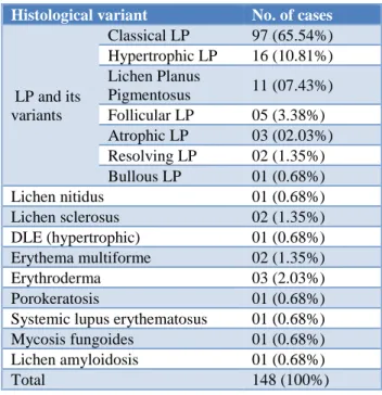
Out of the 148 cases of histologically diagnosed lichenoid tissue reactions studied, the most common entity diagnosed was lichen planus and its variants (135 cases, 91.21%). Among the cases of lichen planus and its variants, classical lichen planus (65.54%) was the most commonly observed entity followed by hypertrophic lichen planus (10.81%). Other variants were also reported.
Table 1:Histological variants of LTR/ID.
Histological variant No. of cases
LP and its variants
Classical LP 97 (65.54%) Hypertrophic LP 16 (10.81%) Lichen Planus
Pigmentosus 11 (07.43%) Follicular LP 05 (3.38%) Atrophic LP 03 (02.03%) Resolving LP 02 (1.35%) Bullous LP 01 (0.68%)
Lichen nitidus 01 (0.68%)
Lichen sclerosus 02 (1.35%)
DLE (hypertrophic) 01 (0.68%) Erythema multiforme 02 (1.35%)
Erythroderma 03 (2.03%)
Porokeratosis 01 (0.68%)
Systemic lupus erythematosus 01 (0.68%) Mycosis fungoides 01 (0.68%) Lichen amyloidosis 01 (0.68%)
Total 148 (100%)
Onset and duration of the disease
In present study, the cases presented with variable duration of the disease, ranging from 15 days to 150 months. Nearly 70% of the cases were seen within 0-6 month’s duration. However, good number of cases (20 cases, 13.51%) was seen between 1 to 5 years duration and few (6 cases, 4.05%) cases were of more than 5 years duration.
Site of involvement
(31.76%), face and neck involved in 8 cases (5.4%),
genitals (glans penis and labia majora) involved in two cases (1.35%) and scalp involvement in 1 case (0.68%). Whole body involvement was seen in 12 cases (8.11%). Genital involvement was seen only in cases of lichen sclerosus (1.35%)
Extracutaneous involvement
In present study, out of 148 cases, only 5 cases showed extracutaneous involvement, with oral cavity and genital involvement with two cases each (1.35% each) and one case (0.68%) showing nail involvement.
Clinical features
In present study, majority of the patients presented with itching (94.59%). Most common presentation of skin lesions was papulo-squamous skin lesions (95.95%), including papules, macules and plaques. Other presentations observed were exfoliative lesions (in erythroderma and one case of lichen planus) and targetoid lesions (in erythema multiforme). Two cases of lichen planus showed presence of follicular papules. Majority of the lesions were violaceous (76.35%), however, erythematous (6.76%), hyperpigmented (17.58%), hypopigmented and skin coloured lesions (1.35% each) were also seen. Koebner’s phenomenon was seen in 45.27% of the cases
Associated factors
Table 2: Histological features.
Histopathological changes No. of cases
Hyperkeratosis 145 (97.97%)
Parakeratosis 18 (12.16%)
Acanthosis 127 (85.81%)
Papillomatosis 17 (11.49%)
Atrophy 20 (13.51%)
Spongiosis 39 (26.35%)
Elongation of rete ridges 119 (80.40%)
Hypergranulosis 133 (89.86)
Basal cell vacuolation 148 (100%)
Max joseph space 114 (77.03%)
Follicular plugging 47 (31.76%)
Civatte bodies 92 (62.16%)
Melanin incontinence 148 (100%) Inflammatory cells in upper dermis 146 (98.65%) Periadnexal inflammatory infiltrates 96 (64.86%)
Out of 148 cases, association of diabetes was seen in 3 cases (2.03%), hypertension in 3 cases (2.03%) and both the group of cases were on treatment. Four cases (2.70%) were on treatment with bronchodilators while one case (0.68%) each of antiepileptics, oral contraceptive pills (OCPs) and non-specified drug were seen. Seven cases (4.73%) showed aggravation of the condition on exposure
to heat and light, and nine cases (6.08%) had history of tobacco intake.
Various histological findings were reported, as in Table 2.
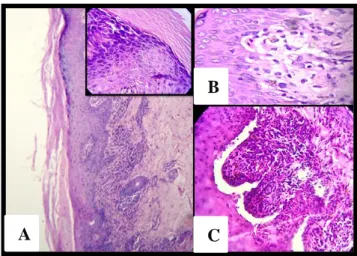
Band like inflammatory infiltrate hugging the dermo-epidermal junction was the most common pattern seen in 123 cases (83.11%) (Figure 1) followed by inflammatory infiltrates at the base of rete ridges were observed in cases with histological diagnosis of hypertrophic lichen planus. Perifollicular inflammatory cell infiltrates observed in cases with histological diagnosis of follicular lichen planus were seen in five cases (3.38%) [Figure 2(A)]. Focal dermal infiltrates were seen in two cases (1.35%). Two cases (resolving lichen planus) did not show any inflammatory infiltrate in the dermis, followed by inflammatory infiltrates seen at the base of rete ridges seen in sixteen cases (10.81%).
Figure 1: (A): Classical lichen planus: hyperkeratosis, acanthosis. Inset: hypergranulosis, (B): Orthokeratosis, basal cell vaculation, civatte bodies,
(C): Melanin incontinence, Max Joseph space.
In present study, majority of the cases showed predominantly lympho-histiocytic infiltration with occasional neutrophils (145 cases, 97.97%). Two cases (1.35%) showed the presence of eosinophils in the inflammatory infiltrates. One case (0.68%, mycosis fungoides) showed presence of only lymphocytes.
Other histological features
Various cases showed histological features which were diagnostic or suggestive of specific entity rather than lichenoid tissue reactions in general. They included marked melanin incontinence was seen in lichen planus pigmentosus [Figure 2(B)], amyloid deposition in the dermis in lichen amyloidosis, subepidermal bulla in bullous lichen planus [Figure 3(A)], inflammatory cells within dermal papilla in lichen nitidus [Figure 3(B)], mucin in upper dermis in SLE and DLE (Figure 4).
A
B
Dermal fibrosis in resolving lichen planus, cornoid lamella in porokeratosis (Figure 5), hyalinisation of upper dermis in lichen sclerosus, extravasation of RBCs in erythema multiforme.
Figure 2:(A): Lichen Planopilaris. Section showing perifollicular inflammatory infiltrates, marked follicular plugging, perifollicular hypergranulosis, melanin incontinence (H&E, 40x), (B) Lichen Planus Pigmentosus. Section shows atrophic epidermis, dense
lymphohistiocytic infiltration at dermoepidermal junction, marked melanin incontinence (H&E, 10x).
Figure 3:(A): Bullous Lichen Planus. Section shows subepidermal bulla, lympho-histiocytic infiltrates
infiltrates at dermoepidermal junction, hypergranulosis, melanin incontinence (H&E, 40x),
(B): Lichen Nitidus. Section shows hyperkeratosis, hypergranulosis, inflammatory infiltrates seen in dermal papilla, melanin incontinence (H&E, 40x).
Clinico-pathological correlation of various variants of LTR/ID
Out of 166 cases, 147 cases were clinico-pathologically concordant with a concordance rate of 88.55%.
Figure 4: DLE (hypertrophic variant). Section shows hyperkeratosis, acanthosis, hypergranulosis, lymphohistiocytic infiltration in upper dermis, mucin in upper dermis, melanin incontinece in upper dermis.
(H&E, 10x).
Figure 5: Porokeratosis. Section shows
hyperkeratosis, acanthosis, cornoid lamella (H&E, 10x).
Out of 153 cases of clinically diagnosed lichen planus, 135 cases were confirmed as lichen planus on histological examination with a concordance rate of 88.24%. Lichen nitidus, lichen amyloidosis, erythema multiforme, dermatitis associated with erythroderma, SLE and DLE, lichen sclerosus and mycosis fungoides showed 100% clinico-pathological concordance. No concordance was seen in case of porokeratosis and it was diagnosed based on histology alone.
Clinico-pathological correlation of various types of lichen planus is shown in Table 3.
DISCUSSION
Histopathology has always been a useful tool in differentiating the various diagnosis with similar clinical presentations and various differential diagnoses can be narrowed by focussing on the distinctive histological features of various entities.2,4 Thus, an appropriate
diagnosis of lichenoid tissue reactions/interface
A
B
dermatitis (LTR/ID) needs integrated approach by
studying histological features of the lesion and
correlating them with the clinical history.
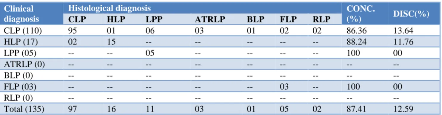
Table 3: Clinico-pathological correlation of variants of lichen planus.
Clinical diagnosis
Histological diagnosis CONC.
(%) DISC(%)
CLP HLP LPP ATRLP BLP FLP RLP
CLP (110) 95 01 06 03 01 02 02 86.36 13.64
HLP (17) 02 15 -- -- -- -- -- 88.24 11.76
LPP (05) -- -- 05 -- -- -- -- 100 00
ATRLP (0) -- -- -- -- -- -- -- -- --
BLP (0) -- -- -- -- -- -- -- -- --
FLP (03) -- -- -- -- -- 03 -- 100 00
RLP (0) -- -- -- -- -- -- -- -- --
Total (135) 97 16 11 03 01 05 02 87.41 12.59
CLP: classical LP; HLP: Hypertrophic LP; LPP: LP pigmentosus; ATRLP: Atrophic LP; BLP: Bullous LP; FLP: Follicular LP; RLP: Resolving LP; CONC: Concordance; DISC: Discordance.
Age distribution
Present study showed a wide age range of involvement from 10 years of age to 78 years of age. This study showed the most common age group as 31 to 50 years of age group with 49.33% cases, which is similar to studies conducted by Hedge et al, Chauhan et al, Garg et al.5-7
Present study reported 13 cases (8.78%) in patients till 18 years of age. Chauhan et al, have showed 16.67% cases of less than 18 years of age.5-7 Thus, lichenoid tissue
reactions can be seen in all age groups, though more common in middle aged individuals.
Sex distribution
In concordance with present study, study conducted by Chauhan et al, also showed slightly higher predilection of the disease for males. Other studies have described nearly equal or more predilection for females.2-9
A higher predilection of males for the disease could be because of either some geographical variation altering the environmental factors or because of inhibition of many females from reporting for treatment due to the social stigma associated with any skin disease.
Prevalence of variants of LTR/ID
Studies done by Kumar et al, Khaled A et al, Hegde et al, and Chauhan et al, have described lichen planus (including its variants) as the most common entity in lichenoid tissue reactions.2,5,7,8 Studies conducted by
Kumar U et al, Chauhan et al, Parihar et al, Garg VK et al, and Nangia A et al, have reported classical lichen planus as the most common variant of lichen planus. All these studies showed hypertrophic lichen planus as the second most common variant of lichen planus, except the study conducted by Kumar MU et al, which has reported
lichen planus pigmentosus as the second most common variant.2,6,8-10 Similar prevalence has been reported in our
study as well.
Clinical features
Onset and duration
Khaled A et al, reported majority of the patients presenting within 6 months of the onset of disease which is in concordance with our study.8
SITE
Lower limb is the most common site in present study. Similar observations were made by Khaled A et al, Chauhan et al, and Parihar et al, who observed lower limb involvement as the most common site.6,8,9 Venous stasis
could be a likely explanation for lower limbs to be the most common site of involvement.11
Extrcutaneous involvement
In present study, out of 135 cases, only 5 patients presented with extra-cutaneous lesions. Oral cavity involvement was 15.38% and genital lesions in 6.84% of cases in study conducted by Maheshwari et al.4
Oral lesions were seen in 16.66% cases of lichenoid eruptions in the study conducted by Chauhan et al.6
Parihar et al reported 3.5% cases of lichen planus with nail involvement.9
Garg et al, has reported mucosal involvement in 24% and nail involvement in 17.3% cases of lichen planus.7
Histological changes
In present study, most common epidermal change is basal cell vacuolation/degeneration followed by hyperkeratosis. Third most common finding was hypergranulosis followed by acanthosis and elongation of rete ridges.
Various studies have shown basal cell vacuolation/degeneration as the most common histological change in the epidermis, followed by hyperkeratosis. These studies also reported good number of cases with acanthosis, hypergranulosis and elongation of rete ridges.2,5,6,7,9,12 However, hyperkeratosis followed
by acanthosis was the most common epidermal change seen in study conducted by Maheshwari et al.4
Histological findings of present study were in concordance with these studies.
In present study, melanin incontinence was the most common dermal change seen in all the cases. This observation is in concordance with various studies which have shown melanin incontinence in the dermis in 100% of the cases.2,5,7 However, study conducted Chauhan et al,
and Maheshwari et al, have shown pigment incontinence in 63.63% and 52.14% of the cases respectively.4,6
Pattern of inflammatory infiltrates
Various studies have described band like inflammatory infiltrates hugging the dermoepidermal junction ranging from 48.48 to 100% of the cases as seen in our study as well.2,4-7,12 Similar to present study, perifollicular
infiltrates have been described in the lesions of lichen planopilaris. Inflammatory infiltrates at the base of rete ridges are classically seen in hypertrophic lichen planus.3
Focal inflammatory infiltrate was seen in a case of lichen nitidus, which showed inflammatory cells in dermal papilla, giving it a claw clutching the ball appearance. Similar findings were reported by Maheshwari et al, and Chauhan et al.4,6
Studies done in past have also shown predominantly lymphocytic infiltrates in the dermis with occasional cases showing neutrophils, eosinophils and plasma cells.2,4-7,12
Various histological features which were leading towards specific diagnosis were also recorded.
Presence of vertical collagen bundles in the upper dermis and inflammatory infiltrates at the base of the rete ridges were seen exclusively in cases of hypertrophic lichen planus. These findings are supported by various literature.3,13
Cases with marked melanin incontinence in the dermis associated with thinned out epidermis were assigned the histological diagnosis of lichen planus pigmentosus. Similar findings have been reported by various other
studies as well which have shown marked pigment incontinence in the dermis in these cases.6,9
In cases reported as resolving lichen planus in present study, there is a possibility to see lichenoid type of infiltrates in biopsy taken from a different lesion in the same patient as lesions can be present in various stages of development.
Amyloid deposition, coronoid lamellae were seen in lichen amyloidosus and porokeratosis respectively. This is in concordance with study done by Hegde et al, which showed presence of amyloid in the dermis in cases of lichen amyloidosis.5,14,15
Subepidermal bullae formation was seen in a case of bullous lichen planus. This finding is supported by various studies which have shown presence of subepidermal bulla in bullous lichen planus.6 The
presence of subepidermal bulla could be due to extensive basal layer degeneration leading to separation of epidermis and dermis leading to bulla formation.
Mucin in dermis was seen in 2 cases in present study, one of DLE and other diagnosed as SLE. Similar observations have been reported by previous literature as well.3
Both the cases of lichen sclerosus showed hyalinisation of the upper dermis. This finding has been reported by Coehlo et al, and CM Ridley.16,17
Extravasation of RBCs in the dermis was seen in two cases diagnosed as erythema multiforme. Similar picture has been reported in literature as well.18
Clinico-pathological correlation
In concordance with present study, study conducted by Hegde et al, 87.2% clinico-pathological concordance with 100% concordance in cases of lichen amyloidosis, erythema multiforme and lichen sclerosus.5 This was
attributed to classical clinical presentation of these entities. In a study conducted by Kumar et al, 78.5% cases of lichenoid tissue eruptions showed clinico-pathological concordance while Maheshwari et al, reported 70.94% of clinico-pathological concordanc.2,4
Single case of porokeratosis was diagnosed solely based on histology in present study. Histology of the skin lesion showed classical cornoid lamella and diagnosis of porokeratosis was made. Similar findings were reported by study done by Maheshwari et al, which reported porokeratosis based on histological features.4 Study
Other diagnosis could be explained by the fact that
lesions can be seen in various stages of development and resolution and thus, atrophic or resolving lichen planus may be seen in some of the cases.
CONCLUSION
Distinguishing various entities in lichenoid eruptions can be challenging for the dermatologist as well as the pathologist because most of the entities present with similar features clinically as well as histologically except for few differences and specific features which help to approach the diagnosis. Histopathology also plays an important role in narrowing the clinical differential diagnoses of the disease. It should be considered that the mere presence of interface dermatitis is not the sole criterion to diagnose lichen planus and other clinical and histological features should be considered.
There are certain limitations in the present study. As a part of study is retrospective, the uniformity in clinical diagnosis and histopathological evaluation could not be assessed. With the limitations, this study still gives information about the importance of histopathology as in few of the cases where disease was not confirmed, or the variant was not diagnosed clinically. Other limitation is that majority of the cases were of lichen planus, with other entities being less in number which undermined the analysis and correlation of the data.
Though definite diagnosis can be made on histopathological examination, size of specimen, site of biopsy, nature and depth of biopsy, quality of sections, treatment history and inter-observer variation (both clinically and histologically) should be kept in mind which may lead to clinico-pathological discordance.
ACKNOWLEDGEMENTS
Authors would like to thank he faculty members and technical staff of the Department of Pathology, KIMS, Hubballi, Karnataka, India.
Funding: No funding sources Conflict of interest: None declared Ethical approval: Not required
REFERENCES
1. Sontheimer RD. Lichenoid tissue reaction/interface dermatitis: Clinical and histological perspectives. J Invest Dermatol. 2009;129(5):1088-99.
2. Kumar MU, Yelikar BR, Inamadar AC, Umesh S, Singhal A, Kushtagi AV. A Clinico-pathological study of lichenoid tissue reactions-A tertiary care experience. J Clin Diagn Res. 2013;7(2): 312-6. 3. Weedon D. Skin Pathology, 3rd ed. China: Elsevier;
2010:35-70.
4. Maheshwari GR, Mehta HH, Nikam V. Clinico-histopathological correlation for diagnosis of lichenoid interface dermatoses. J Dermatol Dermatologic Surg. 2016;20:115-24.
5. Hegde VK, Khadilkar UN. A clinicopathological study of interface dermatitis. Indian J Pathol Microbiol. 2014;57:386-9.
6. Chauhan R, Srinath MK, Ali NM, Bhat RM, Sukumar D. Clinicopathological Study of Lichenoid Reactions: A Retrospective Analysis. J Evolution Med Dental Sci. 2015;4(32):5551-62.
7. Garg VK, Nangia A, Logani K, Sharma RC. Lichen Planus-a Clinico- histopathological. Indian J Dermatol Venereol Leprol. 2000;66(4):193-5. 8. Khaled A, Banu SG, Kamal M, Manzoor J, Nasir
TA. A clinical and histopathological study of lichenoid eruption of skin in two tertiary care hospitals of Dhaka. Pulse. 2011;5(1):12-8.
9. Parihar A, Sharma S, Bhattacharya SN, Singh UR. A clinicopathological study of cutaneous lichen planus. J Dermatol Dermatologic Surg. 2015;19:21-6.
10. Nangia A, Kumar V, Logani KB. An immunopathological study of lichen planus. Indian J Dermatol Venereol Leprol. 2000;66(2):76-8. 11. Bhattacharya M, Kaur I, Kumar B. Lichen planus: a
clinical and epidemiological study. J Dermatol. 2000;27 (9):576-82.
12. Ellis FA. Histopathology of lichen planus based on analysis of one hundred biopsy specimens. J Invest Dermatol. 1967 Feb 1;48(2):143-8.
13. Mobini N, Toussaint S, Kamino H. Noninfectious erythematous, popular, and squamous diseases. In: Elder DE, Elenitsas R, Johnson BL, Murphy GF, Johnson BL, Xu X, editors. Lever’s Histopathology of the Skin. 10th Ed. Philadelphia: Lippincott Williams and Wilkins. 2009;169-204
14. Kamal T, Mufti S, Ahmad TJ. Disseminated superficial actinic porokeratosis: A case report. J Pakistan Assoc Dermatologists. 2015; 25 (1):73-5. 15. Tan LS, Chong WS. Porokeratosis in Singapore: An
Asian perspective. Australasian J Dermatol. 2012;53:e40-4.
16. Coelho WS, Diniz LM, Filho JBDS. Lichen sclerosus et atrophicus. An Bras Dermatol 2006;81:297-300.
17. Ridley CM. Genital lichen sclerosus (lichen sclerosus et atrophicus) in childhood and adolescence. J Royal Soc Med. 1993 Feb;86(2):69. 18. Roujeau JC. Erythema multiforme. In: Goldsmith
LA, Katz SI, Gilchrest BA, Paller AS, Leffell DJ, Wolff K, editors. Fitzpatrick’s Dermatology in General Medicine. 8th ed. New Delhi: McGraw-Hills Companies; 2012:1:431-9.

![Bis[(5RS,11RS) 2,8 dimethyl 5,10 methano 5,6,11,12 tetrahydrodibenzo[b,f][1,5]diazocine 5 ium dihydrogen phosphate] tris(phosphoric acid) methanol solvate](data:image/gif;base64,R0lGODlhAQABAIAAAP///wAAACH5BAEAAAAALAAAAAABAAEAAAICRAEAOw==)