Original Research Article
Comparison of three different techniques of endotracheal tube cuff
inflation: just seal, stethoscope guided and pressure volume loop: a
prospective randomized study
Mamta Bhardwaj
1*, Kiranpreet Kaur
1, Asha Sharma
1, Prashant Kumar
1, Raj Tobria
2,
Savita Saini
1INTRODUCTION
Maintaining airway and ventilating the patients by placement of endotracheal tube (ETT) is common practice for delivery of general anesthesia and in the critical care settings. Cuffed endotracheal tubes (ETT)
not only ensure a proper seal during positive pressure ventilation without volume loss, but at the same time also prevent aspiration of pharyngeal and gastric secretions.1
Both over and under inflation of the cuff might cause postoperative airway morbidity. Over inflation of the ETT cuff might lead to serious complications, ranging
1Department of Anaesthesiology and Critical Care, 2Department of Social and Preventive Medicine, Pt. B.D Sharma
PGIMS at University of Health Sciences, Rohtak, Haryana, India
Received: 19 November 2019
Revised: 04 December 2019
Accepted: 11 December 2019
*Correspondence:
Dr. Mamta Bhardwaj,
E-mail: [email protected]
Copyright: © the author(s), publisher and licensee Medip Academy. This is an open-access article distributed under the terms of the Creative Commons Attribution Non-Commercial License, which permits unrestricted non-commercial use, distribution, and reproduction in any medium, provided the original work is properly cited.
ABSTRACT
Background: Cuffed endotracheal tubes not only ensure a proper seal during positive pressure ventilation, but at the same time also prevent aspiration of gastric secretions. The aim of this prospective, randomized study was to compare three methods of ETT cuff inflation-- palpation of the leak in suprasternal notch (Just seal), a stethoscope guided method of tracheal tube cuff inflation and PVL guided cuff inflation.
Methods: After approval by institutional ethical committee, 192 patients of either sex in age group of 18-50 years belonging to ASA physical status I or II were enrolled. Each patient was randomly allocated into one of three groups: one group received standard 'just seal' method of tracheal cuff inflation (JS), the second group, the stethoscope-guided method (SG) and in third group cuff was inflated using Pressure Volume Loop (PVL). Volume of air introduced into the cuff and pressure within the cuff was recorded.
Results: A total of 192 patients were recruited to the study. The median (IQR [range]) tracheal cuff pressure was 12 (10-22 [6-28]) cm H2O, 16 (12-24[6-38]) and 14 (10-22[8-32]) cmH2O in JS, SG and PVL group respectively. Cuff
pressures within the recommended range of 20-30 cm H2O fell in 25% of the patients in both JS and SG group and
31% patients in PVL group. The mean volumes of air introduced in the cuff and the resultant mean cuff pressure in all groups was found to be statistically insignificant (p= 0.4, 0.18 respectively). Tidal volume discrepancy was more and 75% of cuff pressures were less than the recommended range in JS than the other two groups.
Conclusions: Real time PVL displayed on most modern anaesthesia machine is a good alternative to check for proper ETT cuff inflation, avoid high cuff pressure and monitor air leak.
Keywords: Cuff pressure, Endotracheal tube, Just seal, Pressure volume loop, Stethoscope guided
from tracheal mucosa pressure necrosis to tracheal rupture and tracheoesophageal fistula formation.2-4 Under
inflation of the cuff might result in volume loss, gastric distension and thus regurgitation and aspiration. Adequacy of ETT cuff inflation is usually checked by one of the following techniques: Manual palpation of the pilot balloon, disappearance of audible air leak through the mouth or disappearance of palpable air leak at thyroid cartilage or suprasternal notch (Just seal), auscultation of leak at right lamina of thyroid cartilage (stethoscope guided) or use of either an aneroid manometer or continuous automatic ETT cuff pressure controller.5,6
Pressure–volume loop (PVL) is one of the continuous real time pulmonary graphic incorporated in the monitoring system of anaesthesia machines and mechanical ventilators. Usually, it is used for the assessment of dynamic lung compliance, detection of lung over inflation or presence of air leak.7 It is also used
for endotracheal tube cuff inflation. The aim of this study was to compare three methods of ETT cuff inflation-- palpation of the leak in suprasternal notch (Just seal), a stethoscope- guided method of tracheal tube cuff inflation and PVL guided cuff inflation with regards to adequacy of seal and postoperative cuff related complications if any.
METHODS
This prospective, randomized study was conducted from June 2017 to May 2018 after getting approval from the institutional ethical committee. A total of 192 patients were recruited and informed consent from all the participants was obtained. Inclusion criteria: patients of either sex in the age group of 18-50 years, belonging to American Society of Anesthesiologists (ASA) physical status I or II and scheduled to undergo elective surgery in supine position and requiring tracheal intubation.
Exclusion criteria
Patients with anticipated difficult airway, chronic lung diseases, preoperative cough, hoarseness of voice or sore throat, and an increased risk of aspiration.
All patients were examined and thoroughly investigated preoperatively. All patients were kept fasting for 6 hours and prescribed routine premedication. In the operating room, after the establishment of intravenous (IV) line and attachment of standard monitors [non-invasive blood pressure (NIBP), electrocardiography (ECG) and pulse oximetry (SpO2)], each patient was randomly allocated into one of three groups: one group received the standard ‘just seal’ method of tracheal cuff inflation (JS; n=64), the second group, the stethoscope-guided method (SG; n=64) and in the third group cuff was inflated using pressure volume loop (PVL; n=64). Allocation to one of three combinations was done using sealed coded envelopes.
Before induction of anaesthesia, an automatic leak test was performed on the anaesthesia machine (Primus,
Drager, Lübeck, Germany) to detect any leak in the machine. Anaesthesia was induced with injection glycopyrrolate 0.2 mg, fentanyl (2 µ kg-1) and propofol (2-3 mg kg-1). After checking for ability to ventilate inj vecuronium 0.1 mg kg-1 was administered and three minutes after the trachea was intubated with a portex tracheal tube ((Portex Tracheal Tube, Smith Medical International Ltd., Ashford, Kent, UK). TT of size 7.0 mm ID and 8.0 mm ID was used in female and male patients respectively.
Following successful insertion of the ETT, it was connected to the breathing circuit and volume controlled ventilation was initiated with 10 mL/kg tidal volume and a ventilator rate of 14/ min. The tracheal cuff was then inflated using one of three methods. The ETT cuff was inflated initially by 2 mL of air followed by increments of 0.5 ml. In just seal group air was introduced till the palpable leak disappeared. In stethoscope guided technique the diaphragm of a stethoscope (Littmann 3M, master classic II) was placed over the right lamina of the thyroid cartilage and air was introduced until harsh turbulent breath sounds were replaced by softly pitched sounds. In the group of PVL, ventilation of the lungs and cuff inflation was done in the same fashion as the just seal group till complete closure of the PVL was displayed on the anaesthesia machine monitor i.e. when the expiratory limb reached zero volume and met the starting point of inspiratory limb, cuff inflation was ceased.
In each group, volume of air introduced into the cuff was recorded. The pressure within the cuff was then determined using a calibrated tracheal tube cuff manometer (Mallinckrodt Medical, Athlone, Ireland), which measures tracheal cuff pressure from 0 to 120 cm H2O. In all groups, a three way stopcock was connected
to the pilot balloon and a small manometer was connected to the other end of the stop cock while the third end was connected to a 5 ml syringe. After inflation of the cuff, TV was decreased to 8ml kg-1 and RR to 12
min-1. Any discrepancy between inhaled and exhaled TV
was noted for 5 min and monitored throughout the surgery. After that low flow anaesthesia (2l min-1) with
O2:N2O 50 % and sevoflurane (1.5-2%) was continued
for maintenance. Exhaled TV was monitored for whole of the surgery. Cuff pressure was again recorded 1 hr after start of N2O.
summary tables and compare items within and across various categories. A “p” value of <0.05 considered critical for statistical significance.
RESULTS
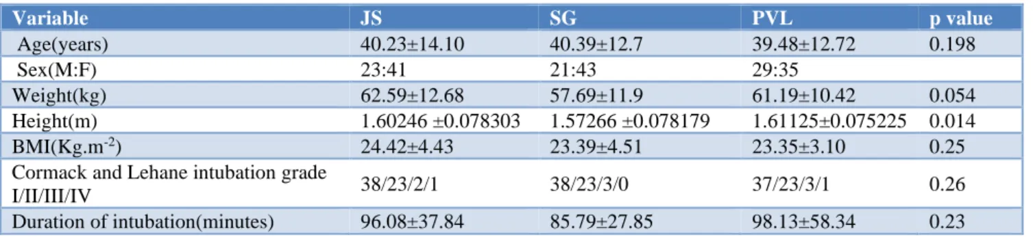
A total of 192 patients were recruited to the study. (Table 1) shows the patients’ characteristics among the three groups. The groups were comparable demographically in respect of age, sex, weight, height and BMI and duration of intubation.
In the ‘just seal’ group, the median (IQR [range]) tracheal cuff pressure was 12 (10-22 [6-28]) cm H2O. In only 25%
of the patients, the cuff pressures fell within the recommended range of 20-30 cm H2O, cuff pressures
were <20 cm H2O in seventy five percent and it did not
exceed the recommended level of 30 cm H2O in any of
the patients (Table 2,3). In the stethoscope-guided group,
the median (IQR [range]) cuff pressure was 16 (12-24[6-38]) cm H2O. Number of patients (26%) with cuff
pressure in recommended range was similar to ‘just seal group, in 67% of patients, pressures were <20 cm H2O
and in 7% pressures >30 cm H2O were recorded. In
pressure volume loop group, the median (IQR [range]) cuff pressure was 14 (10-22[8-32]) cm H2O. Pressures
within in the recommended range of 20-30 cm H2O was
found in 31% of patients, 66% of pressures were <20 cm H2O and 3% of pressures >30 cm H2O were recorded.
(Table 2,3). The mean volumes of air introduced in the cuff and the resultant mean cuff pressure in all groups was found to be statistically insignificant. (p= 0.4, 0.18 respectively) (Table 2). Cuff pressure after 1 hour increased about 7-19% in all groups but increase was not significant among the groups. Tidal volume discrepancy among the groups is shown in (Table 2). It was statistically significant between just seal and pressure volume loop group (p=0.016).
Table 1: Patients’ characteristics among the three groups. Values are mean±SD or number.
Variable JS SG PVL p value
Age(years) 40.23±14.10 40.39±12.7 39.48±12.72 0.198
Sex(M:F) 23:41 21:43 29:35
Weight(kg) 62.59±12.68 57.69±11.9 61.19±10.42 0.054
Height(m) 1.60246 ±0.078303 1.57266 ±0.078179 1.61125±0.075225 0.014
BMI(Kg.m-2) 24.42±4.43 23.39±4.51 23.35±3.10 0.25
Cormack and Lehane intubation grade
I/II/III/IV 38/23/2/1 38/23/3/0 37/23/3/1 0.26
Duration of intubation(minutes) 96.08±37.84 85.79±27.85 98.13±58.34 0.23
Table 2: Volume of air introduced in the tracheal cuff and the resultant cuff pressure in patients in different groups. Values are mean (SD) or median (IQR [range]).
JS (n=64) SG (n=64) PVL (n=64) p value*
Volume of air introduced (ml) 4.0±1.41 4.2±1.26 4.00±1.30 0.56
Cuff pressure (cm H2O) 14.25±6.62
12 (10-22 [6-28])
17.44±8.69 16 (12-24[6-38])
15.91±6.60
14 (10-22[8-32]) 0.053
Cuff pressure after 1 hr. (cm H2O 16.0±6.4 18±7.9 18.3±8.9 0.186
Tidal volume discrepancy(ml) 22.0±36.16 7.39±14.18 3.61±6.33 ** 0.016
*p value <0.05- significant ** significant between JS and PVL group.
Table 3: Cuff pressure in different groups.
Cuff pressure
Study group
Just seal (n=64)
Stethoscope guided (n=64)
PVL guided (n=64)
Normal recommended (20-30)
16(25%) 15(26%) 20(31%)
Below 20 48(75%) 43(67%) 42(66%)
Above 30 0 6(7%) 2(3%)
DISCUSSION
There is agreement that high pressures with in tracheal cuffs may result in significant morbidity and most authorities recommend that pressures should be maintained below 25-30 cm H2O.8,9 A number of studies
have demonstrated that tracheal tube cuffs are commonly inflated to pressures exceeding this range.1,10-13 Tracheal
mucosal capillary flow is impaired at cuff pressures more than 30 cm H2O and that tracheal mucosal flow ceases at
pressures greater than 50 cm H2O.9 The resultant mucosal
tracheal cartilages and can lead to the development of tracheal stenosis, tracheoesophageal fistula and even tracheal rupture.2-4,14 Increased awareness about cuff
pressure related complications has led to change in the design of ETT cuff from a high pressure low volume to a low pressure high volume one.15 Although there is a lack
of consensus regarding an effective lower cuff pressure to ensure an effective seal, most recommend a minimum cuff pressure of 20 cmH2O.1,16
None of currently used clinical method of cuff inflation reliably produces cuff pressures within the recommended range. The standard ‘just seal’ method of cuff inflation has been shown to be an unreliable technique for consistently inflating cuffs to <30 cm H2O. Attempting to
inflate tracheal cuffs to appropriate pressures by palpating the tension in the pilot balloon has been shown to be inaccurate, and often underestimates cuff pressure, with the design of the pilot balloon being a significant influencing factor.12,13,17
Kumar and Hirsch reported median cuff pressure of 34 cm H2O in JS group and; Sixty-four per cent of cuff
pressures exceeded the recommended level of 30 cm H2O
and only 32% of cuff pressures fell within the recommended range; 4% of pressures were < 20 cmH2O.6
While in this study the median tracheal cuff pressure was 12 cm H2O. Only 25% of the patient’s cuff pressures fell
within the recommended range, 75% of cuff pressures were <20 cm H2O and none of cuff pressures exceeded
the recommended level of 30 cm H2O. Similarly, in the
SG group of our study, the cuff pressure was 16 cm H2O.
26 % of pressures fell within in the recommended range of 20-30 cm H2O, 67% of pressures were <20 cm H2O
and 7% of pressures >30 cm H2O were recorded. Kumar
and Hirsch reported median cuff pressure of the 20 (20-26 [16-28]) cm H2O in the stethoscope-guided group,
84% of pressures fell within in the recommended range of 20-30 cmH2O and no pressures >30 cm H2O were
recorded; 16% of pressures were <20 cmH2O.6 The
difference in the results can be because of different methodology used in both studies. Mean volume of air introduced in the cuff was 5.4 ml in both the groups as compared to 4.0 ml and 4.2 ml in this group of patients. They used reinforced tube of smaller size (6.5 mm ID in females and 7.5 mm ID in males) as compared to this study. (Portex tube, 7.0 mm in females and 8.0 mm in males). Moreover, tube cuff was inflated with APL valve fully closed in both groups during manual ventilation in their study. In contrast to their study, cuff was inflated using volume control mode with a set tidal volume of 10 mL/kg and a ventilator rate of 14/ min in all the groups in the present study.
Almarakbi WA reported a cuff pressure of 33 (32-35) cmH2O in JS group. They also used gentle manual
ventilation with APL valve closed at 20 cmH2O. In the
same study cuff pressure in the PV-L group was 18.25 cm H2O with no air leak and volume of air required was
4.05 ml.18 This is similar to our study where cuff
pressure in PVL group is 15.91 cm H2O and volume of
air used is 4.0 ml.
There was a tidal volume discrepancy of 22.0(36.16), 7.39(14.18) and 3.61(6.33) ml in JS, SG and PVL group respectively during first five minutes after inflation of the cuff. It was statistically significant between JS and PVL group. (p<0.001) In two patients in JS group, after inflation of the cuff, there were few secretions and bubbling in the mouth suggesting leak around the cuff. Cuff was further inflated and leak stopped. Monitoring of exhaled tidal volume continued throughout the surgery in all groups and no significant leak noticed. One patient each in JS and SG group and two patients in PVL group complained of sore throat but was non-significant.
In this study, all the methods of cuff inflation are found to be satisfactory, but tidal volume discrepancy was more and 75% of cuff pressures were less than the recommended range in JS than the other two groups. So, stethoscope and pressure volume loop are effective ways to guide endotracheal tube cuff inflation. Real time PVL displayed on most modern anaesthesia machine is a good alternative to check for proper ETT cuff inflation, avoid high cuff pressure and monitor air leak. In situation of non-availability of real time PVL display, stethoscope guided cuff inflation will serve the purpose. Mean cuff pressure was less than the recommended range in more than 60% of patients in all groups and there was no incidence of aspiration. Is this the time to reconsider the recommended cuff pressure range and endotracheal tube size? The limitation of our study is small sample size. So, large scale randomized trials are needed to confirm these results.
Funding: No funding sources Conflict of interest: None declared
Ethical approval: The study was approved by the Institutional Ethics Committee
REFERENCES
1. Sengupta P, Sessler DI, Maglinger P, Wells S, Vogt A, Durrani J, et al. Endotracheal tube cuff pressure in three hospitals, and the volume required to produce an appropriate cuff pressure. BMC Anesthesiol. 2004;4(1):8.
2. Striebel HW, Pinkwart LU, Karavias T. Tracheal rupture caused by overinflation of endotracheal tube cuff. Der Anaesthesist. 1995;44(3):186-8.
3. Luna CM, Legarreta GI, Esteva H, Laffaire E, Jolly EC. Effect of tracheal dilatation and rupture on mechanical ventilation using a low-pressure cuff tube. Chest. 1993;104(2):639-40.
4. Reed MF, Mathisen DJ. Tracheoesophageal fistula. Chest surg clin North Am. 2003;13(2):271-89. 5. Jain MK, Tripathi CB. Endotracheal tube cuff
6. Kumar RD, Hirsch NP. Clinical evaluation of stethoscope‐guided inflation of tracheal tube cuffs. Anaesthe. 2011;66(11):1012-6.
7. Sinha SK, Nicks JJ, Donn SM. Graphic analysis of pulmonary mechanics in neonates receiving assisted ventilation. Arch Dis Childhood Fetal Neonatal Ed. 1996;75(3):F213.
8. Seegobin RD, Van Hasselt GL. Endotracheal cuff pressure and tracheal mucosal blood flow: endoscopic study of effects of four large volume cuffs. Br Med J (Clin Res Ed). 1984;288(6422):965-8.
9. Vyas D, Inweregbu K, Pittard A. Measurement of tracheal tube cuff pressure in critical care. Anaesthe. 2002;57(3):275-7.
10. Chopra M, Jones L, Boulanger C, Benger J, Higginson I, Williamson D, et al. Prospective observational measurement of tracheal tube cuff pressures in the emergency department. Emerg Med J. 2010;27(4):270-1.
11. Galinski M, Tréoux V, Garrigue B, Lapostolle F, Borron SW, Adnet F. Intracuff pressures of endotracheal tubes in the management of airway emergencies: the need for pressure monitoring. Annals Emerg Med. 2006;47(6):545-7.
12. Hoffman RJ, Parwani V, Hahn IH. Experienced emergency medicine physicians cannot safely inflate or estimate endotracheal tube cuff pressure using standard techniques. Am J Emerg Med. 2006;24(2):139-43.
13. Hoffman RJ, Parwani V, Kaban J, Dueffer H, Howell A, Sturmann K. 86: Comparison of Two Common Techniques for Inflating Endotracheal
Tube Cuffs: Set Volume of Air Vs. Palpation of the Pilot Balloon. Annals Emerg Med. 2006;48(4):27. 14. Wain JC. Postintubation tracheal stenosis. Chest
Surg Clin. 2003;13(2):231-46.
15. Carroll RG, Mcginnis GE, Grenvik A. Performance characteristics of tracheal cuffs. Intern Anesthesiol Clinics. 1974;12(3):111-42.
16. American Thoracic Society, Infectious Diseases Society of America. Guidelines for the management of adults with hospital-acquired, ventilator-associated, and healthcare-associated pneumonia.
Am J Respiratory Critical Care Med.
2005;171(4):388.
17. Janossy KM, Pullen J, Young D, Bell G. The effect of pilot balloon design on estimation of safe tracheal tube cuff pressure. Anaesthe. 2010;65(8):785-91. 18. Almarakbi WA, Kaki MA. Tracheal tube cuff
inflation guided by pressure volume loop closure associated with lower postoperative cuff –related complications: Prospective, randomized, clinical trial. Saudi J Anaesth. 2014;8(3):328-34.