Original Research Article
A comparative analysis of mammography breast imaging reporting and
data system score and fine needle aspiration cytology in the evaluation
of palpable breast lump
Arpita Pandia
1, Sagarika Samantaray
1, Jay Sonali Mohapatara
2, Sashibhusan Dash
1*
INTRODUCTION
Breast lump is one of the commonest complaints with which patients present in breast clinics. As most breast lump ultimately leads to a diagnosis of breast cancer, it is
important for women with a breast lump to receive appropriate evaluation.1 Evaluation of breast lumps
involves the rational use of a detailed history, clinical breast examination, imaging modalities and tissue diagnosis. Though the final diagnosis is made by
1Department ofPathology, Acharya Harihar Regional Cancer Centre, Cuttack, Odisha, India 2Department of Bioinformatics, BJB Autonomous College, Bhubaneswar, Odisha, India
Received: 14 February 2019
Revised: 09 April 2019
Accepted: 02 May 2019
*Correspondence:
Mr. Sashibhusan Dash,
E-mail: [email protected]
Copyright: © the author(s), publisher and licensee Medip Academy. This is an open-access article distributed under the terms of the Creative Commons Attribution Non-Commercial License, which permits unrestricted non-commercial use, distribution, and reproduction in any medium, provided the original work is properly cited.
ABSTRACT
Background: Breast lump is one of the commonest complaints with which patients present in breast clinics. As in most cases breast lump ultimately leads to a diagnosis of breast cancer, it is important for women with a breast lump to receive appropriate evaluation. So initial diagnostic approach such as utilization of rapid, inexpensive, presumptive and safe method is utmost important as such methods not only would benefit both patients and clinicians by prompting proper preoperative diagnosis and management but also further limits unnecessary testing and procedures. Aim and objectives of this study was to analyse the diagnostic accuracy of mammogram according to Breast Imaging Reporting and Data System (BI-RADS) score and fine-needle aspiration cytology (FNAC) in patients presented with benign and malignant breast lumps in authors centre.
Methods: During this retrospective study from January 2018 to December 2018, the subjects concerned included all the female patients, who were referred to the department of pathology for FNAC of their breast mass and had radiological reports (mammography) with BI-RADS score according to the latest guidelines. The sensitivity, specificity, accuracy, positive and negative predictive values of BI-RADS scores and FNAC in diagnosis of breast diseases was done on the basis of final diagnosis.
Results: In this study, the sensitivity, specificity, positive predictive value, negative predictive value and accuracy was 88.57%, 82.46%,75.61%,92.16% and 84.78%, respectively for Mammography (BIRADS) score and 100% for all the parameters on FNAC.
Conclusions: According to authors above study findings concluded that FNAC could be considered an ideal initial diagnostic modality in all breast lumps recognized by means of imaging techniques. It is not only considered as safe, cost effective, rapid and presumptive diagnostic method but also further limits unnecessary testing and procedures.
Keywords: BIRADS,Breast Lump, Fine-needle aspiration cytology, Mammography
histopathological examination of the excised tissue, routine excision of all breast lumps would not be rationale, because as much as 80% of lumps are benign.2
So, initial diagnostic approach such as utilization of rapid, inexpensive, presumptive and safe method is utmost important as such methods not only would benefit both patients and clinicians by prompting proper preoperative diagnosis and management but also further limits unnecessary testing and procedures.
In this regard both mammography and fine needle aspiration cytology (FNAC) are used as initial diagnostic modalities in benign and malignant lesions. However confident diagnosis is made in 95% of the cases through a combination of clinical examination, imaging (including mammogram and/or sonomammogram) and FNAC.3
The accuracies of mammography and FNAC in the diagnosis of breast lumps have been tested individually in other studies.3-5 There are paucity of studies reported
from authors state as well as authors center.
So, author aimed to analyze the diagnostic accuracy of mammogram according to Breast Imaging Reporting and Data System (BIRADS) score and FNAC in patients presented with benign and malignant breast lumps in authors center.
METHODS
During this hospital based retrospective study (from January 2018 to December 2018), the subjects concerned included all the female patients, who were complained of breast lump and underwent breast imaging (including BIRADS scoring, according to the latest guidelines) and pathologic examination (i.e. both FNAC and histopathology).6
This study was conducted at Department of Pathology, Acharya Harihar Regional Cancer Centre, Cuttack, Odisha, India.
Recurrence cases, non-cooperative, pregnant and lactating patients were excluded from this study. According to inclusion and exclusion criteria, 92 cases were included in this study.
FNAC slides were retrieved from cytology department and reviewed under light microscopy. The findings were noted and correlated with BI-RADS score. The sensitivity, specificity, accuracy, positive and negative predictive values of BI-RADS scores and FNAC of different breast diseases was done based on histopathological diagnosis. Clinicopathological parameters were also noted. Data was analyzed using an MS Excel worksheet.
RESULTS
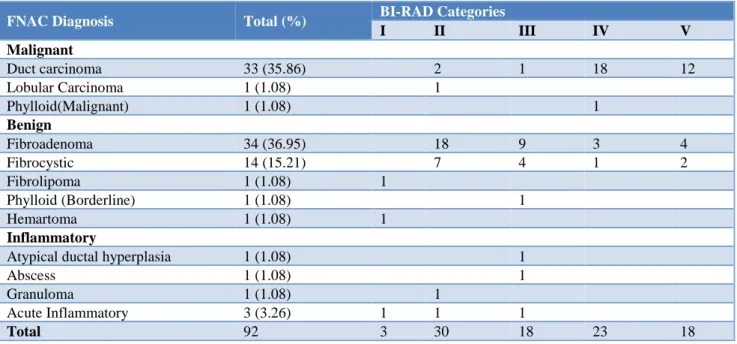
A total number of 92 patients underwent both mammography’s with BI-RADS grading and FNAC procedure in diagnosis of breast lumps in the department of Pathology. There were 51 (55.43%) lesions reported to be benign and 35 (38%) were found to be malignant. Among benign diseases, fibroadenoma was the most common 34 (66.6%) whereas in malignant cases; invasive duct carcinoma was the maximum numbers 33 (94.28%). On mammography, there were no patients in categories 0 and 6. There were 3 (3.26%) patients in category I, 30 (32.6%) patients in category II, 18 (19.5%) in category III, 23 (25%) in category IV and 18 (19.5%) patients in category V.
In this study concordant benign were observed in 47 (51%) cases whereas concordant malignant were observed in 31 (33.6%) cases. Four cases (3 were BI-RADS score II and one was BI-RAD score III) on mammography, of which three were diagnosed as ductal carcinoma in-situ and the other as lobular carcinoma in FNAC.
Both were later confirmed on histopathological examination. These cases constituted the false negatives or disconcordant benign in this study. In this study series, on mammography 10 cases (4 cases were given IV and 6 cases were given V) on BI-RADS i.e. suspicious of malignancy, out of which 7 were diagnosed as fibroadenoma and 3 were as fibrocystic disease on FNAC and later on confirmed on histopathological examination. These cases thus constituted the false positives or disconcordant malignant cases in authors study. In this study, on FNAC diagnosis no false positive and false negative cases were observed showing the comparison of BI-RADS scoring and FNAC findings respectively (Table 1).
The youngest patient in this study was 19 years of age and the eldest was 80 years of age. In this study series maximum numbers of cases were seen in 31-50 years age group. Mean age was 43.1 years.
Maximum numbers of malignant cases 23 (65.71%) were observed above 40 years age where as 31 (54.38%) benign cases were observed below 40 years age. In this study 12 (34.28%) malignant cases (10 duct carcinoma, 1 lobular carcinoma and 1 malignant phylloid tumor) were observed below 40 years age, out of which 2 cases were observed in 21 to 30 years age. The youngest malignant patient was 25 year old (Table 2). Maximum numbers of disconcordant cases were observed in below 40 years age (Table 3).
respectively for BIRADS score and 100% for all the parameters on FNAC.
Table 1: The comparison of mammography BI-RADS scoring and FNAC findings in palpable breast lump.
FNAC Diagnosis Total (%) BI-RAD Categories
I II III IV V
Malignant
Duct carcinoma 33 (35.86) 2 1 18 12
Lobular Carcinoma 1 (1.08) 1
Phylloid(Malignant) 1 (1.08) 1
Benign
Fibroadenoma 34 (36.95) 18 9 3 4
Fibrocystic 14 (15.21) 7 4 1 2
Fibrolipoma 1 (1.08) 1
Phylloid (Borderline) 1 (1.08) 1
Hemartoma 1 (1.08) 1
Inflammatory
Atypical ductal hyperplasia 1 (1.08) 1
Abscess 1 (1.08) 1
Granuloma 1 (1.08) 1
Acute Inflammatory 3 (3.26) 1 1 1
Total 92 3 30 18 23 18
Table 2: Age wise distribution of breast diseases.
FNAC Diagnosis 11-20 21-30 31-40 41-50 51-60 61-70 71-80 Total
Malignant
Duct carcinoma 2 8 13 9 1 33
Lobular Carcinoma 1 1
Phylloid (Malignant) 1 1
Benign
Fibroadenoma 1 6 15 8 2 2 34
Fibrocystic 7 4 1 1 1 14
Fibrolipoma 1 1
Phylloid 1 1
Hemartoma 1 1
Inflammation
Atypical ductal hyperplasia 1 1
Abscess 1 1
Granuloma 1 1
Acute inflammatory 3 3
Total 1 9 39 25 12 5 1 92
Table 3: Age wise distribution of concordant and diasconcordant breast lesions.
BI-RAD Vs FNAC 11-20 21-30 31-40 41-50 51-60 61-70 71-80 Total (%)
Concordant benign (Benign Vs Benign) 1 6 25 10 2 3 - 47 (51%) Disconcordant benign (benign Vs
malignant) - 1 2 1 - - - 4 (4.34%)
Concordant malignant (malignant Vs
malignant) - 1 9 11 9 1 - 31 (33.69%)
Disconcordant malignant (malignant Vs
DISCUSSION
Breast cancer is the most common cancer affecting women worldwide and is the second most common cause of cancer death next to lung cancer.7 Unlike other
countries, in India, it has been rising steadily over the last 10 years and for the first time now; both the incidence and deaths caused by breast cancer are more than cervical cancer in metropolitan cities.8 Although presentation of
breast lump is a symptom/sign for a variety of conditions but most often it cause a great anxiety basically for malignancy.1 So patients with lumps present early and
more to tertiary care hospitals as compared to nipple discharge or any other complaint. In this study maximum numbers of patients with benign diagnosis were in 31-40 years age and 37.14% of malignant were in the 41-50. This finding was consistent with the previous study done by Garg et al.9
Study has shown ,benign breast disease(fibroadenoma) is mostly found in below 20 years group and malignancy is mostly occurring in older than 40 years of age, however, younger women can also be affected especially those with the genetic predisposition.4,10 In India, it has been
observed that the average age of developing breast cancer has undergone a significant shift over the last few decades.8
In a study, 4% were observed in the 20-30 years age group, 16% are in 30-40, and 28% are in 40-50 years age group. Hence, almost 48% patients were below 50 years of age which was similar with authors study findings. An increasing number of patients are in the 25-40 years of age, which is a very disturbing trend.11
According to previous thought those who are literate and conscious about their health and in turn seek medical advice early. Secondly, those who are illiterate neglected patients and inhibition in seeking treatment of the female patients by male doctors.12At this point, the role of
screening mammography and general awareness about breast cancer is very important in authors country as it is designed to detect tumor or other abnormalities.13 In
order to reduce the inter observer variability and standardize the imaging reporting and risk assessment, BI-RADS lexicon was introduced by American college of Radiologists (ACR) in 1993, for mammography imaging of breast, to achieve trick verbal uniformity so as to get clear, unambiguous and standard language, not only among radiologist but also among treating physicians and surgeons whether further specific work up is required or not.
According to latest BI-RADS assessment categories of mammography,
• Category 0: refers to Incomplete- Need Additional Imaging Evaluation and/or Prior Mammograms for Comparison,
• Category 1: Negative,
• Category 2: Benign,
• Category 3: Probably benign,
• Category 4: Suspicious of malignancy,
• Category 4A: Low suspicion for malignancy,
• Category 4B: Moderate suspicion for malignancy,
• Category 4C: High suspicion for malignancy,
• Category 5: Highly Suggestive of Malignancy,
• Category 6: Known Biopsy-Proven malignancy.14
In this study on the basis of the result of mammogram BI-RAD score, 51 cases were in category I, II&III and 41 were in category IV and V and patients who had taken up both FNAC and mammography, (62.9%) patients reported to be having BIRADS I, II and III lesions which revealed 47 (92.15%) benign morphology on FNAC and the majority of these benign cases included Fibro adenoma followed by fibrocystic disease. This finding was consistent with the previous study done by.15
In another study, 88% of BI-RADS III breast lesions were benign and only 6% were malignant, consisting of ductal carcinoma in situ and infiltrating ductal carcinoma.16 Patients reported to be having BIRADS IV
and V lesions which revealed 31(75.6%) malignant morphology on FNAC and the majority of these malignant cases included duct carcinoma insitu.
It has been observed that mammography is one of the most important diagnostic tools in the diagnosis of palpable breast diseases and can successfully clarify the nature of breast lumps especially in older age group (fifty years or above) with less glandular tissue with high incidence of malignant lesions. But it has its limitation especially in younger age as it enhances denser breast density which some time obscure the lesion.17
However, in another study mammography was found a useful imaging technique in providing preoperative detection and diagnosis of breast carcinoma in women below 40 years of age with clinical suspicion of malignancy. Mass and microcalcifications were the most common abnormal mammographic findings and invasive ductal carcinoma were the most common tumor.18
Unfortunately, false-negative mammographic findings in the setting of a palpable breast mass have been estimated at between 4% and 12%.19-21 Therefore, malignancy
cannot be excluded when mammographic findings of a palpable mass are negative.22
Similarly, in another study, among malignant cases, in 99% of the cases turned out to be the correct diagnosis. Four lesions were false positives all of which represented benign proliferative breast diseases.23
to the study done by, who showed total 5 false negative and 8 false positive cases.4 In this study, the diagnostic
accuracy of FNAC was 100 while Rahman et al et al found only 1 false positive and 1 false negative case. On analysis mammography showed 82.76% sensitivity, 90.36% specificity, 75% positive predictive value (PPV), 93.75% negative predictive value (NPV) and 88.39%
accuracy. FNAC showed 97.22% sensitivity, 99.46% specificity, 97.220% PPV, 99.46% NPV and 99.095% accuracy.4 The comparisons of overall diagnostic
accuracy of mammography and FNAC in diagnosis of breast disease with previous study findings are shown in table 4 and 5 respectively.
Table 4: The comparisons of overall diagnostic accuracy of mammography (BI-RADS) score in diagnosis of breast disease with previous study findings.
BI-RADS Mammogram
of breast lump Rahman et al.
4 Bak et al.5 Garg et al.9 Tiwari et al.22 Present study
Sensitivity 82.76% 91% 84.37% 77.7% 88.57
Specificity 90.36% 88% 83.33% 97.72% 82.46%
Positive predictive value 75% 96% - 87.5% 75.61% Negative predictive value 93.7% 71% - 95.5% 92.16%
Diagnostic Accuracy 88.39% - - - 84.78%
Table 5: Overall accuracy of FNAC in diagnosis of palpable breast lump.
FNAC of breast lump Rahman
et al. 4
Bukhari et al. 24
Panjvani et al. 25
Sankaye et al.26
Choi
et al.27 Present study
Sensitivity 97.2% 98 97.82 88.37 77.7 100
Specificity 99.46% 100 100 96.42 99.2 100
Positive predictive value 97.2% 97 97.85 84.37 88 100 Negative predictive value 99.4% 100 100 97.43 98.4 100 Diagnostic accuracy 99.9% 98 98.90 91.54 91.1 100
CONCLUSION
According to authors above study findings, authors concluded that FNAC could be considered an ideal initial diagnostic modality in breast lumps recognized by means of imaging techniques. In experienced hands it is not only give as safe, cost effective, rapid and presumptive diagnostic method but also limits unnecessary testing and procedures.
Funding: No funding sources Conflict of interest: None declared Ethical approval: Not required
REFERENCES
1. Tiwari M. Role of fine needle aspiration cytology in diagnosis of breast lumps. Kathmandu Univ Med J.2007;5(2):215-7.
2. Pruthi S. Detection and evaluation of a palpable breast mass. Mayo Clin Proc. 2001;76(6):641-7;quiz 647-8.
3. Kachewar SS, Dongre SD. Role of triple test score in the evaluation of palpable breast lump. Indian J Med Paediatr Oncol. 2015;36(2):123-7.
4. Rahman MZ, Sikder AM, Nabi SR. Diagnosis of breast lump by fine needle aspiration cytology and
mammography. Mymensingh Med J. 2011;20(4):658-64.
5. Bak M, Konyár E, Schneider F, Bidlek M, Szabó E, Nyári T, et al. Quality assurance of fine-needle aspiration cytology of the organized mammography screening] Orv Hetil. 2010; 151(32):1295-8. 6. Spak DA, Plaxco JS, Santiago L, Dryden MJ,
Dogan BE. BI-RADS®fifth edition: A summary of changes. Diagn Interv Imaging. 2017;98(3):179-90. 7. Bray F, Ferlay J, Soerjomataram I, Siegel RL, Torre
LA, Jemal A. Global cancer statistics 2018: GLOBOCAN estimates of incidence and mortality worldwide for 36 cancers in 185 countries. CA: a cancer J Clin. 2018;68(6):394-424.
8. Singla D, Chaturvedi AK, Aggarwal A, Rao SA, Hazarika D, Mahawar V. Comparing the diagnostic efficacy of full field digital mammography with digital breast tomosynthesis using BIRADS score in a tertiary cancer care hospital. Indian J Radiol Imaging. 2018;28(1):115-22.
10. National Cancer Institute. SEER 1973-2001 public-use data. Accessed online October 14, 2004. Available at: http://seer.cancer.gov/publicdata/. 11. International Agency for Research on Cancer.
GLOBOCAN 2012: estimated cancer incidence, mortality and prevalence worldwide [Internet]. 2012 [Accessed on June 2015]. Available at: http://globocan.iarc.fr/Pages/fact_sheets_cancer.asp x.
12. Jaffary A, Mansuri F, Shani S, Shani S. Knowledge, attitudes and practices regarding breast cancer screening in women of various social strata. J Surg Pak. 2005;10:44-7.
13. Cusheiri A, Steele RJ, Mossa A. Essential surgical practice. 4th ed. Oxford: Butterworth Heinimann; 2002:72-73.
14. Acr.org. 2019. Available at: https://www.acr.org/- /media/ACR/Files/RADS/BI-RADS/BIRADS-Reference-Card.pdf.
15. Bhramaramba K, Padma S, Hemalatha S, Murthy NLN. A comparative study of FNAC breast lesions and BIRADS grading with a focus on grade 4 and grade 5 categories. Int Arch Integrative Med. 2017;4(9):156-63.
16. Engohan-Aloghe C, Hottat N, Cosaert J, Boutemy R, Fayt I, Noël JC. Evaluation of accuracy of fine needle aspiration cytology in BI-RADS3 category breast lesions: cytohistological correlation in 337 cases. Cytopathology. 2010;21(3):161-3.
17. Jatala S, Fitzgerald S, Tietze P, Ramakrishnan K, McCarthy LH, Wickersham E. What Are the Recommended Timing and Screening Modalities in Women at Higher Risk for Developing Breast Cancer? A Clin-IQ. J Patient Cent Res Rev. 2015;2(1):38-42.
18. Muttarak EM, Pojchamarnwiputh S, Chaiwun B. Breast cancer in women under 40 years: preoperative detection by mammography. Ann Acad Med Singapore. 2003;32(4):433-7.
19. Berg WA, Gutierrez L, Nessaiver MS, Carter WB, Bhargavan M, Lewis RS, et al. Diagnostic accuracy of mammography, clinical examination, US, and MR imaging in preoperative assessment of breast cancer. Radiol. 2004;233(3):830-49.
20. Coveney EC, Geraghty JG, O'Laoide R, Hourihane JB, O'Higgins NJ. Reasons underlying negative mammography in patients with palpable breast cancer. Clin Radiol. 1994;49(2):123-5.
21. Kerlikowske K, Smith-Bindman R, Ljung BM, Grady D. Evaluation of abnormal mammography results and palpable breast abnormalities. Ann Intern Med. 2003;139(4):274-84.
22. Tiwari P, Ghosh S, Agrawal VK. Evaluation of breast lesions by digital mammography and ultrasound along with fine-needle aspiration cytology correlation. J Can Res Ther. 2018;14(5):1071-4.
23. Engvad B, Laenkholm AV, Schwartz W, Bak M. Fine needle aspiration cytology of mammography screening. Ugeskr Laeger. 2009;171(34):2379-82. 24. Bukhari MH, Arshad M, Jamal S, Niazi S, Bashir S,
Bakhshi IM. Use of fine-needle aspiration in the evaluation of breast lumps. Pathol Res Int. 2011;2011:689521.
25. Panjvani SI, Parikh BJ, Parikh SB, Chaudhari BR, Patel KK, Gupta GS, et al. Utility of fine needle aspiration cytology in the evaluation of breast lesions. J Clin Diagn Res. 2013;7(12):2777-9. 26. Sankaye SB, Dongre SD. Cytological study of
palpable breast lumps presenting in an Indian rural setup. Indian J Med Paediatr Oncol. 2014;35(2):159-64.
27. Choi YD, Choi YH, Lee JH, Nam JH, Juhng SW, Choi C. Analysis of fine needle aspiration cytology of the breast: A review of 1,297 cases and correlation with histologic diagnoses. Acta Cytol. 2004;48(6):801-6.