Immunophenotypic characterization of acute lymphoblastic leukemia
in a flowcytometry reference centre in Sri Lanka
Manujasri Wimalachandra1, Manurie Prabashika1, Medhavini Dissanayake1, Roshan de Silva2, Lallindra Gooneratne1,2
(Index words: acute lymphoblastic leukemia, acute leukaemia, immunophenotype)
1Faculty of Medicine, University of Colombo, Sri Lanka, 2Asiri Group of Hospitals, Sri Lanka.
Correspondence: MW, e-mail: <[email protected]>. Received 13 February 2019 and revised version 28 June 2020 accepted 02 July 2020.
Abstract
Background Acute lymphoblastic leukemia (ALL), is a biologically heterogeneous disease where diagnosis, treatment and prognosis is heavily dependent on the correct characterization of the immunophenotype of each case.
Objectives To describe the immunophenotypic and laboratory features of a cohort of Sri Lankan children and adults with ALL and to compare them with those reported in other series.
Methods Records of 229 patients who were suspected of having acute leukaemia and referred for flow cytometry to the Asiri Hospital Sri Lanka, between August 2009 and October 2013 were reviewed. Referrals were from across the country including the National Cancer Institute.
Results Sixty seven percent were children below 12 years of age. 80% had B-lymphoblastic leukaemia (B-ALL), 20% T-lymphoblastic leukaemia (T-ALL). In children, the peak age of diagnosis was between 2-6 years in B-ALL (61%) and 3-6 years (43.7%) in T-ALL. Incidence was commoner in males in all age groups and subtypes except in the adult B ALL group where the incidence was equal. Ninety three percent of paediatric B-ALL and 67% adult B-ALL were CD10 positive common ALL. In T-ALL cytoplasmic expression of CD3 was 100%. nTdT was the most commonly expressed immature marker, in B-ALL - 91% and in T-ALL - 69%. At least one myeloid antigen was present in 34% of B-ALL cases and 60% of T-ALL cases.
Conclusions This study represents the first immuno-phenotype based description of ALL in Sri Lanka. Age, sex distribution, ALL subtypes and the immuno-phenotypic profile of each subtype mirrored those of previous studies.
Introduction
Acute lymphoblastic leukemia (ALL), a neoplasm of precursor cells committed to B or T cell lineage has a world wild incidence of 1-4.75/100000 persons per year [1,2]. Seventy five percent of cases occur in children less than 6 years of age, which makes it the most common form of leukaemia in children.
ALL is a biologically heterogeneous disease with clinical features, morphology, immunophenotype, cytogenetic and molecular genetic abnormalities having important implications in the diagnosis, classification, risk stratification, minimal residual disease analysis and prognosis. This was highlighted with the introduction of the WHO system of classification of haemopoetic neoplasms that replaced the purely cytomorphology and cytochemistry based French and British system [2]. Various subtypes of ALL and their different characteristics have shown to differ among geographic distribution, ethnicity, and socioeconomic status [1,3,4,7-10]. As of to date there is a lack of published data on ALL based on this classifi-cation in Sri Lanka.
The objective of this study was to describe the immunophenotypic and laboratory features of a cohort of Sri Lankan children and adults with acute lymphoblastic leukaemia and to compare them with those reported in other series.
Methods
Records of 229 consecutive patients who were diagnosed with ALL at the flow cytometry unit of the Asiri Hospital Sri Lanka, during an 18 month period were reviewed. Immunophenotyping by flow cytometry was performed on bone marrow aspirate or peripheral blood samples. Samples were collected in ethylenediaminetetra acetic acid (EDTA) containing tubes and analysed within 24 hours of collection. Immunophenotyping was done using a Becton Dickinson FACS Calibur flowcytometer. BD Ceylon Medical Journal 2020; 65: 23-27
DOI: http://doi.org/10.4038/cmj.v65i1-2.9133
CellQuest Pro software was used for analysis. The reagents used in order to detect cluster of differentiation markers (CD) were selected in accordance with the guideline issued by the general haematology task force of the British committee for standards in haematology [3]. Immunophenotype of blasts was determined when at least 20% of the blasts expressed the marker at fluorescence intensity above cutoffs established using the corresponding type-matched control antibody.
Results
1. The age and sex distribution at presentation for the B-ALL and T-ALL groups
Out of the total 229 subjects, 157 (68.6%) were children below 12 years of age and 72 (31.4%) were adults. The age and sex distribution in B ALL and T ALL are shown in table 1.
Immunophenotype B-ALL T-ALL
Gender Female Male Female Male
No % No % No % No %
Adult 2 2 9.6 2 1 9.2 7 3 2 2 9.6
Paediatric
<2 yrs 7 3 9 3.4 0 0 0 0
2-5 yrs 1 9 8.3 4 1 17.9 6 2.6 2 0.8
>5 2 5 10.9 4 0 17.5 1 0.4 7 3
*Percentages are out of total cases
Table 1. Age and sex distribution of patients
The median age of presentation among children in the B-ALL group was 4.3 years (range 0.5-12) and in the T-All group 5.5 years (range 1-11). In adults, the median age of presentation was, 27 years (range 13-78) in B All while in T ALL it was 25 (range 13-67).
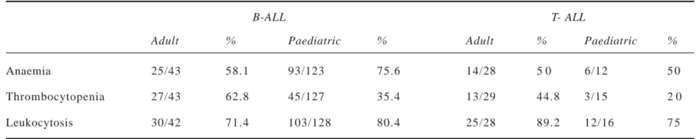
2. The prevalence of anaemia, thrombocytopenia and leukocytosis at presentation
The prevalence of anaemia (Haemoglobin below 100g/L), thrombocytopenia (Platelet count below 100×109/L and
leucocytosis (Leucocyte count > 11×109/l) is shown in table 2.
B-ALL T- ALL
Adult % Paediatric % Adult % Paediatric %
Anaemia 25/43 58.1 93/123 75.6 14/28 5 0 6/12 5 0
Thrombocytopenia 27/43 62.8 45/127 35.4 13/29 44.8 3/15 2 0
Leukocytosis 30/42 71.4 103/128 80.4 25/28 89.2 12/16 7 5 Table 2. The prevalence of anaemia, thrombocytopenia and leukocytosis in different age groups
In the B ALL group 21.4% of adult patients and 27.3% of paediatric patients presented with a total leucocyte of count >100000/µl. 41.37% of adults with T ALL and 25% of children with T ALL had WBC count >100000/µl at presentation.
3. Immunophenotype
cytopenia and total white cell count at presentation have also been studied for their prognostic significance.
In our study both B and T-ALL were commoner in the paediatric group than in adults. B-ALL was more frequent than T-ALL independent of the age group analyzed. The geographic variation of ALL is well established [1,4,5]. This difference has been linked to ethnicity and socioeconomic status. Western data clearly shows a higher incidence of B-ALL over T-ALL. However many older studies done among Asian, African and middle eastern populations have shown a higher incidence of T-ALL [5-7]. Our data also show a higher incidence in the B-ALL category with frequencies similar to that reported in the Americas and Europe. However there is evidence to state that the geographic difference in incidence is more likely due to the socioeconomic status of the population than ethnicity. This was highlighted by two independent studies carried out in south and north India, that showed the change in trends from T-All to B-ALL with the changing socioeconomic status of a single concerned population [5,6]. Similar changes have been observed in other developing nations [4]. We feel our data also supports the same hypothesis as although still considered a low middle income country from south Asia, the Sri Lankan population reaps the benefits of a well organized health care system that has pushed many of its health parameters in par with developed countries.
The 2-5-year age category is considered the peak age of incidence among children with ALL. Similar to the incidence of ALL subtypes, the age of peak incidence has also been linked to the socio economic status of a population with the said peak occurring in populations with higher socioeconomic status. In this cohort, the peak age at diagnosis was between the ages 2-6 years accounting to 61% of B-ALL. The age at first diagnosis is considered a strong predictor of disease outcome [8]. Few cases, 8 (5.7%), presented less than 1 year of age and, more than 10 years, 14 (9.8%) both these categories are associated with an adverse prognosis. Peak age of presentation of T-ALL in childhood was 3-6 years with 43.7% of patients belonging to this group. Two (12.5%) patients were less than 1 year and 2 (12.5%) were >10 years.
Among the adult patients both T-ALL and B-ALL were common in the 13-26 age group amounting to 43% and 72.4% cases respectively. Few adult patients (one B-ALL and 6 T-ALL) were diagnosed after the age of 60 years, which is also associated with a poor prognosis [9]. The commonly described, bimodal distribution and “second lower peak” after 70 years of age was not observed in this cohort.
The incidence of ALL was more in males in all age groups and subtypes except in the adult B-ALL group where the incidence was almost equal (21 males to 22 female). Male sex is described as a risk factor for relapse of disease. [1,2,8,10].
CD4 and CD8 expression was checked in only 18 adult patients and 5 childhood cases. Out of them 47.82% of cases were negative for both CD4 and CD8. CD 4 and CD8 double positivity was seen in only 26.1% of cases. 30.4% cases showed either CD4 or CD8 positivity.
Discussion
ALL is predominantly a disease of childhood amounting to 80% of all childhood leukaemias. The incidence is higher in males and affects Caucasians more than Asian, African and Hispanic populations. Common presenting features include anemia, infections, bleeding and bone pain. Immunophenotyping is an essential tool in the classification of leukaemia as it provides information on the lineage and stage of maturation. It is also useful for identifying subtypes, monitoring of disease and for identifying markers of prognostic relevance. Additional data such as age, sex, presence of anaemia,
thrombo-Immune markers Adult group Paediatric group
CD19 95.3% 100.0%
CD20 27.9% 17.7%
CD10 67.4% 92.9%
CD79 a 81.3% 94.3%
CD34 58.1% 69.5%
n T d T 83.7% 90.8%
CD34 or nTdT 90.2% 96.4%
CD13 20.9% 12.1%
CD33 27.9% 22.9%
CD 13 or 33 36.5% 33.5%
Table 3. The frequencies of positive immune markers in B-ALL
Immune markers Adult group Paediatric group
Cyto CD3 100.0% 100.0%
CD7 100.0% 68.8%
CD5 72.4% 62.5%
CD 2 81.3% 43.8%
CD34 27.6% 6.2%
n T d T 69.0% 68.8%
CD34 or nTdT 75.9% 68.8%
CD13 50.0% 55.6%
CD33 34.5% 18.8%
CD13 or 33 72.4% 55.6%
Leucocytosis is a common presentation in paediatric and adult patients with ALL. We found that adult patients with T ALL had a higher mean WBC count at presentation compared to the paediatric group (p<0.0005). A relatively high prevalence of anaemia was seen with more than half the subjects in all age groups recording a hemoglobin of less than 100g/l at presentation.
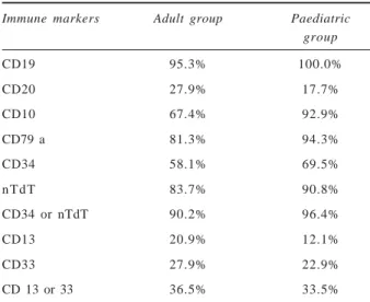
In the immunophenotypic characterization, the most sensitive markers for diagnosis of B-ALL were CD19 and Cyto CD79 a with more than 90% of positivity. Ninety three percent of paediatric B ALL and 67% adult B ALL were CD10 positive common ALL. CD20 expression was seen in only 27.9% of adult B ALL cases and 17.7% of childhood B-ALL cases. These findings roughly mirror those previously presented in most other studies [2,9,10]. The expression of T cell associated antigens CD2, CD7, CD5 and CD3 shows high sensitivity and typifies at least 90% of T ALL cases in both children and adults. Out of these CD3 is lineage specific and cytoplasmic expression was seen in all cases of T ALL. Surface CD3 expression was not checked in most of cases.
There was a relatively high prevalence in the immature markers in B ALL, with nTdT positive in 90.8% of children and 83.7% of adults. CD34 was positive in 69.5% of children and 58.1% of adults. 55.8% adults and 64.5% children with B-ALL showed both CD34 and nTdT positivity.
In the T-ALL group nTdT was positive in 69 % of adult patients and 68.8% of childhood cases. CD34 expression was relatively low and found in 27.6% of adult cases and 6.2% of paediatric cases.
Classification of T-ALL according to maturation stages was difficult in our study as only a limited number of immune markers were analysed. Majority of the analysed cases show negativity for both CD4 and CD8 indicating their origin from more immature pro T or pre T stage. Similar results are reported in previous studies on T-ALL [2]. In contrast T lymphoblastic lymphoma, the tissue counterpart of T-ALL is derived from a more mature thymocyte counterpart as described in previous studies [2,12]. CD4 and CD8 double positivity seen in 26.1% of analysed cases indicated their origin from cortical T cell. Conventionally, development stage of the predominant clone is considered a predictor of outcome [2,8]. Cortical T-ALL carries the best prognosisand early stage T-ALL tend to be worse but once the treatment response is factored into risk alogorithms developmental stage is not considered an independent risk factor [8].
Aberrant expression of myeloid markers in ALL has been found to vary in different studies. And their value as progonostic markers is also a matter of debate [9,13,14]. Clinical outcome studies have shown conflicting data with regard to the prognostic significance of myeloid antigen positivity in terms of achievement of complete remission, response to conventional drug treatment, disease free survival and overall survival at 5 years [13,14]. The overall
incidence of myeloid marker expression in B-ALL was comparable to what is described previously, but in most studies it was found that aberrant myeloid marker expression is more common in B-ALL than T-ALL [10,13,14]. However in our study aberrant myeloid antigen expression was more commonly seen in T-ALL cases. Our results indicate CD13 and CD33 to be the most likely myeloid markers to be expressed with atleast one of them present in 34% of B-ALL cases and 60% of T-ALL cases. Other myeloid antigens like MPO or CD117 were rarely seen. Multiple myeloid antigen expression was also rare. A limitation in our study was that we were able to analyse only limited number of markers some markers of potential interest could not be fully evaluated and sub classification according to the antigen expression corresponding to the maturation stage was not possible. Epidemiological data on ALL show that the incidence, age distribution, sex distribution and cytogenetic features among children can defer on country of origin, ethnicity, and level of socio-economic development. This highlights the importance of maintaining national registries of leukaemia patients for each population and using the data in planning and providing appropriate services in the diagnosis and treatment of leukaemia. We hope that the data reported in our study will serve as a reference for further research in haematological malignancies based on immunophenotyping in Sri Lanka.
List of abbreviations
ALL - acute lymphoblastic leukaemia B-ALL - B-lymphoblastic leukaemia T-ALL - T- lymphoblastic leukaemia EDTA - ethylenediaminetetra acetic acid CD - cluster of differentiation markers
Conflicts of interest
No conflicts of interest
References
1. Redaelli A, et al. A systematic literature review of the clinical and epidemiological burden of acute lymphoblastic leukaemia (ALL). Eur J Cancer Care 2005; 14: 53-62.
2. Swerdlow SH, Campo E, Harris NL, Jaffer ES, Pileri SA, Stein H, Vardiman, JW (Eds.). WHO Classification of Tumours of Haematopoietic and Lymphoid Tissues, Fourth Edition. Lyon, IARC Press. 2008.
3. Bain BJ, Barnett D, Linch D, Matutes E, Reilly JT. Revised guideline on immunophenotyping in acute leukaemias and chronic lymphoproliferative disorders. Clin Lab Haematol 2002; 24: 1-3.
4. Noronha EP, Marinho HT, Thomaz EB, Silva CA, Veras
Maranhão, northeastern Brazil. São Paulo Med J 2011; 129: 392-401.
5. Rajalekshmy KR, Abitha AR, Anuratha N, Sagar TG. Time trend in frequency of occurrence of major immuno-phenotypes in paediatric acute lymphoblastic leukemia cases as experienced by Cancer Institute, Chennai, south India during the period 1989-2009. Indian J Cancer 2011; 48: 310.
6. Sazawal S, Bhargava M, Arya L, Khattar A, Saxena R.
Acute lymphoblastic leukemia in children: Changing immunophenotypic pattern in Northern India. Indian J Med Paediatr Oncol. 2008; 29: 49.
7. Naeem S, Hayee A. Acute lymphoblasticleukaemia – a study of Immunophenotypes. J Pak Med Assoc. 1992; 42: 83-6.
8. Teachy D, Hunger S. Predicting relapse risk of acute lymphoblastic leukaemia. Brit J Haematol 2013; 162: 606-20.
9. Rowe JM. Prognostic factors in adult acute lymphoblastic leukaemia. Brit J Haematol 2010; 150: 389-405.
10. Hann IM, Richards SM, Eden OB, Hill FGH. Analysis of the immunophenotype of children treated on the Medical Research Council United Kingdom Acute Lymphoblastic
Leukaemia Trial XI (MRC UKALLXI). Leukemia 1998;
12: 1249-55.
11. Salem DA, Abd El-Aziz SM. Flowcyto-metric-immunophenotypic profile of acute leukemia: mansoura experience. Indian J Hematol Blood Transfus 2012; 28: 89-96.
12. Jay L. Patel, Lynette M. Smith, James Anderson, Minnie Abromowitch, Dario Campana, Jeffery Jacobson, Mark A Lones, Thomas G. Gross, Mitchell S. Cairo, Sherrie L. Perkins. The immunophenotype of T-Lymphoblastic lymphoma in children and adolescents, Brit J Haematol 2012; 159: 454-61.
13. Vitale A, Guarini A, Ariola C, Meloni G, Perbellini O, Pizzuti M, De Gregoris C, Mettivier V, Pastorini A, Pizzolo G, Vignetti M. Absence of prognostic impact of CD13 and/or CD33 antigen expression in adult acute lymphoblastic leukemia. Results of the GIMEMA ALL 0496 trial. Haematologica 2007; 92: 342-8.