Address for correspondence Dr, Sajad Ahmad Salati
Associate Professor , Department of Surgery, Unaizah College of Medicine , Qassim University, Saudi Arabia.
Email: [email protected]
Review Article
Keloids – an extensive review in the light of recent
literature
Sajad Ahmad Salati
Department of Surgery, Unaizah College of Medicine, Qassim University, Saudi Arabia.
Abstract Keloids are benign but challenging wound healing ailments that manifest clinically in variable and diverse forms. Patients present with one or very few small slow growing lesions on one extreme or else with numerous and very large, life-changing and debilitating lesions covering significant portion of their skin. The keloids require a comprehensive treatment plan and approach and most lesions need more than one modality of treatment. Proper assessment requires accurate quantification of the disease’s impact on the patients in terms of physical symptoms, quality of life and psychosocial status. This review focuses on the natural history, clinical features and therapeutic update of keloids in the light of the recent literature
Key words
Keloid, hypertrophic scars, fibroblast, collagen, treatment, prevention, biology.
Introduction
Wound repair represents interplay between cellular processes that act to restore skin integrity and function after trauma and ultimately end in scar formation. Keloid represents the result of an abnormal wound repair when excessive scar tissue is deposited within and beyond the boundaries of the wound. Keloids pose aesthetic as well as functional problems and are psychologically debilitating. This review discusses the epidemiology, pathophysiology, clinical features, and management of keloids in the light of the recent literature.
Methods
The original articles and case reports dealing with the biology, prophylaxis and treatment
strategies for hypertrophic scars and keloids were searched keloids were reviewed in PubMed, HINARI, Google Scholar, Web of Science and Cochrane library databases after
search on the keywords: Keloid and
hypertrophic scars and treatment and biology. Only the literature published in English was included and all articles in languages other than English were excluded. Time limits were set from 1 January 2012 to till date. In addition, important references from earlier dates and abstracts of non-English articles that appeared as cross references in the included articles were also reviewed and used where ever necessary.
Historical background
in his book “Treatise on Skin Diseases and Things of Mind” published in 1790, probably a misnomer since the clinical description was suggestive of keloid.1
Baron Jean-Louis Alibert (1768–1837), a French pioneer in dermatology , identified the keloid as an entity in 1806 . He called them ‘cancroïde ‘, but later around 1817 changed the name to ‘chéloïde’ to avoid confusion with cancer. The term is derived from the Greek χηλή, chele, meaning "hoof", here in the sense of "crab pincers", referring to the claw like extension of the lesions with a tendency to grow in a lateral direction and the suffix -oid, meaning "like".1
Pathogenesis
Skin injury due to any case such as a surgical incision, a burn or a chronic disease state such as acne, is resolved typically through a process of repair resulting in scar formation.
The classical model of wound repair initially involves hemostasis followed by three distinct, but overlapping phases that follow a time
sequence: the inflammatory phase, the
proliferative phase and the remodeling phase.
The inflammatory phase lasts for approximately 2–3 days after injury and by its end; the process achieves reepithelialization thereby restoring the
skins continuity and protective function.2
As the inflammatory phase resolves, the repair
process progresses beneath the healed
epithelium. Myofibroblasts are formed from the dermal fibroblasts in the dermis adjacent to the wound after they get activated. They migrate into the granulation tissue where they produce Type 3 collagen matrix which leads to wound contraction bringing the wound edges together. The final phase involves remodeling of collagen matrix and replacement with Type 1 collagen,
resulting in the formation and maturation of the visible scar tissue and can last a year or more. For a wound to heal effectively, all phases should occur properly and in the right sequence. Scarring is deemed as abnormal when deposition of dermal collagen and fibrosis is excessive or suboptimal. Uncontrolled deposition of dermal collagen results in formation of benign, fibroproliferative lesions, hypertrophic scars and keloids.3
After the initial injury and the formation of a wound clot, the balance between granulation tissue degradation and biosynthesis becomes essential to adequate healing. Keloids as compared to normal mature scar tissue have an
increased blood vessel density, higher
mesenchymal cell density, a thickened
epidermal layer, and increased mucinous ground substance. The alpha–smooth muscle actin
fibroblasts, myofibroblasts important for
contractile situations are very few. The collagen fibrils in keloids are more irregular, abnormally thick, and have unidirectional fibers arranged in a highly stressed orientation.4
In keloids, TGF-B1 causes an increase in type-1 collagen through activation of COL1 genes, but a decrease in matrix metalloproteinase-1 (MMP-1, interstitial collagenase). The net result is deposition of collagen and fibrosis. Furthermore, it has also been seen that TGF-B1 increases MMP-2 (gelatinase), allowing keloid fibroblasts to migrate 2.5 times faster than normal dermal fibroblasts. This may partially explain the proliferative and spreading nature of keloids over time. Similarly Type III collagen,
chondroitin 4-sulfate, fibronectin and
glycosaminoglycan content are higher in keloids. Collagen cross-linking is greater in normal scars, while keloids have immature cross-links that do not form normal scar stability. Mast cell population and hence histamine production within keloid scars is increased which explains the pruritus associated with these lesions.4-5
Recent advances in understanding of biology of wound healing with relation to keloids
Various factors have been proven to influence the pro-fibrotic or anti-fibrotic pathways and hence determine the outcome of wound repair process.
Hypoxia
Oxygen has been known to be an important factor in wound repair.6 There have been many reports in literature suggesting that a hypoxic environment is associated with keloid formation. A recent study by Touchi et al indicated that the central portion of keloids is severely ischemic as compared to that of normal scar or hypertrophic scar. The investigators found greater expression of hypoxia-induced factor-1α (HIF-1 α) as well as lesser vascular density, in the center than along the periphery of these lesions.7
Zhao et al. conducted a study to investigate if hypoxia drives the transition of dermal fibroblasts to myofibroblasts and to clarify the potential transduction mechanisms involved. They observed that keloids are relatively hypoxic and that hypoxia drives the transition of normal dermal fibroblasts to a myofibroblast-like phenotype [high expression of α-smooth muscle actin (α-SMA) and collagen I and III]. Hypoxia effectively facilitated the nuclear import of the Smad2 and Smad3 complex, while blockade with the Smad3 inhibitor, SIS3,
significantly impaired the expression of
hypoxia-induced fibrosis-related molecules.
Hence the study demonstrated that hypoxia facilitates the transition of dermal fibroblasts to myofibroblasts through the activation of the TGF-β1/Smad3 signaling pathway and these findings are expected to provide a potential target for the treatment of keloids.8
A recent study by Okuno et al suggested that keloids have a prosurvival mechanism in which enhanced autophagy and glycolysis in the fibroblasts of a keloid’s hypoxic central zone aids in the extrusion of lactate to fibroblasts in the normoxic peripheral zone via metabolic coupling.9 Increased autophagy also appears to inhibit central-zone apoptosis. The study results indicate that autophagy inhibitors and MCT4
blockers may have potential therapeutic
implications in keloid treatment.9
Angiogenic imbalance and Periostin
levels were downregulated in keloid patients in comparison to normal controls in both sera and
tissue. The study suggested that the
antiangiogenic therapeutics based on endostatin in combination with current curative strategies could present a scope for the effective
management of keloids.10
Periostin is a secreted extracellular matrix (ECM) protein involved in angiogenesis and was originally identified in osteoblasts, periosteum and periodontal ligament. This matricellular protein has lately been found to be expressed in the basement membrane, dermis and hair follicle.11
Zhang Z et al in an effort to elucidate the underlying regulatory mechanism of keloid angiogenesis and its imbalance conducted a study aimed at examining the impact of periostin on angiogenesis in keloids. The study found that the vessel density was higher in keloids
compared with normal tissue, observed
following staining with CD31 and CD105. Further, the expression of periostin was upregulated and demonstrated a markedly positive correlation with blood vessel density. The upregulated periostin activates the ERK1/2 and FAK signaling pathways leading to increased secretion of vascular endothelial growth factor and angiopoietin-1 in the keloid fibroblasts, thereby promoting angiogenesis. The study suggested that upregulation in the level of periostin may be a key factor in keloid development and hence periostin may, therefore, be a novel therapeutic target in the treatment of keloids.12
MicroRNAs
MicroRNAs (miRNAs) are a class of small non-coding RNAs which play a role in the regulation of gene expression at the posttranscriptional level by degrading their target mRNAs and/or
inhibiting their translation. Recently
researchers have performed miRNA expression microarrays in keloids and reported miRNAs to be deregulated (upregulated or downregulated) in keloids, suggesting a potential in the treatment of keloids.13-15
Decorin
Decorin is a proteoglycan component of dermal connective tissue that binds to type I collagen fibrils and neutralizes the stimulatory effects of
TGF-β on collagen, fibronectin and
glycosaminoglycan synthesis. This protein has been found to be decreased in keloids. Decorin also displays inhibitory action on angiogenesis by interacting with VEGF receptors (VEGFR2) and by inhibiting hepatocyte growth factors and PDGF. Decorin, due to these antifibrotic properties. Hence Decorin is currently being investigated as a potential future therapeutic agent.16-17
Fibroblast activation protein-alpha (FAP-α)
Fibroblasts are proven to be the key cellular mediators of fibrogenesis in keloid .Fibroblast activation protein alpha (FAP-α) and dipeptidyl peptidase IV (DPPIV) are proteases located at
the plasma membrane promoting cell
invasiveness and have been previously
fibroblasts in wound healing and normal adult tissues are generally FAP-α negative, inhibiting FAP-α/DPPIV activity may be a novel treatment option to prevent keloid progression.18
Adiponectin
Adiponectin is an adipocyte-derived protein hormone that is known to exert pleiotropic biological effects on metabolism, inflammation, vascular homeostasis, apoptosis and immunity. Recently, adiponectin has been suggested to attenuate the progression of human dermal fibrosis and studies have been undertaken to understand the underlying mechanism. Limin L et al in their study investigated the expression of adiponectin and adipoRs in keloids and normal skin tissues and revealed the signal transduction pathway by which adiponectin mediated Connective Tissue Growth Factor (CTGF )activity. The study suggested that adiponectin may become a potential focus for studies of the pathogenesis of keloids.19
Wnt/beta-catenin pathway
There are evidences that suggest that Wnt signalling and its effector beta-catenin play an important role in wound healing. Sato M studied the role of Wnt/beta-catenin signaling in TGF-β induced collagen deposition in hypertrophic scars and keloids. The study provided evidence that beta-catenin protein levels are elevated in keloid and that TGF-beta induces activation of beta-catenin mediated transcription in human dermal fibroblasts via the Smad3 and p38 MAPK pathways. These findings may prove in future to be relevant in understanding the
pathogenesis of keloids.20
Epidemiology and Clinical features
Keloids are formed when the pathological scarring process extends beyond the margin of
the original wound and is the most extreme type of scarring. Keloids can develop in predisposed individuals at any site where skin trauma has occurred though they occur more frequently on
shoulders, chest, neck, upper arms and cheeks.21
Keloids have also been reported in literature to occur on eyelids, genitalia, palms, soles, cornea
or mucous membranes.22-24
The precipitating factors include pimples, body piercing, insect bites, scratching, burns, surgical incisions, vaccination or other skin injury. Pruimboom T and Scheltinga M R even reported
keloid formation due to repeated
mammographies.25 Rarely cases are reported in
literature proving that keloids can also develop
spontaneously without any precipitating
injury.26-28
They are more frequent in Africans /African Americans and Asians with incidence rates as high as 15–20% in the black skinned population. Although they can occur in all skin types, there are no reports of keloid formation in albinos 29. Keloids occur in all age groups although mainly seen around the puberty and both the genders are equally affected.
On physical examination, keloids are
characterized by a well-circumscribed, firm, irregular, mildly tender, bosselated and
pink-purple lumps usually accompanied by
hyperpigmentation, and a glossy surface with occasional telangiectasias.30
Keloids are often asymptomatic but may cause pain (throbbing, sharp needle like or else
aching), burning sensation, pruritus, or
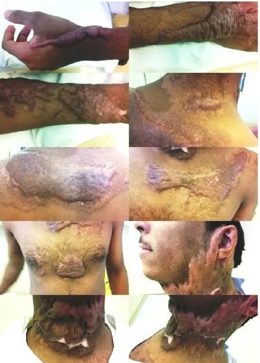
Figure 1 Keloids over multiple areas after nine months of sustaining burn injuries
Individuals may report experiencing some relief
in symptoms when taking a hot shower.25
Besides leadings to significant physical and esthetic disabilities, keloids can have significant psychological and social ill-consequences for patients, and may be associated with substantial emotional and financial burden. Keloids may
lead to sleep disturbances, anxiety, depression, and significant disruption of routine life. Besides, the lesion has a potential to deteriorate
creating contractures, restriction of joint
movements and severe deformities. The
his study concluded that even though keloids do not have a significant negative impact on overall quality of life in communities like a black African population where keloid exists in endemic form but about 47 (35.8%) of the patients believed that keloid swelling limit their social interaction and females (59.1%) felt stigmatized.33
Histopathology of keloids
Butler et al in 2008 summarized four histologic features that are consistently found in specimen
of keloid and therefore are deemed
pathognomonic for keloid diagnosis34:
i. presence of keloidal hyalinized collagen.
ii. tongue-like advancing edge underneath the
normal-appearing epidermis and papillary dermis.
iii. horizontal cellular fibrous bands in the upper
reticular dermis.
iv. prominent fascia-like fibrous bands.
Differential Diagnosis
Hypertrophic scar
Hypertrophic scars are the most important differential diagnosis. They are raised and often discolored, but do not extend beyond the boundary of the initial wound. Hypertrophic scarring typically develops in wounds at locations on the body which are under tension, such as shoulders, ankles, knees, and the neck. Histopathological and immune-histochemical analysis can clear the diagnosis in doubtful case.
Neoplastic lesions
Cases in the literature suggest the presence of various types of malignant tumors that resemble
keloid, including dermatofibrosarcoma
protuberans, trichilemmal carcinoma, and
keloidal basal cell carcinoma. And the careful differential diagnosis is particularly challenging in African-Americans since the skin and tumor color is often similar and hence biopsy is recommended in anomalous cases.
Keloidal blastomycosis (syn:Lobomycosis, lacaziosis, Lobo’s disease )
Keloidal blastomycosis is a chronic keloid-like lesions, slowly growing mycosis of the skin and subcutaneous tissue which produces plaques, nodules, verrucoid lesions, or ulcerated lesions, usually at a site of trauma.35-36
The essential element in the patient's history that should raise suspicion is the exposure to aquatic environments and animals of the Amazon River
basin, particularly fresh water dolphins.
Histopathology reveals abundant fungi and giant cells in the lesions which are granulomatous and devoid of collagenous fibrosis unlike that in keloids.35-36
Nodular contact dermatitis
Contact dermatitis secondary to metallic earrings may produce nodular lesions resembling keloids on the earlobes but histopathological analysis of these lesions shows a dense infiltration of lymphocytes and formation of lymphoid follicles rather than dense collagen tissue. Allergic reaction is more pronounced with nickel and gold is relatively safe material due to its stability and low tendency to ionization. However sporadic reports of allergic contact dermatitis to
gold do appear in literature.37,38 Cases of
palladium induced epithelioid granulomas in the earlobes with a positive patch test are also reported.39
Cutaneous sarcoidosis presents in 9-37% of patients with systemic sarcoidosis, and may mimic keloids or hypertrophic scars. This granulomatous disease of unknown etiology most commonly affects the lungs and is more
prevalent in African-Americans than in
Caucasians though recent studies have shown cutaneous involvement up to 30% in Germany
and about 33% in France.40 Lesions are typically
pink to yellowish-brown or purple, and on diascopy display a classic “apple jelly” appearance. The lesions have a predilection for localization in scars or sites of injury, particularly those containing foreign material (e.g. splinters, suture material, or even silica).40
Atypical mycobacterial infections
Atypical mycobacterial infections are indolent slowly enlarging cutaneous and subcutaneous infections that can mimic keloids. They frequently occur after trauma or surgery, and acid-fast organisms such as Mycobacterium chelonae (subspecies abscessus) have been found in the tap water , disinfectants used in hospital settings and even gentian violet used by surgeons to mark the patients preoperatively.41 Erroneous sterilization of surgical instruments can lead to outbreaks and make it a problem mainly affecting developing countries such as Africa where keloid is otherwise more prevalent. A high index of suspicion is required to accurately diagnose atypical mycobacterial infections, since the usual hallmarks of surgical site infections (pain, fever, worsening erythema) may be absent.
Spitting suture, suture abscess and suture granuloma
These are common causes of postoperative nodularity and hyperplastic growth in surgical wounds and scars. These changes frequently
occur weeks to months after surgery.
Subcuticular sutures may “spit” or cause excess granulation tissue to protrude through the skin and often appear as a shiny red or purple nodule with focal tenderness or pruritus. Spitting sutures may express serosanguinous pus or spicules of partially degraded suture material leading to immediate decompression of the nodule, differentiating it from a keloid. Suture
granuloma may present with classic
inflammatory reaction with erythema, swelling, pain and finally rejection of the suture material or else as a chronic inflammatory reaction with granuloma formation that may present as a solid mass, usually painless and gradually increasing in volume.42
Clinical Genetics of Familial Keloids
Most keloids occur sporadically, but some cases are familial. Various studies have been undertaken to document the genetics and the mode of inheritance of keloid formation and several lines of evidence (increased familial clustering in KD, its increased prevalence in certain races and concordance in identical twins) do suggest that keloid scarring is influenced by genetic factors.43
Marneros et al studied the clinical and genetic characteristics of 14 pedigrees with familial keloids and found the pattern of inheritance to be consistent with an autosomal dominant mode with incomplete clinical penetrance and variable expression. Chen et al arrived at the similar conclusion by studying the clinical and genetic information of six Han Chinese pedigrees with familial keloids with all the family members having no recorded marriage history to other
races except Han people.44,45
among multigenerational families, as a prelude to a positional mapping strategy to identify candidate genes. The study concluded that familial keloids appear to most commonly manifest autosomal dominant or semidominant inheritance, and there may be familial patterns of keloid distribution.46
Brown et al have demonstrated that a genetic association exists between HLA-DRB1 status and the risk of developing keloid scarring in Caucasians of Northern European origin but a similar association is absent in Afro-Caribbean population.47,48
A study by Lu et al supports an association between HLA-DRB1 alleles and susceptibility or resistance to keloids in Chinese Han individuals.49
Systemic Implications and Complications
Hypertrophic scars and keloids alone do not predispose an individual to systemic disorders or complications; however, certain dermatoses may present with an increased number of keloids. Spontaneous keloids have been reported in association with syndromes such as Rubinstein-Taybi syndrome (RSTS), Dubowitz syndrome,
Noonan syndrome, Goeminne syndrome,
Bethlem myopathy, conjunctivo-corneal
dystrophy, X-linked recessive polyfibromatosis, novel X-linked syndrome with flamin A mutation, Ehlers-Danlos syndrome, and the third stage of yaws.31
In Rubinstein-Taybi syndrome (RSTS), keloids
occur in 24% of individuals, either
spontaneously or after a minor trauma, usually starting in early puberty.50
Treatment Strategies
Corticosteroids
Corticosteroids have been a popular treatment for pathological scars since the mid-1960s, and this treatment still continues to play a major role in the management of keloids. Triamcinolone acetonide after its introduction in 1990s has been most popular and is administered intralesional most commonly or else in the form of steroid tapes/ plasters.51
Triamcinolone can suppress vascular endothelial growth factor (VEGF), inhibit fibroblast proliferation, and induce scar regression, which may be the most important mechanism of action. Triamcinolone has also been found to inhibit transforming growth factor (TGF)-β1 expression
and to induce apoptosis of fibroblasts.
Depending on the size and site of the lesion and age of the patient, dosage has varied from 10 to 40 mg/mL, and the dose is administrated at intervals of 3 to 6 weeks for several months or until the scar is flattened. Kontochristopoulos et al. however treated 20 patients with intralesional injections of 5-FU (50 mg/mL) at shorter interval i.e., once weekly for 7 weeks and the follow-up at 12 months revealed that 85% of the
patients experienced greater than 50%
improvement though recurrence rate was 47%.52
Dexamethasone was found by Wu WS et al to cause keloid regression via interaction with the GR and suppression of endogenous VEGF expression and fibroblast proliferation.53
Furthermore, all corticosteroids exhibit
suppressive effects on the inflammatory process in the wound and inhibit collagen and glycosaminoglycan synthesis, fibroblast growth,
and enhance collagen and fibroblast
topical steroid to the classical glucocorticoid receptors.54
Resolution rates for keloids treated with steroids are variable and range from 50-100% and recurrence is seen in 9-50%. Syed F et al. compared the single and combined efficacy of
glucocorticoids-dexamethasone (Dex),
triamcinolone (TAC), and methylprednisolone (Medrol)-on primary keloid fibroblasts (KFs) and normal skin fibroblasts at cellular, protein, and messenger RNA levels in vitro. The study reported the superior efficacy of three well-known steroids on KF and suggested that the combination may be superior than using a single steroid in treating keloids.55
The adverse effects include the pain at injection site, thinning and atrophy of the skin and subcutaneous tissues, the development of steroid acne, capillary dilation, telangiectasia, moulting, the development of secondary lymphogenous
and linear hypopigmentation (temporary/
permanent), high recurrence rates of 9-50%, local skin necrosis /ulceration and Cushing’s syndrome .
Significant pain at injection site is a concern as it causes many patients to discontinue treatment and many studies have been undertaken recently to find means to alleviate the pain. Topical application of anesthetic creams, such as Tetracaine and EMLA cream (lidocaine and prilocaine) to prevent pain on steroid injection site, has limited efficacy in bigger lesions because of poor drug penetration.
Tosa M et al. achieved significant reduction in pain by pretreatment with topical 60% lidocaine
tape.56 Wang X et al applied cryoanesthesia
(cryotip) as pretreatment before injection and found it to be a rapid and effective means of reducing the pain associated with steroid injections.57 Park et al found vibration anesthesia
to be a promising option to effectively and safely alleviate pain, likely by reducing pain transmission from peripheral receptors to the
brain.58 Usanakornkul A and Burusapat C.
administration of mixture of 1% lidocaine and TA did not find it effective in pain relief.59
Park et al. in 2012 in a study concluded that the anatomic location of facial keloids could play a role in re-development, because recurrence rates were greater in perioral regions and that the possible reason for this greater recurrence could be skin tension and wound strain in the highly mobile perioral region.60
To minimize the side effects without
diminishing the effect, steroids are administered in lowest possible doses and in combination with other modalities.
Davison et al. found that the combination of
5-FU and triamcinolone is superior to
triamcinolone alone (15% vs. 40%).61
Furthermore, the triple combination of 5-FU, corticosteroids, and the pulsed-dye laser has been found to be even more effective. Fibroblast activity is suppressed by 5-FU, corticosteroids suppress inflammation and fibroblast activity, and the pulsed-dye laser suppresses angiogenesis and endothelial cells.62
Scar Revision Surgery
During scar revision surgery, tension-free wound closure should be established in order to decrease tension-related inflammation and thereby reduce recurrence and wide range of techniques including three-layered sutures, subcutaneous/fascial tensile reduction sutures, Z-plastics or local flap reconstruction have been
proposed , depending upon the case.63
Cryotherapy
Cryotherapy has been used to treat keloids as a monotherapy or in conjunction with other therapies such as intralesional steroid injections.
Treatments that combine cryotherapy and
intralesional triamcinolone injections
significantly improve keloids.
Delivery methods for cryotherapy are variable and include contact, spray or the needle cryoprobe method. The intralesional-needle cryosurgery device (cryoprobe) was introduced by Zouboulis et al. in 2004 and has since become the most preferred mode of delivery.64
The suggested mechanism underlying
cryotherapy is vascular damage leading to tissue necrosis. The tissues necrotized by cryotherapy secrete unique inflammatory cytokines that alter the responses of myofibroblasts and hence result in transformation of the scar architecture. The collagen fibers become aligned in a more parallel arrangement and the structure tends to
mimic a normal, organized dermis.65 van
Leeuwen et al. in 2015 published the results of a comprehensive review of the articles published on intralesional cryotherapy, based on the Preferred Reporting Items for Systematic Reviews and Meta-Analysis. PubMed and EMBASE. The review showed that this mode produce favorable results in terms of volume
reduction (from 51-63%) and alleviate
complaints of pain and pruritus. However, complete scar eradication was not established, and recurrences are seen in upto 24%. Also, persistent hypopigmentation proved a problem in Fitzpatrick IV-VI skin type patients. Hence, the evidence proved limited and inconsistent resulting in an American Society of Plastic Surgeons grade C recommendation for this type of the treatment of keloid 66.Similar results were found in the study conducted by O’Boyle et al.67
Van Leeuwen et al. in another study have introduced an Argon gas-based system to achieve accurate and highly controlled freezing and found the method to be effective in the treatment of keloid scars, yielding volume reduction and low recurrence rates.68
Radiotherapy
Studies have proven the effectiveness of external beam radiotherapy and brachytherapy in the treatment of keloids. Radiotherapy is generally delivered as an adjuvant treatment one or two days after scar revision surgery, and the recommend radiation dose is 40 Gray over several divided sessions to minimize adverse effects.
Radiotherapy suppresses angiogenesis that results in decreased delivery of inflammatory cytokines, and successive inhibition of fibroblast activity resulting in decreased collagen synthesis and hence suppression of keloid development. The recurrence rate for this modality is around 10%.69,70
Recently, studies have shown effective use of radioactive skin patches that use various kinds of radionuclides like Re-188 and P-32. These patches are used as painless, cost effective monotherapy or else in combination with other modalities. The adverse effects include hyper or else hypopigmentation.72,73
Laser Therapy
Laser treatment involves the use of light energy of a specific wavelength and pulse duration to ablate targeted tissues and vaporize blood vessels, thereby limiting the ability of the inflammatory cytokines to reach the keloids. Studies have found that lasers significantly down-regulates the expression of Connective
Tissue Growth Factor (CTGF). The
proliferation of keloid fibroblasts is thereby significantly inhibited resulting in improved aesthetic and symptomatic outcomes and decreased keloid recurrence.74
Keloids have been treated with several types of lasers including 585-nm pulsed dye laser (PDL),
carbon dioxide (CO2), argon, and neodymium:
yttrium-aluminum-garnet (Nd:YAG) laser. The most popular laser used to treat the keloids is the
585-nm pulsed dye laser (PDL). The
recommended energy is 6.0 to 7.5 J/cm2 (7-mm spot) or 4.5 to 5.5 J/cm2 (10-mm spot) and two to six sessions of treatment may be needed.75-78
The 1064-nm Nd:YAG laser is also popular for treating keloids. For this laser, the recommended energy is 14 J/cm2 (5-mm spot), to be repeated every three to four weeks. Fractional CO2 laser has also been found to be effective.79
Laser therapy is used as a monotherapy or else combined with other modalities like silicone gel sheeting and intralesional steroid injections.80,81 Studies have been conducted to compare lasers with other modalities. Scrimali et al in their
study conclude that in comparison to
radiotherapy, the use of CO2 laser after surgical
excision of keloids has shown great results with no recurrence and without the risk of carcinogenesis.82
One of the most recent developments introduced into laser technology is the nonablative fractional laser (NAFL). Recently NAFL (1540/1550 nm) with a 15 mm handpiece have also been successfully used for the treatment of keloids. However, data regarding recurrence rates in a long-term follow-up is still lacking.83
Possible side effects of laser therapy include hypopigmentation, hyperpigmentation, blister
formation and purpura. CO2 lasers vaporize
tissue, which can aerosolize infectious agents such as the hepatitis and HIV viruses and
thereby potentially endanger health care
personnel.
Intralesional 5-Fluorouracil (5-FU)
5-FU is a pyrimidine analogue, classified as an antineoplastic agent that reduces thymidylate synthase activity thereby inhibiting normal DNA and RNA synthesis. Studies have demonstrated ability of 5-FU to induce fibroblast apoptosis without necrosis and is known to inhibit TGF-β signaling in collagen I production.
Kontochristopoulos et al. in 2005 published the results of 20 patients treated with intralesional injections of 5-FU (50 mg/mL). The results revealed that 85% of the patients experienced
greater than 50% improvement. Biopsy
specimens taken after six injections exhibited a reduction in the amount of hyalinized collagen fibers, regression of the nodular concentric arrangement of the collagen fibers, less prominent vascularity and flattening of the
Since then, further studies have been published proving the efficient role of 5-FU in the management of keloids though adverse effects have also been reported that include wound
ulceration, hyperpigmentation, atrophy,
erythema, tissue sloughing, swelling, pain, moulting, and telangiectasia. However, these complications quickly disappear. To minimize the side effects, studies have suggested lower dosages of the drug or usage in combination with other modalities.85
There is sufficient evidence to suggest that the combination of 5-FU and triamcinolone is superior to triamcinolone alone (15% vs. 40%), as reported by Davison et al.86, Khan et al87, Ren et al88 and Darougheh et al.89 Furthermore, it has been suggested that the triple combination of 5-FU, corticosteroids, and the pulsed-dye laser offer balanced benefit of faster and more efficacious response with lesser adverse effects when compared to individual drug. Fibroblast activity is suppressed by 5-FU, corticosteroids suppress inflammation and fibroblast activity, and the pulsed-dye laser suppresses angiogenesis and endothelial cells.62,90
Bleomycin
Bleomycin, derived from Streptomyces
verticillus, induces fibroblast apoptosis and inhibits lysyl oxidase, a cross-linking enzyme involved in the maturation of collagen and TGF-β1, resulting in net reduction of collagen. Bleomycin blocks the cell cycle via the inhibition of DNA, RNA, and protein synthesis as well as the production of reactive oxygen species.91
Intralesional injection is the preferred method of drug administration. Several studies report the achievement of complete flattening in 54-73% of keloids with resolution of other symptoms
like itching and pain.92,93 Naeini et al. compared
the efficacy of Bleomycin tattoo with that of
cryotherapy combined with intralesional
triamcinolone injection for the treatment of keloids and Bleomycin tattoo to be more effective.94
Manca et al reported use of Bleomycin in combination with electroporation and found it to be an effective treatment for patients affected by large keloid scars or patients who are
non-responders to other treatments.95
Cutaneous side effects, including flagellate
erythema (scratch dermatitis),
hyperpigmentation, Raynaud's phenomenon,
gangrene, fibrosis, eccrine hidradenitis, necrosis of keratinocytes, alopecia, edema, nail changes have been documented in literature. The most common adverse effects are minor ulceration that heal within few days and hyperpigmentation
that may or may not resolve with time.93,96
Interferon (IFN)
Interferon (IFN) α, β and γ are cytokines with antifibrotic, and antiproliferative properties with ability to decrease the synthesis of collagen I and III by inhibition of production of glycosaminoglycans (GAGs) in the fibroblasts, which form the scaffolding for the deposition of dermal collagen. IFN-α2 increases collagenase production whereas IFN-α2b inhibits cell proliferation and TGF-β1 expression. IFN-γ modulates a p53 apoptotic pathway by inducing apoptosis-related genes. IFN-γ inhibits TGF-β and therefore fibrosis, via initial activation of Jak1, which in turn stimulates the negative regulator of collagen YB-1 (Y-box protein-1), which activates Smad7, eventually leading to
TGF-β1 suppression.97
about 85% decrease in depth and volume by treating 20 keloids with a combination of intralesional TAC and IFN alfa-2b compared
with only a nonsignificant improvement
obtained in 20 keloids treated with TAC alone.98
IFN injected into the suture line of keloid excision sites may be prophylactic for reducing recurrences and the use of IFN-α2b has showed
18% recurrence rate when applied to
postsurgical excised keloids as compared to
about 51% with only surgical excision.99 Certain
prospective studies , however have found interferon to be ineffective in the clinical
management of keloids.100
Side effects may be systemic, including flu-like symptoms, fever, headache, arthralgia, fatigue, chills, confusion and other unfavorable reactions such as pain at the injection site, local erythema and edema.
Verapamil
Verapamil is a phenylalkylamine calcium channel antagonist that alters fibroblast shape
from bipolar to spherical, induces
procollagenase expression, inhibits the
synthesis/secretion of extracellular matrix
molecules (including collagen,
glycosaminoglycan and fibronectin) and
increases collagenase.101
Verapamil has also been observed to decrease IL-6 and vascular endothelial growth factor (VEGF) production in the keloid fibroblasts, which translates to decreased cell proliferation, increased apoptosis, and increased expression of decorin, which in turn inhibits fibroblast proliferation and migration.
Wang R et al. in a meta-analysis assessed the effectiveness of verapamil in preventing and treating keloid and the results showed that
verapamil could improve keloid with few adverse effects and was not significantly
different from conventional corticosteroid
injections.102
Alexandrescu et al. found favorable results with 5-FU and verapamil combination in terms of symptomatic relief and decrease in height of the lesions whereas Margaret Shanthi et al. concluded that intralesional verapamil may be a suitable alternative to triamcinolone due to its safer profile.103,104
A recent study by Srivastava et al. revealed that
fractional CO2 laser and verapamil are as
efficient as triamcinolone acetonide (TAC) for treating keloids, except it takes longer for laser and verapamil to act compared to TAC. The study suggested that Verapamil can be used as an alternative treatment modality that is
cost-effective with minimal adverse effects.105
Imiquimod
Imiquimod is a synthetic imidazoquinolone amine, which has potent immunomodulating activity, when topically used. It induces release of interferon and cytokine at the site of skin application, specifically IFN-α, TNF-α, 1, IL-6, and IL-8 that act to decrease excessive collagen production by keloid fibroblasts. Besides, topical application of imiquimod tends to upregulate certain apoptosis-related genes in
keloid fibroblasts.106 In a study by
Chuangsuwanich and Gunjittisomram, the
investigators concluded that Imiquimod 5% cream could effectively prevent recurrence of the excised keloids, especially in the area that had less tension such as pinna. Martin-Garcia and Busquets also conducted a pilot study and the results suggest that imiquimod 5% cream may prove to be a reasonably effective adjuvant therapeutic alternative for the prevention of
Berman and Kaufman also reported that the recurrence rate of excised keloids treated with postoperative imiquimod 5% cream was lower than recurrence rates previously reported in the literature.109
Cacao et al. however suggest that imiquimod 5% cream is not effective in preventing recurrence of keloids over trunk after surgical excision and thereby strongly discourage using imiquimod 5% cream in the prevention of surgically excised trunk keloids.110
When formulated as a 5% cream, imiquimod is a safe and generally well-tolerated drug and the adverse effects include skin erosion, excoriation, flaking, and edema at the site of application.
Future directions of treatment
Interleukin-10 (IL-10)
IL-10 is a cytokine that is known to reduce
inflammatory responses and its
anti-inflammatory effects are mediated through activating AKT and STAT3 phosphorylation, downregulation of IL-10 receptor, and by facilitating crosstalk between the PI3K/AKT and
STAT3 signal transduction pathways.111-113 The
absence of IL-10 is believed to lead to an amplified inflammatory response and abnormal collagen deposition.
Mesenchymal Stem Cell Therapy
Mesenchymal stem cells (MSC) are multipotent stromal cells that can differentiate into a variety of cell types and display immunomodulatory and antifibrotic effects.114
The antifibrotic effects of MSC on various fibrotic diseases such as myocardial infarction, renal or liver cirrhosis have been investigated and reported in literature. On the similar lines,
MSCs are being tried to prevent or attenuate excessive inflammatory processes that are characteristic of keloids. Fang et al. studied impact of Bone marrow derived mesenchymal stems (BMSCs) and found that BMSCs attenuate the proliferative and profibrotic phenotype associated with keloids and inhibit
extracellular matrix synthesis through a
paracrine signaling mechanism.115
Spikeman et al. found that the adipose tissue-derived stromal cells inhibit TGF-β1-induced differentiation and function of adult human
dermal fibroblasts and TGF-β1-induced
contraction in keloid scar-derived fibroblasts, in a paracrine fashion.116
Variable delivery methods and doses are being tried including via systemic injections, local injections (at the wound, intradermal or subcutaneously) or an engineered MSC-seeded tissue scaffold.117-120
The possible mechanisms underlying this mode of keloid treatment include:
i. Promotion of normal angiogenesis that aids
in normal wound healing.
ii. Modulation and inhibition of
proinflammatory cell activity
iii. Antifibrotic activity via downregulation of
myofibroblast differentiation and collagen type I and III production.
More investigations and long-term preclinical studies are still required to apply this method in clinical practice.
Fat Grafting
Autologous fat grafting or lipotransfer,
side effects were rarely reported. Statistical significant improvement of the scar appearance, skin characteristics, pain, and itch with restoration of volume and three-dimensional contour has been reported. The mechanism underlying fat injections is believed to be that transferred fat tissues deliver adipose-tissue
derived MSCs to the wound.121,122
Future randomized controlled trials with a
methodologically strong design are
recommended to confirm the effects of autologous fat grafting on keloids.
Transforming Growth Factor- β (TGF- β)
TGF- β isoforms (TGF-β1,2,3) had long been a
target of anti-keloid therapy. Multiple studies have shown that the ratio of TGF-β3, β1 and 2 is important in scar progression or remission. Various studies had been performed to investigate the effect of exogenous TGF-β1 and 2, neutralizing antibodies and exogenous TFG-β3 and had proven the impact of TGF-β isoforms. TGF-β1 and 2 have been found to
increase fibrosis and TGF-β3 attenuates
fibrosis.123 Recombinant human TGF-β3
(avotermin) showed favorable results in phase I/II clinical trials though results in phase III clinical trials are still unsatisfactory.124-126
Botulinum toxin type A (BoNT-A)
Botulinum toxin is a potent neurotoxin produced by the bacterium Clostridium botulinum. Recent reports have suggested that botulinum toxin type A can minimize scar formation by reducing
muscle tension during wound healing,
modulation of collagen deposition , causing the fibroblast cell cycle to be paused in a non-proliferative state, G0 or G1, influencing TGF- β 1 expression and reducing transcription and expression of profibrotic cytokines in keloid-derived and hypertrophic scar-keloid-derived dermal
fibroblasts.127 BoNT-A may also upregulate
S100A4 gene and downregulate GF-β1, VEGF, MMP-1, and PDGFA genes but there is a
paucity of evidence regarding specific
mechanisms of action.128
Intralesional injection has been used as the preferred delivery method, and treatment outcomes were generally favorable, and patient
satisfaction was high in some studies.
Improvement was also reported in pain,
tenderness and itching sensation.129 Some
studies could not however confirm the suggested clinical efficiency of intralesional BTA for the therapy of keloids.130
Prevention of keloids
Prevention of keloids is undoubtedly an effective measure when there is a wide range of treatment options available but none can qualify to be called an ideal. Hence avoidance of all unnecessary wounds in patients prone to keloid remains an obvious though imperfect solution. Early recognition of keloid formation and prevention of recurrence is also integral in
devising management strategy.131
Various measures mentioned in literature on the subject include:
Tension-Free Primary Closure
Wound epithelialization that is delayed beyond 10–14 days increases the risk of pathological scars, and quick primary closure to achieve rapid epithelialization is mandatory for avoiding inflammation and excess fibrosis. Good surgical technique is important and includes basic wound care steps like:
i. Debridement of dead or severely
ii. Adequate hemostasis to avoid hematoma, seroma or abscess formation.
iii. Tension free closure: The exact molecular
mechanisms that govern the response of skin to physical tension remain uncertain.
However, various pathways potentially
responsible for conversion of mechanical forces
into biochemical responses have been
investigated and reported the phenomenon called
mechanotransduction.132 Gurtner et al. studied
the fibrotic effects of mechanical tension and described the preventive effect of offloading
wound tension on scar formation.133
Passive Mechanical Stabilization
To prevent wound tension and the consequential mechanotransduction, application of prolonged passive mechanical wound stabilization using microporous paper tapes or silicone based products has been reported.
Paper tapes help alleviate scar formation, but silicone-based products continue to be the premier option for prevention because it avoids
repeated epidermal avulsion.134-136 Occlusion
and hydration of scar surface are currently suggested as likely mechanisms of the therapeutic action of silicone gel sheeting rather than an inherent antifibrotic property of silicone.137
Silicone sheets are recommended to be worn for about 12 hours each day for at least 2 months starting from two weeks after primary wound treatment. For areas of consistent movement, where sheeting will not conform, silicone gel is favored and should be applied twice daily.
Recently Fabbrocini et al. assessed the combined efficacy of needling and the use of silicone gel in prevention of keloids and found the results to
be favorable.138 Kwon et al. compared the effect
of topical silicone gel and topical tretinoin cream for the prevention of keloids and found both the modalities to be of help with no significant
difference in outcomes of the two.139
Flavonoids
Flavonoids (or bioflavonoids) are naturally-derived substances from various plants. The efficacy studies testing the ultimate benefit of these flavonoid-containing topical scar creams have provided controversial data. However, quercetin, a dietary bioflavonoid has been recently shown to inhibit fibroblast proliferation, collagen production and contraction of keloid
and hypertrophic scar-derived fibroblasts
through inhibition of the TGF-beta/
Smad-signaling pathway.140
Flavonoids are available in the form of gel and application is started two weeks after primary wound treatment and applied twice daily for four to six months.
Onion Extract (Extractum cepae) and Heparin Gel
Onion extract is believed to have
fibroblast-inhibiting ability that decreases
fibro-proliferative activity and synthesis of
extracellular matrix (ECM), and increases the expression of matrix metalloproteinase MMP-1.141
combination with an occlusive silicon dressing.142
Heparin molecules have a strong tendency to interact with collagen molecules, resulting in the formation of thicker fibrils that and induce
intermolecular bonding in collagen. In
combination, heparin and onion extract have been found to decrease keloid formation
through their inhibitory activity on
inflammation, fibroblast proliferation, and the production capability of fibroblasts.143 Koc et al. found that combination of intralesional TAC and onion extract appears to be superior to TAC alone in the treatment of keloids and hypertrophic scars.144
Pressure Therapy
Cutaneous wound compression has been used not only for prophylaxis but also for treatment of established keloids. Pressure therapy is reported to reduce the signs and symptoms of keloids but the scientific evidence supporting their use is little and their clinical efficacy is also controversial.
The suggested mechanisms underlying pressure therapy include occlusion of blood vessels thus limiting the delivery of inflammatory cytokines, nutrients and oxygen from blood vessels to scar tissue. Increasing apoptosis may be another
mechanism of pressure therapy.145 The
magnitude of desired pressure and the duration of therapy that is used clinically rely on empirical reports as there are no comparative analyses available in literature. The usual recommendation is 15–40 mm Hg for more than 23 hours a day for at least six months while the scar is still active.
The limitations of pressure therapy on sites other than ear lobes is difficulty in adequately fitting the garment to the wounded area and reduced
compliance caused by side effects such as maceration and odor in hot and humid climates.146 Postoperative pressure applied with pressure earrings has been prove to reduce recurrence rates markedly after surgical repair of earlobe keloids.147-149
Long-Term Monitoring
Because of the high rate of recurrence, at least one year of close follow-up is necessary to fully evaluate the effectiveness of therapy and to initiate further management at the earliest sign of recurrence.
Preoperative evaluation and counseling is critical to assess a patient's motivation for treatment and to assess the ability to participate in long-term care and follow-up visits.
Conclusions
Keloid remains a challenging condition, with potential cosmetic and functional consequences to patients. Several therapies exist that function through different mechanisms but there is no universally accepted treatment that can be termed as an ideal. Understanding of the pathogenesis has improved in recent years and it is hoped that in coming years, newer and more targeted therapies would be available.
Before initiating treatment, the physician must educate and inform the patient adequately about the possible recurrence rate and the limitations of current options. Use of various treatment modalities in combination is recommended due to the more favorable outcomes.
Acknowledgments
allowing the usage of images (Figure 1) for academic purposes (including publications).
References
1. Pruimboom T, Scheltinga M.R. Keloid Formation due to Repetitive Mammographies. Case Rep Dermatol 2018;10:257–62
2. Bleasdale B, Finnegan S, Murray K, Kelly S, Percival SL. The Use of Silicone Adhesives for Scar Reduction. Adv Wound Care (New Rochelle). 2015; 4(7):422-30. 3. Andrews JP, Marttala J, Macarak E,
Rosenbloom J, Uitto J. Keloids: The paradigm of skin fibrosis - Pathomechanisms and treatment. Matrix Biol. 2016. 51:37-46
4. Halim A S, Emami A Salahshourifar I, Kannan T P .Keloid Scarring: Understanding the Genetic Basis -Advances, and Prospects. Arch Plast Surg 2012;39:184-9
5. Sidgwick GP, Bayat A. Extracellular matrix molecules implicated in hypertrophic and keloid scarring. J Eur Acad Dermatol Venereol 2012; 26:41-52. 6. Sen CK, Roy S. Oxygenation state as a
driver of myofibroblast differentiation and wound contraction: Hypoxia impairs wound closure. J Investig Dermatol 2010; 130:2701-3.
7. Touchi R, Ueda K, Kurokawa N, Tsuji M. Central regions of keloids are severely ischaemic. J Plast Reconstr Aesthet Surg. 2016; 69(2): e35-41.
8. Zhao B, Guan H, Liu JQ, Zheng Z, Zhou Q, Zhang J, Su LL, Hu DH. Hypoxia drives the transition of human dermal fibroblasts to a myofibroblast-like phenotype via the TGF-β1/Smad3 pathway. Int. J.Mol. Med. 2017; 39: 153-9.
9. Okuno R, Ito Y, Eid N, Otsuki Y, Kondo Y, Ueda K. Upregulation of autophagy and glycolysis markers in keloid hypoxic-zone fibroblasts: Morphological characteristics and implications. Histol Histopathol. 2018; 33(10): 1075-87.
10. Mogili NS, Krishnaswamy VR, Jayaraman M, Rajaram R, Venkatraman A, Korrapati PS. Altered angiogenic balance in keloids: a key to therapeutic intervention. Transl Res. 2012; 159(3): 182-9.
11. Zhou HM, Wang J, Elliott C, Wen W, Hamilton DW, Conway SJ. Spatiotemporal expression of periostin during skin development and incisional wound healing: Lessons for human fibrotic scar formation. J. Cell Commun. Signal. 2010; 4: 99-107. 12. Zhang Z, Nie F, Chen X, Qin Z, Kang C,
Chen B, Ma J, Pan B, Ma Y. Upregulated periostin promotes angiogenesis in keloids through activation of the ERK 1/2 and focal adhesion kinase pathways, as well as the upregulated expression of VEGF and angiopoietin-1. Mol Med Rep. 2015; 11(2): 857-64.
13. Luan Y, Liu Y, Liu C, Lin Q, He F, Dong X, Xiao Z. Serum miRNAs Signature Plays an Important Role in Keloid Disease. Curr. Mol. Med. 2016; 16: 504-14.
14. Wu Z Y, Lu L, Liang J, Guo XR, Zhang PH, Luo SJ. Keloid microRNA expression analysis and the influence of miR-199a-5p on the proliferation of keloid fibroblasts. Genet. Mol. Res. 2014; 13: 2727-38. 15. Kashiyama K, Mitsutake N, Matsuse M,
Ogi T, Saenko VA, Ujifuku K, Utani A, Hirano A, Yamashita S. miR-196a downregulation increases the expression of type I and III collagens in keloid fibroblasts. J. Investig. Dermatol. 2012; 132: 1597-1604.
16. Mukhopadhyay A, Wong M Y, Chan SY, Do DV, Khoo A, Ong CT, Cheong H H, Lim I J, Phan TT. Syndecan-2 and Decorin: Proteoglycans with a difference— Implications in keloid pathogenesis. J. Trauma 2010; 68: 999-1008.
17. Zhang Z, Garron T M, Li X J, Liu Y, Zhang X, Li YY, Xu W S. Recombinant human decorin inhibits TGF- β 1-induced contraction of collagen lattice by hypertrophic scar fibroblasts. Burns 2009; 35: 527–37.
18. Dienus K, Bayat A, Gilmore BF, Seifert O. Increased expression of fibroblast activation protein-alpha in keloid fibroblasts: implications for development of a novel treatment option. Arch Dermatol Res. 2010; 302(10): 725-31.
20. Sato M. Upregulation of the Wnt/ beta-catenin pathway induced by transforming growth factor-beta in hypertrophic scars and keloids. Acta Derm Venereol. 2006; 86(4):300-7.
21. Marneros AG, Krieg T. Keloids--clinical diagnosis, pathogenesis, and treatment options. J Dtsch Dermatol Ges. 2004; 2(11):905-13.
22. Lin-Hai Xie, Sen-Kai Li, Qiang Li. Combined treatment of penile keloid: a troublesome complication after circumcision. Asian J Androl. 2013; 15(4): 575-6.
23. Jung JJ, Wojno TH, Grossniklaus HE. Giant corneal keloid: case report and review of the literature. Cornea. 2010; 29(12):1455-8.
24. Birge O, Akbas M, Ozbey EG, Adiyeke M. Clitoral keloids after female genital mutilation/cutting. Turk J Obstet Gynecol. 2016; 13(3):154-7.
25. Pruimboom T, Scheltinga MR. Keloid Formation due to Repetitive Mammographies. Case Rep Dermatol. 2018; 10(3):257-62.
26. Tiong WHC, Basiron NH: Challenging diagnosis of a rare case of spontaneous keloid scar. J Med Cases 2014; 5:466-9. 27. Jfri A, Rajeh N, Karkashan E. A case of
multiple spontaneous keloid scars. Case Rep Dermatol 2015;7:156-60.
28. Jfri A, Alajmi A. Spontaneous Keloids: A Literature Review. Dermatology. 2018; 234(3-4):127-30.
29. Viera MH, Vivas AC, Berman B. Update on Keloid Management: Clinical and Basic Science Advances. Adv Wound Care (New Rochelle). 2012; 1(5):200-6.
30. Mari W, Alsabri SG, Tabal N, Younes S, Sherif A, Simman R. Novel Insights on Understanding of Keloid Scar: article Review. J Am Coll Clin Wound Spec. 2016; 7(1-3):1-7.
31. Lee SS, Yosipovitch G, Chan YH, Goh CL. Pruritus, pain, and small nerve fiber function in keloids: a controlled study. J Am Acad Dermatol 2004; 51:1002-6. 32. Kakar A K , Shahzad M , Haroon T S .
Keloids: clinical features and management. Part I. J Pak Assoc Dermatol 2006; 16:98-103.
33. Olaitan PB.Keloids: assessment of effects and psychosocial-impacts on subjects in a black African population. Ind J Dermatol Venereol Leprol. 2009; 75(4):368-72.
34. Butler PD, Longaker MT, Yang GP. Current progress in keloid research and treatment. J Am Coll Surg 2008; 206(4):731-41.
35. Ogawa R, Akaishi S, Hyakusoku H. Differential and exclusive diagnosis of diseases that resemble keloids and hypertrophic scars. Ann Plast Surg 2009; 62(6):660-4.
36. Francesconi VA, Klein AP, Santos AP, Ramasawmy R, Francesconi F. Lobomycosis: epidemiology, clinical presentation, and management options. Ther Clin Risk Manag 2014; 10:851-60. 37. Mehta V, Balachandran C. Persistent
nodular contact dermatitis to gold: case report of two cases. Indian J Dermatol Venereol Leprol. 2010; 76(4):397-9. 38. Casper C, Groth W, Hunzelmann N.
Sarcoidal-type allergic contact granuloma: a rare complication of ear piercing. Am J Dermatopathol. 2004; 26(1):59-62. 39. Marsidi N, Beijnen JH, van Zuuren EJ.
Palladium-induced granulomas analysed with inductively coupled plasma mass spectrometry. Contact Dermatitis. 2018; 79(1):41-2.
40. Liu KL, Tsai WC, Lee CH. Cutaneous sarcoidosis: A retrospective case series and a hospital-based case-control study in Taiwan. Medicine (Baltimore). 2017; 96(40):e8158.
41. Chaudhuri S, Sarkar D, Mukerji R. Diagnosis and management of atypical mycobacterial infection after laparoscopic surgery. Ind J Surg. 2010; 72(6):438-42. 42. Secil M, Mungan U, Yorukoglu K. Suture
granuloma after orchiectomy: sonography, doppler and elastography features. Int Braz J Urol. 2015; 41(4):813-6.
43. Glass DA. Current Understanding of the Genetic Causes of Keloid Formation. J Investig Dermatol Symp Proc. 2017; 18(2):S50-3.
44. Marneros AG, Norris JE, Olsen BR, Reichenberger E. Clinical genetics of familial keloids. Arch Dermatol. 2001; 137(11):1429-34.
45. Chen Y, Gao JH, Liu XJ, Yan X, Song M. Characteristics of occurrence for Han Chinese familial keloids. Burns. 2006; 32(8):1052-9.
phenotypic heterogeneity. BMC Dermatol. 2009; 9:8.
47. Brown JJ, Ollier WE, Thomson W, Bayat A. Positive association of HLA-DRB1*15 with keloid disease in Caucasians. Int J Immunogenet. 2008; 35(4-5):303-7. 48. Brown JJ, Ollier WE, Arscott G, Bayat A.
Association of HLA-DRB1 and keloid disease in an Afro-Caribbean population. Clin Exp Dermatol. 2010; 35(3):305-10. 49. Lu WS, Zhang WY, Li Y, Wang ZX, Zuo
XB, Cai LQ, Zhu F, Wang JF, Sun LD, Zhang XJ, Yang S. Association of HLA-DRB1 alleles with keloids in Chinese Han individuals. Tissue Antigens. 2010; 76(4):276-81.
50. Van de Kar AL, Houge G, Shaw AC, De Jong D, van Belzen MJ, Peters DJ, Hennekam RC. Keloids in Rubinstein-Taybi syndrome: a clinical study. Br J Dermatol. 2014; 171(3):615-21.
51. Bijlard E, Steltenpool S, Niessen FB. Intralesional 5-fluorouracil in keloid treatment: a systematic review. Acta Derm Venereol. 2015; 95(7):778-82.
52. Kontochristopoulos G, Stefanaki C, Panagiotopoulos A, Stefanaki K, Argyrakos T, Petridis A, Katsambas A. Intralesional 5-fluorouracil in the treatment of keloids: an open clinical and histopathologic study. J Am Acad Dermatol. 2005; 52(3 Pt 1):474-9.
53. Wu WS, Wang FS, Yang KD, Huang CC, Kuo YR.Dexamethasone induction of keloid regression through effective suppression of VEGF expression and keloid fibroblast proliferation. J Invest Dermatol. 2006; 126(6):1264-71.
54. Ogawa, R. Keloid and Hypertrophic Scars Are the Result of Chronic Inflammation in the Reticular Dermis. Int J Mol Sci 2017; 18: 606.
55. Syed F, Singh S, Bayat A. Superior effect of combination vs. single steroid therapy in keloid disease: a comparative in vitro analysis of glucocorticoids. Wound Repair Regen. 2013; 21(1):88-102.
56. Tosa M, Murakami M, Hyakusoku H. Effect of lidocaine tape on pain during intralesional injection of triamcinolone acetonide for the treatment of keloid. J Nippon Med Sch. 2009; 76(1):9-12. 57. Wang X, Wu X, Liu K, Wang X, Wu X,
Liu K, Xia L, Lin X, Liu W, Gao Z. Topical cryoanesthesia for the relief of pain caused by steroid injections used to treat
hypertrophic scars and keloids. Medicine (Baltimore). 2017; 96(43): e8353.
58. Park KY, Lee Y, Hong JY, Chung WS, Kim MN, Kim BJ. Vibration anesthesia for pain reduction during intralesional steroid injection for keloid treatment. Dermatol Surg. 2017; 43(5):724-7.
59. Usanakornkul A, Burusapat C. A Topical Anesthetic and Lidocaine Mixture for Pain Relief During Keloid Treatment: A Double-Blind, Randomized Controlled Trial. Dermatol Surg. 2017; 43(1):66-73. 60. Park TH, Seo SW, Kim JK, Chang CH.
Clinical characteristics of facial keloids treated with surgical excision followed by intra- and postoperative intralesional steroid injections. Aesthetic Plast Surg. 2012; 36(1):169-73.
61. Davison SP, Dayan JH, Clemens MW, Sonni S, Wang A, Crane A. Efficacy of intralesional 5-fluorouracil and triamcinolone in the treatment of keloids. Aesthet Surg J. 2009; 29:40-6.
62. Wang XQ, Liu YK, Qing C, Lu SL. A review of the effectiveness of antimitotic drug injections for hypertrophic scars and keloids. Ann Plast Surg. 2009; 63:688-92. 63. Ogawa R, Akaishi S, Huang C, Dohi T,
Aoki M, Omori Y, Koike S, Kobe K, Akimoto M, Hyakusoku H. Clinical applications of basic research that shows reducing skin tension could prevent and treat abnormal scarring: The importance of fascial/subcutaneous tensile reduction sutures and flap surgery for keloid and hypertrophic scar reconstruction. J Nippon Med Sch 2011; 78: 68–76.
64. Zouboulis CC, Rosenberger AD, Forster T, Beller G, Kratzsch M, Felsenberg D. Modification of a device and its application for intralesional cryosurgery of old recalcitrant keloids. Arch Dermatol. 2004;140:1293-4.
65. Har-Shai Y, Amar M, Sabo E. Intralesional cryotherapy for enhancing the involution of hypertrophic scars and keloids. Plast Reconstr Surg. 2003; 111(6):1841-52. 66. van Leeuwen MC, Bulstra AE, Ket JC, Ritt
MJ, van Leeuwen PA, Niessen FB. Intralesional Cryotherapy for the treatment of keloid scars: Evaluating Effectiveness. Plast Reconstr Surg Glob Open. 2015; 3(6):e437.