Efraín E. Rivera-Serrano,
a,bEthan J. Fritch,
a*
Elizabeth H. Scholl,
cBarbara Sherry
a,b Department of Molecular Biomedical Sciencesaand Comparative Medicine Institute,bCollege of Veterinary Medicine, North Carolina State University, Raleigh, North Carolina, USA; Bioinformatics Consulting and Service Core, Bioinformatics Research Center, College of Agriculture and Life Sciences, College of Sciences, North Carolina State University, Raleigh, North Carolina, USAcABSTRACT
To replicate efficiently, viruses must create favorable cell conditions and
overcome cell antiviral responses. We previously reported that the reovirus protein
2 from strain T1L, but not strain T3D, represses one antiviral response: alpha/beta
interferon signaling. We report here that T1L, but not T3D,
2 localizes to nuclear
speckles, where it forms a complex with the mRNA splicing factor SRSF2 and alters its
subnuclear localization. Reovirus replicates in cytoplasmic viral factories, and there is no
evidence that reovirus genomic or messenger RNAs are spliced, suggesting that T1L
2
might target splicing of cell RNAs. Indeed, RNA sequencing revealed that reovirus T1L,
but not T3D, infection alters the splicing of transcripts for host genes involved in mRNA
posttranscriptional modifications. Moreover, depletion of SRSF2 enhanced reovirus
replication and cytopathic effect, suggesting that T1L
2 modulation of splicing
benefits the virus. This provides the first report of viral antagonism of the splicing
factor SRSF2 and identifies the viral protein that determines strain-specific
differ-ences in cell RNA splicing.
IMPORTANCE
Efficient viral replication requires that the virus create favorable cell
conditions. Many viruses accomplish this by repressing specific antiviral responses.
We demonstrate here that some mammalian reoviruses, RNA viruses that replicate
strictly in the cytoplasm, express a protein variant that localizes to nuclear speckles,
where it targets a cell mRNA splicing factor. Infection with a reovirus strain that
tar-gets this splicing factor alters splicing of cell mRNAs involved in the maturation of
many other cell mRNAs. Depletion of this cell splicing factor enhances reovirus
repli-cation and cytopathic effect. Our results provide the first evidence of viral
antago-nism of this splicing factor and suggest that downstream consequences to the cell
are global and benefit the virus.
KEYWORDS
RNA processing, SRSF, reovirus, splicing
V
iruses are obligatory intracellular pathogens that require a hospitable cell
environ-ment for their replication. However, viral infection induces cell innate responses
that are antiviral. Accordingly, viruses have evolved numerous strategies to evade these
responses. For example, viral infection of virtually any cell type induces a protective
type I interferon (IFN-
␣
/

) response, and many viruses use at least one mechanism to
suppress this antiviral system (1, 2). Viral proteins that inhibit cell antiviral responses
have been identified for most viruses, as have the cell proteins they target (1, 3). Not
surprisingly, the most commonly identified targets are cell proteins involved in the
IFN-
␣
/

response (1, 2).
Mammalian reoviruses are double-stranded RNA nonenveloped viruses that
repli-cate in membrane-associated cytoplasmic viral factories (VFs) (4, 5). Reovirus strain type
1 Lang (T1L) represses IFN-

signaling, while type 3 Dearing (T3D) does not (6), and this
Received28 December 2016Accepted5 January 2017
Accepted manuscript posted online11 January 2017
CitationRivera-Serrano EE, Fritch EJ, Scholl EH, Sherry B. 2017. A cytoplasmic RNA virus alters the function of the cell splicing protein SRSF2. J Virol 91:e02488-16.https://doi.org/10.1128/ JVI.02488-16.
EditorTerence S. Dermody, University of Pittsburgh School of Medicine
Copyright© 2017 American Society for Microbiology.All Rights Reserved. Address correspondence to Barbara Sherry, [email protected].
difference is determined by the reovirus
2 protein (6, 7) encoded by the
M1
gene.
2
is a minor capsid protein but is expressed abundantly in infected cells and has RNA
binding (8) and NTPase activities (9, 10). It also determines virus strain-specific
differ-ences in the morphology of VFs through its capacity to bind and stabilize microtubules
(10, 11). Strain-specific differences in repression of IFN-

signaling and stabilization of
microtubules are determined by a polymorphism in amino acid 208 of the
2 protein
(6, 7, 11). This may reflect the impact of amino acid 208 on
2 stability (12) or on
2
function. During infection,
2 localizes predominantly to VFs (10–13) but can also be
visualized diffusely in the cytoplasm and nucleus (10, 11, 14). However, the role of
2
in the nucleus remains to be elucidated.
Here, we show that
2 from T1L and reovirus recombinants that encode the T1L
2
amino acid polymorphism form a complex with the pre-mRNA splicing factor SRSF2
(previously known as SC35 [15]) in nuclear speckles. Moreover, T1L but not T3D alters
the splicing of transcripts for genes involved in RNA processing and maturation. Finally,
depletion of SRSF2 enhances reovirus replication and cytopathic effect, suggesting that
T1L
2 modulation of splicing benefits the virus. We provide here the first report of viral
antagonism of the splicing factor SRSF2 and suggest that the consequences to the cell
are global.
RESULTS
2 undergoes constant nuclear shuttling, but the predominant intracellular
localization is strain specific.
Reovirus replication is exclusively cytoplasmic (4), and
yet the reovirus protein
2 can translocate to the nucleus (10, 11, 14). Given that the
capacity for reovirus
2 to repress IFN-

signaling is virus strain specific (6, 7), we
probed
2 intracellular localization further. Notably, we found that the predominant
intracellular localization of
2 is also strain specific. T1L-
2-HA localized primarily to the
nucleus, whereas T3D-
2-HA localized to the cytoplasm (Fig. 1A). Interestingly, nuclear
T1L
2 localized to intranuclear filamentous structures reminiscent of microtubules and
also to discrete aggregates (Fig. 1A and B). T1L
2 nuclear localization is dependent on
the nuclear export signal receptor protein CRM1/exportin 1 (10). To determine whether
CRM1 inhibition (Fig. 1A), it never associated with subnuclear structures, as seen for T1L
2. T1L
2 is known to bind and stabilize microtubules in the cytoplasm (11), but
association with subnuclear aggregates has not been previously described. Although
the role of intranuclear bodies in the life cycle of several viruses is of increasing interest
(17), a role for
2 in the nucleus has not been previously identified.
Intranuclear
2 localizes to nuclear speckles.
To determine the nature of the
intranuclear T1L
2 structures, AD-293 cells were transfected with
2-HA and
immu-nostained for markers of candidate nuclear bodies. Although viruses are known to
modulate promyelocytic leukemia (PML) body function, particularly relating to the IFN
system (reviewed in references 18 and 19), T1L
2 did not colocalize with PML bodies
(Fig. 2A). Similarly,
2 did not colocalize with coilin (Fig. 2B), a marker of Cajal bodies
(20). T1L
2, however, colocalized with the serine/arginine-rich splicing factor 2 (SRSF2;
previously known as SC35 [15]), a marker of nuclear speckles (Fig. 2C). In metazoans,
nuclear speckles are enriched in mature snRNPs, non-snRNP pre-mRNA splicing factors,
and polyadenylated RNA (21–23). Association of
2 with nuclear speckles was specific
to T1L
2, since T3D
2 failed to associate with SRSF2 even after treatment with LMB
to induce its nuclear localization (Fig. 2D).
Amino acid 208 in
2 determines
2 localization to nuclear speckles during
infection.
Amino acid 208 in the
2 protein determines several strain-specific
differ-ences in
2 function (7, 11, 12). To determine whether this amino acid is also a
determinant of
2 localization to nuclear speckles, AD-293 cells were infected with
either the parental reovirus strains T1L or T3D or recombinant mutant viruses and then
immunostained (Fig. 3). As expected (11),
2 localized to cytoplasmic viral factories that
displayed either a filamentous or globular morphology during T1L or T3D infection,
respectively. In addition, T1L but not T3D
2 colocalized with SRSF2 in nuclear speckles
(in 61.5%
⫾
3.3% versus 0.0% of infected cells, respectively), a finding consistent with
our results using plasmid-derived
2 (Fig. 2). Remarkably, substitution of T3D
2 amino
acid 208 with that of T1L (T3D-S208P) induced
2 localization to nuclear speckles (in
50.9%
⫾
2.7% of infected cells), whereas this localization was lost in a T3D virus
expressing T1L
2 with amino acid 208 reverted to that of T3D (T3D T1L-M1 P208S;
0.0% of infected cells). Together, results demonstrate that strain-specific differences in
2 amino acid 208 are both required and sufficient for
2 localization to nuclear
speckles during reovirus infection.
zole reduced the fraction of T1L
2-expressing cells that displayed mislocalized SRSF2,
demonstrating that this TL
2 effect requires microtubules (Fig. 4E and F). However,
stabilization of microtubules with paclitaxel did not alter SRSF2 localization in T3D
2-expressing cells demonstrating that stabilized microtubules are not sufficient to
support
2 effects on SRSF2 localization (Fig. 4E and F). Together, results demonstrate
that T1L
2, but not T3D
2, alters the localization of SRSF2 to filamentous nuclear
structures, likely by interactions with both microtubules and SRSF2.
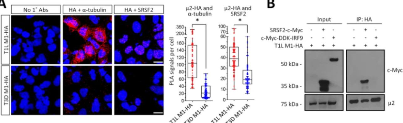
2 forms a complex with the pre-mRNA splicing factor SRSF2.
To determine
fected with T1L-M1-HA and myc-tagged SRSF2 (because endogenous SRSF2 is not
expressed at sufficient levels) or myc-tagged IRF9 as a negative control (Fig. 5B).
Myc-tagged SRSF2, but not myc-tagged IRF9, coimmunoprecipitated with T1L
2-HA.
Together, results demonstrate that T1L-
2 but not T3D-
2 forms a complex with both
microtubules and SRSF2 in the nucleus.
2 nuclear localization requires SRSF2 expression.
Given the impact of T1L
2
on SRSF2 localization, we next sought to determine whether SRSF2 affects
2
local-ization. SRSF2 was depleted by small interfering RNA (siRNA) in L929 cells (Fig. 6A),
T1L-or T3D-M1-HA were transfected, and
2 localization was assessed by
immunofluores-cence (Fig. 6B). As expected, in control siRNA-transfected cells, T1L
2 and T3D
2
localized primarily to the nucleus and the cytoplasm, respectively. However, T1L
2
localization became primarily cytoplasmic in cells with reduced SRSF2 expression,
mimicking T3D
2. Thus, SRSF2 function is required for T1L
2 nuclear localization
where, given the exclusively nuclear location of SRSF2 (26),
2 likely subsequently
forms a complex with SRSF2.
Depletion of SRSF2 increases reovirus replication and cytopathic effect and
impact is strain specific.
The interaction of T1L
2 with SRSF2 suggested the
possi-bility that reovirus modulates SRSF2 function to generate a more favorable cell
envi-ronment. To determine whether SRSF2 affects viral replication and cytopathic effect,
FIG 5T1L2 forms a complex with SRSF2 in the nucleus. (A) AD-293 cells were transfected with T1L M1-HA for 20 h, fixed, and subjected to a proximity ligation assay (PLA) using the indicated primary antibodies. Cells were imaged by confocal microscopy to obtain z-stack images of PLA signals (red) and nuclei (blue) representative of the total volume of the cell in thezaxis. Scale bar, 10m. Quantitation of PLA results expressed as number of PLA dots per cell (n⫽66 to 82 cells per condition) for a representative of at least three independent experiments.*, Significantly different from T3D2-HA (P⬍0.001). (B) AD-293 cells were transfected with the indicated plasmids, whole-cell lysates were immunoprecipitated using anti-HA-conjugated agarose beads, and lysates (input) and immunoprecipitated extracts (IP) were resolved by SDS-PAGE for immunoblotting. The results are representative of at least two independent experiments. It is unclear why overexpressed SRSF2 appears as two distinct bands, but the upper band corresponds to SRSF2 migration in specifications provided with the antibody.
L929 cells were depleted of SRSF2 and then infected with T1L or T3D. SRSF2 depletion
increased T1L but not T3D replication and cytopathic effect in both primary (Fig. 7A and
B) and secondary infections (Fig. 7C and D). The absence of an effect of SRSF2 depletion
on T3D infection suggests that SRSF2-independent events are the predominant
deter-minants of T3D replication in L929 cells. The increased T1L replication and cytopathic
effect suggest that T1L
2 does not inhibit SRSF2 activity entirely and that further
depletion of SRSF2 is beneficial to the virus.
Reovirus T1L alters cellular mRNA splicing.
SRSF2 is a pre-mRNA splicing factor,
but reovirus replication is exclusively cytoplasmic, and there is no evidence that any
reovirus RNAs are spliced (4). We therefore used RNA-seq to determined whether
reovirus T1L but not T3D alters cellular mRNA splicing. Because T3D induces
signifi-cantly more IFN-

than T1L does (6, 7, 27) and because IFN-

alone alters mRNA
splicing of some IFN-stimulated genes (28), we compared mock-infected, untreated
cells to mock-infected or reovirus-infected cells stimulated with IFN-

. This identifies
differences in T1L and T3D effects on splicing separate from differences in their
induction of IFN-

and subsequent IFN-

effects on splicing. The results from RNA
sequencing (RNA-seq) were analyzed by the mixture-of-isoforms (MISO) statistical
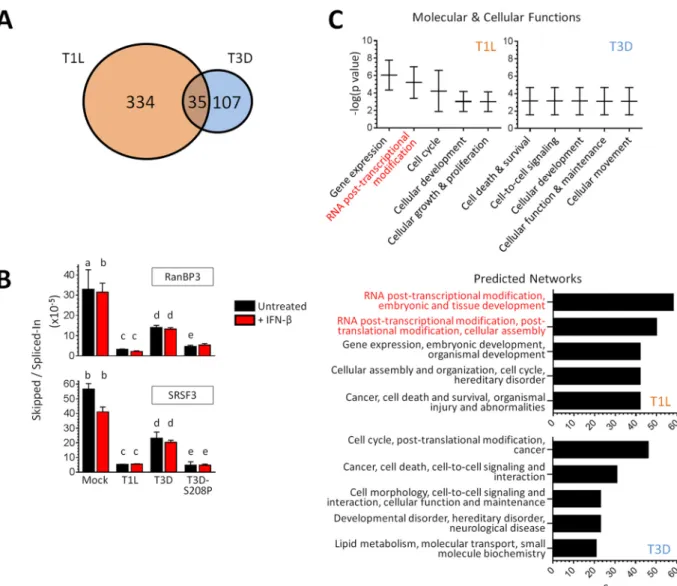
model (29) to assess changes in mRNA alternative splicing (Fig. 8A; see also Tables S1
to S3 in the supplemental material). IFN-

stimulation generated 42 novel splicing
variants in 41 different genes relative to untreated cells. Excluding those IFN-

-induced
variants, T3D infection generated 142 novel splicing variants while, remarkably, T1L
generated 369. Only 35 of these variants were found to overlap between the two
reovirus strains, resulting in a total of 334 and 107 unique splicing events induced in
297 and 97 genes by T1L and T3D, respectively. Splicing events that were induced only
in T1L were confirmed by quantitative reverse transcription-PCR (qRT-PCR) for two
cases (RanBP3 and SRSF3) identified by MISO analysis (Fig. 8B). Whereas T3D also
altered RanBP3 and SRSF3 splicing to some extent (2.4- and 2.5-fold, relative to the
Mock group), T1L altered their splicing much more dramatically (10.4- and 10.8-fold,
respectively). Moreover, these qRT-PCR results confirmed that IFN-

has minimal or no
impact on viral effects on splicing and that the same
2 amino acid that determines
2
association with SRSF2 (Fig. 3) determines reovirus effects on splicing (S208P altered
RanBP3 and SRSF3 splicing 7.0- and 11.5-fold, respectively, Fig. 8B). Thus, while both
reovirus strains induce unique splicing variants, T1L induces more than three times
more than T3D does (RNA-seq results: chi square,
P
⬍
0.001), and a single
2 amino
acid can determine reovirus effects on splicing.
To gain further insights into reovirus-induced global changes in host mRNA splicing,
we used Ingenuity pathway analysis (IPA) and gene ontology (GO) analysis. Specifically,
we performed IPA on the 297 and 97 genes whose splicing was altered uniquely by T1L
or by T3D, respectively. The pathways most affected by T1L involved RNA
posttran-scriptional modifications (Fig. 8C). Indeed, T1L (but not T3D) altered the spicing of
SRSF3, SRSF6, SRSF7, and SRSF11 (see Table S3 in the supplemental material), all of
which have specialized roles in posttranscriptional regulation (30). Moreover, the top
five predicted upstream regulators for altered splicing events induced by T1L (but not
T3D) included SRSF1 (data not shown), another SR family member (31). Importantly, the
minimal changes in splicing that occurred during T3D did not cluster in any of these
categories, and there was no overlap with those obtained for T1L. Lastly, we used the
network of genes affected by T1L with the highest statistical significance to create a
web of known and predicted interactions. One-third of the genes in the network related
to mRNA splicing or mRNA processing (data not shown). These IPA results were
supported by GO analysis of the same data sets. Specifically, the top two processes for
T1L were “mRNA processing” and “ribonucleoprotein complex assembly,” whereas
those were 47th and 8th in priority for T3D, respectively (T1L different from T3D at
P
⬍
0.001 and
P
⬍
0.05, respectively).
In sum, results suggest that SRSF2 is required for T1L
2 nuclear localization
whereupon T1L
2 forms a complex with SRSF2 and antagonizes the organization of
nuclear speckles, resulting in dysregulated splicing of other splicing factors and
regu-lators of mRNA at the posttranscriptional level to benefit viral replication (Fig. 9).
DISCUSSION
To replicate efficiently, viruses must create favorable cell conditions and overcome
cell antiviral responses. Here, we report that the
2 protein from strain T1L localizes to
nuclear speckles, where it forms a complex with and alters the localization of the
pre-mRNA splicing factor SRSF2. Moreover, infection with reovirus T1L alters mRNA
splicing of genes involved in mRNA processing and maturation, likely as a consequence
of T1L
2 antagonistic effects on SRSF2. Finally, depletion of SRSF2 enhances reovirus
replication and cytopathic effect, suggesting that T1L
2 modulation of splicing
benefits the virus. This provides the first report of viral antagonism of the splicing factor
SRSF2 and suggests that by altering mRNA processing and maturation, the virus
induces cell consequences that are global rather than in a single pathway.
Reovirus
2 stability, repression of IFN-

signaling, and stabilization of microtubules
are all determined by a single amino acid polymorphism in the
2 protein (6, 7, 11).
Here, we demonstrate that this amino acid similarly determines
2 localization to
nuclear speckles (Fig. 3). The common dependence on a single amino acid could reflect
its impact on
2 stability or its impact on function. The N terminus of
2 includes a
polybasic region that can function as a nuclear localization signal (10) and, interestingly,
domains that are rich in basic amino acids have been identified as nuclear
speckle-targeting signals (32). However, this
2 polybasic region is conserved between T1L and
T3D strains, indicating that the primary protein sequence alone does not predict its
localization and that other factors contribute to the differential localization of
2. This
complexity in determinants of
2 localization to intracellular compartments is
high-lighted by the observation that SRSF2 is required for
2 nuclear localization (Fig. 6) and
that T3D
2 does not associate with nuclear speckles even when concentrated in the
nucleus after inhibition of nuclear export (Fig. 2D). The requirement for SRSF2 could
reflect trapping of
2 in a complex, an impact on nuclear import or export processes,
or an impact on cell proteins that affect
2 itself (Fig. 9). Posttranslational modifications
of
2, including acetylation (33) and phosphorylation (34), have been reported, and yet
the impact(s) of these modifications on
2 function has not been fully elucidated.
Future studies will address the roles of SRSF2 and posttranslational modifications on
2
subcellular localization.
infection (data not shown). Thus, there is no evidence that reovirus modulation of
SRSF2 affects viral RNAs. Instead, reovirus likely modulates the cell transcriptome to its
benefit.
Alternative splicing of cell mRNAs can affect cell function and disease (45–47), and
several viruses modulate cell splicing machinery to alter the cell transcriptome. The
adenovirus E4-ORF4 antagonizes the function of SRSF1 and SRSF9 by promoting their
dephosphorylation (48, 49). The herpes simplex virus ICP27 protein binds the SR protein
kinase 1 (SRPK1), resulting in its relocalization from the cytoplasm to the nucleus to
dephosphorylate SR proteins and regulate host mRNA splicing events (50–52). The
vaccinia virus VH1 phosphatase appears to directly dephosphorylate SR proteins (53).
Epstein-Barr virus (EBV) itself encodes a splicing factor, SM, that influences splicing and
processing of host pre-mRNAs (54, 55). Splice variants, including those that encode
dominant negative proteins, have been described for several genes involved in the
IFN-
␣
/

response (56–63). Notably, the EBV SM protein skews the mRNA splicing
patterns of STAT1, enhancing production of the dominant-negative STAT1

isoform
(55). Very recently, it has been shown that the NS5 protein of dengue virus binds to and
interferes with components of the U5 snRNP particle and alters mRNA splicing of
several antiviral factors (64). In contrast to our results for reovirus, dengue NS5 does not
appear to affect the intranuclear localization of its splicing targets (64). Finally, in a
recent report reovirus T3D was shown to alter cell mRNA splicing, but the impact on
viral replication and the underlying mechanism were not investigated (65). It is
inter-esting that a single reovirus protein,
2, can modulate both IFN signaling (7) and SRSF2
function to benefit the virus. The results suggest that this protein has evolved to target
both a specific antiviral pathway and mRNA maturation for more global effects.
Several SR proteins play roles in addition to those in splicing that affect mature
mRNA expression and nuclear export (30). For example, SRSF1 participates in the
organization of nuclear speckles and in the promotion of decay of aberrantly spliced
transcripts (31, 66, 67), whereas SRSF2 influences transcriptional elongation of some
mRNAs (68). With the exception of SRSF2, mammalian SR proteins constitutively shuttle
between the nucleus and cytoplasm (26). Once in the cytoplasm, SRSF1 impacts
translation by modulating mRNA entrance into polyribosomes (69, 70). Lastly and
perhaps most intriguingly, SRSF1 has been associated with RIG-I and RNA polymerase
III-dependent sensing of transfected non-self cytosolic DNA to facilitate IFN-

produc-tion (71); however, its role during viral infecproduc-tion remains unclear. Similarly, the
spliceo-somal protein SNRNP200 can also translocate to the cytoplasm, where it modulates the
viral induction of IFN-

(72). Although T1L did not affect splicing of SRSF1 or SNRNP200
mRNA, the loss of SRSF2 can affect SRSF1 function (73), and the regulation of snRNPs
is complex, leaving open the possibility that T1L alters the cytoplasmic function of
spliceosomal proteins to benefit the virus.
isoforms of other SR proteins.
Lastly, reovirus
2 repression of the IFN-

response is a determinant of reovirus
induction of myocarditis (7, 27), and the same
2 amino acid polymorphism that
determines this repression and severity of myocarditis (7) determines
2 association
with SRSF2 (Fig. 3). Together, these results suggest that SRSF2 could participate in
protecting the heart against viral myocarditis. While this could reflect SRSF2 effects on
the IFN response, it has been shown that mutations and/or decreased expression of
splicing factors results in cardiac development defects (45), and cardiac tissue-specific
ablation of SRSF2 results in dilated cardiomyopathy (78). The fact that viruses, such as
adenoviruses, herpesviruses, and reoviruses, known to induce myocarditis (79) can
modulate splicing factors raises the intriguing possibility that altered splicing
repre-sents yet another mechanism by which viruses induce cardiac damage.
MATERIALS AND METHODS
Cells.Mouse L929 cells were maintained in minimal essential medium (MEM; SAFC Biosciences) as a suspension culture supplemented to contain 5% fetal calf serum and 2 mML-glutamine. AD-293 cells were maintained in high glucose Dulbecco MEM (DMEM; Gibco) supplemented to contain 10% fetal calf serum and 1% of sodium pyruvate. Exponentially growing L cells or trypsinized AD-293 cells were plated and incubated for at least 3 h prior to transfection or infection.
Viruses.All viruses were CsCl-purified, low-passage-number stocks originating from plaques of either cultured virus or virus generated by reverse genetics (80) as previously described (6, 7). The results were the same regardless of virus source.
Plasmids and plasmid transfections.A plasmid expressing C-terminus hemagglutinin (HA)-tagged T1L2 in a pCAGGs backbone was described previously (34). The C-terminus HA-tagged T3D2 was generated from pCAGGs-M1-T3D (6) using a similar strategy and the primer 5=-CCCGGGTCAGCTAGCGT AATCTGGAACATCGTATGGGTACGCCAAGTCAGATCGGAAAGCTAGTC-3=. The eGFP-N1 and eGFP-HsCRM1 plasmids were obtained from Clontech Laboratories (catalog no. 6085-1) and GeneCopoeia (catalog no. EX-T0446-M29), respectively. The Myc-DDK-IRF9 plasmid was custom-cloned by OriGene Technologies, Inc., using the murine IRF9 cDNA (NM_001159417) in a pCMV6 expression vector. TheHsSRSF2-c-Myc in a pcDNA3.1 backbone plasmid (81) was a gift from Kathleen Scotto (Addgene; plasmid 44721). Plasmids were transfected using Lipofectamine 3000 (Thermo Fisher Scientific) for 20 to 24 h.
siRNAs and siRNA transfections.L929 cells were transfected for a final 60 nM concentration of control nontargeting siRNA or siRNA against murine SRSF2 (GE Dharmacon; catalog no. D-001810-10 or L-044306) using the transfection reagent RNAiMAX (Thermo Fisher Scientific). At 24 h posttransfection, overlaying medium was replaced with fresh medium.
Antibodies and chemical treatments. The following antibodies and dilutions were used for immunoblotting: anti-2 (generated against two2 peptides by Open Biosystems; 1:1,000), anti--actin (Santa Cruz Biotech, sc-1615-hrp; 1:3,000), anti-SRSF2 (Millipore, 04-1550; 1:1,000), anti-SRSF1 (Abcam, ab129108; 1:350), anti-c-Myc (catalog no. sc-40, 1:3,000; Santa Cruz Biotech), goat horseradish peroxidase (HRP)-conjugated anti-rabbit immunoglobulin (Ig; Millipore, 12-348; 1:2,000), and goat HRP-conjugated anti-mouse Ig (Millipore, 12-349; 1:2,000). For immunoprecipitation experiments, secondary antibodies were either TrueBlot anti-rabbit IgG-HRP or TrueBlot Ultra anti-mouse Ig-HRP (Rockland Immunochemi-cals, 18-8816-33 and 18-8817-33; 1:1,000). Primary antibodies used for immunofluorescence and PLA were anti-coilin (Cell Signaling Technology, 14168; 1:800), anti-SRSF2/SC35 (Abcam, ab11826; 1:1,000), anti-Son (Abcam, ab109472; 1:200), anti-HA epitope tag (GeneTex, 18181; 1:1200; or Sigma, H6908; 1:1,000 or 1:1,200 for PLA), rabbit anti-2 antisera (82) (1:1,000), and a mix of anti-reovirus T1L and anti-reovirus 8B mouse antisera (B. Sherry, unpublished; 1:5,000 each). The secondary antibodies were Alexa Fluor 488- or Alexa Fluor 594-conjugated goat anti-mouse or anti-rabbit IgG (Thermo Fisher Scientific; 1:1,000). The CRM1-inhibitor leptomycin B (LMB; Sigma, L2913) was used at a final concentra-tion of 20 nM in supplemented DMEM. Nocodazole (Sigma, M1404) and paclitaxel (Sigma, T7191) in dimethyl sulfoxide were used at a final concentration of 10M in supplemented DMEM. Recombinant mouse interferon- (PBL Assay Science, catalog no. 12410-1) was used at 1,000 U/ml diluted in supplemented MEM.
abilized with 0.25% Triton X-100 (Sigma). Slides were blocked with normal goat serum (Sigma, catalog no. G9023), incubated in DAPI (4=,6-diamidino-2-phenylindole; Sigma, D8417), immunostained with the indicated primary and secondary antibodies, and preserved with ProLong Gold (Invitrogen). For immu-nostaining of2 and SRSF2 during reovirus infection, the cells were fixed in 100% methanol.
In situproximity ligation assay.Transfected cells in poly-D-lysine-coated chamber slides were fixed with 4% paraformaldehyde, permeabilized in 0.25% Triton X-100, blocked with Duolink blocking solution, incubated with primary antibodies, and then probed using a Duolink PLA kit (Sigma, catalog no. DUO92101). Dried slides were mounted on Duolink mounting medium with DAPI and imaged as z-stacks by confocal microscopy.
Quantitative (real-time) reverse transcription-PCR.For confirmation of the MISO results, oligo-nucleotide primers were designed to amplify transcripts that either skipped or spliced-in a specific exon, as identified by RNA-seq. The primers were as follows: RanBP3 skipped exon (forward, 5=-CCGTCTGTCT TTGTGTTTCAA-3=; reverse, 5=-GCTGCTCTTCTCTTCTGAGTGA-3=), RanBP3 spliced-in exon (forward, 5= -CGACCGTCTGTCTTTGTGTTT-3=; reverse, 5=-AAACATGACCCCATCAAAA-3=), SRSF3 skipped exon (forward, 5=-TGATTACCGCAGGAGGAGTC-3=; reverse, 5=-GATCGAGACGGCTTGTGATT-3=), and SRSF3 spliced-in exon (forward, 5=-TGATTACCGCAGGAGGAGTC-3=; reverse, 5=-TGACGCTGAAAGGGCTAGTT-3=). The total RNA was harvested using an RNeasy kit (Qiagen, Inc.), treated with RNase-free DNase I (Qiagen, Inc.), converted to cDNA by reverse transcription, and used for real-time PCR on a LightCycler 480 fluorescence thermocyler (Roche Life Science). The reaction mixtures contained 1⫻Quantitech SYBR green master mix (Qiagen) and 0.3M (each) forward and reverse primers as previously described (83). The relative mRNA abundance ofRanBP3andSRSF3were normalized to GAPDH (glyceraldehyde-3-phosphate dehydroge-nase) by the ΔΔCp method, and the fold induction was calculated relative to mock-treated cells for each primer pair.
Viral replication and cytopathic effect in siRNA-transfected cells.L929 cells were transfected with siRNA as described above and infected 2 days later at the indicated multiplicity of infection (MOI). The siRNA-mediated depletion of SRSF2 was maintained throughout the experiment (data not shown). Viral replication and cytopathic effect were assessed by plaque assay (84) and MTT assay (27).
Confocal microscopy and image analysis.A Zeiss LSM 710 confocal microscope equipped with a 40⫻C-Apochromat/1.1 NA water immersion objective from the Cellular and Molecular Facility at NC State University was used for all experiments. The pinhole diameter was maintained at 1 Airy unit, and all images were obtained using multitrack sequential scanning for each fluorophore to prevent bleed-through. The excitation/emission wavelengths during micrograph acquisition were as follows: 488 nm/492 to 554 nm for Alexa Fluor 488, 561 nm/584 to 666 nm for Alexa Fluor 594 and PLA Duolink Red, and 405 nm/407 to 507 nm for DAPI. The images were processed for presentation using Photoshop CS4. Intensity plot profiles were generated using ImageJ software (82). Quantification ofin situPLA signals per cell was performed using the particle analysis tool of ImageJ.
RNA-seq.L929 cells were infected at an MOI of 100 PFU per cell, and at 20 h postinfection overlays were replaced with supplemented media or mouse IFN-diluted in supplemented medium to 1,000 U/ml. After 5 h of incubation, the overlays were removed, and the total RNA was harvested, the contaminating DNA was removed, and an aliquot of the RNA was converted to cDNA for qRT-PCR to confirm T1L repression of IFN-signaling (data not shown). Total RNA samples were submitted to the North Carolina State University Genomic Sciences Laboratory (Raleigh, NC) for Illumina RNA library construction and sequencing. mRNA was purified using oligo(dT) beads [NEBNExt poly(A) mRNA mag-netic isolation module (New England BioLabs, USA)], chemically fragmented, and primed with random oligonucleotides for first-strand cDNA synthesis (NEBNext Ultra Directional RNA library prep kit [NEB] and NEBNext Mulitplex Oligos for Illumina [NEB]). Second-strand cDNA synthesis was carried out with dUTPs to preserve strand orientation information. Double-stranded cDNA was “A-tailed” for adaptor ligation and selected for a final size of 250 to 400 bp, including adaptors (AMPure XP bead isolation; Beckman Coulter, USA). Library enrichment was performed, and indexes were added during PCR amplification. Libraries were pooled in equimolar amounts and sequenced on an Illumina NextSeq 500 DNA sequencer, and real-time analysis was used to generate raw base call files, which were then demultiplexed by sample into fastq files.
Supplemental material for this article may be found at
https://doi.org/10.1128/
JVI.02488-16
.
DATA SET S1, xlsx file, 0.01 MB.
DATA SET S2, xlsx file, 0.01 MB.
DATA SET S3, xlsx file, 0.01 MB.
ACKNOWLEDGMENTS
We thank Shannon Chiera, Tiffany Benzine, and Nicole DeAngelis for insightful
discussions and technical assistance. We thank David Andrew Baltzegar at the NC State
University Genomic Sciences Laboratory for his guidance and expertise. We also thank
John L. Parker (Cornell University) and Jennifer Luff (NC State University) and members
of her laboratory for helpful suggestions.
This research was supported by National Institutes of Health (NIH) grant R01
AI083333 (B.S.), a Research Supplement to Promote Diversity in Health-Related
Re-search through a parental NIH grant (B.S. and E.E.R-S.), and a U.S. Department of
Education Graduate Assistance in Areas of National Need (GAANN) Fellowship (E.E.R-S.).
REFERENCES
1. Bowie AG, Unterholzner L. 2008. Viral evasion and subversion of pattern-recognition receptor signaling. Nat Rev Immunol 8:911–922.https:// doi.org/10.1038/nri2436.
2. Hoffmann HH, Schneider WM, Rice CM. 2015. Interferons and viruses: an evolutionary arms race of molecular interactions. Trends Immunol 36: 124 –138.https://doi.org/10.1016/j.it.2015.01.004.
3. Kotwal GJ, Hatch S, Marshall WL. 2012. Viral infection: an evolving insight into the signal transduction pathways responsible for the innate im-mune response. Adv Virol 2012:131457.
4. Dermody T, Parker J, Sherry B. 2013. Orthoreoviruses, p 1304 –1346.In Fields virology, 6th ed. Lippincott/Williams & Wilkins, Philadelphia, PA. 5. Fernandez de Castro I, Zamora PF, Ooms L, Fernandez JJ, Lai CM, Mainou BA, Dermody TS, Risco C. 2014. Reovirus forms neo-organelles for prog-eny particle assembly within reorganized cell membranes. mBio 5:e00931-13.https://doi.org/10.1128/mBio.00931-13.
6. Zurney J, Kobayashi T, Holm GH, Dermody TS, Sherry B. 2009. Reovirus
2 protein inhibits interferon signaling through a novel mechanism involving nuclear accumulation of interferon regulatory factor 9. J Virol 83:2178 –2187.https://doi.org/10.1128/JVI.01787-08.
7. Irvin SC, Zurney J, Ooms LS, Chappell JD, Dermody TS, Sherry B. 2012. A single-amino-acid polymorphism in reovirus protein2 determines re-pression of interferon signaling and modulates myocarditis. J Virol 86: 2302–2311.https://doi.org/10.1128/JVI.06236-11.
8. Brentano L, Noah DL, Brown EG, Sherry B. 1998. The reovirus protein2, encoded by the M1 gene, is an RNA-binding protein. J Virol 72: 8354 – 8357.
9. Kim J, Parker JS, Murray KE, Nibert ML. 2004. Nucleoside and RNA triphosphatase activities of orthoreovirus transcriptase cofactor2. J Biol Chem 279:4394 – 4403.
10. Kobayashi T, Ooms LS, Chappell JD, Dermody TS. 2009. Identification of functional domains in reovirus replication proteinsNS and2. J Virol 83:2892–2906.https://doi.org/10.1128/JVI.01495-08.
11. Parker JS, Broering TJ, Kim J, Higgins DE, Nibert ML. 2002. Reovirus core protein2 determines the filamentous morphology of viral inclusion bodies by interacting with and stabilizing microtubules. J Virol 76: 4483– 4496.https://doi.org/10.1128/JVI.76.9.4483-4496.2002.
12. Miller CL, Parker JS, Dinoso JB, Piggott CD, Perron MJ, Nibert ML. 2004. Increased ubiquitination and other covariant phenotypes attributed to a strain- and temperature-dependent defect of reovirus core protein2. J Virol 78:10291–10302. https://doi.org/10.1128/JVI.78.19.10291-10302 .2004.
13. Broering TJ, Parker JS, Joyce PL, Kim J, Nibert ML. 2002. Mammalian reovirus nonstructural proteinNS forms large inclusions and colocal-izes with reovirus microtubule-associated protein2 in transfected cells. J Virol 76:8285– 8297.https://doi.org/10.1128/JVI.76.16.8285-8297.2002. 14. Mbisa JL, Becker MM, Zou S, Dermody TS, Brown EG. 2000. Reovirus2 protein determines strain-specific differences in the rate of viral inclu-sion formation in L929 cells. Virology 272:16 –26. https://doi.org/ 10.1006/viro.2000.0362.
15. Manley JL, Krainer AR. 2010. A rational nomenclature for serine/arginine-rich protein splicing factors (SR proteins). Genes Dev 24:1073–1074. https://doi.org/10.1101/gad.1934910.
16. Nishi K, Yoshida M, Fujiwara D, Nishikawa M, Horinouchi S, Beppu T. 1994. Leptomycin B targets a regulatory cascade of Crm1, a fission yeast nuclear protein, involved in control of higher order chromosome struc-ture and gene expression. J Biol Chem 269:6320 – 6324.
17. Zakaryan H, Stamminger T. 2011. Nuclear remodelling during viral in-fections. Cell Microbiol 13:806 – 813.https://doi.org/10.1111/j.1462-5822 .2011.01596.x.
18. Moller A, Schmitz ML. 2003. Viruses as hijackers of PML nuclear bodies. Arch Immunol Ther Exp 51:295–300.
19. Regad T, Chelbi-Alix MK. 2001. Role and fate of PML nuclear bodies in response to interferon and viral infections. Oncogene 20:7274 –7286. https://doi.org/10.1038/sj.onc.1204854.
20. Morris GE. 2008. The Cajal body. Biochim Biophys Acta 1783:2108 –2115. https://doi.org/10.1016/j.bbamcr.2008.07.016.
21. Thiry M. 1995. The interchromatin granules. Histol Histopathol 10: 1035–1045.
22. Hall LL, Smith KP, Byron M, Lawrence JB. 2006. Molecular anatomy of a speckle. Anat Rec A Discov Mol Cell Evol Biol 288:664 – 675.
28. Schneider WM, Chevillotte MD, Rice CM. 2014. Interferon-stimulated genes: a complex web of host defenses. Annu Rev Immunol 32:513–545. https://doi.org/10.1146/annurev-immunol-032713-120231.
29. Katz Y, Wang ET, Airoldi EM, Burge CB. 2010. Analysis and design of RNA sequencing experiments for identifying isoform regulation. Nat Methods 7:1009 –1015.https://doi.org/10.1038/nmeth.1528.
30. Anko ML. 2014. Regulation of gene expression programmes by serine-arginine rich splicing factors. Semin Cell Dev Biol 32:11–21. https:// doi.org/10.1016/j.semcdb.2014.03.011.
31. Tripathi V, Song DY, Zong X, Shevtsov SP, Hearn S, Fu XD, Dundr M, Prasanth KV. 2012. SRSF1 regulates the assembly of pre-mRNA process-ing factors in nuclear speckles. Mol Biol Cell 23:3694 –3706. https:// doi.org/10.1091/mbc.E12-03-0206.
32. Salichs E, Ledda A, Mularoni L, Albà MM, de la Luna S. 2009. Genome-wide analysis of histidine repeats reveals their role in the localization of human proteins to the nuclear speckles compartment. PLoS Genet 5:e1000397.https://doi.org/10.1371/journal.pgen.1000397.
33. Swanson MI, She YM, Ens W, Brown EG, Coombs KM. 2002. Mammalian reovirus core protein micro 2 initiates at the first start codon and is acetylated. Rapid Commun Mass Spectrom 16:2317–2324. https:// doi.org/10.1002/rcm.866.
34. Stebbing RE, Irvin SC, Rivera-Serrano EE, Boehme KW, Ikizler M, Yoder JA, Dermody TS, Sherry B. 2014. An ITAM in a nonenveloped virus regulates activation of NF-B, induction of beta interferon, and viral spread. J Virol 88:2572–2583.https://doi.org/10.1128/JVI.02573-13.
35. Dubois J, Terrier O, Rosa-Calatrava M. 2014. Influenza viruses and mRNA splicing: doing more with less. mBio 5:e00070-14. https://doi.org/ 10.1128/mBio.00070-14.
36. Purcell DF, Martin MA. 1993. Alternative splicing of human immunode-ficiency virus type 1 mRNA modulates viral protein expression, replica-tion, and infectivity. J Virol 67:6365– 6378.
37. Schneider PA, Schneemann A, Lipkin WI. 1994. RNA splicing in Borna disease virus, a nonsegmented, negative-strand RNA virus. J Virol 68: 5007–5012.
38. Su TS, Lai CJ, Huang JL, Lin LH, Yauk YK, Chang CM, Lo SJ, Han SH. 1989. Hepatitis B virus transcript produced by RNA splicing. J Virol 63: 4011– 4018.
39. Hernandez-Lopez HR, Graham SV. 2012. Alternative splicing in human tumour viruses: a therapeutic target? Biochem J 445:145–156.https:// doi.org/10.1042/BJ20120413.
40. Fukuhara T, Hosoya T, Shimizu S, Sumi K, Oshiro T, Yoshinaka Y, Suzuki M, Yamamoto N, Herzenberg LA, Herzenberg LA, Hagiwara M. 2006. Utilization of host SR protein kinases and RNA-splicing machinery during viral replication. Proc Natl Acad Sci U S A 103:11329 –11333.https:// doi.org/10.1073/pnas.0604616103.
41. Cavallari I, Rende F, D’Agostino DM, Ciminale V. 2011. Converging strategies in expression of human complex retroviruses. Viruses 3:1395–1414.https://doi.org/10.3390/v3081395.
42. Carter KC, Taneja KL, Lawrence JB. 1991. Discrete nuclear domains of poly(A) RNA and their relationship to the functional organization of the nucleus. J Cell Biol 115:1191–1202. https://doi.org/10.1083/jcb.115.5 .1191.
43. Visa N, Puvion-Dutilleul F, Harper F, Bachellerie JP, Puvion E. 1993. Intranuclear distribution of poly(A) RNA determined by electron micro-scope in situ hybridization. Exp Cell Res 208:19 –34.https://doi.org/ 10.1006/excr.1993.1218.
44. Cardinale S, Cisterna B, Bonetti P, Aringhieri C, Biggiogera M, Barabino SM. 2007. Subnuclear localization and dynamics of the pre-mRNA 3=end processing factor mammalian cleavage factor I 68-kDa subunit. Mol Biol Cell 18:1282–1292.https://doi.org/10.1091/mbc.E06-09-0846.
Akusjarvi G. 1998. Regulation of adenovirus alternative RNA splicing by dephosphorylation of SR proteins. Nature 393:185–187.https://doi.org/ 10.1038/30277.
50. Lindberg A, Kreivi JP. 2002. Splicing inhibition at the level of spliceo-some assembly in the presence of herpes simplex virus protein ICP27. Virology 294:189 –198.https://doi.org/10.1006/viro.2001.1301. 51. Sciabica KS, Dai QJ, Sandri-Goldin RM. 2003. ICP27 interacts with SRPK1
to mediate HSV splicing inhibition by altering SR protein phosphoryla-tion. EMBO J 22:1608 –1619.https://doi.org/10.1093/emboj/cdg166. 52. Hu B, Li X, Huo Y, Yu Y, Zhang Q, Chen G, Zhang Y, Fraser NW, Wu D,
Zhou J. 2016. Cellular responses to HSV-1 infection are linked to specific types of alterations in the host transcriptome. Sci Rep 6:28075.https:// doi.org/10.1038/srep28075.
53. Huang TS, Nilsson CE, Punga T, Akusjarvi G. 2002. Functional inactivation of the SR family of splicing factors during a vaccinia virus infection. EMBO Rep 3:1088 –1093.https://doi.org/10.1093/embo-reports/kvf217. 54. Ruvolo V, Sun L, Howard K, Sung S, Delecluse H-J, Hammerschmidt W,
Swaminathan S. 2004. Functional analysis of Epstein-Barr virus SM protein: identification of amino acids essential for structure, transactiva-tion, splicing inhibitransactiva-tion, and virion production. J Virol 78:340 –352. https://doi.org/10.1128/JVI.78.1.340-352.2004.
55. Verma D, Swaminathan S. 2008. Epstein-Barr virus SM protein functions as an alternative splicing factor. J Virol 82:7180 –7188.https://doi.org/ 10.1128/JVI.00344-08.
56. Lad SP, Yang G, Scott DA, Chao TH, da Correia JS, de la Torre JC, Li E. 2008. Identification of MAVS splicing variants that interfere with RIGI/ MAVS pathway signaling. Mol Immunol 45:2277–2287.https://doi.org/ 10.1016/j.molimm.2007.11.018.
57. Li Y, Hu X, Song Y, Lu Z, Ning T, Cai H, Ke Y. 2011. Identification of novel alternative splicing variants of interferon regulatory factor 3. Biochim Biophys Acta 1809:166 –175. https://doi.org/10.1016/j.bbagrm.2011 .01.006.
58. Koop A, Lepenies I, Braum O, Davarnia P, Scherer G, Fickenscher H, Kabelitz D, Adam-Klages S. 2011. Novel splice variants of human IKKnegatively regulate IKK-induced IRF3 and NF-B activation. Eur J Immunol 41:224 –234.https://doi.org/10.1002/eji.201040814.
59. Liu P, Lu M, Tian B, Li K, Garofalo RP, Prusak D, Wood TG, Brasier AR. 2009. Expression of an IKK␥ splice variant determines IRF3 and canonical NF-B pathway utilization in ssRNA virus infection. PLoS One 4:e8079. https://doi.org/10.1371/journal.pone.0008079.
60. Zhao Y, Xu D, Jiang Y, Zhang L. 2010. Dual functions of interferon regulatory factors 7C in Epstein-Barr virus-mediated transformation of human B lymphocytes. PLoS One 5:e9459. https://doi.org/10.1371/ journal.pone.0009459.
61. Marie I, Smith E, Prakash A, Levy DE. 2000. Phosphorylation-induced dimerization of interferon regulatory factor 7 unmasks DNA binding and a bipartite transactivation domain. Mol Cell Biol 20:8803– 8814.https:// doi.org/10.1128/MCB.20.23.8803-8814.2000.
62. Samarajiwa SA, Mangan NE, Hardy MP, Najdovska M, Dubach D, Braniff SJ, Owczarek CM, Hertzog PJ. 2014. Soluble IFN receptor potentiates in vivo type I IFN signaling and exacerbates TLR4-mediated septic shock. J Immunol 192:4425– 4435.https://doi.org/10.4049/jimmunol.1302388. 63. Deng W, Shi M, Han M, Zhong J, Li Z, Li W, Hu Y, Yan L, Wang J, He Y,
mediated mRNA decay and pre-mRNA splicing in mammalian cells. Curr Opin Cell Biol 17:309 –315.https://doi.org/10.1016/j.ceb.2005.03.002. 68. Lin S, Coutinho-Mansfield G, Wang D, Pandit S, Fu XD. 2008. The splicing
factor SC35 has an active role in transcriptional elongation. Nat Struct Mol Biol 15:819 – 826.https://doi.org/10.1038/nsmb.1461.
69. Sanford JR, Gray NK, Beckmann K, Caceres JF. 2004. A novel role for shuttling SR proteins in mRNA translation. Genes Dev 18:755–768. https://doi.org/10.1101/gad.286404.
70. Sanford JR, Ellis JD, Cazalla D, Caceres JF. 2005. Reversible phosphory-lation differentially affects nuclear and cytoplasmic functions of splicing factor 2/alternative splicing factor. Proc Natl Acad Sci U S A 102: 15042–15047.https://doi.org/10.1073/pnas.0507827102.
71. Xue F, Li X, Zhao X, Wang L, Liu M, Shi R, Zheng J. 2015. SRSF1 facilitates cytosolic DNA-induced production of type I interferons recognized by RIG-I. PLoS One 10:e0115354. https://doi.org/10.1371/journal.pone .0115354.
72. Tremblay N, Baril M, Chatel-Chaix L, Es-Saad S, Park AY, Koenekoop RK, Lamarre D. 2016. Spliceosome SNRNP200 promotes viral RNA sensing and IRF3 activation of antiviral response. PLoS Pathog 12:e1005772. https://doi.org/10.1371/journal.ppat.1005772.
73. Pandit S, Zhou Y, Shiue L, Coutinho-Mansfield G, Li H, Qiu J, Huang J, Yeo GW, Ares M, Jr, Fu XD. 2013. Genome-wide analysis reveals SR protein cooperation and competition in regulated splicing. Mol Cell 50:223–235. https://doi.org/10.1016/j.molcel.2013.03.001.
74. Bedard KM, Daijogo S, Semler BL. 2007. A nucleocytoplasmic SR protein functions in viral IRES-mediated translation initiation. EMBO j 26: 459 – 467.https://doi.org/10.1038/sj.emboj.7601494.
Xiao RP, Ross J, Chen J, Fu XD. 2004. Dilated cardiomyopathy caused by tissue-specific ablation of SC35 in the heart. EMBO J 23:885– 896.https:// doi.org/10.1038/sj.emboj.7600054.
79. Feldman AM, McNamara D. 2000. Myocarditis. N Engl J Med 343: 1388 –1398.https://doi.org/10.1056/NEJM200011093431908.
80. Kobayashi T, Antar AA, Boehme KW, Danthi P, Eby EA, Guglielmi KM, Holm GH, Johnson EM, Maginnis MS, Naik S, Skelton WB, Wetzel JD, Wilson GJ, Chappell JD, Dermody TS. 2007. A plasmid-based reverse genetics system for animal double-stranded RNA viruses. Cell Host Microbe 1:147–157.https://doi.org/10.1016/j.chom.2007.03.003. 81. Shi J, Hu Z, Pabon K, Scotto KW. 2008. Caffeine regulates alternative
splicing in a subset of cancer-associated genes: a role for SC35. Mol Cell Biol 28:883– 895.https://doi.org/10.1128/MCB.01345-07.
82. Schneider CA, Rasband WS, Eliceiri KW. 2012. NIH Image to ImageJ: 25 years of image analysis. Nat Methods 9:671– 675. https://doi.org/ 10.1038/nmeth.2089.
83. Stewart MJ, Smoak K, Blum MA, Sherry B. 2005. Basal and reovirus-induced beta interferon (IFN-) and IFN--stimulated gene expression are cell type specific in the cardiac protective response. J Virol 79: 2979 –2987.https://doi.org/10.1128/JVI.79.5.2979-2987.2005.
84. Virgin HW, Bassel-Duby R, Fields BN, Tyler KL. 1988. Antibody protects against lethal infection with the neurally spreading reovirus type 3 (Dearing). J Virol 62:4594 – 4604.