I n V i t r o a n d I n V i v o C h a r a c t e r i z a t i o n o f a Fu l l y F e l i n i z e d
T h e r a p e u t i c A n t i - N e r v e G r o w t h F a c t o r M o n o c l o n a l A n t i b o d y f o r t h e
T r e a t m e n t o f P a i n i n Ca t s
D.P. Gearing, M. Huebner, E.R. Virtue, K. Knight, P. Hansen, B.D.X. Lascelles, R.P. Gearing, and
A.C. Drew
Background: Limited options are available for the treatment of pain in cats. Monoclonal antibodies (mAbs) that neutral-ize nerve growth factor (NGF) have demonstrated analgesic capacity in rodent models, people with osteoarthritis, and dogs with degenerative joint disease.
Hypothesis/Objectives: This study describes the design and characterization of a fully felinized anti-NGF monoclonal antibody. In vitro potency, pharmacokinetics, and the ability of the antibody to treat pain in a self-resolving, acute inflamma-tion model were investigated in cats.
Animals: Thirty-eight cats at a research colony at Charles River Laboratories, Ireland.
Methods: Felinized anti-NGF mAb, NV-02, was produced using a complementary DNA (cDNA)-based method (PETiza-tion). Purified NV-02 was tested for affinity, potency, and immunoreactivity in vitro, then for safety and plasma pharmacoki-netic distribution in vivo, and analgesic efficacy in a model of kaolin-induced inflammatory pain.
Results: Anti-NGF mAb, NV-02 neutralized NGF with high affinity and potency and did not bind complement. NV-02-administered SC had a plasma half-life of 7–15 days and was well tolerated at dosages up to 28 mg/kg. A dosage of 2 mg/kg NV-02 SC significantly decreased signs of lameness on day 2 (P=.0027), day 3 (P=.016), day 4, (P=.0063), day 5 (P=.0085), day 6 (P=.0014), and day 7 (P=.0034) after induction of inflammation.
Conclusions and Clinical Importance: The high affinity, long plasma half-life, safety, and analgesic efficacy of felinized anti-NGF mAb (NV-02) support further investigation of the analgesic potential of this antibody in the cat.
Key words: Cat pain; Companion animals; Feline analgesia; PETization; Pharmacokinetics.
N
onsteroidal anti-inflammatory drugs (NSAIDs) are
used commonly in other mammals for pain relief,
but are not widely used for control of pain in cats
because of safety concerns.
1In the United States, no
NSAIDs are approved for chronic use in cats, and in
the European Union (EU), only 1 NSAID (meloxicam)
is approved for the treatment of chronic pain. However,
in the United States, meloxicam has a boxed warning,
cautioning against repeated dosing.
Anti-nerve growth factor (NGF) monoclonal
anti-bodies (mAbs) have been shown to have analgesic
effects in rodent models of pain,
2in several human
clin-ical trials
3–5and, more recently, in proof-of-concept
clinical studies in osteoarthritic dogs.
6–8Long-acting
pain
relief
(
>
4 weeks)
and
good
tolerability
was
observed in the dog studies after a single injection.
Although no published data are available linking
NGF and pain in cats, the amino acid sequences of
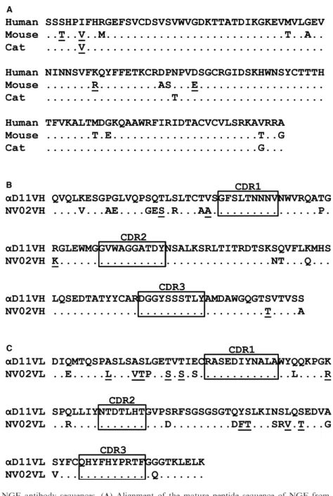
human and feline NGF are highly conserved (Fig 1A).
Given the long half-life of mAbs in mammals in
gen-eral, the favorable safety of humanized antibodies as a
class, and the potential for an equivalent role of NGF
in mediating pain in cats, as in other mammals, we
rea-soned that a felinized mAb (ie, designed to be
recog-nized as self by the feline immune system) that
neutralized feline NGF might have potential as a
long-acting analgesic in cats and potentially would have an
From Nexvet Australia Pty. Ltd., Melbourne, VIC Australia (DP Gearing, Virtue, Knight, Hansen, Drew); ClinData Services Inc., Fort Collins, CO (Huebner); the Comparative Pain Research Program, College of Veterinary Medicine, North Carolina State University, (Lascelles); the Comparative Medicine Institute, North Carolina State University, Raleigh, (Lascelles); the Center for Pain Research and Innovation, UNC Chapel Hill, Chapel Hill, NC (Lascelles); and Ridge Biotechnology, Seattle, WA (RP Gearing).
Corresponding author: D.P. Gearing, Nexvet Australia Pty Ltd, 31 Queen Street, Melbourne, VIC 3000, Australia; e-mail: dave. [email protected]
Submitted January 31, 2016; Revised March 31, 2016; Accepted May 5, 2016.
Copyright © 2016 The Authors. Journal of Veterinary Internal Medicine published by Wiley Periodicals, Inc. on behalf of the Ameri-can College of Veterinary Internal Medicine.
This is an open access article under the terms of the Creative Commons Attribution-NonCommercial License, which permits use, distribution and reproduction in any medium, provided the original work is properly cited and is not used for commercial purposes.
DOI: 10.1111/jvim.13985
Abbreviations:
BBB blood-brain barrier BSA bovine serum albumin
CDR complementarity-determining region CHO Chinese hamster ovary
DJD degenerative joint disease
ELISA enzyme-linked immuno-sorbent assay IV intravenous
mAb monoclonal antibody NGF nerve growth factor
NSAID nonsteroidal anti-inflammatory drug OA osteoarthritis
improved safety profile compared to existing therapies.
In this study, we describe the properties of such a
felin-ized anti-NGF mAb in vitro and in vivo, including its
activity in alleviating signs of pain in a short-term,
self-resolving model of inflammation in cats.
Materials and Methods
Conversion of an Anti-NGF Antibody for Use in the
Cat
Monoclonal antibodies generated by immunization of rodents are immunogenic if injected into other mammals. For use in humans, rodent mAbs are modified for injection by a process
termed “humanizing” or “humanization”. By analogy, a rodent mAb converted for treatment in the cat would be termed “felin-ized”. To decrease the immunogenic potential of rat anti-mouse NGF mAbaD119 in the cat, while retaining its high affinity for NGF, amino acid substitutions were made to the heavy and light chain variable domain framework sequences by alignment with a collection of predicted protein sequences encoded by expressed feline immunoglobulin (IgG) complementary deoxyribonucleic acid (cDNA) sequences. This approach (that we refer to as PETization) previously was used to generate a fully caninized anti-NGF mAb that has a promising efficacy and safety profile in dogs.6–8Where an amino acid in theaD11 sequence corresponded to an amino acid in the collection no change was made. Where it differed, the most similar amino acid (eg, by charge or size) at that position in the collection was substituted. If no similar amino acid was
available, the most abundant feline amino acid residue at that position was chosen. The changes made are illustrated in Fig 1B and C. Sixteen substitutions were made to the heavy chain variable domain, of which 4 were conservative (ie, related by charge, size, or hydrophobicity) and 22 substitutions were made to the light chain variable domain, of which 10 were conservative. By this pro-cess, theaD11 framework amino acid sequences were completely felinized, with minimal changes made from the donoraD11 anti-body. The felinizedaD11 heavy chain variable domain sequence was combined with an immunoglobulin heavy chain signal sequence and the constant domain sequence of feline IgG heavy chain isotype IgG1 to form the feN-HC1 sequence. The felinized NV-02 light chain variable domain sequence was combined with a light chain signal sequence and the constant domain sequence of the feline kappa light chain to form the feN-kLC sequence. The resulting amino acid sequences were converted to Chinese Hamster Ovary (CHO) cell codon-optimized nucleotide sequences and cloned separately into pcDNA3.1+for expression in CHO cells.a
For small-scale work, antibodies (NV-02, NV-01 and caN-HCB-kLC1) were transiently expressed in CHO cellsa and purified by
protein A chromatography from transfected CHO cell super-natant. For larger scale in vivo experiments requiring more mate-rial, NV-02 antibody was stably expressed in CHO cells.bStable CHO cell lines with high productivity were prepared with cDNA encoding NV-02 heavy and light chains, and were cultured in a fed batch system, before harvesting of supernatant containing NV-02. The protein was purified by MabSelectSuRecand Sarto-bind Qdchromatography, then concentrated and formulated into phosphate-buffered saline (PBS) pH 7.2. Endotoxin concentrations were determined using an EndosafeÒ-PTSTMkit.
In Vitro Characterization of the Felinized Anti-NGF
Antibody
Complement C1q binding was assayed as previously described.10 Plates were coated with 2.5lg/mL mouse NGF and
blocked with 5% bovine serum albumin (BSA)/phosphate-buffered saline (PBS). Coated wells were incubated for 1 hour at room tem-perature with NV-02 antibody, or as a positive control a caninized anti-NGF mAb with a IgG-B isotype (complement-binding) heavy chain,6 diluted in PBS/1% BSA. Antibody concentrations ranged
from 10lg/mL to 1.0lg/mL. The plates were washed and incu-bated for 1 hour at room temperature with human serum or heat-inactivated human serum diluted 1/100 in veronal-buffered saline containing 0.05% Tween-20, 0.1% gelatine, and 0.5% BSA. After washing, plates were incubated with a 1/800 dilution of sheep anti-human C1q-HRPe in PBS/1% BSA. After washing, plates were developed by the addition of tetramethylbenzidine (TMB) sub-strate.fDevelopment was stopped by the addition of 2M H2SO4
and absorbance read at 450 nm. The absorbance at 450 nm obtained using heat-inactivated serum was used as background and subtracted from the absorbance at 450 nm obtained using untreated serum.
The binding affinity of NV-02 to mouse NGF was analyzed by surface plasmon resonance assay (SPR) using a ProteOn XPR36 SPRi biosensor equipped with a GLM chipf. The chip was
condi-tioned with 0.5% sodium dodecyl sulfate (SDS), 50 mM NaOH, and 100 mM HCl. After conditioning, the lanes were activated using equal parts of 1-ethyl-3-(-3-demethylaminopropyl) carbodi-imide and N-hydroxysulfosuccincarbodi-imide amine coupling reagents. The NGF protein was immobilized to the chip at a concentration of 50lg/mL in sodium acetate buffer (pH 4.5). After immobiliza-tion all 3 channels were deactivated using ethanolamine. The NV-02 was passed across the surface at 500 nM, 250 nM, 125 nM, 62.5 nM, and 31.25 nM. The binding was displayed as a spectro-gram. Controls were subtracted to give specific binding.
A Langmuir curve fit model then was used to determine the speci-fic affinity. Inhibition of NGF binding by NV-02 was assessed using TF-1 cells as previously described.6 TF-1 cells were starved
for 24 hours, then cultured in 96-well plates in media supple-mented with 1 ng/mL mouse NGF and increasing concentrations of NV-02 or caninized anti-NGF mAb NV-01. The plates were incubated for 48 hour at 37°C/5% CO2before measuring
prolifer-ation using a CellTiter 96 Aqueous One Solution Cell Prolifera-tion Assay.g The assay was performed in triplicate. A mouse IgG2a mAbhwas used as a negative control.
Pharmacokinetics and Safety of NV-02 In Vivo
Pharmacokinetic (PK) studies were conducted in a research colony of cats at Charles River Laboratories (CRL), Ireland, after institutional ethics review panel approval (code CRL/001-15/001). Purified NV-02 was injected SC, at 2.0 mg/kg, 5.6 mg/kg, 16.8 mg/kg, or 28 mg/kg into 8 cats (n=2 animals/group) and plasma samples were taken over the next 42 days. The concen-tration of NV-02 in the plasma samples was determined using a quantitative NGF-binding ELISA. Immunoassay plates (F96 Maxisorpi ) were coated with 100
lL of 0.1lg/mL mouse NGF in PBS and blocked with PBS/0.05% Tween 20/1% BSA. Assay diluent was prepared by diluting pooled normal cat plasma in PBS/0.05% Tween 20/1% BSA. Standards were prepared by diluting NV-02 in assay diluent to 40, 30, 20, 10, 5, 2, and 1 ng/mL. Quality control (QC) samples were prepared from independent stock solutions of NV-02 by diluting in assay dilu-ent to final concdilu-entrations of 30, 15, and 3 ng/mL. Two individ-ual dilutions were prepared for each QC level. Samples were diluted to the required dilution in PBS/0.05% Tween 20/1% BSA.
The NGF-coated wells were incubated for 1 hour at room tem-perature with standard curve, QC and plasma samples. After washing with PBS/0.05% Tween 20, the plates were incubated with a 1/10,000 dilution of goat anti-feline IgG (heavy and light chain specific)-horseradish peroxidase conjugatej in PBS/0.05% Tween 20/1% BSA for 1 hour. Plates were washed and developed by the addition of TMB substratei. Development was stopped by
the addition of 2M H2SO4. The assay was analyzed using a 4
parameter fit. An assay was considered to pass if 4 of 6 QC sam-ples were within 20% of the expected concentration, including 1 of each QC concentration. Plasma half-life was determined using the program PK Solver (11).
Safety was assessed during the course of the study by the CRL veterinarians, observing behavior (daily) and measuring weight (daily), clinical chemistry variables (days 14 and 42: glucose, cre-atine phosphokinase, sodium, chloride, potassium, phosphate, cholesterol, creatinine, total protein, albumin, globulin, calcium), and hematology assessments (days 0, 14, and 42: red blood cells, hemoglobin, hematocrit, mean corpuscular volume, mean corpus-cular hemoglobin, mean corpuscorpus-cular hemoglobin concentration, white blood cells, lymphocytes, monocytes, neutrophils, eosino-phils, basoeosino-phils, large unstained cells).
Model of Inflammatory Pain
NV-02-delivered SC. Vehicle (PBS) was administered SC as a negative control. To maintain blinding, the administration of NV-02 or vehicle was performed by investigators different from those who assessed lameness. The investigators involved in the lameness assessments were masked to the treatments administered to decrease bias. The investigators involved in the administration of NV-02 or vehicle control were not masked.
Thirty cats were used and allocated to 2 treatment groups (n=15 per group). Animals assigned to Group 1 served as a nega-tive control group and were treated with PBS administered by SC injection in the neck region. Animals assigned to Group 2 were treated with NV-02 administered by SC injection at a dosage of 2.0 mg/kg. To facilitate peak plasma concentrations of NV-02 (based on PK data, Fig 4) and optimal distribution to the footpad, NV-02 was given 4 days before a kaolin injection. On the day of kaolin administration to the cats, general anesthesia was induced using ketamine (approximately 7.5 mg/kg [0.075 mL/kg]) and medetomidine (approximately 0.08 mg/kg [0.08 mL/kg]) by IM injection. After kaolin injection, any cat that had not fully recovered after 45 minutes of sedation was given atipamezole hydrochloride (0.2 mg/kg [0.04 mL/kg]) IM to reverse the seda-tion. One cat did not recover from the sedation and this event was attributed to anesthetic death. The cats underwent experimental induction of paw inflammation using 5.4 mL of 250 mg/mL kao-link /PBS solution injected SC in portions at 6 sites in the right hind paw. The degree of lameness induced by kaolin was scored according to 4 levels using a discontinuous scoring system,6,11 where a score of 0=full weight bearing; 1=slightly lame (not fully weight bearing but walking well); 2=moderately lame (slightly weight bearing and not walking well); and 3=severely lame (not weight bearing). All animals used in the study became lame 24 hour after kaolin administration. Lameness assessments were performed at the following times: before kaolin administra-tion, pretreatment on study day 0; and at the following times after dosing: 6h,+1d,+2d,+3d,+4d,+5d,+6d, and+7d. The lameness scores were unblinded and scores from NV-02-treated cats were compared to the placebo control-treated cats as described previ-ously.6 Circumference measurements of the kaolin-injected paw
and rectal temperature measurement were taken daily and aver-aged across each group.
Statistical Methods (in vivo kaolin experiment)
Descriptive statistics (number of subjects, mean, standard devia-tion, standard error of the mean, minimum, median, and maxi-mum values) were determined. Repeated measures analysis of variance (ANOVA) methods were employed. The model included study day as a fixed effect and animal identification as a random effect. Assumptions of normality of residuals were investigated using the Shapiro-Wilk test. If thePvalue was<.05 then it was determined that the distribution cannot be approximated by a nor-mal curve. The values then were ranked in ascending order with tied values given a mean rank before running statistical models. The covariance structure that provided the smallest Akaike’s infor-mation criterion (AIC) was selected. Pairwise comparison of the active dose to placebo was generated from the repeated measures ANOVA model.
Results
Characterization of Felinized Anti-NGF mAb In Vitro
The felinized anti-NGF mAb NV-02 heavy and light
chain cDNA sequences were subcloned into a
mam-malian expression vector and transfected into CHO
cells. Purified NV-02 mAb was isolated from transfected
CHO cell supernatants (previously cultured in animal
component-free chemically defined media) by Protein A
affinity chromatography, ion-exchange chromatography,
and sterile filtration. This procedure resulted in highly
purified preparations of NV-02 (99.3% monomer by
size-exclusion high-performance liquid chromatography
with low endotoxin concentrations (
<
0.1 EU/mg).
Purified NV-02 mAb was assayed by size-exclusion
fast protein liquid chromatography (FPLC) and shown
to consist predominantly of a monomeric species with
an apparent molecular weight of approximately 150 kDa
(Fig 2A), which was confirmed by non-reducing sodium
dodecyl sulfate-polyacrylamide gel electrophoresis
(SDS-PAGE). Analysis by reducing SDS-PAGE identified
heavy and light chains of approximately 50 kDa
and 25 kDa, respectively, as expected (Fig 2B).
Unex-pected heterogeneity of the light chains was assessed by
N-glycanase treatment of NV-02 that resulted in a
decrease in the apparent molecular weight of both the
heavy and light chains (Fig 2C). Analysis of genomic
and expressed cDNA feline kappa light chain sequences
identified an N-linked glycosylation site close to the
C-terminus (Fig 2D) that likely explains the
heterogene-ity, with some light chains more modified than others.
Analysis by mass spectrometry confirmed the presence
of a heterogeneous population of glycans on the light
chains seen with SDS-PAGE (Fig 2C; not shown).
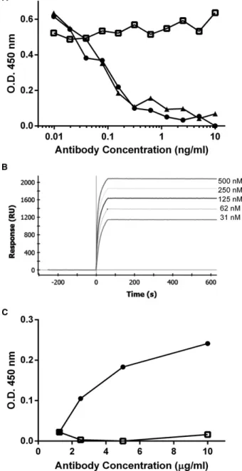
Purified NV-02 mAb was tested for its ability to
neu-tralize NGF in vitro using an NGF-dependent
prolifera-tion assay of TF-1 cells, as previously described.
6The
NV-02 had equivalent potency in this assay as caninized
anti-NGF mAb NV-01 (Fig 3A). Surface plasmon
reso-nance (SPR) assays (Fig 3B) indicated high-affinity
bind-ing of NV-02 to NGF (
K
D=
20 pM), equivalent to
NV-01,
6with no appreciable dissociation of ligand after
switching the flow to buffer, indicating that the high
affinity of binding was caused by a very slow off rate.
Using an ELISA for detection of C1q, the first
compo-nent of the complement cascade, NGF-captured NV-02,
was shown to bind little or no C1q (Fig 3C) by
compar-ison with a C1q-binding control antibody
(caN-HCB-kLC1).
6This result suggests that NV-02 will not initiate
complement-mediated immune damage, an important
safety consideration before its use in vivo.
Pharmacokinetics and Safety of NV-02 In Vivo
The concentration-time plots of NV-02 in the feline
plasma samples are depicted in Fig 4. The PK profiles
are typical of IgG injected to other mammals, with an
absorption phase from the site of injection to plasma
followed by a decay phase corresponding to elimination
from the plasma. After absorption from the injection
site, peak plasma concentrations (Cmax) were achieved
at approximately 3 days (Tmax; range, 1.9
–
4.3 days).
The averaged elimination phase half-life was calculated
to be 9 days (range, 7
–
15 days).
There were no changes in body weights, clinical
chemistry measurements, and hematology assessments
in any of the cats. No adverse reactions or behavioral
changes were noted by the veterinarians.
Effect of NV-02 administration in a Kaolin-induced
Model of Inflammatory Lameness
Lameness scores were ranked before generating the
repeated measures ANOVA because it was determined
that the distribution could not be approximated by a
normal curve. The covariance structure that provided
the smallest AIC was heterogeneous autoregressive.
Pairwise comparisons to placebo yielded statistically
sig-nificant lower lameness scores in the NV-02 group at
day 2 (
P
=
.0027), day 3 (
P
=
.0166), day 4 (
P
=
.0063),
day 5 (
P
=
.0085), day 6 (
P
=
.0014), and day 7
(
P
=
.0034). Mean lameness scores are presented in
Fig 5. No difference was observed in mean rectal
tem-perature or mean paw circumference measurements
between placebo- and NV-02-treated cats on any
treat-ment day (data not shown).
Discussion
Our study represents the initial characterization of a
therapeutic antibody with the potential to treat acute
and possibly chronic pain in cats. No pharmaceutical or
biopharmaceutical products are approved by the United
States Food and Drug Administration (US-FDA) for
long-term control of chronic pain in cats, unlike in
dogs, where several NSAIDs are FDA-approved for
this indication. This situation exists although chronic
pain associated with degenerative joint disease appears
to be very prevalent in cats.
13,14Two NSAIDs,
meloxi-cam and robenacoxib, are FDA-approved for the
treat-ment of postoperative pain in cats, but meloxicam is
restricted to a single administration and robenacoxib to
a maximum of 3 days. In Europe, meloxicam is
approved for unlimited daily use in the cat for the
con-trol of musculoskeletal pain, yet the same drug
(meloxi-cam) carries a boxed warning in the United States,
which reads, “Repeated use of meloxicam in cats has
been associated with acute renal failure and death. Do
not administer additional injectable or oral meloxicam
to cats”. The controversy over repeated administration
of meloxicam, or indeed any NSAID, aside, clearly the
need for new analgesic medications for the control of
chronic pain in cats is high.
Nerve growth factor is a highly conserved dimeric
peptide hormone produced by proteolytic cleavage of a
pre-pro-peptide precursor. During development, NGF is
a neurotrophic factor essential for the survival of
sen-sory and sympathetic neurons.
15,16In the adult, NGF is
expressed at sites of injury and inflammation and is a
major factor promoting pain and hyperalgesia.
4,5Nerve
growth factor acts on 2 receptors, the high-affinity trkA
receptor and low-affinity p75 receptor. The modulating
effect of NGF on nociceptive neurons is mediated by
the trkA receptor, resulting in increased immediate and
long-term excitability
3by modulation of ion channels
such as the transient receptor potential vanilloid
recep-tor (TRPV1).
3Nerve growth factor also causes
sprout-ing of nerve endsprout-ings into the site of inflammation
2and
has been detected in neuromas.
17Administration of
NGF can potentiate pain, and mutations in the trkA
receptor are associated with diminished pain responses.
4Furthermore, antibodies that neutralize NGF are highly
effective analgesics in animal models of inflammatory
pain, arthritis, cancer pain, and bone fracture.
2,3This encouraging biological activity has resulted in
sev-eral NGF antagonists being developed for the treatment
of pain in humans.
3The clinical efficacy of anti-human
NGF mAbs has been demonstrated in several pain states
(eg, OA, lower back pain, cystitis) in clinical trials,
including large Phase 3 studies.
3–5,18,19The NGF
inhibi-tory antibodies were generally very well tolerated
(consis-tent with a benign profile in 6-month primate studies
20)
and mild to moderate, transient peripheral sensation
changes were the only neurological consequences.
3–5In
2010, the FDA instructed several companies to put their
clinical programs on hold (except those for cancer pain)
after observations of worsening of clinical signs in a small
proportion of patients with OA, requiring accelerated
joint replacement.
21The cause of this worsening, termed
“rapidly progressing OA”, was debated, although in
some patients, the accelerated OA appeared to be
associ-ated with concomitant NSAID use. Nonetheless, the
FDA expert review panel overseeing these studies
recom-mended that the clinical halt be lifted in 2012.
22The first veterinary application of an anti-NGF mAb
recently was described in the form of a fully caninized
anti-NGF mAb.
6This high-affinity mAb (NV-01) also
Fig 4. Pharmacokinetic profile of NV-02 in cat plasma following subcutaneous administration at various dose levels. A single dose of NV-02 at 2 mg/kg and 5.6 mg/kg (A) or 16.8 mg/kg and 28 mg/kg (B) was administered to two cats per dose level and the plasma concen-tration of NV-02 was assayed at the times indicated by a quantitative NGF-binding ELISA.
was derived from
a
D11
9and is a potent inhibitor of
NGF in vitro. In dogs, the mAb had a long serum
half-life, did not generate neutralizing anti-drug antibodies
and was effective in decreasing signs of lameness caused
by inflammation. In a pilot clinical study of 9 dogs, a
single dose of NV-01 mAb was well tolerated and
sig-nificantly decreased clinical signs of pain and improved
mobility in OA dogs, as assessed by owners using the
Canine Brief Pain Inventory.
7The magnitude of
improvement in clinical signs was similar to that seen
previously with NSAIDs and the duration of effect was
at least 4 weeks. More recently, in a blinded,
placebo-controlled study of 26 dogs with degenerative joint
dis-ease (including OA), a single dose of NV-01 similarly
was well tolerated and effective at decreasing pain and
improving mobility.
8The NV-01-treated dogs showed
improvement in 3 independent clinical scoring methods,
again in magnitude similar to that observed with
NSAIDs and over a period of 4 weeks. Furthermore,
the NV-01-treated dogs were more active during the
daytime, as assessed by collar-mounted accelerometers.
Given its role in mediating inflammatory pain in
rodents, dogs, and humans, we reasoned that NGF
would be a useful therapeutic target in cats with painful
conditions. Feline NGF and its receptor are closely
homologous to those of other species. For mature
(beta) NGF, there is 100% identity between dog and
cat, 91% identity between human and cat, 85% identity
between rat and cat, and 82% identity between mouse
and cat. Nerve growth factor and its receptor trkA are
expressed in similar tissues in cats and humans, appear
to be under similar control mechanisms, and have
simi-lar functions.
23,24As with other mammals, the feline
immune system shares major immunoglobulin types,
including IgG, IgG-Fc receptors including the
high-affi-nity FcR, and the neonatal FcRn, which potentiates
IgG half-life in vivo.
25–27Felinized anti-NGF mAb NV-02, like the caninized
mAb NV-01 previously described, retains the high
affinity of its parent mAb
a
D11 (Fig 3B) and has
similar ability to neutralize NGF in vitro (Fig 3A),
suggesting that NV-02 retains the structural integrity
of the parent mAb. Because the concentration of
anti-body required to neutralize a given amount of NGF
is decreased with increased affinity and potency, the
results with NV-02 are predictive of low dosage for
in vivo efficacy. Furthermore, NV-02 does not recruit
complement (Fig 3C), which is important for its safe
use in vivo.
We observed no safety signals in cats injected with a
single dose of NV-02 by the SC route at dosages up to
28 mg/kg. Although the safety studies of this small
cohort were limited to observations of behavior, weight
change, blood chemistry, and hematology, we conclude
that NV-02 lacks overt toxicity. Further research will be
necessary to confirm this safety profile. Delivery of
anti-NGF mAbs SC limits their exposure to the periphery,
because mAbs do not cross the blood-brain barrier
(BBB). Consequently, action on the central nervous
sys-tem is avoided. Anti-NGF mAbs from the maternal
cir-culation (via the placenta and developing BBB) cause
fetal abnormalities in rodents,
28,29and in pregnant
non-human primates, caused increased stillbirth, postbirth
infant mortality and morbidity, decreased infant growth,
sensory and sympathetic nervous system changes, and
decreased infant primary antibody responses.
30The use
of anti-NGF mAbs should be avoided in pregnant or
lactating animals. By contrast, in the adult, very high
doses of anti-NGF mAb delivered IV or SC were well
tolerated and extensive analysis identified no effects on
adult peripheral nerves.
31When administered to cats SC, 4 days before kaolin,
NV-02 decreased the severity of lameness and
main-tained protection from lameness over the following
week. As previously observed for anti-NGF mAbs in
other species,
2NV-02 did not decrease inflammatory
pyrexia or swelling of the paw compared with
placebo-treated cats. A similar duration of effect was observed
previously with a caninized anti-NGF mAb in the
kao-lin model in dogs, and longer effects were observed in
dogs with degenerative joint disease (DJD). Studies of
cats with DJD will be necessary to determine whether
NV-02 is effective in providing analgesia in this setting,
as well as whether a duration of effect
>
7 days can be
achieved after a single injection.
Monoclonal antibodies have become an important
part of the therapeutic treatment options for several
dis-eases of humans including inflammation, autoimmunity,
cancer, allergy, and blindness because of their
combina-tion of efficacy, duracombina-tion of effect, and safety. The
PETization approach, described here with the
anti-NGF mAb for cats (NV-02) and previously with dogs
(NV-01) produces mAbs with similar properties to
humanized mAbs and potentially will allow the use of
mAbs for the treatment of many diseases in veterinary
patients.
Conclusions
Our data support the hypothesis that neutralization
of NGF in cats with the mAb NV-02 may have the
potential to provide appropriately safe analgesia in the
cat, potentially for prolonged durations. These
conclu-sions are based on our findings of high affinity for and
potent inhibition of NGF in vitro, no evidence of
bind-ing complement component C1q, a long plasma
half-life, and a lack of observed toxicity in vivo.
Further work will be necessary to assess: (1) the value
of this mAb in treating chronic pain states in the cat,
such as the pain associated with degenerative joint
dis-ease; (2) the optimal route of delivery, dose, and dosing
strategy for best efficacy; and (3) its safety in larger
cohorts of normal or diseased cats.
Footnotes
aGeneart AG, Life Technologies, Regensburg, Germany bLonza Biologics plc, Cambridge, UK
cGE Healthcare, UK dSartorius, Australia eAbD Serotec, UK fBioRad, Hercules gPromega, Madison
heBM2a, eBioscience, San Diego iThermoFisher Scientific j
Jackson Immunoresearch Laboratories
k
Sigma-Aldrich
Acknowledgments
The authors thank the following individuals for their
contributions to this study: Paul Hertzog, Kevin Johnson,
Angel Lopez, and colleagues at Nexvet for technical
advice and support; John Cox, Jane Eagleson, Colin
Giles, Andy Gearing, and Samantha Busfield for critical
review of the manuscript; Michael Spring and colleagues
(Life Technologies, Germany) for gene synthesis services
and small-scale protein expression; Antony Munn and
colleagues (Lonza, UK) for protein expression and
purifi-cation; Edouard Nice and Daniel Layton (MATF,
Mon-ash University) for binding affinity studies; and colleagues
at Charles River Laboratories (Ballina, Ireland) for
per-forming the animal studies and kaolin model.
Conflict of Interest Declaration:
All authors except
MH and BDXL are employees, stockholders, or both
of Nexvet Australia Pty Ltd. MH, BDXL, and RPG
are paid consultants of Nexvet Australia Pty Ltd.
Off-label Antimicrobial Declaration:
Authors declare
no off-label use of antimicrobials.
References
1. Sparkes AH, Heiene R, Lascelles BD, et al. ISFM and AAFP consensus guidelines: Long-term use of NSAIDs in cats. J Feline Med Surg 2010;12:521–538.
2. Ghilardi JR, Freeman KT, Jimenez-Andrade JM, et al. Neu-roplasticity of sensory and sympathetic nerve fibers in a mouse model of a painful arthritic joint. Arth Rheum 2012;64:2223–2232.
3. Cattaneo A. Tanezumab, a recombinant humanized mAb against nerve growth factor for the treatment of acute and chronic pain. Curr Opin Mol Ther 2010;12:94–106.
4. Hefti FF, Rosenthal A, Walicke PA, et al. Novel class of pain drugs based on antagonism of NGF. Trends Pharmacol Sci 2006;27:85–91.
5. Mantyh PW, Koltzenburg M, Mendell LM, et al. Antago-nism of nerve growth factor-TrkA signaling and the relief of pain. Anesthesiology 2011;115:189–204.
6. Gearing DP, Virtue ER, Gearing RP, Drew AC. A fully caninised anti-NGF monoclonal antibody for pain relief in dogs. BMC Vet Res 2013;9:226.
7. Webster RP, Anderson GI, Gearing DP. Canine Brief Pain Inventory scores for dogs with osteoarthritis before and after
administration of a monoclonal antibody against nerve growth factor. Am J Vet Res 2014;75:532–535.
8. Lascelles BD, Knazovicky D, Case B, et al. A canine-specific anti-nerve growth factor antibody alleviates pain and improves mobility and function in dogs with degenerative joint disease-asso-ciated pain. BMC Vet Res 2015;11:101.
9. Ruberti F, Bradbury A, Cattaneo A. Cloning and expression of an anti-nerve growth factor (NGF) antibody for studies using the neuroantibody approach. Cell Mol Neurobiol 1993;13:559–568.
10. Lewis MJ, Wagner B, Woof JM. The different effector function capabilities of the seven equine IgG subclasses have implications for vaccine strategies. Mol Immunol 2008;45:818–827. 11. Giraudel JM, Diquelou A, Laroute V, et al. Pharmacoki-netic/pharmacodynamic modelling of NSAIDs in a model of reversible inflammation in the cat. Br J Pharmacol 2005;146:642– 653.
12. Zhang Y, Huo M, Zhou J, Xie S. PKSolver: An add-in program for pharmacokinetic and pharmacodynamic data analysis in Microsoft Excel. Comput Methods Programs Biomed 2010;99:306–314.
13. Lascelles BD, Henry JB, Brown J, et al. Cross-sectional study of the prevalence of radiographic degenerative joint disease in domesticated cats. Vet Surg: VS 2010;39:535–544.
14. Slingerland LI, Hazewinkel HA, Meij BP, et al. Cross Sec-tional study of the prevelance and clinical features of osteoarthritis in 100 cats. Vet J 2011;187:304–309.
15. Levi-Montalcini R. The nerve growth factor 35 years later. Science 1987;237:1154–1162.
16. Levi-Montalcini R, Skaper SD, Dal Toso R, et al. Nerve growth factor: From neurotrophin to neurokine. Trends Neurosci 1996;19:514–520.
17. Harpf C, Dabernig J, Humpel C. Receptors for NGF and GDNF are highly expressed in human peripheral nerve neuroma. Muscle Nerve 2002;25:612–615.
18. Lane NE, Schnitzer TJ, Birbara CA, et al. Tanezumab for the treatment of pain from osteoarthritis of the knee. New Eng-land J Med 2010;363:1521–1531.
19. Katz N, Borenstein DG, Birbara C, et al. Efficacy and safety of tanezumab in the treatment of chronic low back pain. Pain 2011;152:2248–2258.
20. Zorbas M, Hurst S, Shelton D, et al. A multiple-dose toxic-ity study of tanezumab in cynomolgus monkeys. Regul Toxicol Pharmacol: RTP 2011;59:334–342.
21. Garber K. Fate of novel painkiller mAbs hangs in balance. Nat Biotechnol 2011;29:173–174.
22. United States Food and Drug Administration. Summary minutes of the arthritis advisory committee meeting March 12, 2012. Available from http://www.fda.gov/downloads/Advisory Committees/CommitteesMeetingMaterials/Drugs/ArthritisAdvisory Committee/UCM307879.pdf. Accessed November 2013.
23. Fried K, Risling M, Arvidsson U, Paulie S. Nerve growth factor receptor-like immunoreactivity in nerve fibers in the spinal and medullary dorsal horn of the adult monkey and cat: Correla-tion with calcitonin gene-related peptide-like immunoreactivity. Brain Res 1990;536:321–326.
24. Fried K, Risling M. Nerve growth factor receptor-like immunoreactivity in primary and permanent canine tooth pulps of the cat. Cell Tissue Res 1991;264:321–328.
25. Hinton PR, Johlfs MG, Xiong JM, et al. Engineered human IgG antibodies with longer serum half-lives in primates. J Biol Chem 2004;279:6213–6216.
27. Strietzel CJ, Bergeron LM, Oliphant T, et al. In vitro func-tional characterization of feline IgGs. Vet Immunol Immunopathol 2014;158:214–223.
28. Aloe L, Cozzari C, Calissano P, Levi-Montalcini R. Somatic and behavioral postnatal effects of fetal injections of nerve growth factor antibodies in the rat. Nature 1981;291:413– 415.
29. Pearson J, Johnson EM, Brandeis L. Effects of antibodies to nerve growth factor on intrauterine development of derivatives
of cranial neural crest and placode in the guinea pig. Dev Biol 1983;96:32–36.
30. Bowman CJ, Evans M, Cummings T, et al. Developmental toxicity assessment of tanezumab, an anti-nerve growth factor monoclonal antibody, in cynomolgus monkeys (Macaca fascicu-laris). Reprod Toxicol 2014;53:105–118.