Effect of Fluoride on Parathyroid Hormone Secretion
Chaitanya Prakash Puranik
A dissertation submitted to the faculty of the University of North Carolina at Chapel Hill in partial fulfillment of the requirements for the degree of Doctor of Philosophy in
the School of Dentistry (Oral Biology)
Chapel Hill 2013
Approved by: Eric T. Everett Timothy Wright Mitsuo Yamauchi Bryan Roth
ii © 2013
iii ABSTRACT
Chaitanya Prakash Puranik: Effect of Fluoride on Parathyroid Hormone Secretion (Under the direction of Eric Everett)
iv
v
ACKNOWLEDGEMENTS
vi
Timothy Wright provided me a deep insight in scientific research, clinical problems, and genetic approaches.
I am grateful to my co-workers and friends Dr. Yan Dong, Holly Cheeseman, Kathleen Ryan, Dr. Melanie Alazzam, and Dr. Shijia Hu with whom I shared my research space and time. I would also like to thank Heather Zimmerman, Lauren Katz, Eleni Boukas, Elise McGuire and Staci Love who helped me in my research and made my stay at UNC enjoyable.
I wish to thank Drs. Sylvia Frazier-Bowers, Ching Chang Ko, Patrick Flood, and Asma Khan for their constant help and support. I am grateful for the support and motivation by Dr. Ceib Philips, Oral Biology program director. I am also thankful to the Oral Biology program manager, Ms. Cindy Blake, for patiently guiding me throughout my training.
vii
TABLE OF CONTENTS
TABLE OF CONTENTS ... vii
LIST OF FIGURES AND TABLES ... xii
LIST OF ABBREVIATIONS AND SYMBOLS ... xiii
CHAPTER 1: REVIEW OF LITERATURE ... 1
1.1 Fluoride ... 1
1.1.1 Introduction ... 1
1.1.2 Metabolism ... 2
1.1.2.1 Absorption ... 3
1.1.2.2 Distribution ... 4
1.1.2.2.1 Distribution in the soft tissues ... 5
1.1.2.2.2 Distribution in body fluids ... 6
1.1.2.2.3 Distribution in mineralized tissues ... 6
1.1.2.3 Renal clearance ... 7
1.1.2.4 Fecal clearance ... 9
viii
1.1.2.5.1 Acid-base equilibrium ... 9
1.1.2.5.2 Renal impairments ... 9
1.1.2.5.3 Altitude ... 10
1.1.2.5.4 Physical activity ... 10
1.1.2.5.5 Circadian rhythm ... 11
1.1.2.5.6 Nutrition ... 12
1.1.2.5.7 Genetic influences ... 13
1.1.3 Toxicity ... 15
1.1.4 Fluorides in dentistry ... 16
1.1.4.1 Fluoride’s action on tooth enamel ... 17
1.1.4.2 Enamel fluorosis ... 18
1.1.5 Interactions on bone... 19
1.1.5.1 Bone remodeling, osteoblasts and osteoclasts ... 19
1.1.5.2 Characteristics of fluoride’s actions and hypotheses ... 21
1.1.5.2.1 Tyrosine phosphate hypothesis ... 22
1.1.5.2.2 G protein hypothesis ... 24
1.1.5.5 Skeletal fluorosis ... 25
1.2 Parathyroid hormone (PTH) ... 31
ix
1.2.2 PTH and PTHLH ... 33
1.2.2.1 Synthesis and secretion of PTH ... 34
1.2.2.2 Regulation of PTH secretion ... 35
1.2.2.2.1 Extracellular calcium-mediated regulation ... 35
1.2.2.2.2 1,25(OH)2D3-mediated regulation ... 37
1.2.2.2.3 Phosphorous- FGF23-KLOTHO-mediated regulation ... 37
1.2.2.3 Metabolism of PTH ... 38
1.2.2.4 PTH- and PTHLH-PTH1R ... 39
1.3 Calcium Sensing Regulator (CASR) ... 40
1.3.1 Isolation of Calcium Sensing Receptor ... 40
1.3.2 CASR gene ... 41
1.3.3 Functions of CASR in tissues ... 42
1.3.3.1 Parathyroid gland ... 42
1.3.3.2 Parafollicular cells ... 42
1.3.3.3 Kidney ... 43
1.3.3.4 Intestine ... 43
1.3.3.5 Bone ... 44
1.3.4 CASR signaling ... 44
1.3.5 CASR agonists ... 45
x
CHAPTER 3: FLUORIDE MODULATES PARATHYROID HORMONE
SECRETION IN VITRO AND IN VIVO ... 51
3.1 Abstract ... 52
3.2 Introduction ... 54
3.3 Materials and Methods ... 56
3.3.1 Animals ... 56
3.3.2 Parathyroid gland organ culture ... 56
3.3.3 Gavage and sample collections ... 57
3.3.4 ELISA and biochemistry assays ... 57
3.3.5 RNA extractions and cDNA preparation ... 58
3.3.6 Statistical analysis ... 59
3.4 Results ... 60
3.4.1 Responsiveness of TPC organ culture model ... 60
3.4.2 Effect of fluoride on TPCs ... 61
3.4.3 Serum fluoride levels after gavage ... 61
3.4.4 Serum iPTH after single fluoride dose via oro-gastric gavage ... 62
3.4.5 Serum total calcium, phosphorus, and magnesium levels ... 64
3.4.6 qPCR: Expression of genes involved in BPK feedback loop ... 66
xi
CHAPTER 4: FLUORIDE DOES NOT TRIGGER BETA-ARRESTIN
RECRUITMENT (NON-CANONICAL SIGNALING) BY CASR ... 72
4.1 Abstract ... 73
4.2 Introduction ... 75
4.3 Material and Methods ... 77
4.3.1 CASR and orphan GPCRs constructs ... 77
4.3.2 Cells and transfection ... 77
4.3.3 β-arrestin recruitment assay ... 78
4.3.4 Statistical analysis ... 78
4.4 Results ... 80
4.4.1 Effect of fluoride on human CASR ... 80
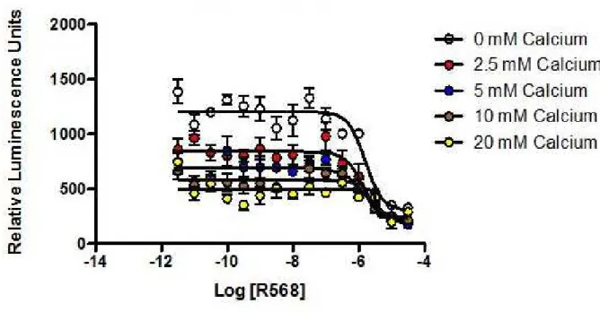
4.4.2 Effect of calcimimetic (R568) on human CASR ... 80
4.4.4 Effect of fluoride on R568-mediated CASR response ... 81
4.4.5 Effect of R568 on CASR is specific ... 82
4.4.5 Effect of R568 extends to CASR related GPCRs ... 83
Figure 4.5: Effect of R568 on GPRC6A ... 84
4.4.6 Effect of fluoride on 126 orphan GPCRs ... 84
4.5 Discussion ... 87
xii
LIST OF FIGURES AND TABLES
Figure 1.1: Fluoride action hypotheses ... 24
Figure 1.2: Ground water fluoride concentration around the world. ... 27
Figure 1.3: Clinical and radiographic features of skeletal fluorosis ... 29
Figure 1.4: Bone-Parathyroid-Kidney (BPK) axis ... 33
Figure 1.5: CASR signaling cascade ... 45
Figure 3.1: iPTH levels in media supernatant ... 60
Figure 3.2: Serum fluoride levels ... 62
Figure 3.3: Serum iPTH levels 0-24 hrs ... 63
Figure 3.4: Serum iPTH levels 48-96hrs ... 64
Figure 3.5: Serum calcium, phosphorus, and magnesium levels... 65
Figure 3.6: Expression of genes involved in BPK feedback loop ... 67
Figure 4.1: Effect of fluoride on CASR ... 80
Figure 4.2: Effect of calcimimetic (R568) on CASR ... 81
Figure 4.3: Effect of fluoride on R568-mediated CASR response ... 82
Figure 4.4: Effect of R568 on CASR is specific ... 83
Table 4.1: Percentage RLU for screened orphan GPCRs ... 85
Table 4.2: Orphan GPCRs activated by fluoride ... 86
xiii
LIST OF ABBREVIATIONS AND SYMBOLS
[F-] fluoride ion
1,25(OH)2D3 1, 25 dihydroxy vitamin D3
AC adenylyl cyclase
ALP alkaline phosphatase
B6 C57BL/6J mouse strain
BIPK bone-intestine-parathyroid-kidney
BMM bone marrow macrophages
BPK bone-parathyroid-kidney
C3H C3H/HeJ mouse strain
Ca2+ ionic calcium
cAMP cyclic adenosine monophosphate CASR calcium sensing receptor
cDNA complimentary deoxyribonucleic acid CLCN5 chloride channel 5
c-Src tyrosine protein kinase
CT calcitonin
DC-STAMP dendritic cell-specific transmembrane protein ELISA enzyme-linked immunosorbant assay
ER endoplasmic reticulum
ERK extracellular signal-regulated kinase
xiv
FGF1Rc fibroblast growth factor receptor 1c FGF23 fibroblast growth factor 23
FMBD fluorotoxic metabolic bone disease
Gd2+ ionic gadolinium
GI tract gastro-intestinal tract GPCR G-protein coupled receptor
Grb2 growth factor receptor-bound protein 2 GTP guanosine triphosphate
HAP hydroxyapatite
HF hydrofluoric acid
HTL HEK293T-cloned cells containing integrated luciferase reporter
IP3 inositol triphosphate
iPTH intact parathyroid hormone MAPK mitogen-activated protein kinase
MC3T3-E1 osteoblastic cell line form mouse calvaria M-CSF macrophage colony-stimulating factor
Mg2+ ionic magnesium
MMP9 matrix metalloproteinase 9 mRNA messenger ribonucleic acid
NFATc-1 nuclear factor of activated T-cells, cytoplasmic 1 NSHPT neonatal severe hyperparathyroidism
xv PDZK1 PDZ containing domain 1
PKC protein kinase C
PLA2 phospholipase A2
PLC phospholipase C
PLD phospholipase D
PPI polyphosphoinositide
PTH parathyroid hormone
PTH1R parathyroid receptor 1 PTH2R parathyroid receptor 2
qPCR quantitative real time polymerase chain reaction Raf serine/threonine protein kinase
Ras family of small GTPase
SF skeletal fluorosis
SLC34A1 solute carrier family 34, member 1
SLC9A3R1 solute carrier family 9 member 3 regulator 1
SOS son of sevenless
Sr2+ ionic strontium
sRANKL soluble receptor-activator of NF-κB ligand (current name TNFSF11, tumor necrosis factor (ligand) superfamily, member 11)
TPC thyro-parathyroid complex
xvi WHO World Health Organization
1
CHAPTER 1: REVIEW OF LITERATURE
1.1 Fluoride 1.1.1 Introduction
2 1.1.2 Metabolism
Ingested fluoride is primarily absorbed from the stomach and intestine. Fluoride ion can complex with calcium ion to form fluorite (CaF2) (Kanbur et al., 2009). In the body, fluoride can combine with various elements to form insoluble complexes which can accumulate in the extracellular compartments (Mittal and Flora, 2006). Fluoride absorption (stomach and intestine), distribution (hard and soft tissue) and excretion (kidney) are pH dependent process. Hydrofluoric acid (HF) is a weak acid having an acid dissociation constant (pKa) of 3.4. Therefore at 3.4 pH, 50% of fluoride exists as un-dissociated form (HF) while the remaining 50% is in the dissociated or ionic form [F-]. When pH decreases from 3.4, the concentration of HF increases and when pH increases above 3.4, the concentration of [F-] increases (Whitford, 1996). HF has a high coefficient of permeability as compared to fluoride and hence HF can penetrate lipid bilayer more easily (Gutknecht and Walter, 1981). HF movement occurs in response to a pH gradient between body compartments (HF moves from the acidic compartment to the alkaline compartment along the pH gradient).
3
Plasma fluoride concentrations reach peak after ingestion within 20-60 minutes (Whitford, 1996). The fluoride is then deposited in the newly mineralized tissues or is excreted through urine. In adults, approximately half of the absorbed fluoride is deposited in the mineralized tissues (bone, enamel, and dentin). Mineralized tissue holds approximately 99% of body’s fluoride (Whitford, 1994). However, fluoride is not irreversibly bound to bone. Fluoride can be released back into the plasma during bone remodeling (Whitford, 2008). A small fraction of fluoride absorbed is found in soft tissues (Whitford, 1984). Most of the absorbed fluoride, not taken up by mineralized tissues, is excreted in urine. Very small fraction (<1%) of absorbed fluoride is excreted in sweat and feces. The plasma fluoride concentrations return to the baseline after 3-6 hours (Whitford, 1996).
1.1.2.1 Absorption
More than 80% of the ingested fluoride is absorbed from gastro-intestinal (GI) tract. (Whitford, 1996). Presence of high amounts of bi- and tri-valent cations (calcium, magnesium, and aluminum) which form insoluble complexes with fluoride reduces net fluoride absorption. (Whitford, 1996). Although fluoride absorption from the stomach occurs rapidly, the rate of absorption is determined by gastric acidity (Messer and Ophaug, 1993; Whitford and Pashley, 1984) and velocity of gastric emptying (Messer and Ophaug, 1991; Nopakun et al., 1989).
4
gastric lumen, it is converted into HF which is an uncharged molecule that can readily cross the gastric mucosa. Higher the acidity of the gastric content, faster will be the fluoride absorption from the stomach.
Additionally, gastric emptying interferes with gastric fluoride absorption. Animal studies have shown that delayed gastric emptying might result in slower and smaller increases in plasma fluoride levels (Messer and Ophaug, 1991; Nopakun et al., 1989). More than half of the fluoride which escapes gastric absorption is absorbed in the proximal part of small intestine (Nopakun et al., 1989). The small intestine has a huge capacity for fluoride absorption and fluoride is rapidly absorbed following emptying from the stomach. Ionic fluoride crosses the leaky epithelia through tight junctions between the paracellular channels (Nopakun et al., 1989). Fluoride absorption from the small intestine compensates for the low gastric absorption due to high pH to ensure that overall fluoride absorption is relatively unaffected by gastric pH (Messer and Ophaug, 1993; Whitford and Pashley, 1984).
1.1.2.2 Distribution
Plasma is the central compartment for fluoride distribution. Fluoride achieves steady-state distribution in the soft tissues where less than 1% of absorbed fluoride is present. More than half of absorbed fluoride is taken up by the calcified tissues, where fluoride is reversibly bound, and can be released back into the plasma in case of demand. (Whitford, 1994).
5
in the plasma is twice as high as that found in the blood cells. The biological function of non-ionic fluoride has not been established yet. However, its concentration is usually higher than that of ionic fluoride. The non-ionic and ionic fluoride together constitute the total plasma fluoride (Whitford, 1996). Ionic fluoride concentrations in the plasma are dynamic; it depends on the amount of fluoride intake, deposition/removal (in/from soft and hard tissues), and urinary excretion (Whitford, 1996). Therefore, plasma fluoride levels have been used as ‘contemporary biomarkers’ of fluoride exposure.
1.1.2.2.1 Distribution in the soft tissues
6 1.1.2.2.2 Distribution in body fluids
Fluoride concentrations in body fluids are dynamic and different from those found in plasma. Cerebrospinal fluid and milk have fluoride concentrations 50% lower than that of plasma. Gingival crevicular fluid and ductal saliva (submandibular and parotid) fluoride levels are slightly higher and lower, respectively, than those in plasma. Reported ratio of fluoride concentrations in ductal saliva to plasma fluoride concentrations are around 0.9 and 0.8 for submandibular and parotid secretions, respectively (Whitford, 1994). Ductal saliva has also been employed as a ‘contemporary biomarker’ of fluoride exposure (Olympio et al., 2007; Wilson and Bawden, 1991; Zero et al., 1992). Concentrations of fluoride in the whole saliva are more variable and higher than those seen in ductal saliva. Exogenous fluoride contamination of whole saliva renders it unsuitable for estimation of plasma fluoride concentrations as a ‘contemporary biomarker’. (Whitford et al., 1999).
1.1.2.2.3 Distribution in mineralized tissues
7
It is estimated that approximately 36% of the absorbed fluoride becomes associated with the bone, while the remainder is excreted. In children, the degree of retention is much higher (55%) (Villa et al., 2010) due to the rich blood supply and large surface area of the growing bones (Whitford, 1994).
Fluoride concentrations in the dentin are similar to bone fluoride concentrations and increase with age. Dentin fluoride levels are higher close to the pulp and reduce progressively towards the dentino-enamel junction (Rao et al., 1995). Enamel fluoride concentrations are usually lower than the levels found in dentin. No correlation was found between the concentrations of fluoride in dentin and enamel (Vieira et al., 2004; Vieira et al., 2006). Concentration of fluoride in the dental enamel decreases with age, especially in areas where dental biofilm accumulates. The fluoride concentrations in the dental enamel reflect the level of fluoride exposure during amelogenesis (process of enamel formation) (Rao et al., 1995). A significant correlation between the severity of dental fluorosis and fluoride concentrations has been found only for the dentin. (Vieira et al., 2004; Vieira et al., 2006).
1.1.2.3 Renal clearance
8
bone remodeling in adults. Fluoride in the plasma and urine reflects a physiologic balance between fluoride intake and removal.
The concentrations of fluoride in the glomerular filtrate and plasma are comparable. After entering the renal tubules, a variable amount of the ion is reabsorbed and returned to the systemic circulation (Whitford, 1996). Tubular reabsorption of fluoride and glomerular filtration rate are the principal determinants of the urinary fluoride excretion. Compared to the renal clearance of other halogens (~1-2 mL/min), fluoride has high renal clearance (35mL/min). However, individual variations in the renal clearance of fluoride are common (Whitford, 1994) and are attributed to the three major factors; differences in the glomerular filtrations rates (Spak et al., 1985), pH of urine (Jarnberg et al., 1983) and urinary flow rate (Ekstrand et al., 1982).
9 1.1.2.4 Fecal clearance
Fecal fluoride represents less than 10% of the ingested fluoride. In other words, more than 90% of ingested fluoride is usually absorbed (Ekstrand et al., 1994). When diets containing high amounts of calcium are consumed, unabsorbed calcium forms complexes with fluoride in the stomach. The complexes of calcium and fluoride do not easily diffuse through the GI lining and thereby reduces the total amount of fluoride absorbed. (Whitford, 1994).
1.1.2.5 Factors modifying metabolism 1.1.2.5.1 Acid-base equilibrium
Acid-base equilibrium plays an important role on the metabolism and tissue concentrations of fluoride. Factors that alter the acid-base equilibrium include diet composition (vegetarian diet increases urinary pH), altitude, drugs, metabolic and respiratory disorders. Acute respiratory acid-base disorders affect renal excretion of fluoride in the same manner as the metabolic disorders (Whitford, 1996).
1.1.2.5.2 Renal impairments
10 1.1.2.5.3 Altitude
Rats raised in hypobaric chambers to stimulate high altitudes demonstrated increased enamel disturbances, regardless of the levels of ingested fluoride (Whitford, 1996). Alterations in acid-base balance, caused by hypobaric hypoxia due to high altitude of residence were responsible for decreased urinary fluoride clearance. This decrease in urinary fluoride clearance resulted in greater retention of fluoride (Whitford, 1997). A significantly higher prevalence of fluorosis has been observed in Tanzanian communities at a high altitude (1,463 m), in contrast with a low altitude area (100m), with similar food habits and low levels of fluoride in the drinking water (Yoder et al., 1998). The severity of enamel disturbances at the high altitude area was not consistent with the fluoride concentration in drinking water. Such a finding suggested that altitude, along with other factors, may contribute to the severity of enamel disturbances. Studies conducted in other high altitude locations around the world confirmed that physiological changes associated with residence at high altitude are able to exacerbate the effects of fluoride on mineralized tissues (Akosu and Zoakah, 2008; Martinez-Mier et al., 2004; Pontigo-Loyola et al., 2008).
1.1.2.5.4 Physical activity
11
associated with either decrease or increased circulating fluoride levels (Whitford, 1996). Although physical activity may alter the pattern of fluoride excretion, the impact on the development of dental fluorosis seems negligible.
1.1.2.5.5 Circadian rhythm
Daily variations are observed in levels of calcium and phosphorus which are involved in bone remodeling (the process involving a balance between deposition and resorption of bone) under the influence by mineral homeostatic hormones. Fluoride, similar to calcium and phosphorous, is primarily stored in the skeleton and therefore, it was hypothesized that plasma fluoride levels would exhibit circadian rhythm too (Talmage et al., 1975). Such rhythmicity was verified in dogs, with a mean peak fluoride concentration around 9 AM, followed by a decrease around 9 PM (Whitford, 1996).
12 1.1.2.5.6 Nutrition
A correlation between malnutrition and prevalence of dental fluorosis prevalence has been suggested in the past; however, the evidence for such a relationship is controversial. In a study with Tanzanian children, Yoder et al suggested a direct relationship between malnutrition and dental fluorosis (Yoder et al., 1998) whereas another study demonstrated that dental fluorosis is independent of nutritional status (Buzalaf and Whitford, 2011). Nutritional status is commonly assessed by the height-for-age and weight-height-for-age indices as per the recommendations by the World Health Organization (WHO). It is hypothesized that a malnourished child may absorb fluoride more quickly due to the inexistence of ions to form complexes in an empty stomach. On the other hand, a malnourished child may have low fluoride deposition due to slower bone growth.
13
to remain in a dissociated form as [F-] and hence fluoride cannot be reabsorbed (as HF) in the kidneys. This ultimately leads to fluoride wasting.
High dietary concentrations of certain cations, especially calcium, can reduce the extent of fluoride absorption (Vieira et al., 2004; Vieira et al., 2006). A study reviewed by Buzalaf et al, conducted in the province of Jaingxi, China, where the prevalence of dental fluorosis was reported to be above 50%, the consumption of milk was a critical factor influencing the incidence of dental fluorosis in children. The incidence of dental fluorosis among the children consuming milk was lower (7.2%) as compared to the children not consuming milk (37.5%). (Buzalaf and Whitford, 2011). In India, where approximately 62 million people have dental fluorosis, studies have been conducted to identify increased risk of dental fluorosis due to dietary components, other than fluoride (Bhargavi et al., 2004). Low calcium concentrations in the drinking water and prevalence of dental fluorosis are inversely related. Therefore, calcium supplementation in areas with endemic fluorosis will minimize the absorption of fluoride through water and will avoid excessive incorporation of fluoride in the mineralized tissues (Khandare et al., 2005).
1.1.2.5.7 Genetic influences
14
similar between those two groups. Differences between two homogeneous tribes raised the possibility of genetic influence on fluorosis prevalence.
15
partly explained with genetics. The differences in fluorosis susceptibility may involve physiological, biochemical, and molecular differences in theses mouse strains.
1.1.3 Toxicity
16 1.1.4 Fluorides in dentistry
17 1.1.4.1 Fluoride’s action on tooth enamel
Dental enamel is primarily composed of minerals of calcium and phosphate commonly termed as hydroxyapatite (HAP) (McCann, 1953). The HAP crystals form a closely packed lattice structure. Fluoride can replace hydroxyl (OH), carbonate (CO32-) and phosphate (PO43-) ions in HAP lattice to form fluorapatite (FAP). FAP renders enamel hard and resistant to decay and acid attack.
Ca10(PO4)6(OH)2 + 2F- Ca10(PO4)6F2 +2OH (Hydroxyapatite) (Fluorapatite)
Calcium present on the outer surface of the enamel complexes with fluoride to form fluorite (CaF2). Fluorite is resistant to acid dissolution and hence renders the outer surface of enamel resistant to demineralization after bacterial acid attack. Fluorite also acts as a source for fluoride ions. Reversibly bound fluoride to the calcium can be released in saliva to raise salivary fluoride concentration. Fluoride also diffuses to the underlying enamel layers where it replaces the OH, CO32- or PO43-ions and forms FAP.
The rate of formation of FAP is dependent on the pH of saliva and concentration of fluoride in the saliva (McCann, 1953). Apart from the role of fluoride in reducing demineralization by formation of FAP, fluoride also has an antimicrobial effect (Marquis, 1995; Yoshihara et al., 2001). Fluoride slows down plaque metabolism by inhibiting glycolysis. Reduced metabolism decreases reproduction in bacteria which reduces the bacterial count and acid production. To summarize:
18
• Fluoride promotes remineralization of demineralized tooth enamel by precipitation of fluorapatite from saliva
• Fluoride inhibits bacterial growth in the plaque and prevents acid attack on the teeth
1.1.4.2 Enamel fluorosis
19
triphosphate binding proteins (such as heterotrimentric G proteins) regulates vesicle formation in the golgi complex (Leyte et al., 1992).
1.1.5 Interactions on bone
1.1.5.1 Bone remodeling, osteoblasts and osteoclasts
Bone remodeling is a process that involves a balance between osteoclast and osteoblast activity. Osteoblasts increase bone mass and osteoclasts resorb bone. This fine balance between the osteoclasts and osteoblasts activity is essential to maintain bone homeostasis. Fluoride actions on bone are influenced by the concentration of fluoride. Physiological concentrations of fluoride promote osteoblast proliferation and increased matric production. Fluoride concentrations higher than physiological concentrations suppresses proliferation by inducing apoptosis of differentiated osteoblast (Allolio and Lehmann, 1999). The effects of fluoride on the osteoclasts are contradictory. Rodent bone marrow osteoclast precursor cells demonstrated increased tartrate-resistant acid phosphatase activity after fluoride treatment suggestive of increased fluoride-mediated osteoclast number and activity (Debinski and Nowicka, 2004; Yan et al., 2007). Contrary to this finding, another study demonstrated a fluoride-mediated reduction in osteoclast number and bone resorption (Okuda et al., 1990). The underlying molecular mechanism for fluoride’s action on osteoclasts is not clearly understood.
20
number of differentiated osteoclast are very few long the bone surfaces. (Tanaka et al., 2006; Yavropoulou and Yovos, 2008). Osteoclasts can be induced from bone marrow macrophages (BMM) using two cytokines: receptor activator of nuclear factor-κB ligand (RANKL) and macrophage colony-stimulating factor (M-CSF) (Boyle et al., 2003). Osteoclast precursors fuse together to form multinucleated, terminally differentiated osteoclast. NFATc1 upregulates DC-STAMP to stimulate osteoclast precursor fusion (Kim et al., 2008) which triggers formation of functional osteoclast. Functional, multinucleated osteoclasts attaches to the bone and forms a tight seal underneath which hydrogen ions are pumped to create an acidic environment for bone resorption (Song et al., 2009). Acidic environment help osteoclasts to degrade the inorganic bone matrix. Osteoclasts also secrete matrix metalloproteinase (MMP9) which is involved in the degradation of organic matrix (Kim et al., 2008; Song et al., 2009).
21
DNA synthesis and ALP activity in osteoblasts (Farley et al., 1983). Fluoride also demonstrated an increase in patients with osteoporosis (Riggs et al., 1980).
Fluoride action induced cell proliferation in bone cells at a concentration of 10
µM. This concentration is comparable to the serum fluoride concentration in humans (Farley et al., 1983). Mitogenic effects of fluoride on bone cells from other species including human was confirmed by various researches (Burgener et al., 1995; Hall, 1987; Kassem et al., 1993; Khokher and Dandona, 1990; Modrowski et al., 1992; Reed et al., 1993; Wergedal et al., 1988).
There is now compelling evidence that fluoride at mitogenic (µM) concenrations stimulates mature osteoblast activities such as alkaline phosphatase expression (Khokher and Dandona, 1990; Wergedal et al., 1988), collagen synthesis (Kassem et al., 1993; Modrowski et al., 1992) and osteocalcin production (Kassem et al., 1994) in monolayer cultures. Fluoride at mitogenic concentrations also stimulated calcium uptake (Farley et al., 1983; Zerwekh et al., 1990) and sodium-dependent phosphate transport (Selz et al., 1991) in osteoblasts. Therefore, it is widely accepted that at low doses, fluoride stimulates osteoblast proliferation leading to increased bone formation.
1.1.5.2 Characteristics of fluoride’s actions and hypotheses
22
al., 1988; Reed et al., 1993). Phosphate concentration in the media used for in vitro experiments often influence the fluoride sensitivity of the cells to induce mitogenic changes (Farley et al., 1988). Fluoride acts on various cell types, however the effects of fluoride on the osteoblast precursor cells are more pronounced (Bellows et al., 1990; Farley et al., 1990). Fluoride’s mitogenic actions on the osteoblasts are commonly mediated through increased activation of various signaling proteins, including mitogen-activated protein kinase (MAPK) (Lau et al., 1989; Thomas et al., 1996; Wu et al., 1997)
Fluoride is interestingly known to skew the balance between bone formation and resorption towards the bone formation. This overall shift results in increased bone deposition resulting in overall increase in bone mass. Several laboratories around the world are trying to uncover the mechanistic aspect of fluoride’s action on cell types involved in biomineralization especially osteoblast cells. The understanding the exact mechanism of fluoride’s action on bone formative cell, osteoblast, will further our understanding of the pathophysiology of degenerative bone diseases and thereby help development of efficient therapeutic strategies. Based on some literature findings, researchers have proposed two possible mechanism of fluoride’s action on bone cells. These two hypotheses are presented below.
1.1.5.2.1 Tyrosine phosphate hypothesis
23
Baylink, 1998). These findings suggest that the effects of fluoride are mediated through the tyrosine phosphate pathway. Proteins harvested from chicken calvarial cells, which were exposed to fluoride, demonstrated increased amount of phosphorylated plasma proteins (Lau et al., 1989). We know that the effects of most growth factors are mediated through tyrosine kinase receptors present on the cell surface. Fluoride sensitizes the cells to readily respond to mitogenic actions of growth factors (Lau et al., 1987). These observations led to the formulation of this first hypothesis for the mechanism of fluoride’s action.
24
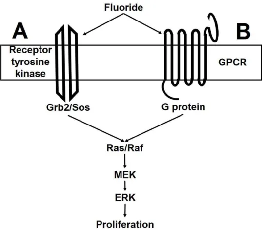
Figure 1.1: Fluoride action hypotheses
Fluoride inhibits the tyrosine phosphatase leading to increased phosphorylation of signaling proteins such as ERK 1/2 triggering proliferation (A) (Lau et al., 1989; Thomas et al., 1996). Fluoride along with other multivalent ions stimulates GDP to convert into GTP which is required for the activation of G proteins which in turn activates and phosphorylated signaling proteins including ERK 1/2 which are required for cell proliferation (B) (Caverzasio et al., 1996; Caverzasio et al., 1997).
1.1.5.2.2 G protein hypothesis
25
Heterotrimeric G proteins (Gi and Gq), triggers cell proliferation is widely accepted. However, the mechanism underlying this triggering of cell proliferation is not clear (Post and Brown, 1996; van Biesen et al., 1996). The involvement of Ras pathways leading to ERK phosphorylation by G protein has been studied in the past (Post and Brown, 1996) along with the involvement of the Grb2 and Sos (Post and Brown, 1996; van Biesen et al., 1996). Similar to the first hypothesis, this second hypothesis needs additional experimental evidence is required to understand the fluoride’s mode of action (van Biesen et al., 1996).
1.1.5.5 Skeletal fluorosis
Fluoride is predominantly stored in the mineralized tissues of the body such as enamel, dentin, and bones. In the bone fluoride demonstrate a biphasic actions (Collier, 1980). Fluoride, at low doses, provides hardness to the bone and renders enamel less susceptible to acid dissolution (Parnell et al., 2009). However, excess systemic exposure to fluoride can lead to skeletal and dental fluorosis. Skeletal fluorosis (SF) is a bone condition characterized by spectrum of bone changes ranging from osteoporosis to osteosclerosis. SF is clinically characterized by stiffness and rigidity of bone, restricted joint movements, bending deformities of spine and rotational deformities of long bones and calcification of ligaments and tendons (Ismail and Hasson, 2008). Fluoride reduces mineral solubility in the bone thereby affecting calcium metabolism in the body (Messer et al., 1973).
26
after fluoride exposure. Fluoride-mediated proliferation of bone forming cells is indicative of anabolic effect of fluoride on the bone. It was previously speculated that fluoride renders bone hard and resistant to resorption. The increase in bone mass after fluoride exposure is attributed to excessive bone formation rather than decreased bone resorption (Briancon and Meunier, 1981; Riggs et al., 1980). Although fluoride exposure increased bone mass and bone mineral density, it did not reduce the incidence of fractures undermining the effectiveness of fluoride treatment (Kleerekoper et al., 1991; Pak et al., 1994; Riggs et al., 1990; Riggs et al., 1994). Thus effectiveness of fluoride as a therapeutic agent can be enhanced if the benefit-to-risk profile of fluoride is improved. This improvement in benefit-benefit-to-risk profile can be achieved by understanding the mechanism of fluoride’s action.
27
without bone deformity), osteomalacia (osteoporosis with bone deformity), intermittent growth lines and diaphyseal widening and soft tissue calcification.
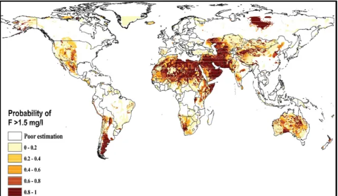
Figure 1.2: Ground water fluoride concentration around the world.
Reprinted with permission from Amini M, Mueller K, Abbaspour KC, Rosenberg T, Afyuni M, Møller KN, Sarr M, Johnson CA., Statistical modeling of global geogenic fluoride contamination in groundwaters., Environ Sci Technol. 2008 May 15;42(10):3662-8. Copyright 2008 American Chemical society.
28
Clinical findings of SF (Fig. 1.3) includes stiffness and rigidity of spine and joints, flexional deformities of bones, rotational deformities of legs, neurological complications, arthralgia, pseudo-hyperparathyroidism and restricted movements (Teotia et al., 1998). Teotia et al also found that pseudo-hyperparathyroidism was a common finding in patients with SF (Teotia and Teotia, 1973). Another study undertaken in Turkish population indicated that PTH was elevated in SF patients along with a decrease in serum calcium (Koroglu et al., 2011). Harinarayan et al reported that in FMBD patients demonstrated serum phosphorus and 1,25(OH)2D3 values comparable to the control group. Serum alkaline phosphatase (ALP) and PTH mid-molecule (PTH-MM) levels were higher (Harinarayan et al., 2006). Additionally, serum calcium, creatinine clearance and phosphorus excretion index were decreased. It is interesting to note that although, the patients in this study demonstrated similar clinical picture, they were exposed to variable amount of fluoride through drinking water, ranging from 0.23-14ppm (Harinarayan et al., 2006). Collectively, the findings can be summarized as:
• SF demonstrates variable radiographic and clinical picture • Persistent elevation of PTH in fluoride endemic areas
• Pseudo-hyperparathyroidism is a common finding in fluoride endemic area • Patients demonstrating similar clinical and radiographic picture were exposed
Figure 1.3: Clinical and radiographic features of skeletal fluorosis Reprinted with permission from
changes in children with fluorosis. 1980 Radiological Society of North
Whitford GM., Skeletal fluorosis from instant t
May;23(5):759-69. Copyright 2008 American Society of Bone and Mineral Research.
1.1.5.6 Fluoride and parathyroid h
Yan et al in 2007 (Yan et al., 2007
on bone homeostasis in two strains of mice: C3H/HeJ
These mice are extensively studied for their bone and bone cell properties. C3H has high bone mass, peak bone density, serum alkaline phosphatase activity and lower bone resorption rate as compared to B6
Linkhart et al., 1999; Turner et al., 2000 female mice were exposed to fluoride
29
: Clinical and radiographic features of skeletal fluorosis
Reprinted with permission from: 1. Christie DP., The spectrum of radiographic bone changes in children with fluorosis., Radiology. 1980 Jul;136(1):85
Radiological Society of North America. 2. Whyte MP, Totty WG, Lim VT, Skeletal fluorosis from instant tea., J Bone Miner Res. 2008 Copyright 2008 American Society of Bone and Mineral Research.
Fluoride and parathyroid hormone (PTH)
Yan et al., 2007; Yan et al., 2011) studied the effect of fluoride in two strains of mice: C3H/HeJ (C3H) and C57BL/6J
These mice are extensively studied for their bone and bone cell properties. C3H has high bone mass, peak bone density, serum alkaline phosphatase activity and lower bone resorption rate as compared to B6 (Dimai et al., 1998; Judex et al., 2004
Turner et al., 2000; Turner et al., 2001). Young C3H and B6 male mice were exposed to fluoride for 3 weeks. Bone fluoride content, serum
: Clinical and radiographic features of skeletal fluorosis
The spectrum of radiographic bone Radiology. 1980 Jul;136(1):85-90. Copyright Whyte MP, Totty WG, Lim VT, J Bone Miner Res. 2008 Copyright 2008 American Society of Bone and Mineral Research.
30
osteoclast biomarkers, in situ osteoclast numbers, osteoclast potential and hematopoietic colony forming cell assay, micro-computed tomography (µCT) analysis and biomechanical properties of bone were studied. After 3 weeks of fluoride exposure, C3H and B6 mice demonstrated variable effects. B6 mice began with higher bone fluoride and accumulated more fluoride in bone as compared to C3H. Anabolic actions of fluoride were favored in B6, including increase in serum ALP activity, increase in mineralized bone volume and trabacular thickness. However, in C3H, fluoride favored osteoclastogenesis demonstrated by an increase in bone marrow osteoclast potential and increase in osteoclast number along with an increase in serum osteoclast biomarkers including intact PTH (iPTH), sRANKL, TRAP5b and decrease in OPG. Similar results were found when the same strains of mice were exposed to long term fluoride exposure (Yan et al., 2011). Interesting finding of both the studies was elevated iPTH which was consistent with findings in fluoride endemic areas (Harinarayan et al., 2006; Teotia et al., 1998; Teotia and Teotia, 1973).
1.2 Parathyroid hormone (PTH)
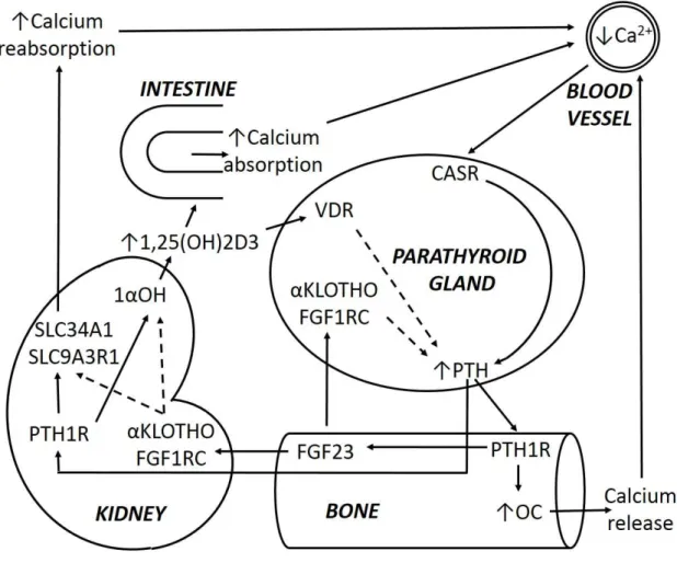
1.2.1 Mineral homeostasis and bone-intestine-parathyroid-kidney (BPK) axis
31
change in the ionized calcium levels triggers secretion of PTH by the Chief cells. PTH actions are required to restore the normocalcemic state in the body. PTH normalizes plasma ionic calcium levels by three defined actions:
1. PTH mediates production of osteoclast in the bone which resorbs bone and release calcium in the blood
2. PTH acts on kidney to induce calcium reabsorption
3. PTH has an indirect action on intestine. In the kidneys PTH signals to increase 1αhydroxylase production which promotes calcium absorption in the intestine.
FGF23 and 1,25(OH)2D3 are involved in feedback regulation to check levels of PTH. The interaction of these three hormones is required for the calcium homeostasis. The cross-talk between these three tissues to achieve calcium homeostasis has been traditionally referred as bone-parathyroid-kidney (BPK) axis. However, the indirect involvement of intestine in the absorption of calcium is crucial for successful calcium regulation. Hence we now refer this control mechanism for calcium homeostasis as the BPK as bone-intestine-parathyroid-kidney (BIPK) axis (Fig. 1.4)
32
production is sensed by vitamin D receptor (VDR) on parathyroid gland which signals production of PTH.
33
Figure 1.4: Bone-Parathyroid-Kidney (BPK) axis
(Adapted from Donate-Correa et al., 2012; Landry et al., 2011; Razzaque, 2009; Strom and Juppner, 2008)
1.2.2 PTH and PTHLH
34
Therefore, similar to human PTH, these primitive forms of PTH might be involved in the development of skeletal and neural systems in these fishes (Guerreiro et al., 2007).
The sequence of parathyroid hormone like hormone (PTHLH) in the amino terminal region (1-34) is homologous to PTH. PTHLH is required for endochondral bone formation and also mediates hypercalcemia in malignancy (Broadus et al., 1988; Strewler et al., 1987; Suva et al., 1987). PTHLH is necessary for development of teeth and mammary glands due to its involvement in epithelial-mesenchymal interactions (Gensure et al., 2005). Due to the sequence similarity between PTH and PTHLH, both these may have a common ancestral origin (Abbink and Flik, 2007).
1.2.2.1 Synthesis and secretion of PTH
35
hypercalcemia, calcium-sensitive proteases cleave PTH inside the secretory vesicles leading to the production of small PTH fragments which are devoid of classical PTH response. (Habener et al., 1975). PTH can be removed from the circulation after secretion through its cleavage in the kidneys and the liver (D'Amour, 2006). Peripheral clearance of PTH by cleavage is required for abolishing the prolonged activity of PTH in the body. Peripheral clearance ensures that the PTH response is transient. The amino- and carboxyl-terminal fragments, generated after cleavage, may have distinct biologic roles (Murray et al., 2005).
1.2.2.2 Regulation of PTH secretion
1.2.2.2.1 Extracellular calcium-mediated regulation
The primary function of the parathyroid gland Chief cells is to secrete PTH, a calcitropic hormone. Calcium sensing receptors (CASR) present on the Chief cells of parathyroid gland senses ionized calcium in the blood. Based on the blood ionized calcium levels, Chief cells modify PTH secretion. Increased ionized calcium levels lead to decreased PTH secretion, decreased ionized calcium levels increases PTH secretion. The response of the parathyroid gland chief cells to blood ionized calcium level is steep sigmoidal. Minor fluctuations in the blood ionized calcium levels can trigger appreciable changes in PTH secretion by the chief cells (Potts, 2005).
36
mechanisms. When there is an increase in the blood ionized calcium levels the proteases in the PTH vesicles are activated which then cleaves the PTH and generates functionally inactive small size PTH fragments. Similarly, when there is an increase in the blood ionized calcium levels it leads to the inhibition of PTH vesicle fusion with the parathyroid Chief cell membrane leading to a decrease in PTH secretion. (Potts, 2005). The sensitivity of parathyroid Chief cells is achieved by a unique calcium sensing receptors, CASRs, present in the cell membrane. CASR have high affinity for ionized calcium. CASR dimer can bind to ionized calcium (at mM concentration) which leads to the receptor (CASR) activation (Ward et al., 1998).
37
Alternative form of CASR is expressed in the bone, cartilage, kidneys, intestine and thyroid gland parafollicular cells. (Brown and MacLeod, 2001). Increase in blood ionized calcium leads to activation of CASR in the parathyroid Chief cells which inhibits PTH release. In the kidneys, activation of CASR leads to inhibition of calcium reabsorption. Thus blood ionized calcium can individually regulate functions of these two tissues to achieve normal blood ionized calcium level. (Grant et al., 2012).
1.2.2.2.2 1,25(OH)2D3-mediated regulation
From the BIPK axis it is clear that 1,25(OH)2D3 negatively regulates PTH levels. Hence deficiency of 1,25(OH)2D3 can lead to PTH overproduction (Metzger et al., 2013). 1,25(OH)2D3 binds to the vitamin D receptor (VDR) present of the parathyroid Chief cells. VDR is associated with a transcriptional repressor of PTH (Ritter and Brown, 2011; Steingrimsdottir et al., 2005). Therefore, 1,25(OH)2D3 binding to the VDR can down-regulate PTH expression. VDR binding of 1,25(OH)2D3 can also mediate inhibition of proliferation of the parathyroid Chief cells which can lead to reduced PTH secretion.
1.2.2.2.3 Phosphorous- FGF23-KLOTHO-mediated regulation
38
secreted by osteocytes in response to hyperphosphatemia. FGF23 acts on the kidney to inhibit reabsorption of phosphorus. FGF23 binds to its receptor couple
αKLOTHO and FGF1Rc (Hu et al., 2013). Interestingly, αKLOTHO and FGF1Rc are also expressed in parathyroid Chief cells. FGF23 acts through αKLOTHO and FGF1Rc on the parathyroid Chief cells to inhibit PTH secretion (Silver and Naveh-Many, 2012). The integrated actions of phosphorous, FGF23, αKLOTHO, and FGF1Rc on the parathyroid gland Chief cells and kidney form an important physiological mechanism for calcium-phosphorous regulation and PTH regulation.
1.2.2.3 Metabolism of PTH
After secretion, PTH is quickly removed from the circulation by peripheral clearance. PTH has a very short half-time of approximately 4-8 minutes (Koshikawa et al., 2010). Peripheral clearance of PTH is achieved in liver (60-70%) and kidneys (20-30%) (D'Amour et al., 2005; D'Amour, 2006). Hepatic clearance of PTH is mediated by the Kupffer cells through rapid and extensive cleavage and proteolysis (D'Amour et al., 1996). Renal clearance is achieved by reabsorption of PTH from the urine followed by PTH cleavage in the renal tubules. The small degraded fragments of the PTH appears in the urine ultra-filtrate (Hruska et al., 1981). Rapid renal and hepatic clearance of PTH ensures that the available PTH in the receptor proximity is exclusively determined by the PTH secretory rate.
N-39
terminal fragments which might have biological functions (Nguyen-Yamamoto et al., 2002). Small fraction of carboxyl- and mid-region fragments of degraded PTH, which lack classic PTH activity, reappears in the blood. Biologically inactive fragments of degraded PTH via intra-glandular mechanism are also released by the parathyroid Chief cells. The biologic actions of carboxyl- and mid-region fragments of PTH may act on tissues via receptors other than classical PTH receptors (Bringhurst et al., 1988).
1.2.2.4 PTH- and PTHLH-PTH1R
40 1.3 Calcium Sensing Regulator (CASR)
The CASR is a type C G protein-coupled receptor (GPCR) which form homodimers (Ward and Riccardi, 2012). The CASR contains a large N-terminal extracellular calcium-binding domain. The intracellular C-terminal domain is small and associate with signaling proteins. The CASR has a seven transmembrane domain. When calcium binds to CASR it leads to receptor activation and subsequent activation of heterotrimeric G proteins such as Gq (Magno et al., 2011). This results in intracellular calcium increase and cAMP decrease in parathyroid gland Chief cells. Intracellular calcium release suppressed the PTH vesicle fusion with the parathyroid Chief cell membrane and thereby decreased PTH secretion. Deletion of G proteins, Gq and Gi, in mice resulted in features similar to the NSHPT, which suggested a possible involvement of these heterotrimeric G proteins in CASR signaling (Wettschureck et al., 2007).
1.3.1 Isolation of Calcium Sensing Receptor
41
CASR exists as a dimer with a disulphide bond linking corresponding cysteine at 129 and 131 position in the extracellular domains. (Ray et al., 1999). Although the receptor is heavily glycosylated, glycosylation is not essential for functionality of CASR (Brauner-Osborne et al., 1999). Intracellular loops of the seven transmembrane and cytoplasmic-tail of the human CASR have five protein kinase C sites. Phosphorylation at threonine 888 inhibits the CASR-mediated signaling thereby serving a feedback regulation (Davies et al., 2007).
Flask-shaped invaginations of the parathyroid Chief cell membrane harbor functional CASR dimers (Kifor et al., 1998). These invaginations are called, ‘caveolae’. In the caveolae, cholesterol binding protein, caveolin-1, binds to CASR (Chakravarti et al., 2012). The C-tail of CASR associates with filamin-A, an actin binding protein. Filamin-A acts as a scaffold for other intracellular proteins (Pi et al., 2002). The CASR’s ability to activate MAPK, ERK1 and ERK2 is dependent on its affinity to bind filamin-A. The resistance of CASR to desensitization on repetitive exposure to calcium is due to its binding to the cytoskeletal filamin-A. This resistance to desensitization of CASR is important for its calcium sensing function in various cell types including parathyroid gland Chief cells.
1.3.2 CASR gene
42
extracellular domain while the seventh exon codes for the transmembrane and intracellular C-terminus region.
1,25(OH)2D3 mediates upregulation of CASR expression in the parathyroid Chief cells (Brown and MacLeod, 2001). High extracellular calcium also initiates upregulation of CASR expression in the parathyroid gland Chief cells. The high The upregulation of CASR expression might be responsible for the inhibition of PTH secretion mediated by calcium and 1,25(OH)2D3 (Brown and MacLeod, 2001).
1.3.3 Functions of CASR in tissues 1.3.3.1 Parathyroid gland
The parathyroid glands express highest levels of CASR. CASR stimulates proliferation of parathyroid Chief cells. Humans with homozygous or heterozygous CASR inactivating mutations exhibit parathyroid hyperplasia. Similarly, mice homozygous for CASR inactivating mutation exhibited marked hyperparathyroidism and parathyroid hyperplasia (Brown and MacLeod, 2001; Liu et al., 2011).
1.3.3.2 Parafollicular cells
43
exocytosis (Brown and MacLeod, 2001). In contrast, extracellular calcium-mediated activation of CASR on parafollicular cells stimulates increase in CT exocytosis. Sequence similarity exist between CASR isolated form humans, rabbit and rat (Quarles, 2003). CASR in parathyroid gland helps to normalize low ionized calcium in blood; whereas CASR on parafollicular cells helps to normalized increased blood ionized calcium levels. (Fudge and Kovacs, 2004).
1.3.3.3 Kidney
In the kidney, expression of CASR is predominantly seen in the cells of the ascending loop of Henle. The ascending loop of Henle is responsible for the reabsorption of cations under the control of PTH. The CASR is also expressed in the cells of the distal convoluted tubule, which is involved PTH-mediated calcium reabsorption. In the proximal convoluted tubule, the CASR activation inhibits PTH-induced phosphorus wasting and stimulates expression of VDR.
1.3.3.4 Intestine
PTH indirectly stimulate 1,25(OH)2D3 production which leads to increased calcium absorption through the intestine. The duodenum is the primary site for 1,25(OH)2D3-mediated, trans-cellular calcium absorption (Brown and MacLeod, 2001). The human intestine can absorbs high amounts of calcium by 1,25(OH)2D3-dependent and in1,25(OH)2D3-dependent pathways.
44
Activation of the CASR leads to marked reduction in fluid secretion in the colon (Cheng, 2012).
1.3.3.5 Bone
The concentration of calcium in the bone fluctuates during remodeling. Calcium underneath resorbing osteoclasts can be as high as 8-40mM (Lam et al., 2011). High calcium concentrations may activate CASR resulting in increased chemotaxis of pre-osteoblasts to the recent bone resorption site in vitro. High calcium-mediated CASR activation also leads to proliferation of pre-osteoblasts and promotes their differentiation into mature osteoblasts (Sanchez-Fernandez et al., 2008). Enhanced proliferation and differentiation increase the capacity of mature osteoblasts to mineralize bone proteins. However, these responses due to CASR activation are not documented in vivo.
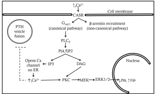
1.3.4 CASR signaling
45
stimulation also leads to activation of MAPKs, including ERK1/2 and p38 MAPK (Magno et al., 2011).
Ubiquitously expressed (β-arrestin1 and β-arrestin2), bind to phosphorylated C-terminal of CASR (Fig.1.5), resulting in both an uncoupling of the receptor from its cognate G proteins and intracellular signaling (non-canonical pathway) (Doucette et al., 2009). This β-arrestin recruitment by CASR will be discussed in detail in chapter 4.
Figure 1.5: CASR signaling cascade
PTH: parathyroid hormone, eCa2+: extracellular calcium, CASR: calcium sensing receptor, Gαq11: heterotrimeric G protein, PLC: phospholipase C, IP3: Inositol
triphosphate, DAG: diacyl glycerol, PKC: protein kinase C, iCa2+: intracellular calcium, ERK: extracellular signal-regulated kinase, VDR: vitamin D receptor (complete lines: stimulation, dotted lines: inhibition).
1.3.5 CASR agonists
46
than calcium, CASR respond to various di- and tri-valent cations including Mg2+, Sr2+ and Gd3+. CASR also interacts with organic polycations, such as spermine (Brown et al., 1993; Brown and MacLeod, 2001). Most of the ligand receptor interactions occur at the extracellular domain (‘venus fly trap’ structure) of the CASR. Ca2+, Mg2+ and spermine, have the potential to be physiologically relevant CASR activators. In the microenvironments of intestine and CNS, where calcium concentrations are comparatively low, spermine levels are sufficiently high to activate the CASR.
Type II agonists of CASR are also known as positive allosteric modulators (PAM) of CASR. Type II CASR agonists such as R567, S567, R568, S568 and Cinacalcet Hydrochloride are currently being used clinically to treat hyperparathyroidism. These are small hydrophobic molecules, which interact with transmembrane region of the CASR and increases CASR affinity for calcium. Type II agonists of CASR are also called as calcimimetics since they mimic calcium and binds at a site other than calcium binding site. Calcimimetics activate CASR in presence of calcium and are referred as ‘modular’ rather than as ‘agonists’ (Nemeth et al., 2004). Calcimimetics, such as R568 hydrochloride and Cinacalcet Hydrochloride, decrease PTH synthesis (Levi et al., 2006) and secretion (Fox et al., 1999; Nemeth et al., 1998; Nemeth et al., 2004), and reduce parathyroid cell proliferation (Colloton et al., 2005; Wada et al., 1997; Wada et al., 2000).
47
CHAPTER 2: SPECIFIC AIMS
48
also unclear what role PTH plays in fluoride-induced osteoclastogenesis in C3H mice. Understanding the impact of fluoride on PTH is critical due to the involvement of PTH in mineral homeostasis, which is tightly regulated by BPK axis (Bergwitz and Juppner, 2010; Lavi-Moshayoff et al., 2010; Torres and De Brauwere, 2011). Unfolding of the unknown mechanistic aspects of fluoride-mediated PTH modulation will help us to better define treatment strategies and develop drug for treatment of resorptive bone diseases.
To understand the pathophysiology of fluoride-induced osteoclastogenesis we hypothesize that:
Fluoride directly acts on parathyroid gland to modulate PTH secretion. To address this hypothesis we proposed two specific aims focused to determine the effect of fluoride on PTH secretion and involvement of CASR in fluoride-mediated PTH modulation.
Sub-hypothesis 1: Fluoride leads to increased PTH secretion.
49
Specific aim 1B: Investigate PTH secretion in sera of C3H and B6 mice after single fluoride dose via oro-gastric gavage. C3H and B6 mice were given a single gavage dose via oro-gastric route with fluoride or distilled water and serum fluoride, calcium, phosphorus, magnesium, and PTH levels were measured at serial time points. Expression of suite of genes involved in BPK axis was determined to understand the effect of fluoride on PTH secretion.
Sub-hypothesis 2: Effect of fluoride on PTH secretion is mediated through CASR.
Specific aim 2A: Determine the effect of fluoride on β-arrestin recruitment in HTL cells transfected with human CASR cDNA. HTL cells were transfected with human CASR and treated with fluoride to determine arrestin recruitment in the transfected cells using a modification of Tango® assay (β-arrestin recruitment assay) to identify fluoride-mediated CASR activation.
50
51
CHAPTER 3: FLUORIDE MODULATES PARATHYROID HORMONE SECRETION
IN VITRO AND IN VIVO
FLUORIDE MODULATES PARATHYROID HORMONE SECRETION IN VIVO AND
IN VITRO
Chaitanya P. Puranika, Kathleen A. Ryanb, Shijia Hua, E. Angeles Martinez-Mierc and Eric T. Everettd
a
Oral Biology PhD Curriculum, University of North Carolina at Chapel Hill. b
Dental Research, University of North Carolina at Chapel Hill c
Department of Preventive and Community Dentistry, Indiana University School of Dentistry
d
52 3.1 Abstract
53
54 3.2 Introduction
Fluoride has recognized actions on bone homeostasis (Everett, 2011). Yan et al investigated in two inbred strains of mice, C3H/HeJ (C3H) and C57BL/6J (B6), fluoride actions on bone homeostasis (Yan et al., 2007). Short term systemic exposure to fluoride for C3H resulted in increased osteoclastogenesis as evidenced by increases in serum osteoclast biomarkers: intact parathyroid hormone (iPTH), soluble receptor activator of nuclear factor kappa-B ligand (sRANKL) and tartrate-resistant acid phosphatase 5b (TRAP5b) along with decrease in serum osteoprotegerin (OPG) levels (Yan et al., 2007). Osteoclast numbers along bone surfaces and osteoclast potential of bone marrow cells were increased in C3H as well. However, similar fluoride exposure in B6 mice favored anabolic responses with increases in serum alkaline phosphatase (ALP) activity, proximal tibia trabecular and vertebral bone mineral density.
55
56 3.3 Materials and Methods
3.3.1 Animals
Ninety-six, 5-6 week old male C3H/HeJ (C3H) and C57BL/6J (B6) mice (The Jackson Laboratory, Bar Harbor, ME, USA) were placed on a constant nutrition no fluoride diet #5861 (Test Diet®, Richmond, IN, USA; fluoride: 0ppm, calcium: 0.60%, phosphorus: 0.57%, Vit D3: 2.2 IU/g, and Energy: 4.09 kcal/g) and provided distilled drinking water, ad libitum, for one week to acclimatize. Mice were housed in The Division of Lab Animal Medicine facility at The University of North Carolina at Chapel Hill, an AAALAC accredited unit. Mice were placed in 12:12 light and dark cycles at 21oC ambient temperature. All experimental procedures were approved by the Institutional Animal Care and Use Committee at The University of North Carolina at Chapel Hill.
3.3.2 Parathyroid gland organ culture
57
each strain of mice: C3H (calcium: 10.6mg/dL, phosphorus: 8.7mg/dL, and magnesium: 2.1mg/dL) and B6 (calcium: 10.4mg/dL, phosphorus: 10.3mg/dL, and magnesium: 2.9mg/dL). The TPC from C3H and B6 were randomly divided into 3 groups (n=6) based on fluoride concentrations 0, 250 or 500µM. Media supernatants were collected at 24hrs and stored at -80oC.
3.3.3 Gavage and sample collections
Following acclimatization, mice from both strains were randomly divided in 6 groups based on time of sacrifice after gavage dose. Mice were further divided into control and test group (n=6) based on fluoride levels (0 or 100ppm) in gavage. Both the groups of mice were sacrificed at 0.5, 1, 3, 6, 12 or 24hrs. At the time of gavage, mice were weighed and the gavage dose was calculated for each mouse (0.001mg [F-]/g body weight). Mice were anesthetized with ketamine-HCl (90mg/kg) and xylazine (14mg/kg) i.p. 15mins prior to the determined time of sacrifice. Sera were collected from mice and stored at -80oC until use. TPCs, kidneys, and humerii were harvested, snap-frozen in liquid nitrogen, and stored at -80oC.
3.3.4 ELISA and biochemistry assays
58
extracted over a light table using the micropipette/micropipette holder and dispensed onto the surface of an inverted fluoride electrode (F-ISE) under a layer of mineral oil to prevent loss of fluoride. Using a micromanipulator and microscope for viewing, the reference electrode is touched to the standard or sample drop, resulting in an electrometer mV reading. Both the F-ISE and hand-pulled reference electrode are connected to an electrometer and computer with plot program monitoring/recording software. Samples are run in triplicate, with periodic electrode conditioning and standard checks. Calcium, phosphorus, and magnesium were determined by the Animal Clinical Laboratory, University of North Carolina at Chapel Hill and the Clinical Pathology Laboratory, College of Veterinary Medicine at North Carolina State University.
3.3.5 RNA extractions and cDNA preparation
RNA was extracted from 24hrs TPCs, kidneys, and humerii samples using RNeasy Midi kit (Qiagen Inc., Valencia, CA, USA). Quantitative analysis of RNA sample was performed using Agilent Bioanalyzer using Nano Labchip at the Genomic Core Facility, University of North Carolina at Chapel Hill. All RNA sample had RIN values 7.5-10 range. High Capacity Reverse transcription kit (Applied Biosciences, Carlsbad, CA, USA) was used to prepare cDNA. After preparation, cDNA was stored at -80oC.
qPCR
59
receptor (Vdr), parathyroid hormone like hormone (Pthlh), tumor necrosis factor 11 (Tnfs11), fibroblast growth factor 23 (Fgf23), parathyroid hormone receptor 1 (Pth1r), chloride channel 5 (Clcn5), Solute carrier 9 member 3 regulator 1 (Slc9a3r1), PDZ domain containing 1 (Pdzk1) and Solute carrier family 34 member 1 (Slc34a1). Expression of these genes was normalized using two house-keeping genes comprising of beta actin (Actb) and glyceraldehyde 3-phosphate dehydrogenase (Gapdh). All the primers were ordered from SA Biosciences (Qiagen Inc., Valencia, CA, USA). Data were analyzed using 2-∆∆Ct method as previously described (Livak and Schmittgen, 2001; Yuan et al., 2006).
3.3.6 Statistical analysis
3.4 Results
3.4.1 Responsiveness of TPC organ culture model The TPC organ culture model was
effects of fluoride on the
model to changes in the extracellular
determined, was used to validate the culture model calcium concentrations (hypocalcemic:
hypercalcemic: 25mg/dL) demonstrated changes in iPTH secretion into the media
Figure
(A) iPTH ± SE (pg/mL) in C3H TPCs at three calcium concentrations; 1.1, 10.5 or 25mg/dL (n=3 in each group). (B) iPTH ± SE (pg/mL) in C3H and B6 TPCs after 24hrs of incubation at three fluoride concentrations; 0, 250 or 500µM (grey bar=C3H and black bar=B6, n=6 in each group; **p<0.005).
After 24hrs of incubation at hypocalcemic condition, iPTH secretion (129.43±27.14pg/mL) was significantly increased (
normocalcemic condition (49.28±6.21pg/mL)
conditions lead to a modest iPTH decline. Although not shown, TPCs in culture remain responsive to changes in calcium for 3 days.
60
Responsiveness of TPC organ culture model
The TPC organ culture model was utilized in order to determine
the parathyroid gland. Responsiveness of the organ culture in the extracellular calcium, a known PTH
, was used to validate the culture model. TPCs cultured hypocalcemic: 1.1mg/dL, normocalcemic:
demonstrated changes in iPTH secretion into the media
Figure 3.1: iPTH levels in media supernatant
iPTH ± SE (pg/mL) in C3H TPCs at three calcium concentrations; 1.1, 10.5 or 25mg/dL (n=3 in each group). (B) iPTH ± SE (pg/mL) in C3H and B6 TPCs after 24hrs of incubation at three fluoride concentrations; 0, 250 or 500µM (grey bar=C3H
6 in each group; **p<0.005).
After 24hrs of incubation at hypocalcemic condition, iPTH secretion (129.43±27.14pg/mL) was significantly increased (p<0.001) as compared to the normocalcemic condition (49.28±6.21pg/mL) (Fig. 3.1A). The hypercalcemic ns lead to a modest iPTH decline. Although not shown, TPCs in culture remain responsive to changes in calcium for 3 days.
to determine the direct Responsiveness of the organ culture mediator was tured in varying normocalcemic: 10.5mg/dL or demonstrated changes in iPTH secretion into the media.
iPTH ± SE (pg/mL) in C3H TPCs at three calcium concentrations; 1.1, 10.5 or 25mg/dL (n=3 in each group). (B) iPTH ± SE (pg/mL) in C3H and B6 TPCs after 24hrs of incubation at three fluoride concentrations; 0, 250 or 500µM (grey bar=C3H