Detection and Analysis of Common Fragile Sites in Drosophila melanogaster
Matthew C. LaFave
A dissertation submitted to the faculty of the University of North Carolina at Chapel Hill in partial fulfillment of the requirements for the degree of Doctor of Philosophy in the Curriculum in Genetics
& Molecular Biology.
Chapel Hill 2011
Approved by:
Jeff Sekelsky
Shawn Ahmed
Corbin Jones
Steve Matson
Abstract
Matthew C. LaFave: Detection and Analysis of Common Fragile Sites in Drosophila melanogaster
(Under the direction of Jeff Sekelsky)
Common fragile sites (CFSs) are regions of DNA exceptionally prone to breakage.
While these regions have implications in cancer, the causes of chromosome fragility remain
poorly understood. This is partially due to relatively low-resolution cytological mapping of
CFSs, and the use of exogenous agents to induce chromosome breakage. In an effort to better
understand the causes of fragility, I have developed Drosophila melanogaster as a model for CFS
study. In doing so, I have developed approaches to identify CFSs at a high resolution, based on
both spontaneous chromosomal events and breakage induced by the inhibition of replication. In
the first approach, I used a mutant form of mus309, the ortholog of human BLM helicase, to
locate CFSs by visualizing sites of DNA breakage as mitotic crossovers. High rates of breakage
correspond to CFSs, and results from my study indicate that there is a significantly non-uniform
rate of mitotic crossovers across the left arm of chromosome 2. This work constitutes the first
report of specific regions sensitive to endogenous damage in D. melanogaster. Further, the
resolution of damage detection can be brought even higher with SNP mapping. My second
approach is a novel assay that uses S2 cell culture to detect preferential sites of exogenous DNA
integration, a hallmark of CFSs. This allows me to survey the entire genome, while using
high-throughput sequencing to obtain a resolution of CFS detection orders of magnitude better than
previous studies. I obtained numerous integration events under a variety of conditions, providing
evidence of putative fragile regions. I anticipate that the assays I developed will serve as valuable
of the CFSs detected from both of these approaches will lead to a better understanding of the
Acknowledgements
I would first like to thank my advisor, Dr. Jeff Sekelsky. He has been unfailingly
supportive and encouraging, and has made graduate school something to be excited about. I
thank the members of my committee, Dr. Shawn Ahmed, Dr. Corbin Jones, Dr. Steve Matson,
and Dr. Dale Ramsden, for their helpful advice pertaining to both my experiments and my career.
I thank the members of the Sekelsky lab, for making the lab an ideal work environment. I’d like
to give particular recognition to Jeannine LaRocque, who mentored me as a rotation student, and
Susan McMahan Cheek, whose appreciation of Ratt was one of the first signs that the Sekelsky
lab was the right fit. I’d like to recognize all of the Sekelsky lab undergrads, as well, but
particularly my mentees Lewis Overton and Kelly Wolfe. Both intelligent and hard-working,
they endured my first forays into mentoring, in which I learned at least as much as I taught them.
I’d also like to thank Dr. Bob Duronio and his lab for helpful discussions in lab meetings. I’d like
to specifically thank Bob for our meetings when I was in my first year, when he confirmed that,
yes, it would be a good idea to do a rotation in the Sekelsky lab. I’m grateful to the UNC Center
for Bioinformatics, particularly Dr. Hemant Kelkar and Dr. Xiaojun Guan. The assistance of Dr.
Piotr Mieczkowski and the UNC High-Throughput Sequencing Facility was instrumental in this
project, as well. I’d like to thank some of my influential professors from my time as an undergrad
at Notre Dame: Dr. Michelle Whaley, Dr. Gary Lamberti, Dr. Paul Helquist, and Dr. William
Ramsey. Among my undergrad professors, I’d most like to thank my first research advisor, Dr.
David Hyde, as well as Dr. Christopher Burket, the postdoc who served as my mentor. I’d also
like to thank Doug Baltz, my excellent high school physics teacher. I acknowledge my sources of
Research. Thanks to the friends who have made my time at UNC so much more than just work,
especially my comrades-in-arms in Team Venture. I’d like to recognize the motivational prowess
of Steve Harris, Bruce Dickinson, Kai Hansen, Sam Totman, and ZP Theart. Of course, I thank
my family, who has supported me in everything I’ve done. My mother’s use of a Punnett square
to explain why I was the only one in the family with blue eyes encouraged me to take my first
step into a larger world. Finally, I thank my fiancée, Kelly. She is the most loving and giving
Table of Contents
List of Tables ... xi
List of Figures ... xii
List of Abbreviations ... xiii
Chapter I: General Introduction ... 1
Genome stability & fragile sites ... 1
Rare fragile sites ... 1
Common fragile sites ... 2
CFSs appear to be evolutionarily conserved ... 4
Proposed causes of CFS fragility... 5
Primary sequence characteristics ... 6
Chromatin environment ... 7
Replication timing & origin density ... 7
Issues of resolution ... 8
Induced vs. natural CFSs ... 9
Using Drosophila to study CFSs ... 9
Chapter II: Common Fragile Sites and Mitotic Crossovers ... 11
Results ... 14
Discussion ... 19
Non-uniform breakage indicates CFSs ... 19
Sources of breaks in the crossover assay ... 20
Implications of replication inhibition and endogenous breaks ... 21
Connections between DmBLM & CFSs ... 23
Materials and Methods ... 24
Chapter III: Common Fragile Sites and Exogenous DNA Insertions ... 27
Introduction ... 27
Results ... 29
The integration assay identifies putative fragile regions ... 29
Integration analysis: insert-genome junctions ... 37
Integration analysis: local genomic environment ... 41
Discussion ... 44
Aneuploidy in S2 cells... 44
Replication inhibition may influence DNA repair ... 45
Integration data suggests fragmentation of the construct ... 46
Interpretation of insert environment ... 46
Factors affecting the insertion assay ... 47
Materials and Methods ... 49
Evolutionary conservation ... 54
Differences in the crossover and integration assays ... 55
Use of Eureqa for future studies ... 57
Direct tests of fragility ... 58
Useful modifications to assays ... 60
Conclusion ... 61
Appendix Chapter: Transcription Initiation from Within P Elements Generates Hypomorphic Mutations in Drosophila melanogaster ... 62
Preface ... 62
Results & Discussion ... 62
List of Tables
Table 3.1. Location of unambiguous integrations in aphidicolin-treated
cells selected with hygromycin ... 32
Table 3.2. Location of unambiguous integrations in aphidicolin-treated cells enriched via splinkerette PCR ... 35
Table 3.3. Location of unambiguous integrations in cells selected with hygromycin ... 37
Table 3.4. Sizes of insertions and microhomology at integration-genome junctions ... 38
Table 3.5. Local genomic environment of aphidicolin-induced inserts ... 42
Table 3.6. Local genomic environment of inserts incurred without aphidicolin ... 43
List of Figures
Figure 2.1. Detection of fragile sites ... 13
Figure 2.2. Cross scheme to visualize mitotic crossovers ... 15
Figure 2.3. The rate of crossovers is non-uniform ... 16
Figure 2.4. Cross scheme to visualize and recapitulate mitotic crossovers for future SNP sequencing... 17
Figure 2.5. The rate of crossovers is non-uniform in a heteroallelic mus309 background ... 18
Figure 3.1. Cell-based DNA integration detection scheme ... 30
Figure 3.2. Integration of aphidicolin-induced DNA inserts ... 31
Figure 3.3. Splinkerette PCR enriches for insert-containing fragments ... 34
Figure 3.4. Integration of DNA inserts without aphidicolin ... 36
Figure 3.5. Microhomology at insert-genome junctions ... 39
Figure 3.6. Position of insert reads suggest fragmented integrations ... 40
Figure 5.1. A cryptic transcript initiates from within a P element in nbs ... 64
List of Abbreviations
BAC – Bacterial artificial chromosome
BrdU – Bromodeoxyuridine
CFS – Common fragile site
ChIP-seq – Chromatin immunoprecipitation followed by high-throughput sequencing
CO – Crossover
DmBLM – Drosophila melanogaster ortholog of BLM (encoded by mus309)
DMSO – Dimethyl sulfoxide
DSB – Double-strand break
FHA – Forkhead-associated
GA IIx – Genome analyzer IIx
HTS – High-throughput sequencing
Polα – Polymerase alpha (encoded by DNApol-α180) RACE – Rapid amplification of cDNA ends
RFS – Rare fragile site
ssDNA – single-stranded DNA
Chapter I
General Introduction
Genome stability & fragile sites
DNA serves as the master plan for the cell, encoding the information for processes
necessary to begin and sustain life. Just as the information encoded by the genome is important,
the physical integrity of the DNA is critical to ensure successful implementation of the coded
information. When genome stability is compromised, the negative effects on the cell or organism
can be varied and severe (DILLON et al. 2010). It is critical, therefore, for the cell to maintain the
integrity of its genome. The cell has evolved numerous mechanisms to cope with this issue
(SANCAR et al. 2004). However, damage can occur, and often does. Damage can come from
exogenous sources, such as UV radiation or chemicals, or endogenous sources, such as
byproducts of cellular metabolism or errors during DNA replication (DE BONT and VAN
LAREBEKE 2004). Interestingly, some regions of DNA appear to be more prone to damage than
others.
Rare fragile sites
Regions of DNA that are exceptionally prone to chromosomal abnormalities – chiefly
breakage – have been termed “fragile sites” (DURKIN and GLOVER 2007). When a fragile site
forms a break or gap, it is said to be expressed. Fragile sites have two classifications: rare and
common. Rare fragile sites (RFSs) are found infrequently in the human population, and are
associated with specific disease alleles (SUTHERLAND et al. 1998). Most RFSs are
folate-deficient media (LUKUSA and FRYNS 2008). The few RFSs that are not folate-sensitive have their
expression induced by bromodeoxyuridine (BrdU) and/or distamycin A, agents that integrate into
DNA and interfere with replication (LUKUSA and FRYNS 2008).
RFS only exhibit fragility in an individual with the disease. For example, the cultured
cells of an individual with Fragile X disease would exhibit a chromosome constriction at the
FRAXA RFS, whereas those of a healthy individual would not (MCBRIDE 1979; VERKERK et al.
1991). The causes of RFS fragility are relatively well understood: the expansion of di- or
trinucleotide repeats leads to non-B DNA structures, which interfere with DNA replication and
nucleosome assembly. The wild type copy number of such repeats is tolerated by the cell, but
expansion beyond a threshold value specific to each RFS leads to chromosomal instability
(LUKUSA and FRYNS 2008). The increased prevalence of non-B DNA makes these regions prone
to DNA gaps and breaks (FREUDENREICH 2005).
Common fragile sites
Common fragile sites (CFSs), on the other hand, are generally considered to be a normal
component of chromosome structure. There have been 88 CFSs identified in the human genome,
thirteen of which have been characterized at a resolution higher than that of chromosome bands
(LUKUSA and FRYNS 2008). CFSs are presumed to be ubiquitous in the human population,
although some studies have indicated variability in the fragility of sites between individuals, and
between different tissues (DENISON et al. 2003; TEDESCHI et al. 1992). In addition, CFSs are not
all equally fragile; the first study of CFSs found that, in humans, 80% of breaks at CFSs were due
to only 20 CFSs (GLOVER et al. 1984). The CFSs FRA3B and FRA16D are the most highly
expressed.
Human CFSs have a medically relevant connection to cancer. When expressed in the
presence of aphidicolin, CFSs have been shown to serve as significant predictor of breast,
rearrangements implicated in cancer, including loss of heterozygosity (DURKIN and GLOVER
2007). Recurrent breaks at CFSs have been found in prostate, breast, lung, and pancreatic cancer,
among many other types (CARTER et al. 1990; CHEN et al. 1996; HIBI et al. 1992; NEGRINI et al.
1996; SHRIDHAR et al. 1996). In fact, most cancer-specific translocations have at least one
breakpoint in a fragile site (BURROW et al. 2009). Furthermore, the human CFSs most prone to
breakage, FRA3B and FRA16D, lie within tumor suppressor genes (HUEBNER and CROCE 2003;
O'KEEFE and RICHARDS 2006). This indicates that breaks in these regions, especially if they are
repaired incorrectly or go unrepaired, could contribute to downstream events involved in
tumorigenesis. Indeed, breaks at CFSs have been shown to be capable of initiating gene
amplification via breakage-fusion bridge cycles (COQUELLE et al. 1997).
The strict definition of what constitutes a CFS comes from the way they are traditionally
detected. CFSs are defined as choromsomal regions that form reproducible breaks on metaphase
chromosomes after partial inhibition of DNA replication (DURKIN and GLOVER 2007). Breaks
can be observed in cells grown in folate-deficient media or in the presence of fluorodeoxyuridine,
although the level of breakage is low (GLOVER 1981; YUNIS and SORENG 1984). Efficient
inhibition of replication is therefore usually achieved by use of aphidicolin, and occasionally by
agents such as BrdU or 5-azacytidine (GLOVER et al. 1984; SUTHERLAND et al. 1985).
Aphidicolin acts to inhibit polymerases involved in DNA replication, such as polymerase α, δ, and ε (CHENG and KUCHTA 1993; GOSCIN and BYRNES 1982; IKEGAMI et al. 1978). While aphidicolin can be used to completely halt the cell cycle in S phase, CFSs are induced using a low
concentration of the agent, typically 0.4 µM (GLOVER et al. 1984). At this concentration,
aphidicolin makes the replication of DNA somewhat more difficult, without causing it to stop
altogether.
It has been observed that down-regulation of components of homologous recombination,
such as RAD51, or of non-homologous end joining, such as DNA-PKcs, results in a significant
indicates that expressed CFSs can be repaired both by homologous repair and non-homologous
end-joining. CFSs have been reported to be prone to sister chromatid exchanges after treatment
with aphidicolin, a result of homologous repair of the double-strand break (DSB) incurred at the
CFS (GLOVER and STEIN 1987; HIRSCH 1991). Of course, not all expressed CFSs are repaired
after exposure to replicative stress, leading to the characteristic breaks on metaphase
chromosomes.
CFSs appear to be evolutionarily conserved
An interesting, and somewhat counter-intuitive, characteristic of CFSs is that they appear
to be evolutionarily conserved. Most studies of CFSs are done with human chromosomes, but
CFSs have also been detected in several other mammals. It has been shown that mice have CFSs
that correspond to known human CFSs (HELMRICH et al. 2006; HELMRICH et al. 2007). These
studies in mice showed conservation of specific CFSs, but regions analogous to CFSs – that is,
regions prone to breakage, especially when under replicative stress – have also been detected in
yeast.
Reducing levels of polymerase α in Saccharomyces cerevisiae has been shown to increase the rate of breaks leading to translocations, due in large part to retrotransposons called
Ty elements (LEMOINE et al. 2005). A site at which Ty elements formed an inverted repeat was
found to be prone to double-strand breaks under these conditions; therefore, it has the
characteristics of a CFS. A separate study found that a region containing tRNA genes was prone
to DNA breaks (ADMIRE et al. 2006). Disruption of replication by exogenous agents or mutation
of a helicase resulted in increased instability in this region, and removal of the region reduced the
overall genomic instability of the cell. Finally, a study of spontaneous mitotic recombination in
yeast identified a 1.3 kb region that was significantly more prone to crossovers than surrounding
out that such a region might constitute a CFS prone to endogenous damage (LAFAVE and
SEKELSKY 2009).
Proposed causes of CFS fragility
It is important to note that the reasons CFSs are prone to damage are not well understood.
CFSs do not appear to share the nucleotide repeats characteristic of RFSs, so the reason for CFS
fragility is likely to be somewhat different (ARLT et al. 2002; RASSOOL et al. 1996; SCHWARTZ et
al. 2006). CFSs can be induced by agents that interfere with replication, so it is generally thought
that some aspect of fragile regions that make them inherently difficult to replicate.
This claim is bolstered by genetic evidence, which involves the checkpoint kinase
Ataxia-telengiectasia and Rad3 Related (ATR). This protein is involved in signaling the response
to stalled or collapsed replication forks, as well as the response to single stranded DNA exposed
by damage. In human cell culture, ATR has been shown to be necessary for CFS stability
(CASPER et al. 2002). CFS expression is enhanced in the presence of aphidicolin in ATR mutant
cells, and such cells spontaneously express CFSs even in the absence of aphidicolin. ATR has
been found to preferentially interact with FRA3B after treatment with aphidicolin (WAN et al.
2010). Depletion of CHK1, the downstream effector of ATR, also results in CFS expression
(DURKIN et al. 2006). Similar results have been reported in yeast, in which cells mutant for the
ortholog of ATR, Mec1, display an increase in DNA breaks in replication slow zones (CHA and
KLECKNER 2002). These reports suggest that a key early step in CFS expression is the stalling of
a replication fork, consistent with the notion that CFSs are inherently difficult to replicate. What
causes the forks to stall, however, remains an open question. Here, I discuss some of the major
Primary sequence characteristics
Many characteristics of CFSs have been proposed as the reasons for their fragility.
Primary sequence attributes have received a considerable amount of attention in this regard. One
model posits that aphidicolin causes uncoupling of polymerases from the helicase-topoisomerase
complex at the replication fork, resulting in exposed single stranded DNA (ssDNA) (DURKIN and
GLOVER 2007). Some of the sequences in CFSs have the potential to form stable secondary
structures – therefore, exposure of such sequences as ssDNA may lead to hairpins and other
structures that disrupt replication. Interestingly, the breaks and ssDNA at CFSs can be reduced
by low-dose camptothecin, even in the presence of aphidicolin (ARLT and GLOVER 2010). It is
thought that low concentrations of camptothecin may slow the helicase-topoisomerase complex,
and that this may reduce the polymerase uncoupling normally induced by aphidicolin.
No sequence motifs have been detected in CFSs, although it is unclear if the relatively
low resolution of CFS detection makes such regions difficult to identify (MISHMAR et al. 1998;
SCHWARTZ et al. 2006). Still, there is evidence that the sequence of CFSs is inherently unstable.
It was shown in a study in which bacterial artificial chromosomes (BACs) carrying sequence
from FRA3B were inserted at ectopic sites of the human genome that the FRA3B BACs could
recapitulate fragility, while control BACs did not (RAGLAND et al. 2008).
In addition, DNA flexibility has been proposed to be a major cause of CFS fragility
(SCHWARTZ et al. 2006). Flexibility calculations are based on the maximum potential angular
twist that two consecutive bases could achieve (SARAI et al. 1989). A program, TwistFlex, was
developed to calculate the average potential twist of DNA in a sliding window; averages that
surpass a threshold value are reported as flexibility peaks (MISHMAR et al. 1998). Several CFSs
have been analyzed in this way, and found to have a high number of flexibility peaks (DURKIN
and GLOVER 2007). Interestingly, a highly flexible region of human FRA16D inserted into the S.
cerevisiae genome was shown to induce fragility, while non-flexible regions did not (ZHANG and
nature of CFSs, as the A to T step is more than twice as flexible as any other possible step (SARAI
et al. 1989). These regions could contribute to fragility via secondary structure formed by AT
repeats. However, if DNA flexibility is involved in CFS fragility, it is unlikely to be the sole
cause. Not all CFSs have more flexibility peaks than controls, and BACs with a high number of
flexibility peaks are not sufficient to recapitulate fragility in ectopic regions (HELMRICH et al.
2007; RAGLAND et al. 2008).
Chromatin environment
The state of the chromatin surrounding CFSs has also been considered to contribute to
fragility. Though not strictly related to replication problems caused by aphidicolin, the banding
pattern of metaphase chromosomes has raised an interesting issue regarding fragility. The dark
G-bands tend to be AT-rich, late replicating, and have few genes; the light R-bands are just the
opposite: early-replicating and gene- and GC-rich (GARDINER 1995). Some CFSs, including
FRA3B, have the characteristics of the dark G-bands, but map to R-bands. It has been suggested
that CFS fragility may be related to the discrepancy between the chromatin context of CFSs and
their surrounding environment (SCHWARTZ et al. 2006). Others have suggested that histone
modifications may play a role in fragility. Histones at CFSs have been found to be
hypoacetylated, and FRA3B is more resistant to micrococcal nuclease than nearby non-fragile
regions (JIANG et al. 2009). These findings suggest that CFS chromatin might be more compact
than flanking regions, and raise questions about the role of chromatin in fragility.
Replication timing & origin density
The timing of replication, and the related factor of the distance from a firing replication
origin, may have a substantial impact on fragility. CFSs are often associated with large,
late-replicating genes in humans (LE BEAU et al. 1998). It has been reasoned that these regions are
presence of replication-inhibiting aphidicolin. FRA16D, for example, was found to have a
slow-moving replication fork (PALAKODETI et al. 2004). Recently, it was determined that the center of
FRA3B is origin-poor in JEFF lymphocytes (LETESSIER et al. 2011). Moreover, in cells in which
FRA3B was not origin-poor, such as MRC-5 fibroblasts, fragility was not observed. This
suggests that the combination of replication timing and origin density play a major role in CFS
fragility, and may explain tissue-specific differences in CFS expression. However, studies that
have found that insertions of CFS DNA in ectopic regions are sufficient to recapitulate fragility,
while control insertions are not, suggest that origin density is insufficient to fully explain fragility
(RAGLAND et al. 2008; ZHANG and FREUDENREICH 2007).
Issues of resolution
While much has been learned about the characteristics of CFSs, studies of CFSs have
suffered from the use of relatively low-resolution approaches. The initial mapping of CFSs were
accomplished by observing breaks on metaphase chromosome spreads, and were thus limited to
the resolution of chromosome bands (GLOVER et al. 1984). Fluorescent in situ hybridization
probes have been used to achieve a somewhat higher resolution (BECKER et al. 2002). These
studies score breaks qualitatively – proximal, distal, or within – relative to a probe of known
location. By doing this for many probes, a distribution of breaks can be generated. This
technique is good for determining the outer boundaries of fragility and general shape of the
distribution, but the effective resolution is still in the hundreds of kilobases. This is because the
precise location of any given break cannot be inferred from its position relative to a probe.
Because of this, CFSs have been classified as large, fragile regions, often a megabase or more in
length (BECKER et al. 2002; HANDT et al. 2000).
This resolution makes it difficult to ascertain which factors truly contribute to fragility.
For example, it leaves us unable to distinguish between a single large region of fragility, and
fragility, it would be advantageous to focus on what I will refer to as the minimal fragile region –
the most fragile portion of the CFS. Including in the analysis regions that are less fragile may
have the effect of drowning out the truly relevant information from the minimal fragile region.
Induced vs. natural CFSs
Almost all of what we know about CFSs is based on damage induced by exogenous
sources, typically aphidicolin. The use of such inducers of fragility has been necessitated by the
difficulty of locating sites of endogenous damage in human cell culture. The physiological
relevance of these aphidicolin-induced regions is well-established; the most fragile CFSs, such as
FRA3B and FRA16D, have been implicated in recurrent tumorigenic breakpoints (HUEBNER and
CROCE 2003; O'KEEFE and RICHARDS 2006). Still, I felt it would be useful to determine the
location of CFS breaks caused by endogenous lesions. This constitutes DNA damage that the
genome normally incurs, but typically repairs in a way that makes the initial break difficult to
detect. I designed assays in a way that allowed me to examine damage incurred under both types
of conditions. I will refer to CFSs corresponding to endogenous breaks as “natural CFSs”; those
that have their fragility induced by exogenous sources will be referred to as “induced CFSs”. By
analyzing both types of regions, one can determine if induced CFSs are exceptionally
aphidicolin-sensitive, or if aphidicolin simply increases the expression of natural CFSs.
Using Drosophila to study CFSs
To accomplish the tasks of studying CFSs at a high-resolution, induced by endogenous or
exogenous means, I chose to work with Drosophila melanogaster. To my knowledge, there have
been no previous studies to detect CFSs in Drosophila. Most studies of CFSs have taken place in
human cell culture, and a few have used yeast; this study gives me an opportunity to develop D.
melanogaster as the first live, whole-organism in vivo metazoan model of CFSs. There exists an
which is a mutation that greatly increases the frequency of mitotic crossovers (MCVEY et al.
2007). My implementation of an assay that takes advantage of the characteristics of this mutant
to detect endogenous damage and verify the presence of CFSs in D. melanogaster is detailed in
Chapter II. This is the first report of CFSs detected in Drosophila. Having established the
presence of such sites, I developed a cell-based assay that couples selectable DNA integration
with high-throughput sequencing (HTS) to identify putative CFSs at a high resolution, and on a
genome-wide scale. The assay, as well as my analysis of the integration sites, is detailed in
Chapter II
Common Fragile Sites and Mitotic Crossovers
Introduction
Common fragile sites (CFSs) are chromosomal regions that are prone to breakage,
particularly when under DNA replication stress. While the existence and location of human CFSs
are well-documented (DURKIN and GLOVER 2007), the relatively low resolution at which CFSs
have been studied means that an understanding of the causes of fragility has remained elusive. I
aimed to determine the causes of fragility by employing high-resolution approaches in
Drosophila melanogaster. By increasing the resolution at which CFSs are detected and analyzed,
we increase the chance of determining features unique to CFSs. In addition, it permits one to
distinguish between a CFS as a single, large region of fragility, or several smaller regions
clustered close together.
This study established D. melanogaster as a metazoan in vivo model of CFSs. This
particular combination has not been available to the CFS field before – studies are typically
carried out in human cell culture (GLOVER et al. 1984), and occasionally in sacrificed mice
(HELMRICH et al. 2006). So far, the only truly in vivo studies of CFSs have been in budding yeast
(ADMIRE et al. 2006; LEMOINE et al. 2005). Working in Drosophila allows us to bridge studies
in yeast and mammals, and to ask questions about the evolutionary conservation of CFSs.
The first step was to determine the location of D. melanogaster CFSs – indeed, to see if
flies have CFSs at all. Data in yeast, as well non-human mammals such as mice, dogs, and cats,
al. 1991; STONE et al. 1993). However, no one had looked for CFSs in Drosophila, and so
detection of the sites became my primary task.
I chose to use a novel genetic assay to determine the presence and location of CFSs (Fig.
2.1). My assay allowed me to map sites of DNA breakage by taking advantage of a characteristic
of flies mutant for mus309, which encodes Drosophila BLM (DmBLM). DmBLM is the
Drosophila homolog of human BLM, a RecQ helicase (ADAMS et al. 2003; BACHRATI and
HICKSON 2003). Humans that lack BLM have Bloom’s Syndrome (BS), characterized by short
stature, sterility, sensitivity to sunlight, and increased susceptibility to a broad spectrum of
cancers. This last phenotype underscores the role of BLM as a fundamental protein in DNA
repair. BS cells display an increased rate of sister chromatid exchanges (CHAGANTI et al. 1974).
BLM can act on structures formed during homologous repair, such as migrating Holliday
junctions and unwinding D-loops in vitro (KAROW et al. 2000). BLM also acts to maintain the
integrity of stalled replication forks (DAVIES et al. 2007; MANKOURI and HICKSON 2007). In
Drosophila, there is evidence suggesting that DmBLM acts to free newly-synthesized DNA from
the template D-loop during homologous repair (MCVEY et al. 2004).
The feature of mus309 mutants most relevant to this study is the greatly increased rate of
mitotic crossovers (MCVEY et al. 2007). Such crossovers are associated with sites of endogenous
breaks (Fig. 2.1). In flies with wild type mus309, DNA damage in mitotically dividing cells is
typically repaired to yield a non-crossover product. Mitotic crossover frequencies in this situation
are typically below 0.002% between dp and bw, a region covering a little under 30% of the
genome (WOODRUFF and THOMPSON 1977). In flies homozygous mutant for mus309, however,
mitotic crossover frequencies are about 2% between st and e, a region that covers a little over
20% of the genome (MCVEY et al. 2007). I designed an assay in which these crossovers would
occur in the male germline, so the location of the crossover – and therefore the initial site of
endogenous damage – could be detected via mapping of recessive markers in the progeny of the
A key feature of the assay is that it allows one to map breaks incurred from endogenous
sources. Although such damage, which cells normally incur, is the most biologically relevant,
most studies of CFSs have focused on damage induced in the presence of exogenous agents,
usually aphidicolin (DURKIN and GLOVER 2007). This is because endogenous damage normally
occurs at a frequency that is difficult to detect with traditional, cytology-based approaches to
mapping CFSs (GLOVER et al. 1984). My assay detects endogenous damage at a sufficiently high
frequency to make the analysis of such damage straightforward. This permits me to determine
the location of natural CFSs.
In addition, the assay can be modified to detect breakage in the presence of replicative
stress. Direct treatment with aphidicolin is possible, but would be impractical – flies would have
to be fed the chemical, and it would be very difficult to ensure that each fly consumed exactly the
same dosage. However, I was able to create a mutant that genetically mimics the effect of
aphidicolin. Since aphidicolin acts to inhibit replicative polymerases, I removed one copy of
DNApol-α180, the gene encoding for the catalytic subunit of polymerase α (LAROCQUE et al.
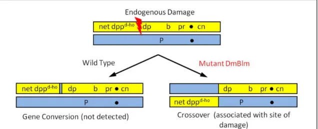
Figure 2.1. Detection of fragile sites. Genomic damage incurred in mitotic cells of wild-type flies is repaired to yield a non-crossover product, making the site of damage difficult to
detect. The mus309 mutant flies lack DmBLM protein, which results in damage being
2007a); DNApol-α180 will henceforth be referred to as Polα). In this way, the assay can be used
to detect either endogenous damage, or damage like that induced by aphidicolin.
The assay is also able to recover both halves of a DNA damage repair event. This is
noteworthy, because one typically is only able to recover and examine one half of a mitotic
crossover in metazoans, limiting the amount of information that can be gleaned from such a
study. In my assay, mitotic crossovers occur pre-meiotically in the male germline, meaning that
both halves of the crossover are produced as gametes. Analysis of the repair event from
reciprocal crossover siblings gives us the ability to determine the nature of repair in a mus309
mutant background.
I employed this assay to determine if CFSs are present on the left arm of chromosome 2
in D. melanogaster. Here, I treat CFSs as regions that are prone to DNA breakage; the source of
this damage can be either endogenous or the result of replication inhibition. My assay allows one
to distinguish between the two sources of damage, and lends itself to insights regarding the nature
of chromosome fragility. I argue that D. melanogaster does contain CFSs, both induced and
natural, and identify fragile regions on 2L.
Results
It was first necessary to determine if the fly genome contains regions that are especially
prone to DNA breakage. I accomplished this through the use of a mitotic crossover assay (Fig.
2.2). In these mus309 mutants, sites of DNA breakage can be repaired to produce a mitotic
crossover. The crossover is located at the site of initial damage, so by using phenotypic markers
to map a distribution of crossovers, I was able to essentially generate a visualization of the
In the first iteration of the assay, I analyzed 532 crossovers from 313 mus309N1 males.
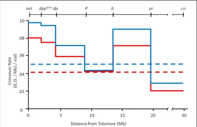
By examining the distribution of mitotic crossovers on 2L, I was able to determine that the rate of
crossing over, and therefore the rate of endogenous damage, is significantly non-uniform
(bootstrapping, P < 0.0001; Fig. 2.3). This suggests that regions that produced the highest rates
of crossing over – specifically, the regions between net and dp, and between b and pr – constitute
CFSs, or that they contain more CFSs than the other intervals. In release 5 of the D.
melanogaster genome, these regions cover the regions 2L:87,382..4,479,471 and
2L:13,823,894..20,073,719, respectively. These results were recapitulated in additional
experiments in which stocks with different markers were used to derive the 2nd chromosome
homologs, and where a different combination of mus309 alleles was used (P = 0.0002; Figs. 2.4
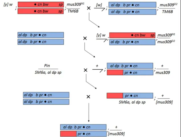
and 2.5). In this version of the assay, I analyzed 634 crossovers from 391 mus309N1/mus309D2 Figure 2.2. Cross scheme to visualize mitotic crossovers. Virgin females heterozygous for
mus309N1 and homozygous for a P element on 2L were crossed to mus309N1 males that
carried a 2nd chromosome with six recessive phenotypic markers (Cross 1). From the progeny
of this cross, males that did not carry TM6B, and were therefore homozygous for mus309N1,
were collected. These flies carried one copy of each of the parental chromosomes, and the crossovers I was able to analyze in the following generation were produced in the pre-meiotic germline of these males. Each male was crossed to three virgin females homozygous for the marker chromosome (Cross 2). The progeny of this cross were scored for mitotic crossovers.
One or both halves of the crossover could be recovered, and the P element could be detected
males. The heteroallelic combination of mus309 alleles demonstrates that the crossovers detected
in the assay are due to the lack of functional DmBLM, and not to homozygous second-site
mutations. The al-dp fragile region detected in this version of the assay covers the region 2L:
387,439..4,479,471.
Figure 2.3. The rate of crossovers is non-uniform. Five phenotypic markers on 2L (
net-pr) and one on 2R (cn) were used to determine the rate of mitotic crossovers in the male
germline. One homolog of chromosome 2 carried the markers, and the other carried a P
element to increase the resolution of the 9.2 Mb between dp and b. PCR was used to detect
the P element. Crossover data was collected in vials of three females crossed to a single
male; each vial therefore represents crossovers in the germline of one male. The red line
represents males homozygous mutant for mus309N1 (532 crossovers, 313 vials), while the
blue line represents males that were both homozygous mutant for mus309N1 and heterozygous
mutant for DNApol-α180, the gene for Polα (334 crossovers, 157 vials). Dotted lines are the
average crossover rates over the entire 40.6 Mb interval. The region between pr and cn
Figure 2.4. Cross scheme to visualize and recapitulate mitotic crossovers for future SNP sequencing. The initial steps are as described in Fig. 2.2, with the following relevant differences. Parental virgin females carry the Celera reference sequence chromosome,
marked with cn bw sp. Males carry a slightly different marker chromosome, the alleles of
which facilitate production of a crossover (CO) stock two generations later. Males also carry
a different allele of mus309; the heteroallelic arrangement in the following generation
prevents second-site mutations from affecting the experiment. COs are again generated in 2nd
generation males, which are visualized by a cross to three to five marker chromosome virgin females (Cross 2). Again, one or both products of the CO can be recovered; one possible CO is shown here. If the fly in which the CO is visible is a male, the fly is crossed to a stock
containing the SM6a 2nd chromosome balancer (Cross 3). The al dp sp markers carried on
SM6a allows one to distinguish between the CO chromosome and the marker chromosome; in
The mus309 mutant background provides information about the rate of endogenous
breakage. While it would be useful to modify this assay – by adding aphidicolin – to obtain
information about the rate of breakage due to an exogenous source, this would not be practical.
Instead, I chose to use a genetic means of mimicking exogenous damage. I introduced replicative
stress by using one mutant copy of Polα; 334 crossovers were analyzed from 157 mus309N1
+/mus309N1 Polα males. By doing so, I found that the rate of mitotic crossing over on 2L was
again significantly non-uniform (P < 0.0001). In most regions, the rate was elevated above the
rate detected in the version of the assay that was designed to detect purely endogenous damage. Figure 2.5. The rate of crossovers is non-uniform in a heteroallelic mus309 background. The DmBLM crossover assay was repeated in a different genetic background, as depicted in
Fig. 2.4. Crossovers were accrued between a marker chromosome carrying al dp b pr cn, and
a reference chromosome with cn bw sp. The crossover rate displayed here only takes into
account vials in which at least one crossover was detected. Crossover data was collected in vials of three to five females crossed to a single male; each vial therefore represents crossovers in the germline of one male. The solid green line represents males mutant for
In both cases, the regions between net and dp, and between b and pr, appear to be more prone to
DNA breakage than the other regions of 2L.
I found that my assay often produced reciprocal crossovers. For example, if a crossover
occurred between b and pr, the following generation might produce both net dppd-ho dp b + +
flies, as well as their + + + + pr cn siblings. Since this presented a unique opportunity to learn
about DNA repair in a mus309 mutant background by examining both products of a crossover,
reciprocal crossover flies from the second iteration of the assay (the al dp b pr cn version) were
crossed to produce multiple flies that recapitulated the original crossover (Fig 2.4). The DNA of
seven of these flies has been submitted for high-throughput sequencing and SNP mapping.
Discussion
Non-uniform breakage indicates CFSs
I undertook this study to determine if D. melanogaster had CFSs, and to identify the
location of such sites. To do so, I developed an assay that allowed me to locate sites of
endogenous double-strand breaks (DSBs) by visualizing these sites as mitotic crossovers. In
addition, I have laid a foundation for further studies that are expected to increase the resolution of
this initial mapping by orders of magnitude.
I have demonstrated that the rate of mitotic crossing over, and therefore the rate of DNA
damage repaired by a crossover, is significantly non-uniform on 2L. This observation holds true
in all backgrounds tested, and indicates that 2L has regions that are more prone to DSBs than
other regions. It is important that similar crossover distributions were observed in genetic
backgrounds with different 2nd chromosomes, as it shows these damage-sensitive regions to not
be an isolated phenomenon. These regions – specifically, net-dp and b-pr – therefore constitute
CFSs. This is noteworthy, as this is the first report of CFSs in Drosophila. The apparent
evolutionary conservation of CFSs as a feature of chromosomes is consistent with the report of
fact that I was able to locate regions prone to endogenous DSBs has important implications for
our understanding of fragility.
Sources of breaks in the crossover assay
The natural and induced versions of the assay both detect DSBs associated with
replication; the difference in the two methods lies in the initial source of the break. In the assay
presented here, natural CFSs are regions that incur DSBs in cells that lack DmBLM. In general,
endogenous DSBs are likely to be the result of single-strand DNA lesions, such as single-strand
breaks, apurinic/apyrimidinic sites, and oxidation products (VILENCHIK and KNUDSON 2003).
Such lesions can be converted to DSBs during DNA replication. Therefore, regardless of when
the initiating lesion is acquired, the actual DSB is likely to be produced during S phase. In the
context of mus309 mutant cells, lesions that block replication fork progression are likely to be the
main contributors to endogenous DSBs, because DmBLM likely acts to prevent the collapse of
stalled forks (DAVIES et al. 2007). Such fork stalling can result from the presence of an abasic
site on the leading strand (HIGUCHI et al. 2003). Without DmBLM to promote fork regression or
stabilization of the fork, encountering such a lesion could lead to fork collapse and a DSB
(MANKOURI and HICKSON 2007). Therefore, DmBLM mutant cells should not have any more
stalled forks than wild type cells, but the forks that do stall should be much more likely to become
DSBs. These DSBs, in turn, are repaired as mitotic crossovers, due to the role of DmBLM in
preventing crossovers during DSB repair.
Breaks at induced CFSs, on the other hand, are the result of inhibition of the replicative
polymerases. Such inhibition leads to increased ssDNA at the fork and an overall reduction in
fork speed, and may result in polymerase-helicase uncoupling (ARLT and GLOVER 2010;
LETESSIER et al. 2011). These features could lead directly to DSBs; for example, a nick on
ssDNA would result in a one-ended DSB. Alternatively, these features, particularly
of active polymerases but which are wild type in all other respects, such as cells treated with
aphidicolin, the increased number of stalled forks may exceed the cell’s ability to stabilize them
all. This results in increased DSBs.
In cells with reduced Polα and mutant mus309, the crossovers observed are from breaks likely to be due to a combination of these effects. Polα reduction increases the number of forks that stall, and the lack of DmBLM results in a greater percentage of those stalled forks resulting
in DSBs. The DSBs are then repaired to produce COs. The crux of the issue, of course, is where
the forks stall. As previous studies have focused on induced CFSs, it was not yet known if the
regions of fork stalling due to polymerase inhibition are similar to those in which stalling
normally occurs. The similarity of the natural and induced damage distributions detected by my
crossover assay implies that fork stalling occurs at the same sites regardless of the state of
replication inhibition.
Implications of replication inhibition and endogenous breaks
The distribution obtained from flies mutant for both mus309 and Polα is interesting, in
that it is a similar shape as the distribution detected with wild type Polα. The regions from net-dp
and b-pr appear to be fragile in both of these genetic backgrounds. The similarity of these
distributions is consistent with the notion that regions affected by endogenous lesions – that is to
say, the natural CFSs – are the same as those damaged while under replicative stress, the induced
CFSs. This supports the hypothesis that DNA sequence plays a substantial role in CFS fragility.
While the timing of replication, for example, is likely to be different in flies with reduced levels
of Polα, the sequence of the chromosomes remained constant between the two backgrounds. The implication that natural and induced CFSs may be the same is important, as it would
indicate that CFSs are more than aphidicolin-sensitive regions. It may be, for example, that CFSs
are not simply sensitive to the polymerase-slowing effects of aphidicolin alone; rather, it appears
region that leads to chromosome breaks. In this scenario, the aphidicolin serves to push the
regions that are already sensitive to endogenous fork stalling above the threshold of detection.
This is consistent with a study that found CFSs in human cells lacking ATR exhibited instability
even in the absence of aphidicolin (CASPER et al. 2002).
This connects to previous work from our lab, in which the reduction of the levels of Polα was studied (LAROCQUE et al. 2007a). Mutation of Polα alone did not produce any detectable
genome instability phenotypes. However, when Polα was mutant in a background mutant for
mei-41, which encodes the D. melanogaster ortholog of ATR, increases in apoptosis, loss of
heterozygosity, and male germline mitotic crossovers were detected. Mutation of mei-41 by itself
was also found to have a significantly higher rate of genome instability relative to wild type. This
indicates another situation in which cells are prone to endogenous damage, and where this effect
can be exacerbated by the replicative stress of Polα reduction.
Interestingly, in that same study, mutation of one of the mitotic cyclins, cyclin A (CycA),
was found to rescue the apoptosis phenotype of mei-41; Polα/+ back to the levels observed in
mei-41 mutants. One interpretation of this result is that mutation of cyclin A slows the cell cycle,
giving the cell more time to deal with the damage and stalled forks incurred from the reduction of
Polα and lack of mei-41. CycA mutation could not, however, reduce the levels of apoptosis
observed in mei-41 mutants. This suggests that Polα and mei-41, both of which have been shown
to induce fragile regions in other organisms even without aphidicolin treatment, may affect
genome stability in different ways. Polα reduction appears to result in a deleterious effect that can be ameliorated with enough time; perhaps it causes replication forks to slow or stop in a
manner that doesn’t require MEI-41 to fix. On the other hand, flies mutant for mei-41 do not
appear to receive any benefit from a slower cell cycle. This effect of the mei-41 mutation may be
due to a possible DNA repair function that has been proposed for mei-41, beyond its role as a cell
Connections between DmBLM & CFSs
The DmBLM mutation may have had additional effects beyond simply allowing us to
visualize DSB repair events as mitotic crossovers. Human BLM has been shown to be associated
with ultra-fine DNA bridges in normal cells (CHAN et al. 2007). The presence of these bridges is
elevated in cells that lack BLM; it has been inferred that the bridges represent catenated,
intertwined DNA between sister chromatids, and that BLM is involved in promoting the
necessary decatenation. Strikingly, it was later shown that these ultra-fine bridges are associated
with many CFSs after treatment with aphidicolin (CHAN et al. 2009). This supports a model in
which CFSs are among the last portions of the genome to be replicated, and require BLM to
disentangle from the sister chromatid. If replication is slowed by an exogenous agent, CFSs are
left either entangled or unreplicated – consistent with work showing that BS cells have slow
replication (RASSOOL et al. 2003). Either way, this could easily lead to DNA breaks. Indeed,
spontaneous chromosome breaks in BS patients have been found to be significantly associated
with CFSs (FUNDIA et al. 1995). In my assay, regions exceptionally prone to breaks in mus309
Polα mutants are the same as those prone to breaks in flies mutant for mus309 alone. Due to the
association of BLM with CFSs detected in human cells, the similarity between the distribution of
induced and natural CFSs presented here suggests that this relationship is present in Drosophila,
as well. As BLM has been proposed to prevent the collapse of stalled replication forks, this
relationship provides further evidence that fork stalling occurs at CFSs even in the absence of
replication inhibition.
It is interesting to note that the distribution of mitotic crossovers I detected is very
different from the distribution of meiotic crossovers (e.g. MCVEY et al. 2007). For example,
based on the meiotic crossover rate, the region distal to the centromere, net-dppd-ho, would be
expected to have a very low rate; the region between P-b would be expected to have a high rate. I
mitotic crossovers in fragile regions is truly due to the repair of DSBs, and not due to any
inherent propensity for forming crossovers.
Having determined the location of CFSs due to replication-associated damage, I aim to
resolve these mitotic crossovers at a higher resolution. Examining the sites at a high resolution
will allow me to determine if the sites I detected display clustering beyond what I was able to
detect with my megabase-resolution phenotypic markers. Detection of such clustering would
allow one to not only identify the boundaries of the fragile regions, but also to focus on the most
fragile portion of the region in subsequent analyses of the causes of fragility. To facilitate such
future studies, I have prepared crossovers for SNP mapping via high-throughput sequencing.
This has entailed additional crosses of crossover flies, the creation of stocks containing balanced
crossovers, and the freezing of flies in which I’ve recapitulated the genotype of the original
crossover male (e.g.,al dp b pr cn/CO) (Fig 2.4). I have commenced sequencing of reciprocal
crossover products; as it is often difficult to recover both halves of a mitotic crossover in
metazoans, this will give us a unique opportunity to study DNA repair in a mus309 mutant
background.
In summary, I have demonstrated that CFSs are present in D. melanogaster. Two fragile
regions appear to be present on 2L, based on natural and induced replication difficulties.
Comparison of the results obtained with normal and reduced levels of Polα suggest that inhibition of replication pushes natural CFSs above the threshold of detection in tranditional CFS assays.
Future studies will use higher resolution approaches to ascertain the distribution of endogenous
DSBs within the 2L fragile regions. I have laid the foundation for such studies by freezing CO
flies in anticipation of SNP mapping of the COs via high-throughput sequencing. Such analyses
will aid in determining the causes of fragility.
Materials and Methods
Flies were reared on standard medium at 25 C, and virgined at 18 C overnight. The
marker chromosome stock used for initial crossover experiments was net dppd-ho dp b pr cn;
mus309N1/TM6B. Males of this stock were crossed to females of genotype
P{SUPor-P}GlcAT-SKG01446; mus309N1/TM6B. Male progeny that were homozygous for mus309N1 and heterozygous
for the 2nd chromosome were crossed to net dppd-ho dp b pr cn females; the progeny of that cross
were scored for mitotic crossovers between the phenotypic makers. Crossovers that occurred
between dp and b were further characterized via PCR to determine if they occurred proximal or
distal to the P element. For crosses in which one copy of Polα was removed, the marker
chromosome stock was changed to net dppd-ho dp b pr cn; ru mus309N1 DNApol-α180 ca/TM6B. I
used the DEVIAT program developed by Mohamed Noor to perform bootstrapping to test if CO
distributions were significantly non-uniform (CIRULLI et al. 2007).
Crossover flies designed to be used for SNP mapping were obtained in a similar fashion,
with the following differences. Parental males were al dp b pr cn/SM6a; mus309D2/TM6B, and
parental females were w; cn bw sp; mus309N1/TM6B. The second chromosome of the females
was derived from the reference sequence stock, available from the Bloomington stock center.
Male progeny that were heteroallelic for mus309 and heterozygous for the 2nd chromosome were
crossed to al dp b pr cn females; progeny of that cross were scored for mitotic crossovers.
If at least one crossover fly of that cross was a male, an attempt was made to make a
balanced stock of the crossover chromosome. The crossover-bearing male was crossed to y;
Pin/SM6a, al dp sp; the al, dp, and sp on SM6a were used in the following generation to
distinguish the crossover chromosome from the marker chromosome. Siblings that carried both
the crossover chromosome and SM6a were crossed to each other to make a stock.
If al and dp were both present on the initial crossover chromosome, there was a
possibility that sp had been crossed off in an unrelated mitotic crossover. To avoid this situation,
which would make it difficult to distinguish between the marker and crossover chromosomes, the
not balanced on the 2nd chromosome were crossed to y/y+Y; Pin/SM6a, al dp sp females, and the
appropriate progeny were crossed to make a stock, as above.
Males that were to be used for SNP mapping via high-throughput sequencing were
crossed to al dp b pr cn. Typically, this was done from a balanced crossover stock, but it could
also be performed directly from the initial male that manifested the crossover. All progeny of the
genotype al dp b pr cn/CO were collected and frozen at -80 C to await library preparation. The
purpose of this approach was to generate multiple flies that had identical 2nd chromosome content,
thus providing additional DNA for sequencing library preparation.
PCR analysis
The first crossover detection scheme used PCR to determine if crossovers between dp
and b occurred proximal or distal to the P element. DNA was obtained via single-fly squishes
(GLOOR et al. 1993). The primers were GTCTAGTGCCAGGCTACTCG and
GCGGACCACCTTATGTTATTTC; the annealing step was 65 C, the extension step was 30
Chapter III
Common Fragile Sites and Exogenous DNA Insertions
Introduction
I showed in Chapter II that D. melanogaster has common fragile sites (CFSs) on the left
arm of chromosome 2. This encouraged me to look for CFSs across the genome as a whole. I
approached this study aiming to design a CFS detection assay that was both high-resolution and
genome-wide. To accomplish this, I took advantage of a property that CFSs have in addition to –
and likely because of – their propensity to incur DNA breaks.
It has been demonstrated that CFSs have a tendency to take up DNA from exogenous
sources. This has been shown to be the case with aphidicolin-treated human/hamster hybrid cells,
in which a selectable DNA cassette was found to preferentially integrate in FRA3B, a known
human CFS (RASSOOL et al. 1991). In this study, the authors used fluorescence in situ
hybridization to identify the chromosome band in which the integration took place. They found a
significant hybridization in FRA3B in aphidicolin-treated cells; cells that had not been treated
with aphidicolin had a more diffuse integration pattern, but did contain a site of non-random
integration in the hamster portion of the DNA. It is unclear if the non-random integration in cells
without aphidicolin was due to sequence characteristics of the construct, or if the integration site
constitutes a natural CFS. Either way, the work represents the first experimental investigation of
DNA integration at CFSs.
Similar tendencies to take up exogenous DNA, can be found in viral integrations
(POPESCU et al. 1990). In this study, the authors reviewed the literature on integration sites of
a CFS. Additional studies have found integrations of HPV16 within numerous CFSs (THORLAND
et al. 2000; WILKE et al. 1996).
Non-random integrations at CFSs have also been detected in breast cancer cell culture
(MATZNER et al. 2003). The MDA-MB-436 cell line was known to spontaneously express CFSs;
the authors used fluorescent in situ hybridization to tag integrations of a selectable construct at
these sites. Many of the integrations were found to co-localize to canonical CFSs, while the
others integrated at other spots of known spontaneous breakage in the unstable cell line.
This propensity to take up exogenous DNA is likely due to the instability of CFSs,
especially when under conditions that put the genome under stress; DSBs have been shown to
take up ectopic DNA in S. cerevisiae (HAVIV-CHESNER et al. 2007; MOORE and HABER 1996).
Several human CFSs were first able to have their sequence analyzed due to an approach based on
the cloning of inserted DNA (MISHMAR et al. 1998; RASSOOL et al. 1996).
I designed an assay to take advantage of the integration-prone characteristic of fragility.
My goal was to identify putative fragile regions, rather than to characterize known CFSs. I
incorporated the idea of introducing a selectable construct to cells, but also included
high-throughput sequencing (HTS) to efficiently identify integration sites at a high resolution. The
assay can be used with or without aphidicolin, and may thus be used to gauge the effect of
replicative stress on DNA integration.
My assay has been successfully used to identify multiple DNA integration sites, thus
providing evidence for the possible location of CFSs. Analysis of these sites has provided
information about the type of DNA repair used to integrate the foreign DNA, as well as
Results
The integration assay identifies putative fragile regions
I designed a novel assay to locate integration sites of a linear, selectable DNA construct
into the genome of D. melanogaster S2 cells (Fig 3.1). Briefly, the construct was transfected into
cells in the presence of aphidicolin, and the construct was selected for; the insert-containing
genomic DNA was then harvested and used for either HTS or TOPO cloning; either method
could be used to identify the location of multiple insertions with a very high resolution. The HTS
method relies on paired-end sequencing to identify fragments that contain both insert and
When cells transfected with 500 ng of construct were analyzed via HTS, I was able to
detect 23 unique, unambiguous integrations of the construct with an initial resolution of less than
400 bp (Fig 3.2 A, Table 3.1). The high resolution is due to the size to which DNA fragments are
sheared during HTS library prep. Five additional insertion events from this initial pool were
identified, but due to their integration into natural transposable elements, their exact location Figure 3.1. Cell-based DNA integration detection scheme. I have designed an assay that will allow one to map DNA integration sites across the entire genome, indicating putative fragile regions. A modified, linear version of the pCoHygro vector was used. It contains a
gene conferring hygromycin resistance (light blue box), flanked by Drosophila gypsy
could not be ascertained from HTS alone. The possible sites of their integration are presented,
relative to the D. melanogaster reference sequence (Fig 3.2 B).
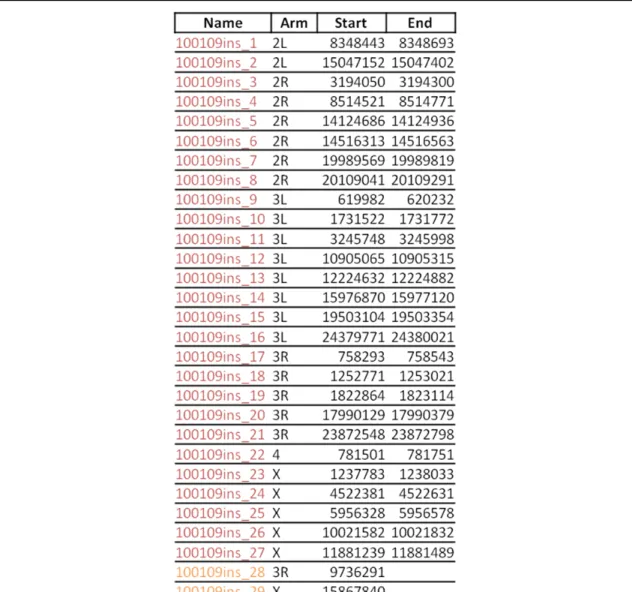
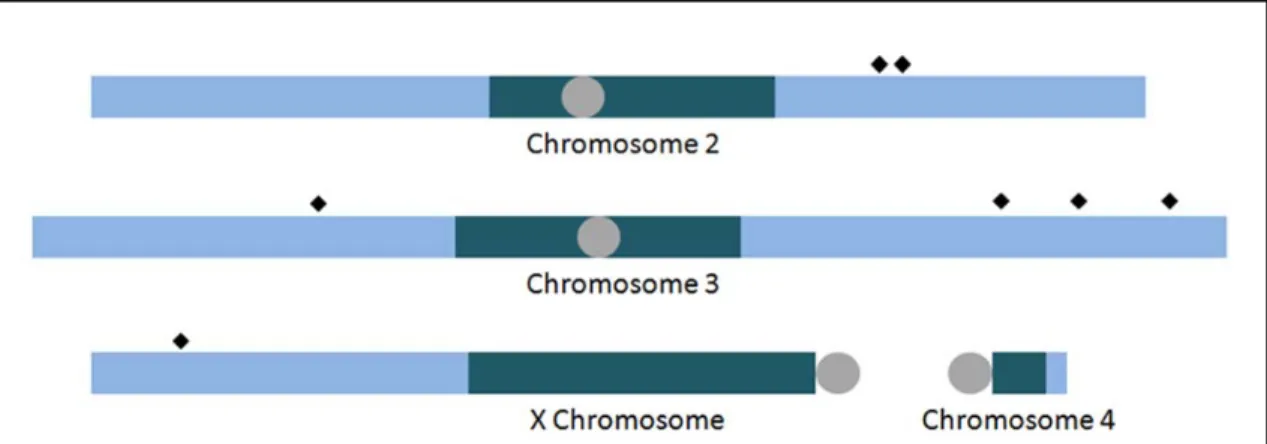
Figure 3.2. Integration of aphidicolin-induced DNA inserts. Integration events of pCoHygro into S2 cell DNA in the presence of aphidicolin were mapped with a variety of techniques. Those that could be unambiguously mapped to a single location are presented
here relative to the D. melanogaster reference sequence assembly (A). Depicted are
I found that, even when transfecting 100-fold less DNA, I was able to detect
unambiguous integrations. In this iteration of the experiment, splinkerette PCR was used to
enrich for insert-containing sequences after the extracted DNA had been sheared (Fig 3.3).
Splinkertte PCR involves the annealing of a “splink” adapter to both ends of each sheared
fragment (DEVON et al. 1995; UREN et al. 2009). The hairpin structure on the adapter is designed Table 3.1. Location of unambiguous integrations in aphidicolin-treated cells selected with hygromycin. These integrations came from the first electroporation of S2 cells with 500 ng of the construct. Displayed are the possible ranges that contain the insert-genome junction for a given integration. In instances in which the precise location was determined, only one number is given. An asterisk indicates that a precise junction of the insert and genome was determined, but that there was microhomology. The colors correspond to those
used in Fig. 3.2: blue inserts were detected via HTS, and the green inserts were detected via
TOPO cloning of splinkerette PCR products. The green insert on 2L landed in the histone
to prevent “end-repair” priming during PCR, in which unligated DNA anneal and amplify
fragments that do not contain the insert. In the first round of PCR, one of the two primers used
matches the sequence in the single-stranded region of the splink; that is, it has no complementary
sequence, and can therefore not initiate DNA synthesis. The other primer used is an
insert-specific primer, which accomplishes first-strand synthesis only in fragments that contain the
corresponding portion of the insert. Synthesis from the insert-specific primer produces the
complementary sequence that the splink primer needs to continue the PCR. Therefore, even
though the majority of the DNA fragments are nothing but genomic DNA, splinkerette PCR can
enrich for DNA fragments that contained insert DNA. In this manner, I detected an additional 27
Figure 3.3. Splinkerette PCR enriches for insert-containing fragments. Following sonication of the DNA, blunt splinkerette adapters (red) are ligated to the ends of the sheared fragments. An insert-specific PCR primer is employed to enrich for insert-containing fragments, while the hairpin on the splinkerette adapters prevent end-repair priming (1). A second round of PCR further amplifies sequences of interest, while shortening the terminal non-genomic DNA (2). Sequencing adapters are added to the ends of the PCR products, allowing them to be sequenced on the GA IIx (3,4). The resulting 76 bp paired-end reads allow us to pinpoint the integration site of multiple insertions (5). Alternatively, the product
of the nested PCR from (2) can be cloned into a TOPO-TA vector and transformed into E.
coli (6). Colony PCR is used to detect successful cloning, and sequencing of the PCR
The products of splinkerette PCR were also used for TOPO cloning, a system that allows
direct cloning of PCR products. By doing so, I was able to maximize the resolution of the
insert-genome junction, at the cost of some of the throughput. TOPO cloning of the 500 ng transfection
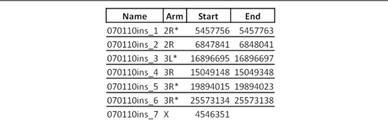
revealed two additional inserts: an unambiguous integration on 3R, and an insertion in the Table 3.2. Location of unambiguous integrations in aphidicolin-treated cells enriched via splinkerette PCR. These integrations came from the second electroporation of S2 cells, which used 5 ng of the construct. Displayed are the possible ranges that contain the insert-genome junction for a given integration. In instances in which the precise location was determined, only one number is given. The colors correspond to those used in Fig. 3.2: red
inserts were detected via HTS of splinkerette PCR products, and the orange inserts were
detected via TOPO cloning of splinkerette PCR products. All locations refer to release 5 of