The mammalian nuclear matrix is now recognized as a site of lipid biosynthesis and signaling in both normal and pathological states (reviewed in 1–8). Radioisotope and stable isotope tracer studies consistently identify glycero-phospholipids (GPLs), and the enzymatic activities that metabolize them, within purified nuclear fractions pur-portedly devoid of nuclear envelope and other cellular membrane contaminants (9–15). These findings suggest the existence of an independently-regulated GPL pool within the nuclear matrix, one distinct from the nuclear envelope and from bulk cellular membranes. In support of this interpretation, nuclear isoforms of a number of GPL metabolic enzymes have been described (10, 11, 16– 22). The STAR-PAP RNA poly-A polymerase, the direct modification of a phosphoinositide headgroup presented to an inositide kinase by a nuclear receptor, and nuclear phosphoinositide control of the basal transcription ma-chinery all provide compelling examples of GPL-regulated activities that discharge their functions within the nuclear matrix (23–27). At issue, however, is the scale of nuclear GPL metabolism and nuclear GPL load.
This difficult question can now be addressed using quantitative mass spectrometric methods. The single ma-jor study on this topic estimates that phosphatidylcholine (PtdCho) alone occupies 10–16% of the nuclear matrix of IRB-32 cells by volume (14). This is a startling conclusion Abstract A reliable method for purifying envelope-stripped
nuclei from immortalized murine embryonic fibroblasts (iMEFs) was established. Quantitative profiling of the glyc-erophospholipids (GPLs) in envelope-free iMEF nuclei yields several conclusions. First, we find the endonuclear glycerophospholipidome differs from that of bulk mem-branes, and phosphatidylcholine (PtdCho) and phosphati-dylethanolamine species are the most abundant endonuclear GPLs by mass. By contrast, phosphatidylinositol (PtdIns) represents a minor species. We also find only a slight enrich-ment of saturated versus unsaturated GPL species in iMEF endonuclear fractions. Moreover, much lower values for GPL mass were measured in the iMEF nuclear matrix than those reported for envelope-stripped IMF-32 nuclei. The collective results indicate that the nuclear matrix in these cells is a GPL-poor environment where GPL occupies only approximately 0.1% of the total nuclear matrix volume. This value suggests GPL accommodation in this compart-ment can be satisfied by binding to resident proteins. Finally, we find no significant role for the PtdIns/PtdCho-transfer protein, PITP, in shuttling PtdIns into the iMEF nuclear matrix.—Tribble, E. K., P. T. Ivanova, A. Grabon, J. G. Alb, Jr., I. Faenza, L. Cocco, H. A. Brown, and V. A. Bankaitis. Quantitative profiling of the endonuclear glycero-phospholipidome of murine embryonic fibroblasts. J. Lipid Res. 2016. 57: 1492–1506.
Supplementary key words cell signaling • lipids • nuclear receptors/ lipid ligands • phospholipids/metabolism • phospholipids/
phosphatidylinositol
This research was supported by Foundation for the National Institutes of Health Grants R01-NS37723 and R01-GM112591, and Welch Foundation Grant BE-0017 awarded to V.A.B. P.T.I. and H.A.B. were supported by National Institutes of Health Large Scale Collaborative Initiative LIPID MAPS Grant U54 GM069338 and the James S. McDonnell Foundation. I.F. and L.C. were supported by Italian MIUR-FIRB Grant RBAP10447J. The content is solely the responsibility of the authors and does not necessarily represent the official views of the National Institutes of Health. The authors declare no financial conflicts of interest.
Manuscript received 22 April 2016 and in revised form 23 May 2016. Published, JLR Papers in Press, June 2, 2016
DOI 10.1194/jlr.M068734
Quantitative profiling of the endonuclear
glycerophospholipidome of murine embryonic
fibroblasts
Emily K. Tribble,1,* Pavlina T. Ivanova,† Aby Grabon,*,§ James G. Alb, Jr.,* Irene Faenza,** Lucio Cocco,** H. Alex Brown,† and Vytas A. Bankaitis1,*,§
Lineberger Comprehensive Cancer Center,* University of North Carolina School of Medicine, Chapel Hill, NC; Departments of Pharmacology and Biochemistry,† Vanderbilt University School of Medicine, Vanderbilt Institute of Chemical Biology, Nashville, TN; Department of Molecular and Cellular Medicine,§ Texas A&M Health Science Center, College Station, TX; and Cellular Signaling Laboratory,** Department of Biomedical Sciences, University of Bologna, Bologna, Italy
Abbreviations: DAPI, 4′,6-diamidino-2-phenylindole; EM, electron microscopy; ER, endoplasmic reticulum; GPL, glycerophospholipid; iMEF, immortalized murine embryonic fibroblast; MEF, murine embry-onic fibroblast; PL, phospholipid; PtdCho, phosphatidylcholine; PtdEtn, phosphatidylethanolamine; PtdGro, phosphatidylglycerol; PtdIns, phosphatidylinositol; PtdOH, phosphatidic acid; PtdSer; phosphatidylserine.
1 To whom correspondence should be addressed.
e-mail: [email protected] (E.K.T.); [email protected] (V.A.B.)
The online version of this article (available at http://www.jlr.org)
Assay Designs, Ann Arbor, MI; product number SPA-860), an anti-histone H3 antibody (generous gift of Brian Strahl, Univer-sity of North Carolina-Chapel Hill), an anti-NURIM antibody (Santa Cruz Biotechnology, Santa Cruz, CA; product number sc-133260), and an anti-fibrillarin antibody (Abcam Inc., Cam-bridge, MA; product number ab5821). Goat-anti-mouse or goat-anti-rabbit HRP-conjugated secondary antibodies (Bio-Rad, Hercules, CA) were used for development in ECL assays. Donkey-anti-rabbit secondary antibody conjugated to IR Dye 800 was purchased from Rockland Immunochemicals Inc. (Gilbertsville, PA; product number 611-731-127) for use in Odyssey immunob-lotting experiments.
Cell culture and transfection
iMEFs were derived from E14-E16 embryos, and immortalized iMEF lines were generated using the SV40 large T-antigen method (28). Unless otherwise specified, all primary and immor-talized cell lines were cultured in complete DMEM containing
4.5 g/l glucose and supplemented with 10% FBS, 1 U/ml penicil -lin G, and 100 g/ml streptomycin (complete DMEM). All cell
culture was performed at 37°C in a 10% CO2 incubator.
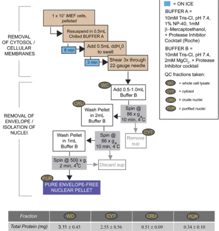
Initial steps in purifying envelope-stripped iMEF nuclei Envelope-stripped nuclei were prepared using the method of Martelli et al. (11) with essential modifications: iMEF cells were seeded to 150 mm tissue culture dishes and grown for 24–48 h. Approximately 107 iMEFs were pelleted (for a 1× preparation) and washed three times in Dulbecco’s PBS solution. After com-plete removal of PBS, the cell pellet was resuspended thoroughly in 500 l of chilled buffer A [10 mM Tris-HCl (pH 7.4), 1% NP-40, 10 mM -mercaptoethanol, 0.5 mM PMSF] supplemented with Complete Protease Inhibitor cocktail (Roche Biopharma-ceuticals). Cells were incubated on ice for 8 min with occasional agitation. An equal volume of ice-cold ddH2O was added to swell the cells. Following a 3 min incubation, swollen cells were subse-quently subjected to three passages through a 22 gauge needle. Removal of cellular debris was monitored by examination of sev-eral microliters of triturate by phase contrast microscopy. In suf-ficiently sheared samples, minimally 40 of 50 nuclei examined lacked significant cytosolic or membranous debris. After addi-tion of 0.5 ml of chilled buffer B [10 mM Tris-HCl (pH 7.4), 2 mM MgCl2] supplemented with protease inhibitors, the nuclei were gently triturated and again examined by microscopy. Nuclei were returned to ice for 1 min in preparation for centrifugation, during which a portion of lysate (representing the whole cell fraction) was saved for immunoblot analysis of cellular markers and total protein quantification.
Ultimate steps in purifying envelope-stripped iMEF nuclei
Nuclei were sedimented at 86 g for 10 min at 4°C. The superna-tant (cytosolic fraction) was either discarded or saved for quality control analysis as needed. The crude pellets were washed once with an excess of buffer B and sedimented again at 86 g for 10 min at 4°C. During an unscaled preparation, the purified nuclei were resuspended in buffer B and distributed as necessary for quality control protein analyses. When generating three times purified nuclear pellets for analysis of PLs by ESI LC-MS, the purified pellet was instead resuspended in 1 ml total of buffer B. Of this suspen-sion, 85% of the final material was pelleted at 500 g for 2 min at 4°C. Following complete removal of the supernatant, the pellet was snap-frozen in liquid nitrogen and stored at 80°C in preparation for PL analysis by MS. Aliquots (150 l) were collected for protein measurements and quality controls before the final pelleting step. considering that the genome itself is estimated to occupy
some 39% of the nuclear volume in the IMF-32 cell line. Furthermore, the endonuclear GPL pool is reported to be unusual in that it is dominated by saturated PtdCho mo-lecular species (14). The abundance of endonuclear GPLs, when coupled with their predominantly saturated nature, motivates speculation that phospholipids (PLs) profoundly influence the chemical properties of the nu-clear matrix, i.e., by contributing to the formation of gel-like regions within the nuclear matrix (1, 14). This concept raises a fundamental question of how does the nuclear matrix accommodate such a large PL load? That is, how are lipids organized within the nuclear matrix?
Given the mounting evidence for endonuclear lipid sig-naling and the lingering questions regarding how lipids are organized in nuclear matrix, we reinvestigated the problem of endonuclear lipidomics. To this end, we devel-oped a reliable and reproducible method for purification of envelope-free nuclei from immortalized murine embry-onic fibroblasts (iMEFs). This method adheres to a strin-gent quality-control regime for assessing purity of isolated endonuclear compartments. Quantitative GPL profiling of these highly purified fractions describes an endonu-clear GPL composition that is indeed distinct from that of bulk cellular membrane. Contrary to previous reports, however, the profile shows no particular enrichment of saturated GPL molecular species. Moreover, the mass measurements record drastically lower GPL contents in endonuclear compartments than those previously reported, at least for a neuroblastoma cell line. While the collective data confirm the nuclear matrix harbors a GPL pool of distinct composition from that of bulk membrane, the data also identify the iMEF nuclear matrix as a PL-poor environment that requires no unusual provisions for PL accommodation other than binding to resident proteins.
MATERIALS AND METHODS
Reagents and general notes
Chemicals and reagents were purchased from Fisher Scientific (Pittsburg, PA) or from Sigma-Aldrich (St. Louis, MO), unless otherwise stated. All lipid standards were purchased from Avanti Polar Lipids (Alabaster, AL). Organic solvents and supplies used to prepare samples for electron microscopy (EM) were obtained from Electron Microscopy Sciences (Hatfield, PA). The mass la-bels, myo-inositol-d6 and choline-d9, were obtained from C/D/N Isotopes (Pont-Claire, Quebec, Canada) and Sigma-Aldrich, respectively.
Media and antibodies
DMEM and antibiotics were obtained from Gibco/Invitrogen
OR), equipped with a Gatan model 794 multiscan digital camera.
To examine nuclei at different stages of purification, samples were fixed during extraction, shearing, and “unveiling” stages (see Results) of separate 1× preparations. Nuclei undergoing the initial extraction in buffer A (500 l total volume) were diluted to 10 ml with an excess of fixative [2% glutaraldehyde, 1% tannic acid, 2 mM CaCl2 in 0.1 M sodium cacodylate (pH 7.4)]. Nuclei were fixed at the 6 min time point. Sheared nuclei (1 ml) and “unveiled” nuclei (1.5 ml) were also diluted to the same final volume in fixative. One milliliter of each dilution was pelleted at 100 g for 10 min at 4°C. Fixed samples were washed two times for 10 min in 1 ml fixative. After washing thoroughly in buffer [2 mM CaCl2, 0.1 M sodium cacodylate( pH 7.4)], samples were pro-cessed for EM as described above.
Profiling of bulk iMEF PL
To generate iMEF pellets for bulk cellular GPL analysis, 1.2 × 10⁶ cells were seeded onto 150 mm tissue culture dishes and grown for 24 h to 60–70% confluence. Some 107 cells were har-vested by trypsinization, pelleted, and washed two times in HBSS without Ca2+ or Mg2+. Cells were then transferred to microfuge tubes in 1 ml of the same buffer and pelleted at 1,000 g for 5 min. Following complete removal of the supernatant, pellets were fro-zen in liquid nitrogen and stored at 80°C prior to lipid extrac-tion and global PL quantificaextrac-tion by MS.
GPL extraction and analyses
GPLs from whole cells or nuclear pellets were extracted using a modified Bligh and Dyer procedure (29). Approximately 1 × 10⁷ iMEF cells or 3 × 10⁷ nuclei per pellet in cold 1.5 ml mi-crofuge tubes (Laboratory Product Sales, Rochester, NY) were vortexed with 800 l of cold 0.1 N HCl:CH3OH (1:1) and 400 l of cold CHCl3 was added. The extraction proceeded with vortex-ing (1 min) and centrifugation (5 min, 4°C, 18,000 g). Quantifi-cation of GPLs was achieved by the use of ESI LC-MS employing synthetic (non-naturally occurring) diacyl and lysophospholipid standards as communicated elsewhere (30). Typically, 200 ng of each odd-carbon standard was added to each sample.
Identifica-tion of the individual PLs was accomplished by LC-MS/MS using an MDS SCIEX 4000 QTRAP hybrid triple quadrupole/linear
ion trap mass spectrometer and a Shimadzu HPLC system with a normal phase Luna Silica column (2 × 250 mm, 5 m) using a gradient elution (30). Identification of the individual species was based on their chromatographic and mass spectral characteris-tics and comparison to these of chemically defined standards (30, 31). This analysis allows identification of both fatty acid moi-eties, but does not determine position on the glycerol backbone (sn-1 vs. sn-2).
Statistical analyses
Data are presented as means plus standard errors. Differences between percentages of the total GPL pool represented by differ-ent classes for whole cells versus nuclei were determined by Stu-dent’s t-test.
Molecular biology and site-directed mutagenesis
Rat PITP cDNA was amplified by PCR and subcloned as a 0.85 kb HindIII-BamHI fragment into pEGFP-N1 (Clontech, Palo Alto, CA). A HA-tagged rat PITP cDNA (C-terminal tag) was generated by amplification of the rat PITP cDNA and insertion of the 0.85 kb product into the unique BamHI-NotI sites of pEF3HA, a derivative of pEF4 (Invitrogen, Carlsbad, CA). This construct contains a HA epitope that was incorporated into that Immunoblot analyses of envelope-free nuclei
In preparation for immunoblot analysis, whole cell lysates, wash fractions, and nuclear pellets were homogenized in M-Per lysis buffer (Thermo Scientific) supplemented with Complete Protease Inhibitor Cocktail (Roche Biopharmaceuticals). Sam-ples were triturated vigorously through a 25 gauge needle until complete sample disruption was achieved (as confirmed by phase contrast microscopy). Samples were clarified by centrifugation, and the supernatant was separated to a fresh tube for protein precipitation using the SDS-PAGE Clean-up kit (GE Life Sci-ences, Piscataway, NJ) according to the manufacturer’s direc-tions. Precipitates were resuspended in CHAPS buffer (8 M urea, 2% CHAPS, and 50 mM DTT). These samples were solubilized in Laemmli sample buffer resolved by SDS-PAGE (10% gels), and nuclear and membrane markers were visualized by immunoblot-ting. Sample loading was normalized by “cell equivalents.” For Odyssey Westerns, the range of signal linearity was determined for each fraction with each antibody.
Resolved proteins were transferred to nitrocellulose mem-branes by standard methods. Memmem-branes were blocked in the appropriate blocking reagent (as recommended by the manufac-turer) for 1 h at room temperature and probed with primary an-tibody overnight at 4°C. Decorated membranes were washed three times for 10 min in TTBS and incubated for an additional 1–2 h with the corresponding HRP-conjugated secondary anti-body diluted in 2% BSA in TTBS. Blots were again washed three times in TTBS and once in PBS before development using the ECL method (Amersham Biosciences). We define the threshold for acceptable purity as lack of detectable calnexin immunoreac-tivity in a nuclear preparation of 2.4 × 10⁵ cell equivalents.
For detection of blotted proteins using the Odyssey platform, transferred membranes were blocked for 1 h at room tempera-ture in Odyssey blocking buffer (LI-COR Biotechnology, Lin-coln, NE). The appropriate primary and secondary antibodies were diluted in a 1:1 solution of Odyssey blocking buffer and PBS. Secondary incubations and terminal wash steps were per-formed in the dark. Decorated membranes were analyzed on the Odyssey® infrared imaging system using Odyssey® 2.0 software (LI-COR Biotechnology). Scan settings were high image quality, resolution was set to 169 m, and the intensity of the scan was 5.0. Antibody signals were quantified as integrated intensities of the areas above and below the bands of interest.
Quantification of cellular protein
Prior to protein quantification, cellular fractions were recon-stituted in 1% SDS and incubated at 95°C for 10 min. Cooled samples were homogenized by at least 30 passages through a 25 gauge needle. Satisfactory sample disruption was confirmed by phase microscopy and samples were subsequently clarified by centrifugation. In preparation for BCA analysis (Thermo Scien-tific, Rockland, IL), samples were diluted 1:10 or 1:20. Diluted fractions were analyzed in triplicate according to the protocol for BCA assay for microplate reader.
EM of envelope-free nuclei
Purified nuclei were pelleted and fixed in 2% glutaraldehyde, 1% tannic acid in 0.1 M sodium cacodylate, 2 mM CaCl2, and postfixed in 2% OsO4 in 0.1 M sodium cacodylate, 2 mM CaCl2. Samples were stained in 4% uranyl acetate in 50% ethanol,
fibroblast (MEF) system for addressing questions related to nuclear signaling, we developed a method for prepar-ing highly purified envelope-free nuclei from these cells. The method generated purified endonuclear compart-ments suitable for quantitative lipidomic analyses and yielded reproducible data. The procedure employed serial stripping manipulations in the presence of detergent (1% NP-40), and hypotonic swelling and mechanical shearing in progressively more dilute detergent environments (1%
→ 0.5% → 0.33% → 0% NP-40) to arrive at envelope-free nuclear preparations.
As a general comment, our protocol for purification of envelope-free nuclei worked optimally when 1 × 10⁷ pri-mary or iMEF cells were used as starting material (termed a 1× scale). The efficacy of the method is sensitive to scale and applications that require larger mass quantities of nu-clei are best served by generating several smaller nuclear preparations in parallel and pooling the corresponding PL extracts. We recommend processing no more than 3 × 10⁷ cells during a single purification. Buffer volumes used in preparing larger samples should be scaled accord-ingly. A methodological flowchart is depicted in Fig. 1. From nine independent 1× preparations of envelope-free nuclei, an average of 0.34 ± 0.10 mg of endonuclear pro-tein was recovered. As the averaged 1× quantity of total cellular starting material was 3.11 ± 0.45 mg of total cellu-lar protein, the final protein yield in the purified endo-nuclear fractions was approximately 10% of total starting material.
Visual landmarks for monitoring purification quality Several morphological transformations accompanied separation of the nuclear particle from contaminating cel-lular membranes and nuclear envelope. These transfor-mations were readily observed during the purification and reliably diagnosed quality of the final preparation. As such, these morphological landmarks served as valuable real-time reporters of processing efficacy. The stages of processing at which these transformations were monitored are highlighted in Figs. 2, 3. Typical nuclear morphologies observed during the initial detergent extraction step (1% NP-40) are shown in Fig. 2B. The nuclear particles were readily distinguished from assorted cytosolic and organ-elle debris by light microscopic examination at 40× magni-fication. The efficacy of the hypotonic swelling step (NP-40 diluted to 0.5% at this step) was similarly interpretable as nuclei increased slightly in diameter (Fig. 2C).
A subsequent and effective shearing step was an essen-tial component of the purification. Rapid and vigorous trituration of crude nuclear fractions through a 22 gauge needle liberated nuclei of associated debris and left the sheared nuclei as oblate particles. Satisfactory outcomes at this stage were defined by lack of associated large debris in at least 40 of 50 nuclear particles examined. Examples of typical nuclear morphologies at this stage are shown in Fig. 2D, E. EM analyses demonstrated that cellular debris attached to the detergent-extracted nuclear particle (Fig. 2F) was mostly removed by the shearing step (Fig. 2G). Although a small amount of debris remained attached to construct as an XbaI-PmeI cassette (DNA sequence 5′
-TATCCT-TACGAC GTTCCAGACTATGCA-3′). Site-directed mutagenesis primers used in this study were from Fisher Scientific, and rat PITP cDNAs were mutagenized according to QuickChange mu-tagenesis kit specifications (Stratagene, La Jolla, CA). All mutant constructs were confirmed by nucleotide sequence analysis.
Transient transfection of mammalian cell lines
Immortalized cell lines were transfected using the FuGene transfection reagent (Roche, Indianapolis, IN). Briefly, Cos7 cells or HeLa lines were seeded onto plastic dishes in complete DMEM 24 h prior to transfection. Once cells settled on the plas-tic surface, the medium was exchanged for antibioplas-tic-free DMEM. One microgram aliquots of PITP-EGFP constructs were incubated in 100 l Opti-MEM (Invitrogen) premixed with 3 l transfection reagent according to the manufacturer’s instructions. The com-plete transfection cocktail was incubated at room temperature for 1 h before distribution to the medium. At 12 h posttransfection, cells were split onto coverslips and cultured in complete DMEM.
Immunocytochemistry
HeLa or Cos7 cells, transiently transfected with appropriate PITP-EGFP or PITP-HA expression plasmids, were fixed 16–20 h posttransfection in 4% PFA in PBS. After permeabilization in 0.2% Triton X-100, cells expressing PITP-EGFP constructs were counterstained with 4′,6-diamidino-2-phenylindole (DAPI) (Mo-lecular Probes) for 1 min. Cells transfected with PITP-HA con-structs were permeabilized similarly, then incubated for 1 h at room temperature in blocking buffer (2% BSA in PBS). Primary anti-HA antibodies (1:2,000 dilution in blocking buffer) were ap-plied to blocked coverslips. After a 12–15 h incubation of cover-slips with primary antibody at 4°C, cells were serially washed three times for 10 min in 1% BSA in PBS. Secondary antibodies (1:8,000 in blocking buffer) were then applied onto coverslips and fixed cells were incubated at 4°C for 5–8 h. After several washes (1% BSA in PBS), cells were counterstained with DAPI. Coverslips were mounted on glass slides, imaged on a Zeiss 510 META scanning laser confocal microscope, and images were pro-cessed using Adobe Photoshop 6.0.
Labeling iMEF cells in culture with deuterated GPL precursors
PITP+/+ or pitp0/0 iMEFs were seeded in complete DMEM and grown to a subconfluent (60%) density. Cells were washed in PBS and labeled for the desired time period in antibiotic-free DMEM containing 10% FBS, 80 g/ml choline-d9, and 50 g/ml
myo-inositol-d6. For whole cell measurements, PITP+/+ or pitp0/0 iMEFs were collected in 800 l HBSS for GPL extraction and
analysis by LC-MS/MS. In experiments involving purified nuclei
stripped of membranes, 3 × 10⁷ cells were pelleted and washed in PBS prior to detergent-mediated removal of the nuclear enve-lope. Extracts pooled from three samples (9 × 10⁷ nuclei) were isolated and analyzed by LC-MS.
RESULTS
Purification of envelope-free iMEF nuclei
higher magnification (>10,000×) by EM, the peripheries of individual nuclear particles presented a fuzzy border and the obvious membrane bilayer structures that consti-tute the nuclear envelope were no longer visible at these peripheries (compare Fig. 5C, E with Fig. 5D, F, respec-tively). However, the high resolution EM analyses detected two classes of impurities that commonly evade detection by lower magnification EM, i.e., the types of analyses typi-cally presented as evidence for the envelope-free status of purified nuclear particles (14). First, small copurifying membrane-like strands, attached to the isolated nuclear particles, were occasionally observed (Fig. 5H). Second, these structures were also occasionally observed in the ab-sence of an attached nucleus, and those were counted as well (Fig. 5G). Typically, two strands were recorded per 100 nuclei, and one small patch (average length 500 nm) was identified per 34 nuclei (Fig. 5H). We found this low level of potential contamination to be unavoidable. For purposes of reference, supplementary Table 1 com-piles the contamination data over a series of independent nuclear preparations.
Biochemical criteria for purified envelope-free nuclei The accepted biochemical standard for envelope-free nu-clei is the absence of the abundant cytosolic protein, -tubulin, in the most purified fractions (14, 21, 32, 33). Unfortunately, this criterion does not adequately interro-gate the preparation for contamination by the nuclear en-velope, a membrane system physically contiguous with the highly abundant endoplasmic reticulum (ER). To control nuclei at this stage, closer examination of nuclear borders
revealed that shearing removed the majority of the nu-clear envelope (Fig. 2H).
Upon further dilution of suitably sheared nuclei into Mg2+-containing buffer B (final NP-40 concentration at this stage is 0.33%), the nuclear envelope was effectively removed. This stripping event was monitored via phase contrast microscopy by what we term an unveiling process. Unveiling was marked by the nuclei becoming less opaque and the nucleoli assuming much sharper contrast relative to nucleoplasm (Fig. 3B). The loss of nuclear envelope was visible in electron micrographs of nuclei fixed immedi-ately after addition of buffer B (Fig. 3C, left two panels). Borders of stripped nuclei appear fuzzy and without dis-cernable traces of nuclear envelope (Fig. 3C, right two panels). Subsequent pelleting and washing steps in buffer B (no detergent present) completed the envelope strip-ping process (Fig. 4). Purified nuclei retained their mor-phologies and appearance throughout the purification.
Imaging criteria for envelope-free nuclei
Visualization of envelope-stripped nuclei by EM directly interrogated the membrane content of nuclear prepara-tions. When compared with in situ nuclear morphologies (Fig. 5A), low-magnification (<2,000×) electron micro-graphs of purified envelope-free nuclei (Fig. 5B) demon-strate efficient removal of extraneous material. Nuclear structure was nicely preserved, and contaminating organ-elles or other heterogeneous membrane debris were not apparent at this level of resolution. When inspected at
described herein exhibited undetectable levels of the abun-dant integral ER membrane protein, calnexin, and the in-ner nuclear envelope integral membrane protein, NURIM, in addition to undetectable levels of -tubulin (Fig. 6A). We for these primary sources of contaminating membranes, we
monitored membrane-free nuclear preparations for abun-dant integral membrane proteins of the ER and nuclear envelope. Endonuclear fractions purified by the protocol
Fig. 2. Phase contrast monitoring of nuclear particles. A: Flow chart of the critical steps for removing ex-tranuclear debris is indicated. All steps are executed on ice using chilled solutions and tubes. Eye symbols identify points where extraction efficacy is monitored by phase contrast microscopy. B–E: Phase contrast images of extracting nuclei at various steps during the purification. All images were taken using a 40× objec-tive, and 3–5 l of sample was typically viewed under a coverslip. B: Cells are resuspended in buffer A to initi-ate the extraction process. At this stage, nuclei are visible as the phase dense center of each cellular particle.
Partially solubilized cytoplasm/plasma membrane contaminants are discerned as the less dense material
surrounding the nuclear particle. C: Addition of an equal volume of chilled ddH2O swells the nuclei and
further enhances contaminating material. D, E: Images of swollen nuclei sheared three times through a 22 gauge needle. D: Successfully sheared nuclei exhibit little to no visible debris attached. E: Nuclei marked with an asterisk retain attached debris. Nuclei marked with arrowheads are scored as at a suitable stage of purification. F–H: Electron micrographs of nuclear preparation stages. F: Electron micrograph of nucleus (1,100×) fixed after 6 min of extraction in buffer A (scale bar = 2 m). G: Sheared nucleus (1,100×). Minor amounts of debris are still attached to the lamin boundary, which is clearly visible (scale bar = 2 m). H: Higher magnification (15,000×) image of a nuclear border that has been successfully stripped of envelope (scale bar = 0.2 m).
defined the threshold for acceptable nuclear matrix purity as lack of detectable calnexin immunoreactivity in a sample of 2.4 × 10⁵ nuclei in ECL immunoblotting experiments. Odyssey immunoblotting experiments indicated that less than 0.3% of cellular calnexin remained in nuclear prepa-rations (Fig. 6B), reporting a <300-fold enrichment of nu-clear particles from contaminating ER membranes.
The endonuclear fractions were highly enriched for the nuclear matrix constituent, lamin A, however (Fig. 6A). Enrichment of the nucleolar protein, fibrillarin, was also monitored. Fibrillarin localizes to the nucleoplasm as well as to nucleoli (34), and thus served as a marker for loss of both nucleoplasm and nucleoli. Based on quantitative im-munoblotting data, we again estimated at least a 250-fold purification of nuclear matrix components (i.e., fibrilla-rin) with respect to contaminating ER membrane protein (i.e., calnexin).
Total PL in cells and nuclei
The resident GPLs were quantified and their composi-tions profiled in purified endonuclear compartments. Based on a signal-to-noise ratio of >3 as limit of detection threshold, we found the nuclear matrix to be a GPL-poor compartment. PtdCho, phosphatidylethanolamine (PtdEtn), phosphatidylserine (PtdSer), phosphatidylinositol (PtdIns), phosphatidic acid (PtdOH), and phosphatidylglycerol (PtdGro) were all detected in the endonuclear compart-ment, in rank order of mass abundance. The total GPL mass measured per unit endonuclear compartment (0.077 ±
0.0075 nmol/106 cells) projected an estimated load of
ap-proximately 5.9 × 10⁷ GPL molecules per nuclear particle. Based on this value, and our estimate that the average iMEF nucleus occupies a volume of approximately 600 m3, we calculated that the GPL component occupies <0.1% of the iMEF nucleoplasm. As expected, endonuclear GPL load was small relative to whole cell amounts. The endonuclear GPL mass was almost two orders of magnitude less than
that measured per unit iMEF cell (5.1 ± 0.43 nmol/106 cells).
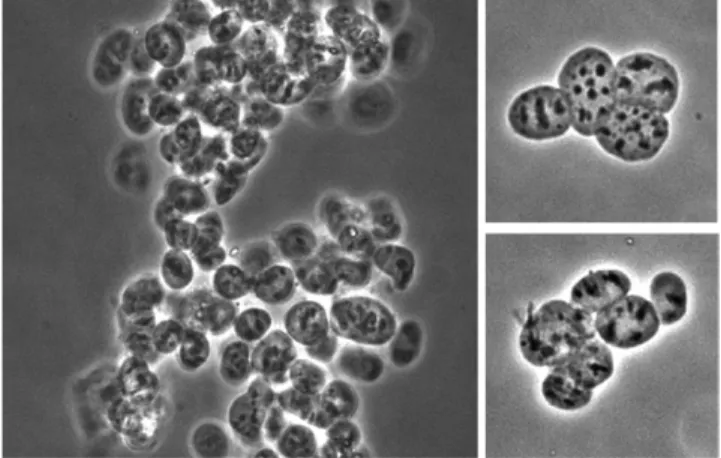
Fig. 4. Morphology of purified nuclei. Purified envelope-free nuclear fractions were imaged by phase contrast using a 40× objec-tive. Left panel: Nuclear particles are prone to aggregation after envelope removal. Right panels: Inspection of smaller groups of purified nuclear particles confirms that individual particles do not change shape or appearance during the final centrifugation steps of the purification process.
the 32:0 and 30:0 PtdCho were the only saturated PtdCho molecular species of sufficient abundance to justify quan-tification in the endonuclear fractions. Reciprocally, the polyunsaturated PtdCho species were only modestly re-duced in abundance in the nuclear matrix relative to their representation in bulk GPL. The relative pool contribu-tions of monounsaturated PtdCho species did not signifi-cantly differ in endonuclear compartments versus bulk cell material (Fig. 7B, middle row).
These profiling data were inconsistent with strong enrichments of saturated PtdCho molecular species in envelope-free nuclear preparations. Indeed, unsaturated PtdCho molecular species compromised 79% of the total endonuclear PtdCho mass (Fig. 7). This pattern was ob-served across the other GPL classes as well. Inspection of the rank order of the 42 most abundant endonuclear GPLs revealed a general paucity in both diversity and mass abun-dance of saturated species (supplementary Table 2). For example, only 30% of the PtdSer mass was represented by saturated molecular species, and this fraction was much larger than for any other quantifiable endonuclear GPL class. PtdSer represented the third most abundant nuclear GPL class, comprising some 15% of the total endonuclear GPL mass. The preponderance of unsaturated GPLs was encouraging given the legitimate concern that detergent-mediated stripping of the nuclear envelope might gener-ate artifactual detergent-insoluble lipid domains expected to be enriched in saturated GPL molecular species and sphingolipids. In that regard, SM was at the limit of detec-tion in endonuclear fracdetec-tions, indicating this sphingolipid Thus, the iMEF endonuclear phospholipidome accounts
for 1.4% of the total cellular GPL mass.
Lipidomic profiling of the major endonuclear PLs With regard to composition, the bulk iMEF phospho-lipidome presented a diverse signature, with the leading GPL classes (by mass) represented by PtdCho, PtdEtn, and PtdSer molecular species, respectively (supplementrary Table 2). We note that 38:4 PtdIns was also represented within the 10 most abundant bulk GPL species. By con-trast, the iMEF endonuclear phospholipidome was domi-nated (in mass terms) by PtdCho, PtdEtn, and PtdSer molecular species; whereas PtdIns, PtdOH, and PtdGro were poorly represented in endonuclear compartments. Interestingly, and as discussed in greater detail below, the differences in fractional representation of these minor PLs in bulk membranes versus the endonuclear compart-ment were highly significant (P < 0.001). By contrast, the PtdCho/PtdEtn ratios for these two systems were similar (approximately 1.85). We interpret the relative paucity of PtdIns, PtdOH, and PtdGro to argue against the measured endonuclear GPLs representing mere contamination from bulk ER (see Discussion).
PtdCho was the most abundant endonuclear PL class, represented by 9 of the top 10 most abundant molecular species (supplementary Table S2). Greater than half of the endonuclear GPL mass was accounted for by PtdCho species (53.1 ± 2.8%). The proportion of the PtdCho pool represented by saturated molecular species was only modestly increased in nuclei relative to whole cells. In that regard,
Fig. 6. Immunoblot analyses of nuclear fractions. A: ECL immunoblot analysis of whole cell (WC) and purified nuclear fractions (NUC). Fractions collected from three separate nuclear preparations were prepared for gel electrophoresis as described in the Materials and Meth-ods. Gels were loaded by cell equivalents based on the amount of whole cell lysate needed for blotting in the linear range. Comparison to
whereas the most abundant endonuclear PtdIns was the 38:4 molecular species, the only detectable PtdOH in this compartment was the 36:2 molecular species. In that re-gard, none of the three acyl chain configurations identi-fied for endonuclear PtdIns were of the 36:2 variety. Those data were inconsistent with a simple nucleoplasmic phos-pholipase C-diacylglycerol kinase pathway. The action of such a metabolic sequence is expected to yield endonu-clear 38:4 PtdOH (see Discussion).
Considerations for how PtdIns is imported into the nuclear matrix
The existence of endonuclear phosphoinositide biosyn-thetic and signaling pathways begs the question of how PtdIns is incorporated into the nuclear matrix. The nu-clear matrix has no capacity to produce PtdIns in situ given that PtdIns synthase is an integral membrane pro-tein of the ER. The PtdIns/PtdCho-transfer protein, PITP, exhibits both cytoplasmic and endonuclear pools (33, 35, 36), raising the possibility that PITP shuttles PtdIns into the nuclear matrix. The low steady-state mass quantities of PtdIns we measured in the nuclear matrix suggested that maintenance of a suitable endonuclear PtdIns supply could be achieved via the action of a shut-tling lipid transfer protein, such as PITP. Moreover, when coupled with the measured mass excess of endonu-clear PtdCho to PtdIns in the nuendonu-clear matrix, a coupled endonuclear PtdIns-import/PtdCho-export cycle was also plausible. This shuttle hypothesis predicted that PITP
localization to the nucleus is facilitated by PtdIns binding, and that PITP-deficient cells will be compromised for PtdIns import into the nucleus. We tested these two pre-dictions in turn.
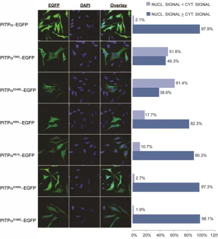
PtdIns-binding and PITP localization in the nucleus A combination of genetic and structural data identifies a set of residues critical for coordination of the PtdIns Fig. 7. A: GPL composition of iMEF cells and puri-fied envelope-free nuclei. Top row: GPL distribution by class for whole iMEFs and envelope-free iMEF nu-clei (mean of n = 9 shown). The difference in compo-sition between whole cells and nuclei for PtdOH (PA), PtdGro (PG), and PtdIns (PI) is highly signifi-cant (P < 0.001), while the PtdCho (PC)/PtdEtn (PE)
ratio is 1.85 for both whole cells and nuclei, with no significant difference. PtdSer (PS). B: Percent contri-butions of saturated, monounsaturated, and polyun-saturated species to PtdCho pool. The proportions of polyunsaturated and saturated PtdCho differ (P < 0.01) between whole iMEFs and nuclei. C: The rela-tive mass contribution of ether-linked PtdEtn species to the PtdEtn pool is substantially greater in whole iMEFs than in nuclei (P < 0.001).
class is a very minor constituent, at best, of the iMEF endo-nuclear PL pool (data not shown).
PtdEtn, the second most abundant GPL in the nuclear matrix, accounted for some 28% of total endonuclear GPL mass. We observed interesting distinctions between nonnuclear and nuclear PtdEtn species. Ether-linked PtdEtn was substantially reduced in endonuclear fractions when evaluated as a percentage of the overall PtdEtn pool. In comparison to a fractional representation of 40.4 ± 1.0% of bulk iMEF PtdEtn molecular species, only 27.7 ± 1.5% of the mass of endonuclear PtdEtn species was ether-linked (Fig. 7C, bottom row). The PtdCho/PtdEtn mass ratio, however, remained similar in endonuclear compart-ments when compared with that ratio in bulk iMEF GPL, further confirming ether-linked PtdEtn species were genu-inely segregated from the nuclear matrix.
Lipidomic profiling of the minor endonuclear PLs While PtdCho and PtdEtn were the most highly repre-sented GPLs in the endonuclear PL profile, envelope-free nuclei were relatively deprived of other GPL species (sup-plementary Table 2). For example, PtdIns was depleted in the envelope-free nuclear fractions (1.5 ± 0.2% of nuclear GPL) as compared with whole iMEFs (5.6 ± 0.6% of bulk cellular GPL). The distribution by headgroup class (Fig. 7, top row) shows that the minor GPL classes (PtdOH, PtdIns, PtdGro) were all significantly reduced (in terms of fractional representation) in the most purified endonu-clear fractions relative to PtdCho and PtdEtn. For pur-poses of comparison, endonuclear PtdCho, PtdEtn, and PtdSer species accounted for 1.63, 1.77, and 1.13% of the corresponding total cellular pools. By contrast, PtdGro was particularly sparse, only 0.02% of the total iMEF pool was recovered in the nuclear matrix.
experiments performed in HeLa cells, the localization profiles of all of the chimeras were also evaluated in pitp0/0 iMEFs and in Cos7 cells with similar results (data not shown).
The effects of the various missense substitutions on PITP localization profiles were not strictly dependent on the EGFP tag. HA-epitope-tagged versions of PITP, PitpT59D, PitpK61A, PitpN90L, and PitpE248K were also in-dividually expressed in HeLa cells. The cells were fixed, permeabilized, and HA-PITP visualized by indirect im-munofluorescence. The results largely recapitulated those obtained with EGFP-tagged chimeras (supplementary Fig. 2). The nuclear signal was dominant in cells expressing PITP-HA, and this localization pattern was observed in
97.6% of transfected cells (980/1,004). Predominantly cy
-tosolic distributions were observed for the PitpT59D-HA
(78.3%; 548/700), and PitpE248K-HA expressing cells. For
approximately 78% of cells expressing the PitpT59D-HA fusion protein, the fluorescence signal was predominantly cytoplasmic, and a nuclear localization defect was ob-served in approximately 95.1% of cells transiently express-ing the PitpE248K-HA fusion proteins (448/471 cells; supplementary Fig. 2). Nuclear localization defects were less prominent in cells expressing PitpK61A-HA, with only 15.6% of cells demonstrating a nuclear localization de-fect. We do note that the nuclear signal in cells express-ing PitpK61A-HA was most frequently of similar intensity to the cytoplasmic signal. It was rarely the dominant sig-nal, as was observed for the PITP-HA control. Nuclear headgroup in the PITP binding pocket (37–39). Four
mutant versions with specific defects in PtdIns-binding were employed in these experiments, PitpT59D, PitpK61A, PitpN90L, and PitpE248K. The corresponding missense substitutions were incorporated into the PITP-EGFP re-porter for transient expression in HeLa cells and imaged. These constructs all produced stable proteins when ex-pressed in cells, as confirmed by immunoblotting experi-ments (data not shown). Individual cells were scored as to whether the chimera localized predominantly to the nu-cleus or to the cytoplasm.
The PITP-EGFP reporter exhibited a predominant nuclear localization in 97.9% of the cells imaged (Fig. 8A, B). By contrast, PitpT59D-EGFP displayed a nuclear
localiza-tion defect in 51.6% of the cells imaged (190/368). Simi
-lar localization defects were scored for cells expressing PitpE248K-EGFP (61.4%, 189/308 cells). Milder, but nev -ertheless significant, nuclear localization defects were also recorded for the PitpN90L-EGFP and PitpK61A-EGFP chi-meras (17.7%, 57/322 and 10.8%, 40/370 of cells imaged, respectively). Those data suggested that PtdIns-binding influences either PITP import into or PITP export from the nucleus. The relationship was not simple, however. PitpS166A-EGFP and PitpS166E-EGFP chimeras [i.e., pro-teins defective in both PtdIns and PtdCho-transfer activity (36, 39)] were fully competent for localization to the nu-cleus. Some 2.7% (11/404) and 1.9% (8/425) of the cells imaged scored as showing defects in nuclear EGFP fluores-cence, respectively. While the collective data reported
Fig. 8. Nuclear localization patterns of PITP-EGFP and lipid binding mutants transiently overexpressed in HeLa cells. A: Confocal images of HeLa cells tran-siently expressing PITP-EGFP and PITP PL bind-ing and transfer mutants. Cells were grown on coverslips, transfected, and fixed 18 h posttransfec-tion. Cells are counterstained with DAPI to examine nuclear localization of mutant constructs. WT PITP
constructs produce nuclear fluorescence that is greater than or equivalent to cytoplasmic fluores-cence in the vast majority of expressing cells. PtdIns-binding defective point mutants, PitpT59D, PitpK61A, PitpN90L, PitpE248K, and the PtdIns/PtdCho-binding
of MEFs to efficiently incorporate these soluble head-group precursors into GPL. However, we reproducibly detected reduced fractional incorporation of Ins-d6 into
newly synthesized PtdIns in pitp0/0 MEFs. We do not pres-ently understand either the underlying basis or the physi-ological significance of this observation. By contrast, fractional rates of incorporation of newly synthesized PtdIns in endonuclear compartments was much slower in both PITP+/+ and pitp0/0 MEFs relative to fractional rates of incorporation of newly synthesized PtdCho (supple-mentary Fig. 5C, D). Very little newly synthesized PtdIns was recovered from PITP+/+ or pitp0/0 endonuclear com-partments during a 1 h pulse. Detectable incorporation required a pulse of at least 2 h. We interpret the data from these experiments to reflect the intrinsic ability of endo-nuclear compartments to produce PtdCho in situ, while PtdIns is only slowly imported from extranuclear sources. Those rates of fractional incorporation of newly synthe-sized PtdIns into pitp0/0 MEF endonuclear compartments were only modestly reduced (at best) relative to those measured for the PITP+/+ control (supplementary Fig. 5C), and we do not consider these modest differences to be significant.
DISCUSSION
Herein, we describe a facile and reliable method suit-able for purifying quality envelope-free nuclei from iMEFs and from primary MEFs. The choice of cell model was driven by the wealth of MEF lines that can be derived from mutant strains of mice. The method incorporated an ex-panded repertoire of quality controls and defined criteria for purity. Particular attention was paid to the abundant integral membrane constituents of the membranes that constitute the greatest reservoirs for contamination (e.g., ER and nuclear envelope). This method reproducibly yielded purified nuclear particles that exhibited the dual properties of preserved ultrastructure and the essential ab-sence of contaminating envelope. Quantitative phospho-lipidomics of the iMEF endonuclear compartment leads us to three basic conclusions: First, in agreement with pre-vious reports (9, 13, 14), the composition of the endonu-clear GPL pool is distinct from that of bulk cell GPL. Second, the endonuclear GPL pool is not substantially enriched in saturated molecular species relative to the saturated/unsaturated GPL-composition of the bulk cellular pool. Third, the nucleoplasm is a GPL-poor envi-ronment. Those latter two conclusions represent sig-nificant departures from previous ideas regarding the fundamental properties of the mammalian endonuclear phospholipidome.
A GPL-deprived nucleoplasm
Mass measurements of the 42 detectable endonuclear GPL species demonstrated the nuclear matrix to be a PL-poor environment. The endonuclear phospholipidome accounted for 1.4% of the total cellular GPL mass, and the GPL component occupied <0.1% of the volume of the localization defects were also scored in 25% of cells
ex-pressing PitpS166E-HA (34/137 cells imaged; supplemen -tary Fig. 2).
Quantitative lipidomics of pitp0/0 iMEF nuclei
The capability to produce highly purified envelope-free MEF nuclei, when coupled with the availability of mutant MEF lines, made it possible to address previously intrac-table questions regarding mechanisms of endonuclear lipid signaling. To that end, we first compared endonu-clear GPL composition and content in PITP+/+ versus pitp0/0 iMEFs to test whether the mutant GPL profile was
dramatically altered relative to that of the WT case. By the criteria of marker protein enrichment (supplementary Fig. 3A) and examination by EM (supplementary Fig. 3B, C), nuclei from pitp0/0 iMEFs were recovered to a compa-rably high purity as from WT cells. Satisfyingly, composi-tions of the whole cell and endonuclear phospholipidomes of both cell lines were also essentially indistinguishable (supplementary Fig. 4A, B). Moreover, the total PL con-tent of the nuclear preparations was very similar for WT versus pitp0/0 iMEFs (1,039.5 ± 226.7 pmol/mg vs. 1,094.2 ± 217.5 pmol/mg nuclear protein, respectively).
Dynamic lipidomics analysis of PtdIns import into nuclei The general requirement for PtdIns binding for effi-cient localization of PITP to the nucleus was consistent with a nuclear PtdIns-supply activity for this protein, perhaps as a mechanism for sustaining a nuclear phosphoinositide signaling cycle. Although qualitative inspection of steady-state nuclear PtdIns-4,5-P2 content by immunofluores-cence staining did not suggest a major defect in nuclear pools of this phosphoinositide in PITP-deficient cells, a direct test of the PtdIns shuttle model required kinetic analysis of the nuclear PtdIns-import process in the pres-ence and abspres-ence of PITP. To that end, standard pulse-chase strategies were modified to estimate endonuclear dynamics of PtdIns and PtdCho lipidomics in PITP+/+ or pitp0/0 MEFs. Deuterated PL precursors (Cho-d9 and
Ins-d6) were used to label newly synthesized iMEF PtdCho and
PtdIns in a pulse regimen, and precursor scan ESI-MS was used as readout to analyze the data. The ESI-MS analyses were conducted on whole cell lipid extracts as bulk con-trol, and on lipids extracted from purified envelope-free nuclei as test case. Precursor scans of the Cho head-group-derived m/z 184+ and m/z 193+ fragments over the mass range of 600–920 amu identified endogenous (i.e., preexisting) and newly synthesized PtdCho and SM spe-cies, respectively. Precursor scans of the Ins headgroup-derived m/z 241
and m/z 247
fragments over the mass range of 600–1,000 amu identified preexisting and newly synthesized PtdIns species, respectively. The light (pre- existing) species defined steady-state baselines for these measurements.
The fractional rates of Cho-d9 and Ins-d6 incorporation
contamination from these systems dominated the GPL mass. As a result, the reported mass and compositional measurements were misinterpreted as reporting strictly endonuclear GPL pools. Such contamination issues lead to large overestimates of endonuclear GPL mass if the nu-cleoplasm is a PL-poor environment. Indeed, as demon-strated herein, simple detergent solubilization steps do not efficiently remove ER and nuclear envelope contami-nants from nuclear fractions.
Implications for organization of nuclear GPLs
The reported abundance of endonuclear GPL, particu-larly saturated PtdCho (14), in IMR-32 neuroblastoma cells posed the central question of how such a large PL load might be organized within the nuclear matrix. Standard solutions to the “PL-accommodation” problem, such as in-corporation into membrane bilayers, are untenable. While invaginations of the nuclear envelope do extend into the nuclear interior, those structures are topologically distinct from the nuclear matrix itself (40, 41). Indeed, a hallmark feature of the nucleus is that it does not contain internal membrane-bound sub-compartments, despite the pres-ence of morphologically and functionally distinct endonu-clear domains (42). Alternatively, it is speculated that the large nuclear lipid-load provokes assembly of endonuclear PL into large aggregates or liquid crystalline phases (1, 3). In that regard, endonuclear PL “rafts” have been reported in hepatocytes (43–46), as have intranuclear lipid droplets, most strikingly under pathological conditions (47–49).
Nuclear lipid rafts and droplets are not common fea-tures of other cell types, however, so hepatocytes seem un-usual in this regard. Even so, the purported nuclear rafts are seen only in heavily processed hepatocyte nuclei (43– 46), and their identities as genuine rafts have not been directly confirmed by independent methods. If protein binding is a primary mechanism for nuclear lipid-organi-zation, then the reported high nuclear abundance of GPL demands that a large fraction of nuclear protein be GPL-associated. All of these general ideas lead to speculations that PtdCho and other GPLs may play significant struc-tural roles within the nuclear matrix (14, 50). Such com-plex issues surrounding mechanisms of PL-accommodation largely evaporate if the nuclear matrix proves to be a PL-poor compartment, as our data indicate it to be. With the stoichiometries we measure, GPL-binding to resident pro-teins alone could satisfy the PL-accommodation problem without the need for occupying a major fraction of nuclear protein with bound GPL. A GPL-deprived nuclear matrix also circumvents issues associated with a global gel-like physical environment formed by an abundance of satu-rated GPL. Our conclusions are consistent with the results of multiple studies that describe the nucleoplasm as a highly dynamic environment where naive molecules, such as GFP, exhibit comparable diffusion coefficients in the cytoplasm versus the nuclear matrix [e.g., (34, 51)]. While involvements of GPLs in structural or organizational ca-pacities within the nuclear matrix remain tenable, our data indicate any such involvements are most likely highly constrained and of a small scale.
iMEF nucleoplasm. Strict interpretations of those values are subject to several unavoidable caveats. First, removal of the nuclear envelope demanded manipulation of the or-ganelle in progressively dilute detergent environments ranging from 1% NP-40 for the initial extraction, to 0.5 and 0.33% NP-40 in the intermediate stages, and no deter-gent in the final step. It is not clear how much of the base-line endonuclear GPL pool was lost during those obligate NP-40-containing steps. We view this as an unknowable factor because it is not possible to assess the ground state of endonuclear GPLs prior to purification of envelope-free nuclei for the comparison. But, the excellent repro-ducibility of the data through independent preparations suggests the perturbations imposed are consistent and manageable.
Second, high resolution EM analyses consistently de-tected trace amounts of membrane-like material in the most purified nuclear particle fractions. If those repre-sented membrane remnants, such contaminants would have contributed to the GPL mass measured in the puri-fied endonuclear fractions. The under-representation of PtdIns in the endonuclear GPL profiles relative to bulk cell membranes argues against contamination by bulk ER representing a major source of membrane contamination in these preparations, as ER is a PtdIns-rich membrane sys-tem. Rather, the inner nuclear envelope would be a more likely source of membrane contamination, and our analy-ses could not determine the extent to which the matrix leaflet of the inner nuclear envelope was removed. Be-cause the lipids of this leaflet contact lamins and other abundant proteins of the nuclear periphery, this lipid pool is likely to be more resistant to stripping. What morpho-logical state such remnants would assume during the prep-aration of envelope-free nuclei is unknown. These might represent the membrane remnants discussed above, or we might have failed to recognize such contamination alto-gether. Thus, the endonuclear GPL values reported herein, although low, may yet overestimate endonuclear GPL mass. However, we are encouraged with the repro-ducibility of the quantitative measurements.
The values we measured for endonuclear GPL load di-verge from those reported for the IMR-32 neuroblastoma cell line. That is, the only other cell line of which we are aware for which a quantitative analysis of the nuclear ma-trix phospholipidome is described. A load of
approxi-mately 4 nmol PtdCho/106 nuclei was reported for those
cells, along with the remarkable estimate that PtdCho alone occupies 12–16% of the IMR-32 nuclear volume (14). Why this large discrepancy between the two analyses? As both studies used 0.5–1.0% concentrations of nonionic detergent for solubilization of nuclear envelope, one for-mal possibility is that different cell lines genuinely exhibit manifestly different endonuclear GPL loads. While cell line-specific variations will almost certainly prove to be the case, we consider it unlikely that such variations fully ac-count for the approximately two order of magnitude range
that separates the two measurements. Rather, as ER/nu
signaling lipids by channeling these into production of more inert molecular species. Our identification of 38:4 PtdIns as, by far, the most abundant endonuclear PtdIns molecular species, when only 36:2 PtdOH is detected in this same compartment in an essentially 1:1 stoichiometry, is interesting from this perspective. A simple nucleoplas-mic phospholipase C-diacylglycerol kinase pathway will yield endonuclear 38:4 PtdOH. Perhaps this PtdOH is generated, but is rapidly and quantitatively remodeled to 36:2 PtdOH in the nucleoplasm. Such a remodeling could occur via an endonuclear Lands cycle employing the sequen-tial actions of phospholipase A2 and lyso-PtdOH acyltrans-ferase (57). Alternatively, 38:4 PtdOH may be channeled into 38:4 PtdCho or 38:4 PtdEtn synthesis by nuclear isoforms of enzymes of the CDP-choline and CDP-ethanolamine pathways (14, 18). In either case, the physical context for how such enzymes register their substrates in the GPL-depleted nucleoplasm remains an open and fundamental question. How (or even whether) the corresponding bio-synthetic products are exported from the nucleoplasm also remains to be established.
Endonuclear lipid dynamics and metabolism
The open questions regarding the scale and nature of GPL signaling and metabolism in the nucleus, and how (or whether) GPLs shuttle into and out of the nucleo-plasm, are difficult ones to experimentally address. As suit-ably noninvasive methods with which to investigate these issues do not exist, the most tractable experimental ap-proaches demand reliable methods to rapidly and effec-tively purify envelope-stripped nuclear particles. These methods must yield endonuclear fractions with consistent properties, and the analytical platforms must be chaper-oned by rigorously defined sets of quality controls. Herein, we describe one such method. Whether this particular pro-tocol translates to other cell types, or whether it requires modification as a function of the specific application, re-quires further investigation. The method does, however, provide a useful blueprint, and a set of quality control pa-rameters, for development of nuclear matrix purification regimes suited for cell lines of interest to individual re-searchers.
E.K.T. and V.A.B. thank Alan Hunt and Anthony Postle (University of Southampton, UK) for introducing them to this interesting biological problem, and for their interest and encouragement in the early stages of this work. David Myers and Stephen Milne (Vanderbilt) are also acknowledged for technical assistance with MS and data processing.
REFERENCES
1. Irvine, R. F. 2006. Nuclear inositide signalling–expansion, struc-tures and clarification. Biochim. Biophys. Acta.1761: 505–508. 2. Clarke, J. H., A. J. Letcher, C. S. D’Santos, J. R. Halstead, R. F.
Irvine, and N. Divecha. 2001. Inositol lipids are regulated during cell cycle progression in the nuclei of murine erythroleukaemia cells. Biochem. J.357: 905–910.
3. Hunt, A. N. 2006. Dynamic lipidomics of the nucleus. Biochim. Biophys. Acta.1761: 577–587.
Endonuclear PL molecular species
In our hands, iMEF endonuclear GPLs are predomi-nantly of the unsaturated variety with few saturated mo-lecular species. Indeed, of the major endonuclear GPL classes (PtdCho, PtdEtn, PtdSer), >70% of the total PL mass is represented by unsaturated molecular species and most of these are of the polyunsaturated variety. While the relative contributions of unsaturated molecular species to the total pool are modestly reduced for iMEF nucleoplas-mic GPLs (as compared with bulk GPLs), the collective data do not support previous conclusions that saturated PtdCho dominates the endonuclear phospholipidome (14).
Implications for nuclear PL signaling
There is an abundance of evidence demonstrating that the nucleus is an active compartment of lipid signaling, however. The nucleus contains the necessary components of a complete phosphoinositide cycle (10, 11, 22): PtdOH and diacylglycerol metabolism (15, 20, 52), phosphoinosit-ide-dependent enzymatic activities that require the intact GPL as a cofactor (21, 53, 54), and SM-dependent sphin-golipid signaling pathways (9, 11, 12, 55–60). The barren landscape of the endonuclear phospholipidome, at least under steady-state conditions, holds interesting implica-tions for nucleoplasmic signaling in iMEFs. First, the data suggest nuclear signaling in these cells may involve only small numbers of GPL molecules. Execution of such “pico-scale” signaling implies either an intimate channeling be-tween signaling PLs (or their products) and effector, or that PL action is mediated by direct association with a cata-lytic activity that efficiently amplifies signaling. One exam-ple of the latter scenario is the STAR-PAP poly-A RNA polymerase that uses PtdIns-4,5-P2 as essential cofactor (23, 24). Other examples include direct modification of a phosphoinositide bound to the nuclear receptor by inosit-ide multikinase kinase (25, 26) and control of the basal transcription machinery by nuclear phosphoinositides (27).
Second, if the signaling scheme consumes many GPL molecules, then the bulk of the endonuclear GPL-driven signaling events likely occur on the nucleoplasmic surface of the inner nuclear envelope. It remains formally possi-ble, however, that in situ GPL synthesis also fuels endonu-clear lipid signaling pathways. In that regard, the nucleus houses isoforms of the PtdCho-biosynthetic enzymes of the CDP-choline pathway (14, 18). Moreover, robust path-ways for GPL import into the nucleoplasm might also exist. PtdIns-driven signaling pathways are outstanding candidates for this last type of supply mechanism, as it is generally accepted that PtdIns synthase activity is excluded from the nuclear compartment (14). Similarly, the paucity of SM in iMEF nucleoplasm implies a requirement for ac-tive import pathways if these cells genuinely execute endo-nuclear SM-signaling pathways.
4. Monserrate, J. P., and J. D. York. 2010. Inositol phosphate synthe-sis and the nuclear processes they affect. Curr. Opin. Cell Biol.22:
365–373.
5. Barlow, C. A., R. S. Laishram, and R. A. Anderson. 2010. Nuclear phosphoinositides: a signaling enigma wrapped in a compartmen-tal conundrum. Trends Cell Biol.20: 25–35.
6. Albi, E., and M. P. Viola-Magni. 2004. The role of intranuclear lip-ids. Biol. Cell.96: 657–667.
7. Fiume, R., Y. Stijf-Bultsma, Z. H. Shah, W. J. Keune, D. R. Jones, J. G. Jude, and N. Divecha. 2015. PIP4K and the role of nuclear phos-phoinositides in tumour suppression. Biochim. Biophys. Acta.1851:
898–910.
8. Divecha, N. 2016. Phosphoinositides in the nucleus and myogenic differentiation: how a nuclear turtle with a PHD builds muscle.
Biochem. Soc. Trans.44: 299–306.
9. Viola-Magni, M. P., P. B. Gahan, and J. Pacy. 1985. Phospholipids in plant and animal chromatin. Cell Biochem. Funct.3: 71–78. 10. Cocco, L., R. S. Gilmour, A. Ognibene, A. J. Letcher, F. A. Manzoli,
and R. F. Irvine. 1987. Synthesis of polyphosphoinositides in nuclei of Friend cells. Evidence for polyphosphoinositide metabolism in-side the nucleus which changes with cell differentiation. Biochem. J. 248: 765–770.
11. Martelli, A. M., R. S. Gilmour, V. Bertagnolo, L. M. Neri, L. Manzoli, and L. Cocco. 1992. Nuclear localization and signalling activity of phosphoinositidase C beta in Swiss 3T3 cells. Nature.358: 242–245. 12. Albi, E., and M. V. Magni. 1999. Sphingomyelin synthase in rat liver
nuclear membrane and chromatin. FEBS Lett.460: 369–372. 13. Divecha, N., A. J. Letcher, H. H. Banfic, S. G. Rhee, and R. F. Irvine.
1995. Changes in the components of a nuclear inositide cycle dur-ing differentiation in murine erythroleukaemia cells. Biochem. J. 312: 63–67.
14. Hunt, A. N., G. T. Clarke, G. S. Attard, and A. D. Postle. 2001. Highly saturated endonuclear phosphatidylcholine is synthesized in situ and colocated with CDP-choline pathway enzymes. J. Biol. Chem.276: 8492–8499.
15. Jones, D. R., C. S. D’Santos, I. Merida, and N. Divecha. 2002. T lymphocyte nuclear diacylglycerol is derived from both de novo synthesis and phosphoinositide hydrolysis. Int. J. Biochem. Cell Biol. 34: 158–168.
16. Smith, C. D., and W. W. Wells. 1983. Phosphorylation of rat liver nuclear envelopes. I. Characterization of in vitro lipid phosphoryla-tion. J. Biol. Chem.258: 9360–9367.
17. Smith, C. D., and W. W. Wells. 1983. Phosphorylation of rat liver nuclear envelopes. II. Characterization of in vitro lipid phosphory-lation. J. Biol. Chem.258: 9368–9373.
18. Wang, Y., T. D. Sweitzer, P. A. Weinhold, and C. Kent. 1993. Nuclear localization of soluble CTP:phosphocholine cytidylyltransferase. J. Biol. Chem.268: 5899–5904.
19. Freyberg, Z., D. Sweeney, A. Siddhanta, S. Bourgoin, M. Frohman, and D. Shields. 2001. Intracellular localization of phospholipase D1 in mammalian cells. Mol. Biol. Cell.12: 943–955.
20. Boronenkov, I. V., J. C. Loijens, M. Umeda, and R. A. Anderson. 1998. Phosphoinositide signaling pathways in nuclei are associated with nuclear speckles containing pre-mRNA processing factors.
Mol. Biol. Cell.9: 3547–3560.
21. Topham, M. K., M. Bunting, G. A. Zimmerman, T. M. McIntyre, P. J. Blackshear, and S. M. Prescott. 1998. Protein kinase C regulates the nuclear localization of diacylglycerol kinase-zeta. Nature.394:
697–700.
22. Divecha, N., S. G. Rhee, A. J. Lechter, and R. F. Irvine. 1993. Phosphoinositide signalling enzymes in rat liver nuclei: phos-phoinositidase C isoform beta 1 is specifically, but not predomi-nantly, located in the nucleus. Biochem. J.289: 617–620.
23. Mellman, D. L., M. L. Gonzales, C. Song, C. A. Barlow, P. Wang, C. Kendziorski, and R. A. Anderson. 2008. A PtdIns4,5P2-regulated nuclear poly(A) polymerase controls expression of select mRNAs.
Nature.451: 1013–1017.
24. Gonzales, M. L., D. L. Mellman, and R. A. Anderson. 2008. CKIalpha is associated with and phosphorylates star-PAP and is also required for expression of select star-PAP target messenger RNAs.
J. Biol. Chem.283: 12665–12673.
25. Blind, R. D., M. Miyuki Suzawa, and H. A. Ingraham. 2012. Direct modification and regulation of a nuclear receptor-phosphoinosit-ide complex by the inositol-lipid kinase IPMK and PTEN. Sci. Signal.5: ra44.
26. Sablin, E. P., R. D. Blind, R. Uthayaruban, H. J. Chiu, A. M. Deacon, D. Das, H. A. Ingraham, and R. J. Fletterick. 2015. Structure of liver
receptor homolog-1 (NR5A2) with PIP3 hormone bound in the ligand binding pocket. J. Struct. Biol.192: 342–348.
27. Stijf-Bultsma, Y., L. Sommer, M. Tauber, M. Baalbaki, P. Giardoglou, D. R. Jones, K. A. Gelato, J. van Pelt, Z. Shah, H. Rahnamoun, et al. 2015. The basal transcription complex component TAF3 transduces changes in nuclear phosphoinositides into transcrip-tional output. Mol. Cell.58: 453–467.
28. Shay, J. W., and W. E. Wright. 1989. Quantitation of the frequency of immortalization of normal human diploid fibroblasts by SV40 large T-antigen. Exp. Cell Res.184: 109–118.
29. Bligh, E. G., and W. J. Dyer. 1959. A rapid method of total lipid extraction and purification. Can. J. Biochem. Physiol.37: 911–917. 30. Ivanova, P. T., S. B. Milne, M. O. Byrne, Y. Xiang, and H. A. Brown.
2007. Glycerophospholipid identification and quantitation by elec-trospray ionization mass spectrometry. Methods Enzymol.432: 21–57. 31. Milne, S., P. Ivanova, J. Forrester, and H. A. Brown. 2006.
Lipidomics: an analysis of cellular lipids by ESI-MS. Methods.39:
92–103.
32. Manzoli, L., A. M. Billi, R. S. Gilmour, A. M. Martelli, A. Matteucci, S. Rubbini, G. Weber, and L. Cocco. 1995. Phosphoinositide signal-ing in nuclei of Friend cells: tiazofurin down-regulates phospholi-pase C beta 1. Cancer Res.55: 2978–2980.
33. Rubbini, S., L. Cocco, L. Manzoli, J. Lutterman, A. M. Billi, A. Matteucci, and K. W. A. Wirtz. 1997. Phosphoinositide signalling in nuclei of Friend cells: DMSO-induced differentiation reduces the association of phosphatidylinositol-transfer protein with the nucleus. Biochem. Biophys. Res. Commun.230: 302–305.
34. Phair, R. D., and T. Misteli. 2000. High mobility of proteins in the mammalian cell nucleus. Nature.404: 604–609.
35. De Vries, K. J., J. Westerman, P. I. Bastiaens, T. M. Jovin, K. W. Wirtz, and G. T. Snoek. 1996. Fluorescently labeled phosphatidylinositol transfer protein isoforms (alpha and beta), microinjected into fetal bovine heart endothelial cells, are targeted to distinct intracellular sites. Exp. Cell Res.227: 33–39.
36. Phillips, S. E., K. Ile, M. Boukhelifa, R. P. H. Huijbregts, and V. A. Bankaitis. 2006. Specific and nonspecific membrane binding determinants cooperate in targeting phosphatidylinositol transfer protein -isoform to the murine trans-Golgi network. Mol. Biol. Cell. 17: 2498–2512.
37. Alb, J. G., Jr., A. Gedvilaite, R. T. Cartee, H. B. Skinner, and V. A.
Bankaitis. 1995. Mutant rat phosphatidylinositol/phosphatidylcho -line transfer proteins specifically defective in phosphatidylinositol transfer: implications for the regulation of phosphatidylinositol transfer activity. Proc. Natl. Acad. Sci. USA.92: 8826–8830.
38. Yoder, M. D., L. M. Thomas, J. M. Tremblay, R. L. Oliver, L. R. Yarbrough, and G. M. Helmkamp, Jr. 2001. Structure of a mul-tifunctional protein. Mammalian phosphatidylinositol transfer protein complexed with phosphatidylcholine. J. Biol. Chem.276:
9246–9252.
39. Tilley, S. J., A. Skippen, J. Murray-Rust, P. M. Swigart, A. Stewart, C. P. Morgan, S. Cockcroft, and N. Q. McDonald. 2004. Structure-function analysis of phosphatidylinositol transfer protein alpha bound to human phosphatidylinositol. Structure.12: 317–326. 40. Fricker, M., M. Hollinshead, N. White, and D. Vaux. 1997.
Interphase nuclei of many mammalian cell types contain deep, dynamic, tubular membrane-bound invaginations of the nuclear envelope. J. Cell Biol.136: 531–544.
41. Echevarría, W., M. F. Leite, M. T. Guerra, W. R. Zipfel, and M. H. Nathanson. 2003. Regulation of calcium signals in the nucleus by a nucleoplasmic reticulum. Nat. Cell Biol.5: 440–446.
42. Misteli, T. 2005. Concepts in nuclear architecture. BioEssays.27:
477–487.
43. Rossi, G., M. V. Magni, and E. Albi. 2007. Sphingomyelin-cholesterol and double stranded RNA relationship in the intranuclear com-plex. Arch. Biochem. Biophys.459: 27–32.
44. Rossi, G., M. Viola-Magni, and E. Albi. 2007. Signal transducer and activator of transcription 3 and sphingomyelin metabolism in intra-nuclear complex during cell proliferation. Arch. Biochem. Biophys. 464: 138–143.
45. Albi, E., R. Lazzarini, and M. Viola Magni. 2008.
Phos-phatidylcholine/sphingomyelin metabolism crosstalk inside the
nucleus. Biochem. J.410: 381–389.
46. Cascianelli, G., M. Villani, M. Tosti, F. Marini, E. Bartoccini, M. Viola Magni, and E. Albi. 2008. Lipid microdomains in cell nu-cleus. Mol. Biol. Cell.19: 5289–5295.