www.impactjournals.com/oncotarget/
Oncotarget, Vol. 7, No. 37
The functions and clinical applications of tumor-derived
exosomes
Yingkuan Shao
1, Yanwei Shen
1, Ting Chen
1, Fei Xu
1, Xuewen Chen
2and Shu Zheng
11 Cancer Institute (Key Laboratory of Cancer Prevention and Intervention, China National Ministry of Education, Key Laboratory of Molecular Biology in Medical Sciences, Zhejiang Province, China), The Second Affiliated Hospital, Zhejiang University School of Medicine, Hangzhou, Zhejiang, China
2 Department of Biology, University of North Carolina, Chapel Hill, North Carolina, United States of America
Correspondence to: Shu Zheng, email: [email protected]
Keywords: exosomes, solid tumor, metastasis, immunoregulation, clinical applications
Received: May16, 2016 Accepted: July 13, 2016 Published: August 10, 2016
ABSTRACT
Exosomes are extracellular vesicles with diameters ranging from 30 to 150 nm. They can be secreted by all cell types and transfer information in the form of their contents, which include proteins, lipids and nucleic acids, to other cells throughout the body. They have roles in normal physiological processes as well as in disease
development. Here, we review recent findings regarding tumor-derived exosomes,
including methods for their extraction and preservation. We also describe the actions of exosomes in tumorigenesis. The exosomal antigen-presenting effect during antitumor immune responses and its suppressive function in immune tolerance are discussed. Finally, we describe the potential application of exosomes to cancer therapy and liquid biopsy.
INTRODUCTION
All prokaryotic and eukaryotic cells secrete
extracellular vesicles (EVs) in order to exchange
information [1]. Johnstone et al. first described EV
formation during reticulocyte maturation [2]. Currently,
EVs are classified into at least three main subgroups
including microvesicles, apoptotic bodies, and exosomes.
Exosomes in particular have been shown to play important
roles in cardiovascular disease [3], neurological disease
[4], and pain sensation [5]. Yáñez-Mó et al. provided a
comprehensive review of our current understanding of the
biological properties and physiological roles of EVs [1].
The role of exosomes in cancer development is
of particular interest to oncologists because cancer cells
secrete at least 10-fold more exosomes than normal cells,
and tumor-derived exosomes (TDEs) can facilitate
cell-cell communication through the transport of growth
factors, chemokines, microRNAs, and other small
molecules. Exosomes are protected by a lipid bilayer,
which enables them to carry genetic information (e.g.
miRNAs) to distant sites through the bloodstream. They
may induce metastatic niche formation in target organs,
which facilitates cancer cell colonization. Here, we first
review the basic methodology for exosome extraction
and preservation. Next, we discuss the exosomal
antigen-presenting effect in the antitumor immune response and
its suppressive function in immune tolerance. Finally, we
describe potential applications for exosomes in cancer
therapy and liquid biopsy.
PREPARATION, IDENTIFICATION, AND
PRESERVATION OF TUMOR-DERIVED
EXOSOMES
Initially, differential centrifugation was used
to purify reticulocyte exosomes from tissue culture
medium [2]. Taylor et al. first isolated circulating
TDEs using modified magnetic-activated cell sorting
protocol with anti-EpCAM in order to identify miRNA
signatures in TDEs that could be used as diagnostic
biomarkers in ovarian cancer [6]. The gold standard of
exosome preparation is sucrose gradient enrichment
after ultracentrifugation (UC). Thery et al. developed a
protocol for the isolation and characterization of exosomes
from both cell culture medium and biological fluids [7,
8]. However, Abramowicz et al. determined that UC
combined with iodixanol density gradient centrifugation
or gel filtration yielded higher quality exosomes.
Importantly, this method was reliable and suitable for
mass spectrometry [9]. Commercial exosome extraction
kits also exist. Interestingly, Deun et al. performed a
comparative analysis of UC, ultrafiltration, and several of
these kits. The results indicated centrifuge-based methods
for exosome concentration were optimal. Although
commercial precipitation protocols could generate higher
yields of concentrated exosomes, they provided the least
pure preparations [10]. A summary of the advantages and
disadvantages of the different extraction techniques is
shown in Table 1.
Recently, a new method for exosome purification
was developed based on the precipitation of EVs
with polyethylene glycol. This was referred to as the
“ExtraPEG” method [11]. This protocol is interesting
because it is faster than ultrafiltration and costs less than
commercial kits. We have obtained similar results in our
lab, but still use the ultrafiltration and sucrose gradient
method as the last step of our preparation to achieve a
more pure population of exosomes. Weng et al. used a
similar approach to isolate exosomes from cell culture
supernatants for protein identification [12]. Overall, we
believe that the most effective method is UC combined
with density gradient centrifugation. Paolini et al. showed
that UC alone had less of an ability to induce NF-κB
nuclear translocation in endothelial cells, underscoring
the need for density gradient centrifugation in addition to
UC [13].
There are several alternative methods to extract
exosomes if the original source is difficult to obtain. For
example, exosomes can be isolated from various body
fluids including serum, urine and cerebrospinal fluid
[19-24]. Yoshioka et al. developed a rapid and accurate liquid
biopsy technique called Exoscreen to identify and quantify
exosomes in blood samples [25]. In addition, Musante
et al. developed a hydrostatic dialysis method for the
isolation of exosomes from urine samples. The Musante
method is highly cost-effective and maximizes the benefits
of biobanking [26].
Following exosome isolation, the next step
is to ensure the purity of the preparation. This step
requires morphological analysis. Exosomes typically
have diameters of 30-150 nm. Transmission electron
microscopy is therefore essential to obtain high-resolution
images of exosomes. We recommend labeling exosomal
membrane proteins such as CD9 prior to imaging [8].
Additional exosomal markers can be found here:
http://
exocarta.org/exosome_markers_new. Negative controls
are also recommended by the International Society for
Extracellular Vesicles. These include Grp94 (HSP90B1)
and calnexin (CANX), which are both markers of the
endoplasmic reticulum, GM130 (a Golgi marker),
cytochrome C (a mitochondrial marker), histones (nuclear
markers), and Argonaute (AGO), which marks the RISC
complex [27]. Fluorescence active cell sorting (FACS)
and enzyme-linked immunosorbent assays (ELISA) are
advantageous for high-throughput analysis. However,
because other antigens can interfere with the assays,
ELISA is not ideal for exosome detection. FACS analysis
Table 1: Standard methods for exosome extraction
Extraction
principle
Affinity precipitation
Size exclusion Membrane
filtration
affinity
Ultracentrifugation (UC)
+
Density gradient
centrifugation
Representative
commercial
kits
Invitrogen™
[14]
SBI™
ExoQuick-TC
[15]
101Bio™
P100
[16]
iZON™
qEV
[17]
QIAGEN™
exoEasy
[18]
/
Cell
media
required
5–10 mL
Up to 500 µL
Up to 500 µL Up to 36 mL 25–125 mL, depending on cell
type
Time
Mix well overnight +1 hour
2 days
Approximately 30 min
12–24 hours
Advantage
(Based on our
concept)
Widely and easily used; high yield
Less time and less impure
protein
Reliable
Disadvantage
(Based on our
concept)
Impure protein
Valuable
Contains few
large vesicles
Valuable
Selects
for
a
specific
subgroup
Recourse large
Time cost Instrument required
Purity*
+ to ++
++ to +++
UC + DGC: ++++++
UC: +++
Cost
(one-time)
$10
$20
$30
$40
$40
Ultracentrifuge daily use
involves an advanced device designed for very small
particles, but it has additional associated costs. Further,
it can be difficult to detect the exact number of exosomes
and estimate the concentration. Normally, direct cleavage
of exosome surface proteins can be used to estimate the
total protein concentration, but it is ideal to obtain a more
precise number of exosomes. Therefore, Nanoparticle
Tracking Analysis and qNano are typically used in
these applications. There has also been progress in the
development of microfluidics [28-31].
New insights regarding exosome preservation are
emerging. Previous reports have indicated that exosomal
size decreases by approximately 60% after storage at
37°C for 2 days. However, the original size is preserved
when exosomes are stored at -80°C for 2 days. It is also
possible to preserve exosomes in either culture medium
or phosphate-buffered saline at -80°C for longer periods
of time [32, 33]. However, exosomes isolated using the
ExoQuick kit are only stable for up to 18 h, even if they are
stored at -80°C [3]. Exosomes extracted by ultrafiltration
or UC will begin to degrade and release their contents
after 2 h of storage at 37°C [34]. Because the proteins and
nucleic acids in exosomes are relatively unstable, storage
at -80°C is recommended.
THE ROLES OF EXOSOMES IN THE
METASTASIS OF SOLID TUMORS
Cell-cell communication can occur through various
signaling molecules including chemical and electrical.
Valadi et al. proposed that communication could also be
mediated by exosomal RNA (mRNAs and microRNAs)
[35]. Interestingly, amplification of oncogenes was
observed in EVs [36]. More recently, mitochondrial
DNA (mtDNA) was detected in exosomes [37]. Finally,
retrotransposon RNA transcripts and single-stranded DNA
were detected in exosomes [38]. Detailed analyses of the
nucleic acid content of TDEs has revealed the presence
of both double-stranded DNA [39] and circular RNA
[40]. It is important to note that mRNA mutants/variants
and miRNAs have been detected in serum microvesicles
[36]. Exosomal DNA was representative of the entire
genome and the mutational status of the corresponding
parental tumor cell [38, 39]. Recently, exosome transfer
from cancer cells to other cell types was observed
in vivo
.
Using a Cre-LoxP-based approach, Zomer et al. observed
uptake of EVs by tumor cells. Following uptake of EVs by
more malignant cells, less malignant tumor cells displayed
enhanced migratory behavior and metastatic capacity
[41]. Malignant cells have the ability to transfer genetic
information to other cells in the tumor microenvironment
through exosomes. Examples of microRNA transport
between cancer cells and tumor-associated cells through
exosomes are shown in Table 2. Collectively, the data
indicate that exosomal miRNAs contribute to cancer cell
proliferation, metastasis, dormancy, and drug resistance.
The main functions of exosomes are described in
Table 2. Here, we focus on the roles of exosomes in solid
tumor metastasis. There are three general mechanisms by
which cancer cells can communicate with other cells in
the microenvironment. First, less invasive tumor cells can
become more malignant by taking up microRNAs secreted
by invasive tumor cells in exosomes. Melo et al. found that
exosomes derived from either a malignant breast cancer
cell line or serum from breast cancer patients instigated
nontumorigenic epithelial cells to form tumors in mice
via
RISC-associated miRNAs. They observed an increase in
cell proliferation and viability in non-malignant cells after
treatment with exosomes derived from malignant cells
[42]. Singh et al. found that metastatic MDA-MB-231
breast cancer cells had high expression of miR-10b and
actively secreted exosomes containing this miRNA into
the culture medium, which was not observed in cultures
of non-metastatic or non-malignant cells. Exosomal
miR-10b suppressed the protein level of target genes such as
HOXD10 and KLF4. Treatment with exosomes derived
from MDA-MB-231 cells promoted invasion of
non-malignant cells [43]. RNA sequencing confirmed that
exosomes contained more miRNAs than expected [38, 44,
45] and indicated that they were not sorted into exosomes
randomly [46, 47]. Therefore, it is not surprising that
malignant cells could alter the behavior of less malignant
or non-malignant cells.
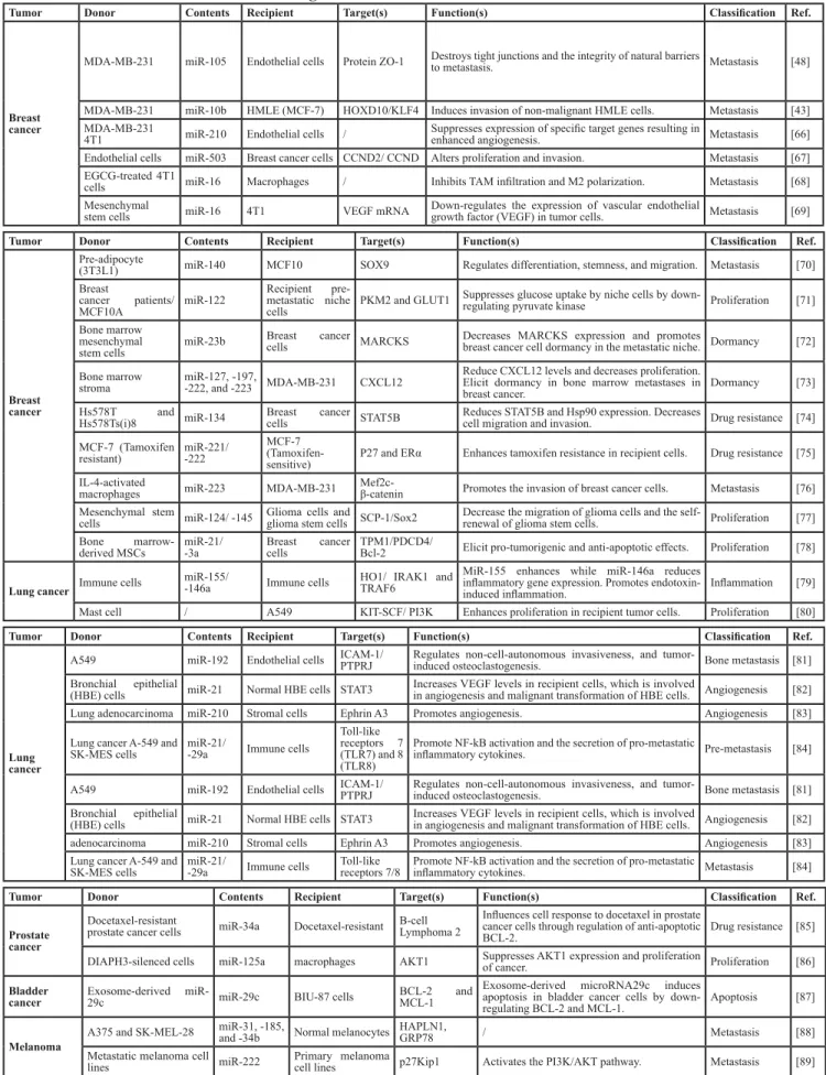
Table 2: Intercellular communication through exosome-derived microRNAs in different cancer models.
Tumor Donor Contents Recipient Target(s) Function(s) Classification Ref.
Breast cancer
MDA-MB-231 miR-105 Endothelial cells Protein ZO-1 Destroys tight junctions and the integrity of natural barriers to metastasis. Metastasis [48]
MDA-MB-231 miR-10b HMLE (MCF-7) HOXD10/KLF4 Induces invasion of non-malignant HMLE cells. Metastasis [43]
MDA-MB-231
4T1 miR-210 Endothelial cells / Suppresses expression of specific target genes resulting in enhanced angiogenesis. Metastasis [66] Endothelial cells miR-503 Breast cancer cells CCND2/ CCND Alters proliferation and invasion. Metastasis [67]
EGCG-treated 4T1
cells miR-16 Macrophages / Inhibits TAM infiltration and M2 polarization. Metastasis [68] Mesenchymal
stem cells miR-16 4T1 VEGF mRNA Down-regulates the expression of vascular endothelial growth factor (VEGF) in tumor cells. Metastasis [69]
Tumor Donor Contents Recipient Target(s) Function(s) Classification Ref.
Breast cancer
Pre-adipocyte
(3T3L1) miR-140 MCF10 SOX9 Regulates differentiation, stemness, and migration. Metastasis [70] Breast
cancer patients/
MCF10A miR-122
Recipient pre-metastatic niche
cells PKM2 and GLUT1 Suppresses glucose uptake by niche cells by down-regulating pyruvate kinase Proliferation [71]
Bone marrow
mesenchymal
stem cells miR-23b
Breast cancer
cells MARCKS Decreases MARCKS expression and promotes breast cancer cell dormancy in the metastatic niche. Dormancy [72]
Bone marrow
stroma miR-127, -197, -222, and -223 MDA-MB-231 CXCL12
Reduce CXCL12 levels and decreases proliferation. Elicit dormancy in bone marrow metastases in
breast cancer. Dormancy [73]
Hs578T and
Hs578Ts(i)8 miR-134 Breast cells cancer STAT5B Reduces STAT5B and Hsp90 expression. Decreases cell migration and invasion. Drug resistance [74]
MCF-7 (Tamoxifen
resistant) miR-221/-222
MCF-7
(Tamoxifen-
sensitive) P27 and ERα Enhances tamoxifen resistance in recipient cells. Drug resistance [75]
IL-4-activated
macrophages miR-223 MDA-MB-231 Mef2c-β-catenin Promotes the invasion of breast cancer cells. Metastasis [76]
Mesenchymal stem
cells miR-124/ -145 Glioma cells and glioma stem cells SCP-1/Sox2 Decrease the migration of glioma cells and the self-renewal of glioma stem cells. Proliferation [77] Bone
marrow-derived MSCs miR-21/-3a Breast cells cancer TPM1/PDCD4/Bcl-2 Elicit pro-tumorigenic and anti-apoptotic effects. Proliferation [78]
Lung cancer Immune cells miR-155/-146a Immune cells HO1/ IRAK1 and TRAF6
MiR-155 enhances while miR-146a reduces inflammatory gene expression. Promotes
endotoxin-induced inflammation. Inflammation [79]
Mast cell / A549 KIT-SCF/ PI3K Enhances proliferation in recipient tumor cells. Proliferation [80]
Tumor Donor Contents Recipient Target(s) Function(s) Classification Ref.
Lung cancer
A549 miR-192 Endothelial cells ICAM-1/ PTPRJ Regulates non-cell-autonomous invasiveness, and tumor-induced osteoclastogenesis. Bone metastasis [81]
Bronchial epithelial
(HBE) cells miR-21 Normal HBE cells STAT3 Increases VEGF levels in recipient cells, which is involved in angiogenesis and malignant transformation of HBE cells. Angiogenesis [82] Lung adenocarcinoma miR-210 Stromal cells Ephrin A3 Promotes angiogenesis. Angiogenesis [83]
Lung cancer A-549 and
SK-MES cells miR-21/-29a Immune cells
Toll-like
receptors 7 (TLR7) and 8 (TLR8)
Promote NF-kB activation and the secretion of pro-metastatic
inflammatory cytokines. Pre-metastasis [84]
A549 miR-192 Endothelial cells ICAM-1/ PTPRJ Regulates non-cell-autonomous invasiveness, and tumor-induced osteoclastogenesis. Bone metastasis [81]
Bronchial epithelial
(HBE) cells miR-21 Normal HBE cells STAT3 Increases VEGF levels in recipient cells, which is involved in angiogenesis and malignant transformation of HBE cells. Angiogenesis [82]
adenocarcinoma miR-210 Stromal cells Ephrin A3 Promotes angiogenesis. Angiogenesis [83]
Lung cancer A-549 and
SK-MES cells miR-21/-29a Immune cells Toll-likereceptors 7/8 Promote NF-kB activation and the secretion of pro-metastatic inflammatory cytokines. Metastasis [84]
Tumor Donor Contents Recipient Target(s) Function(s) Classification Ref.
Prostate cancer
Docetaxel-resistant
prostate cancer cells miR-34a Docetaxel-resistant B-cellLymphoma 2
Influences cell response to docetaxel in prostate
cancer cells through regulation of anti-apoptotic
BCL-2. Drug resistance [85]
DIAPH3-silenced cells miR-125a macrophages AKT1 Suppresses AKT1 expression and proliferation of cancer. Proliferation [86]
Bladder
cancer Exosome-derived miR-29c miR-29c BIU-87 cells BCL-2 MCL-1 and
Exosome-derived microRNA29c induces
apoptosis in bladder cancer cells by
down-regulating BCL-2 and MCL-1. Apoptosis [87]
Melanoma A375 and SK-MEL-28
miR-31, -185,
and -34b Normal melanocytes HAPLN1, GRP78 / Metastasis [88]
Metastatic melanoma cell
An analogous mechanism was discovered in
pancreatic cancer. Exosomes from pancreatic ductal
adenocarcinomas fused with resident cells at the metastatic
location, which included liver Kupffer cells and lung
epithelial cells. In addition, exosome integrin uptake by
resident cells activated Src and altered the expression of
pro-migratory and pro-inflammatory S100 genes, which
are associated with metastasis [52]. Grange et al. reported
that in human renal cell carcinoma, microvesicles released
by CD105 positive tumor-initiating cells promoted
angiogenesis and enhanced lung metastasis [53]. All of
the work we have described is based on
in vitro
studies of
purified TDEs. Whether TDEs perform these functions
in
vivo
is still not clear [54]. Our data indicate that colorectal
cancer-derived exosomes could be used to predict
organ-specific metastasis (unpublished data).
A third mode of communication involves exosomes
derived from normal cells, which can alter the behavior
of tumor cells. Zhang et al. revealed that both human and
mouse tumor cells lost PTEN expression after metastasis
to the brain, but not to other organs. They determined that
PTEN expression was regulated by microRNAs from
brain astrocytes [55]. These results indicate that
organ-specific metastasis is not only determined by TDEs, but
Colorectalcancer HCT-15, SW480 and WiDr miR-21, -192 and -221 HepG2 and A549 /
Regulate the expression of target genes in
HepG2 and A549 cells. May promote various
functions. / [90]
Gastric
cancer Macrophage miR-21 BGC-823 PDCD4
MiR-21 inhibitor-loaded exosomes promote
migration and reduce apoptosis. Metastasis [91] Mesenchymal stem cells miR-221 HGC-27 / Promotes HGC-27 growth and migration. Metastasis [92]
Tumor Donor Contents Recipient Target(s) Function(s) Classification Ref.
Gastric cancer
AZ-P7a Let-7 AZ-521 RAS HMGA2 and Induces tumorigenesis and metastasis. Metastasis [93]
OCUM-2MD3 miR-21 and -1225-5p OCUM-2M /
MiR-21 and miR-1225-5p may prepare a pre-metastatic niche in the peritoneum for the dissemination and colonization of metastatic cancer cells.
Metastasis [94]
Liver cancer
Macrophages miR-142 and -223 Hepatocellular carcinoma cells
(HuH7 and HepG2)
Stathmin-1/
IGF1R Inhibits proliferation of cancer cells. Inhibitor [95]
Huh7 cells miR-122 HepG2 cells IGF1R mRNA Reduced growth and proliferation of recipient HepG2 cells. Inhibitor [96]
Hep3B, HepG2,
and PLC/PRF/5 miR-584 Hep3B, HepG2 and PLC/PRF/5 TGF-β
activated kinase-1
(TAK1)
HCC cell-derived exosomes modulate TAK1
expression and associated signaling. They also
enhance the growth of transformed recipient cells. Proliferation [97]
Cholangiocarcinoma KMBC HuCCT1 and / Mesenchymal stem cells / Enhance MSC migratory capability and expression of alpha-smooth muscle actin mRNA.
Promote the release of CXCL-1, CCL2, and IL-6. Metastasis [98]
Tumor Donor Contents Recipient Target(s) Function(s) Classification Ref.
Hematological malignancies
K562 under
hypoxic
conditions miR-210
Umbilical vein
endothelial cells EFNA3
Exosomal miRNAs derived from cancer cells under hypoxic conditions may affect angiogenic
activity in endothelial cells. Metastasis [99]
LAMA84 miR-126 Endothelial cells CXCL12 and VCAM1 HUVECs with a miR-126 inhibitor reversed the decrease in CXCL12, restores motility and
adhesion in LAMA84 cells. Metastasis [100]
Chronic lymphocytic leukemia (MEC1)
miR-202-3p Human stromal cells c-Fos and ATM Enhances proliferation of recipient cells. Proliferation [101]
K562 cells miR-92a Umbilical endothelial cellsvein Integrin α5 Enhances endothelial cell migration and tube formation. Metastasis [102]
CLL cells miR-21 MSCs endothelial cellsand / Induce differentiation of stromal cells into cancer-associated fibroblasts. Metastasis [103]
Multiple
myeloma cells miR-135b endothelial cells FIH-1
Exosomal miR-135b from HR-MM cells enhances endothelial tube formation under hypoxic conditions via the HIF-FIH signaling pathway.
Metastasis [104]
Neuroblastoma NBL cells miR-21 Human monocytes TLR8-NF-кB / Drug resistance [105] Monocytes miR-155 NBL cells TERF1
Tumor Donor Contents Recipient Target(s) Function(s) Classification Ref.
Ovarian cancer
SKOV-3 let-7 family OVCAR-3 / Exosome release varies between ovarian cancer cell lines and is correlated with invasive potential. Metastasis [106] CP70 miR-21-5p A2780 NAV3 Increases platinum-resistance in A2780 cells. Drug resistance [107]
High-grade ovarian cancer ATF2, MTA1, and ROCK1/2 Endothelial cells / Exosomes derived from high-grade ovarian cancer alter angiogenesis compared to non-high-grade
ovarian cancer cells. Metastasis [108]
The serum of patients with
NPC or TW03 cells
miR-24-3p,
-891a, and
also by specific organ-associated cells. In Table 2 we
have concluded other models of this kind of transferring.
Interestingly, TDEs can fuse with non-parenchymal cells
in various organs, leading to inflammation, anoxia, and
vascularization in the metastatic microenvironment.
Collectively, the data indicate that exosomes play
important roles in pre-metastatic niche formation.
Quantitative proteomic analysis of EVs has resulted
in the identification of proteins in exosomal membranes
and lumens, which may contribute to metastasis
[56-59]. Public databases including Vesiclepedia (
www.
microvesicles.org
) [60], EVpedia (
www.evpedia.info
) [61,
62], and ExoCarta (
www.exocarta.org
) [63] can be used to
identify exosomal proteins. For example, Timothy et al.
searched all of the proteins in the Vesiclepedia database
and used a gene ontology approach to identify regulatory
factors involved in cancer initiation and progression [64].
Additionally, Ostenfeld et al. determined that exosomes
derived from a metastatic human bladder carcinoma
cell line had high expression of vimentin and
hepatoma-derived growth factor in the membrane, and casein kinase
II and annexin A2 in the lumen using quantitative isobaric
tags for relative and absolute quantitative proteomics
[57]. Finally, Lee et al. performed a proteomic analysis to
identify differences in protein expression between MCF-7
and MDA-MB-231 cells, and described a new function
for EDIL3 on EVs, which enhanced cell invasion and lung
metastasis
in vivo
[65].
THE ROLES OF TUMOR-DERIVED
EXOSOMES IN THE ANTI-TUMOR
IMMUNE RESPONSE
Exosomal antigen presentation in the antitumor
immune response
TDEs contain tumor-associated antigens (TAAs)
and major histocompatibility complex (MHC) class I
molecules [110, 111]. Exosomes deliver TAAs to dendritic
cells (DCs), which results in the induction of
antigen-specific CD8 T-cells and tumor rejection in various
prophylaxis and therapeutic murine tumor xenograft
models. Importantly, coupling of TAAs to exosomes
elicited a more efficient antitumor immune response and
had a stronger therapeutic effect compared to subcutaneous
delivery of TAAs in a mouse fibrosarcoma model
[112]. DCs loaded with syngeneic or allogeneic TDEs
stimulated regression of pre-established tumors in mice
[110, 111]. Another approach to exosome-based cancer
immunotherapy involves the applications of DCs pulsed
with tumor peptides [113-116]. Both mouse and human
TAA-loaded DCs can secrete exosomes that express
functional MHC class I, II, and T-cell co-stimulatory
molecules. These exosomes have been reported to
stimulate tumor-specific CD8 T-cells
in vivo
and inhibit
the growth of transplanted tumors in mice. Clinical grade
exosomes were first isolated from DCs and characterized.
They were then evaluated in clinical trials for various
cancers [116-118]. In a phase I clinical trial, exosomes
derived from autologous DCs loaded with MAGE 3
peptides were investigated in stage III/IV melanoma
patients [117]. This was the first trial to demonstrate the
feasibility of large-scale exosome production and safety
of exosome administration. Several other phase I or phase
II clinical trials involving exosome-based regimens have
been initiated in patients with non-small cell lung cancer,
malignant glioma, breast cancer, and gastric cancer [118].
The immunosuppressive effects of tumor-derived
exosomes
TDEs are a major source of tumor antigens.
However, recent studies have shown that TDEs can also
suppress antigen-specific or non-specific anti-tumor
responses. For example, TDEs are rich in FasL, TRAIL,
and galectin-9, which stimulate T-cell apoptosis [119-121].
Moreover, TDEs suppress CD3-ζ chain expression in
T-cells, which prevents activation [122]. They also inhibit
NKG2D-dependent cytotoxicity in natural killer cells and
CD8 T-cells [123].
In addition to the effects of TDEs on T-cells and
natural killer cells, TDEs also modulate antigen-presenting
cells, which controls differentiation. For example, they
can induce monocyte differentiation into myeloid-derived
suppressor cells (MDSCs), which inhibit the antitumor
immune response [123]. The prostaglandin E2, TGF-β,
Hsp70, and miRNAs contained in tumor-derived vesicles
play important roles in monocyte differentiation
[124-126]. Moreover, in tumor-bearing mice, blood-borne
exosomes positive for CD11b could suppress tumor
Ag-specific responses through a MHC Class-II dependent
and MHC Class-I independent mechanism [127]. These
observations suggest that TDEs initially stimulate
CD11-positive antigen presenting cells in the tumor
microenvironment, which then secrete immunosuppressive
MHC Class-II, CD11b-positive vesicles into the
circulation. Exosomes released by human melanoma and
colorectal carcinoma cells impair differentiation of blood
CD14+ monocytes into immune-stimulating DCs and
replace them with highly immunosuppressive MDSCs that
inhibit T-cell functions through secretion of TGF-b [123,
128]. Thus, the MDSC generation, expansion, migration,
and activation are controlled by various mediators of
chronic inflammation [129-132].
favor of immunosuppressive MDSCs at metastatic sites
[129, 130, 133]. MDSCs then induce or support the
functions of regulatory T-cells (Tregs), which have key
roles in the tumor-suppressive microenvironment. In
addition, TDEs can enhance Treg function and inhibit
apoptosis. For example, the expression of
membrane-bound TGF-b and other cytokines, growth factors, and
matrix metalloproteinases from MDSCs are capable of
directing CD4+ T-cells towards the Th2 and Treg lineages
[134-137]. Finally, exosomes have been shown to convert
conventional CD4+FoxP3- T-cells into highly suppressive
and apoptosis-resistant Tregs
via
TGF-b and IL-10, and to
promote Treg expansion [138, 139].
TUMOR-DERIVED EXOSOMES IN
CLINICAL PRACTICE
The concentrations of exosomes cancer patient
blood
The levels of exosomes in blood have been
correlated with tumor development. Logozzi et al.
observed an increase in CD63+ exosomes in melanoma
patients compared to healthy donors [140]. In a study of
lung adenocarcinoma, both the mean exosome and miRNA
concentrations were higher in the lung adenocarcinoma
compared to the control group [141]. The expression
levels of exosomal miR-21 were correlated with advanced
tumor stage, positive lymph node status, and metastasis
in patients with esophageal squamous cell cancer [142].
In oral cancer, Ren et al. suggested that elevated levels
of circulating microparticles were closely correlated with
oral squamous cell carcinoma [143]. Using nanoparticle
tracking analysis, Ayelet et al. determined that the
concentration of exosomes in blood samples from oral
cancer patients was higher than in healthy individuals.
Additionally, they observed differences in the size
distributions of the exosomes and marker expression
between the two groups [144]. It is possible that surgery
or treatment with cancer therapeutics could decrease the
number of exosomes in blood. Indeed, exosomal miRNAs
(a panel) decreased in the blood of patients with lung
squamous cell carcinoma surgery [145]. Thus, exosome
concentrations in blood may help physicians evaluate the
results of surgery and detect relapse in cancer patients.
Applications of exosomes in cancer diagnosis
and as prognostic markers in patients with solid
tumors
Blood-based tumor markers such as cancer antigen
199 and alpha-fetal protein are widely used in cancer
diagnosis. Recently, Melo et al. demonstrated a high
degree of specificity in detecting early pancreatic cancer
through the use of glypican-1 circulating exosomes as
markers [146]. This is just one advantage of exosomes
in detecting early-stage cancers. TDEs can reach cells in
distant organs and drive genetic alterations [36, 93, 147,
148]. Eichelser et al. observed an increase in circulating
exosomal miRNA-373 in receptor-negative breast cancer
patients [149]. Additionally, Huang et al. determined
that miR-1290 and miR-375 were prognostic markers
in castration-resistant prostate cancer [150]. Finally,
Matsumura et al. found that the exosomal miR-19a cluster
expression level in serum was correlated with recurrence
in colorectal cancer [151]. The results of a meta-analysis
suggested that plasma miR-21 may be a reliable and
non-invasive biomarker for colorectal cancer diagnosis [152].
In addition to circulating exosomes, three fecal microRNA
levels were significantly higher in colorectal cancer
patients [153].
New technology has been developed to capture
circulating exosomes [28], which can serve as tumor
markers for personalized diagnostics. The use of exosomes
in liquid biopsy is also currently under investigation [154].
However, as Thery et al. noted, the testing will be more
reliable and less complex if the contributions of exosomes
and exosomal miRNA to cancer progression are elucidated
[155]. In addition, circulating tumor cells contribute to
cancer metastasis [156]. Therefore, a combination of
exosomes and circulating tumor cell detection could
improve the precision of cancer diagnosis.
Circulating miRNAs can be (1) passively
transported out of cells, (2) actively secreted by membrane
vesicles, or (3) actively secreted by complex formation
with lipoproteins (e.g. high-density lipoprotein) and
RNA-binding proteins (e.g. AGO2 and nucleophosmin 1) [157].
Arroyo et al. hypothesized that circulating miRNAs may
not be not restricted to vesicles. Instead, most miRNAs
are associated with circulating Ago2 complexes. Less
than 10% of miRNAs are vesicle-associated, whereas
it is possible that 90% of miRNAs in the circulation
are present in a non-membrane bound form (e.g. in a
ribonucleoprotein complex) [158]. However, Gallo et al.
found that the majority of miRNAs that were detectable
in serum and saliva were concentrated in exosomes.
The differences in results could be explained by lysis of
exosomes during the isolation process [159].
stable in the blood, they are still regarded as non-specific
by-products of cell activity and death. In addition, the
biological functions of miRNAs have not been fully
elucidated [160, 161]. Overall, exosomes and exosomal
miRNAs in blood may be useful markers of early-stage
cancer and may be predictive of prognosis.
Drug delivery
Exosomes can also be used for drug delivery. The
development of nanoformulations has improved the
therapeutic efficacy of drugs. Unfortunately, none of the
nanotechniques avoid toxicity, and the drugs are typically
cleared by the immune system immediately [162].
Exosomes are advantageous in that they can function as
both synthetic nanocarriers and as cell-mediated drug
delivery vehicles [163]. It is generally difficult to deliver
drugs into the brain because of the selectivity of the
blood-brain barrier. However, exosomes are lipid soluble and
can easily cross the blood-brain barrier [164]. There are
at least three ways that drugs can be loaded into exosomes
for delivery: 1) naïve exosomes isolated from parental
cells can be loaded ex vivo, 2) parental cells can be loaded
with a drug, which is then released in exosomes, or 3)
parental cells can be infected/transfected with DNA that
encodes therapeutically active compounds, which are then
released in exosomes [163]. Batrakova et al. first delivered
the enzyme catalase (a large therapeutic protein) to the
brain by loading exosomes extracted from immune cells
with the enzyme [165]. Importantly, exosomes have the
natural ability to home to tumors without eliciting an
immune response. Exosome-encapsulated paclitaxel is
50 times more potent against drug-resistant lung cancer
tumors [166].
CONCLUSIONS
There is compelling evidence for the roles of
exosomes in cancer. Exosomes are distinct from other
EVs. To date, most studies have analyzed mixed EV
populations, and not all EV subgroups have been
characterized. The formation of exosomes is tightly
regulated to ensure content stability and maintain
biological activity. Like seeds from soil (primary tumor),
exosomes may serve as “Trojan Horses”. There are
thousands of miRNAs with potential functions in cancer.
However, significantly fewer are present in exosomes.
The molecular mechanisms underlying the sorting and
release of cellular contents into exosomes are not yet clear.
Because exosomes comprise an information transduction
pathway in cancer, they have the potential to educate and
compel other cell types (e.g. immune cells) to build a niche
suitable for CTC colonization. An increasing number of
clinical tests have been performed to study exosomes,
which contain specific miRNAs or surface proteins. It
is important to note that TDEs are qualitatively different
from those derived from non-cancerous cells. Additional
studies are required to distinguish TDEs from exosomes
secreted by normal cells. Finally, advances in technology
will result in new insights into exosome function and
therapeutic potential.
ACKNOWLEDGMENTS
The work was supported by the National Natural
Science Foundation of China (81472666), the Key
Projects in the National Science & Technology Pillar
Program during the Twelfth Five-year Plan Period
(2014BAI09B07) and the Fundamental Research Funds
for the Central Universities (2016FZA7011).
CONFLICTS OF INTEREST
The authors declare that there are no conflicts of
interest.
REFERENCES
1. Yáñez-Mó M, Siljander PRM, Andreu Z, Bedina Zavec A, Borràs FE, Buzas EI, Buzas K, Casal E, Cappello F, Carvalho J, Colás E, Cordeiro-da Silva A, Fais S, Falcon-Perez JM, Ghobrial IM, Giebel B, et al. Biological
properties of extracellular vesicles and their physiological functions. Journal of extracellular vesicles. 2015; 4.
2. Johnstone RM, Adam M, Hammond JR, Orr L and Turbide
C. Vesicle formation during reticulocyte maturation.
Association of plasma membrane activities with released vesicles (exosomes). The Journal of biological chemistry. 1987; 262:9412-9420.
3. Danielson KM and Das S. Extracellular Vesicles in Heart Disease: Excitement for the Future ? Exosomes and
microvesicles. 2014; 2.
4. Yelamanchili SV, Lamberty BG, Rennard DA, Morsey BM, Hochfelder CG, Meays BM, Levy E and Fox HS. MiR-21 in Extracellular Vesicles Leads to Neurotoxicity
via
TLR7 Signaling in SIV Neurological Disease. PLoS pathogens. 2015; 11:e1005032.5. Winkler CW, Taylor KG and Peterson KE. Location is everything: let-7b microRNA and TLR7 signaling results in a painful TRP. Science signaling. 2014; 7:pe14. 6. Taylor DD and Gercel-Taylor C. MicroRNA signatures
of tumor-derived exosomes as diagnostic biomarkers of ovarian cancer. Gynecologic oncology. 2008; 110:13-21. 7. Théry C, Amigorena S, Raposo G and Clayton A. (2001).
Isolation and Characterization of Exosomes from Cell
Culture Supernatants and Biological Fluids. Current Protocols in Cell Biology: John Wiley & Sons, Inc.). 8. Thery C, Amigorena S, Raposo G and Clayton A. Isolation
supernatants and biological fluids. Current protocols in cell biology / editorial board, Juan S Bonifacino [et al]. 2006; Chapter 3:Unit 3 22.
9. Abramowicz A, Widlak P and Pietrowska M. Proteomic analysis of exosomal cargo: the challenge of high purity vesicle isolation. Molecular bioSystems. 2016;
12:1407-1419.
10. Van Deun J, Mestdagh P, Sormunen R, Cocquyt V, Vermaelen K, Vandesompele J, Bracke M, De Wever O and Hendrix A. The impact of disparate isolation methods for extracellular vesicles on downstream RNA profiling.
Journal of extracellular vesicles. 2014; 3.
11. Rider MA, Hurwitz SN and Meckes DG, Jr. ExtraPEG: A Polyethylene Glycol-Based Method for Enrichment of Extracellular Vesicles. Scientific reports. 2016; 6:23978. 12. Weng Y, Sui Z, Shan Y, Hu Y, Chen Y, Zhang L and
Zhang Y. Effective isolation of exosomes with polyethylene
glycol from cell culture supernatant for in-depth proteome
profiling. The Analyst. 2016.
13. Paolini L, Zendrini A, Noto GD, Busatto S, Lottini E, Radeghieri A, Dossi A, Caneschi A, Ricotta D and Bergese
P. Residual matrix from different separation techniques
impacts exosome biological activity. Scientific reports. 2016; 6:23550.
14. Zeringer E, Li M, Barta T, Schageman J, Pedersen KW, Neurauter A, Magdaleno S, Setterquist R and Vlassov AV. Methods for the extraction and RNA profiling of exosomes. World journal of methodology. 2013; 3:11-18.
15. King HW, Michael MZ and Gleadle JM. Hypoxic enhancement of exosome release by breast cancer cells. BMC cancer. 2012; 12:421.
16. Bonafede R, Scambi I, Peroni D, Potrich V, Boschi F, Benati D, Bonetti B and Mariotti R. Exosome derived from murine adipose-derived stromal cells: Neuroprotective
effect on
in vitro
model of amyotrophic lateral sclerosis.Experimental cell research. 2016; 340:150-158.
17. Lobb RJ, Becker M, Wen SW, Wong CS, Wiegmans AP, Leimgruber A and Moller A. Optimized exosome isolation
protocol for cell culture supernatant and human plasma.
Journal of extracellular vesicles. 2015; 4:27031.
18. Enderle D, Spiel A, Coticchia CM, Berghoff E, Mueller R, Schlumpberger M, Sprenger-Haussels M, Shaffer JM, Lader E, Skog J and Noerholm M. Characterization of RNA from Exosomes and Other Extracellular Vesicles Isolated by a Novel Spin Column-Based Method. PloS one. 2015; 10:e0136133.
19. Xu R, Greening DW, Rai A, Ji H and Simpson RJ. Highly-purified exosomes and shed microvesicles isolated from the human colon cancer cell line LIM1863 by sequential centrifugal ultrafiltration are biochemically and functionally distinct. Methods (San Diego, Calif). 2015.
20. Witwer KW, Buzas EI, Bemis LT, Bora A, Lasser C, Lotvall J, Nolte-’t Hoen EN, Piper MG, Sivaraman S, Skog J, Thery C, Wauben MH and Hochberg F. Standardization
of sample collection, isolation and analysis methods in extracellular vesicle research. Journal of extracellular vesicles. 2013; 2.
21. Gao W, Chen L, Ma Z, Du Z, Zhao Z, Hu Z and Li Q.
Isolation and Phenotypic Characterization of Colorectal
Cancer Stem Cells With Organ-Specific Metastatic Potential. Gastroenterology. 2013; 145:636-646.e635. 22. Tauro BJ, Greening DW, Mathias RA, Ji H, Mathivanan
S, Scott AM and Simpson RJ. Comparison of
ultracentrifugation, density gradient separation, and
immunoaffinity capture methods for isolating human colon cancer cell line LIM1863-derived exosomes. Methods (San Diego, Calif). 2012; 56:293-304.
23. Verweij FJ, van Eijndhoven MA, Middeldorp J and Pegtel
DM. Analysis of viral microRNA exchange
via
exosomesin vitro
andin vivo
. Methods in molecular biology (Clifton, NJ). 2013; 1024:53-68.24. Kalra H, Adda CG, Liem M, Ang CS, Mechler A, Simpson RJ, Hulett MD and Mathivanan S. Comparative proteomics
evaluation of plasma exosome isolation techniques and
assessment of the stability of exosomes in normal human blood plasma. Proteomics. 2013; 13:3354-3364.
25. Yoshioka Y, Kosaka N, Konishi Y, Ohta H, Okamoto H, Sonoda H, Nonaka R, Yamamoto H, Ishii H, Mori M, Furuta K, Nakajima T, Hayashi H, Sugisaki H, Higashimoto H, Kato T, et al. Ultra-sensitive liquid biopsy of circulating extracellular vesicles using ExoScreen. Nature communications. 2014; 5:3591.
26. Musante L, Tataruch D, Gu D, Benito-Martin A, Calzaferri G, Aherne S and Holthofer H. A simplified method to
recover urinary vesicles for clinical applications, and
sample banking. Scientific reports. 2014; 4:7532.
27. Lotvall J, Hill AF, Hochberg F, Buzas EI, Di Vizio D, Gardiner C, Gho YS, Kurochkin IV, Mathivanan S, Quesenberry P, Sahoo S, Tahara H, Wauben MH, Witwer KW and Thery C. Minimal experimental requirements for definition of extracellular vesicles and their functions: a position statement from the International Society for
Extracellular Vesicles. Journal of extracellular vesicles.
2014; 3:26913.
28. Im H, Shao H, Weissleder R, Castro CM and Lee H. Nano-plasmonic exosome diagnostics. Expert review of molecular diagnostics. 2015; 15:725-733.
29. Yoon J, Jo W, Jeong D, Kim J, Jeong H and Park J. Generation of nanovesicles with sliced cellular membrane fragments for exogenous material delivery. Biomaterials. 2015; 59:12-20.
30. Taller D, Richards K, Slouka Z, Senapati S, Hill R, Go DB and Chang HC. On-chip surface acoustic wave lysis and ion-exchange nanomembrane detection of exosomal RNA for pancreatic cancer study and diagnosis. Lab on a chip. 2015; 15:1656-1666.
9:2321-2327.
32. Yu B, Zhang X and Li X. Exosomes derived from
mesenchymal stem cells. International journal of molecular
sciences. 2014; 15:4142-4157.
33. Sokolova V, Ludwig AK, Hornung S, Rotan O, Horn PA, Epple M and Giebel B. Characterisation of exosomes derived from human cells by nanoparticle tracking analysis and scanning electron microscopy. Colloids and surfaces B, Biointerfaces. 2011; 87:146-150.
34. Malik ZA, Kott KS, Poe AJ, Kuo T, Chen L, Ferrara KW and Knowlton AA. Cardiac myocyte exosomes: stability, HSP60, and proteomics. American journal of physiology Heart and circulatory physiology. 2013; 304:H954-965. 35. Valadi H, Ekstrom K, Bossios A, Sjostrand M, Lee JJ and
Lotvall JO. Exosome-mediated transfer of mRNAs and
microRNAs is a novel mechanism of genetic exchange
between cells. Nature cell biology. 2007; 9:654-659. 36. Skog J, Wurdinger T, van Rijn S, Meijer DH, Gainche L,
Sena-Esteves M, Curry WT, Jr., Carter BS, Krichevsky AM and Breakefield XO. Glioblastoma microvesicles transport RNA and proteins that promote tumour growth and provide diagnostic biomarkers. Nature cell biology. 2008; 10:1470-1476.
37. Guescini M, Genedani S, Stocchi V and Agnati LF. Astrocytes and Glioblastoma cells release exosomes
carrying mtDNA. Journal of neural transmission (Vienna,
Austria : 1996). 2010; 117:1-4.
38. Balaj L, Lessard R, Dai L, Cho YJ, Pomeroy SL, Breakefield XO and Skog J. Tumour microvesicles contain retrotransposon elements and amplified oncogene sequences. Nature communications. 2011; 2:180.
39. Thakur BK, Zhang H, Becker A, Matei I, Huang Y, Costa-Silva B, Zheng Y, Hoshino A, Brazier H, Xiang J, Williams C, Rodriguez-Barrueco R, Silva JM, Zhang W, Hearn S, Elemento O, et al. Double-stranded DNA in exosomes: a novel biomarker in cancer detection. Cell research. 2014; 24:766-769.
40. Li Y, Zheng Q, Bao C, Li S, Guo W, Zhao J, Chen D, Gu J, He X and Huang S. Circular RNA is enriched and stable in exosomes: a promising biomarker for cancer diagnosis. Cell research. 2015; 3:82.
41. Zomer A, Maynard C, Verweij FJ, Kamermans A, Schafer R, Beerling E, Schiffelers RM, de Wit E, Berenguer J, Ellenbroek SI, Wurdinger T, Pegtel DM and van Rheenen
J.
In Vivo
imaging reveals extracellular vesicle-mediatedphenocopying of metastatic behavior. Cell. 2015; 161:1046-1057.
42. Melo SA, Sugimoto H, O’Connell JT, Kato N, Villanueva A, Vidal A, Qiu L, Vitkin E, Perelman LT, Melo CA, Lucci A, Ivan C, Calin GA and Kalluri R. Cancer exosomes perform cell-independent microRNA biogenesis and promote tumorigenesis. Cancer cell. 2014; 26:707-721. 43. Singh R, Pochampally R, Watabe K, Lu Z and Mo YY.
Exosome-mediated transfer of miR-10b promotes cell
invasion in breast cancer. Molecular cancer. 2014; 13:256. 44. Neerincx M, Sie DL, van de Wiel MA, van Grieken NC,
Burggraaf JD, Dekker H, Eijk PP, Ylstra B, Verhoef C, Meijer GA, Buffart TE and Verheul HM. MiR expression profiles of paired primary colorectal cancer and metastases by next-generation sequencing. Oncogenesis. 2015; 4:e170. 45. Ji H, Chen M, Greening DW, He W, Rai A, Zhang W and
Simpson RJ. Deep sequencing of RNA from three different extracellular vesicle (EV) subtypes released from the human LIM1863 colon cancer cell line uncovers distinct miRNA-enrichment signatures. PloS one. 2014; 9:e110314. 46. Cha DJ, Franklin JL, Dou Y, Liu Q, Higginbotham JN,
Demory Beckler M, Weaver AM, Vickers K, Prasad N, Levy S, Zhang B, Coffey RJ and Patton JG. KRAS-dependent sorting of miRNA to exosomes. eLife. 2015; 4:e07197.
47. Ostrowski M, Carmo NB, Krumeich S, Fanget I, Raposo G, Savina A, Moita CF, Schauer K, Hume AN, Freitas RP, Goud B, Benaroch P, Hacohen N, Fukuda M, Desnos C, Seabra MC, et al. Rab27a and Rab27b control different steps of the exosome secretion pathway. Nature cell biology. 2010; 12:19-30; sup pp 11-13.
48. Zhou W, Fong MY, Min Y, Somlo G, Liu L, Palomares MR, Yu Y, Chow A, O’Connor ST, Chin AR, Yen Y,
Wang Y, Marcusson EG, Chu P, Wu J, Wu X, et al.
Cancer-secreted miR-105 destroys vascular endothelial barriers to promote metastasis. Cancer cell. 2014; 25:501-515. 49. Peinado H, Lavotshkin S and Lyden D. The secreted factors
responsible for pre-metastatic niche formation: old sayings and new thoughts. Seminars in cancer biology. 2011; 21:139-146.
50. Peinado H, Aleckovic M, Lavotshkin S, Matei I, Costa-Silva B, Moreno-Bueno G, Hergueta-Redondo M, Williams C, Garcia-Santos G, Ghajar C, Nitadori-Hoshino A, Hoffman C, Badal K, Garcia BA, Callahan MK, Yuan J, et al. Melanoma exosomes educate bone marrow progenitor cells toward a pro-metastatic phenotype through MET. Nature medicine. 2012; 18:883-891.
51. Costa-Silva B, Aiello NM, Ocean AJ, Singh S, Zhang H, Thakur BK, Becker A, Hoshino A, Mark MT, Molina H, Xiang J, Zhang T, Theilen TM, Garcia-Santos G, Williams
C, Ararso Y, et al. Pancreatic cancer exosomes initiate
pre-metastatic niche formation in the liver. Nature cell biology.
2015.
52. Hoshino A, Costa-Silva B, Shen TL, Rodrigues G, Hashimoto A, Tesic Mark M, Molina H, Kohsaka S, Di Giannatale A, Ceder S, Singh S, Williams C, Soplop N, Uryu K, Pharmer L, King T, et al. Tumour exosome
integrins determine organotropic metastasis. Nature. 2015;
527:329-335.
53. Grange C, Tapparo M, Collino F, Vitillo L, Damasco C, Deregibus MC, Tetta C, Bussolati B and Camussi G.
Microvesicles released from human renal cancer stem cells stimulate angiogenesis and formation of lung premetastatic
54. Tkach M and Thery C. Communication by Extracellular Vesicles: Where We Are and Where We Need to Go. Cell. 2016; 164:1226-1232.
55. Zhang L, Zhang S, Yao J, Lowery FJ, Zhang Q, Huang WC, Li P, Li M, Wang X, Zhang C, Wang H, Ellis K, Cheerathodi M, McCarty JH, Palmieri D, Saunus J, et al. Microenvironment-induced PTEN loss by exosomal microRNA primes brain metastasis outgrowth. Nature.
2015.
56. Schey KL, Luther JM and Rose KL. Proteomics characterization of exosome cargo. Methods (San Diego,
Calif). 2015.
57. Jeppesen DK, Nawrocki A, Jensen SG, Thorsen K, Whitehead B, Howard KA, Dyrskjot L, Orntoft TF, Larsen MR and Ostenfeld MS. Quantitative proteomics of fractionated membrane and lumen exosome proteins from isogenic metastatic and nonmetastatic bladder cancer cells
reveal differential expression of EMT factors. Proteomics.
2014; 14:699-712.
58. Epple LM, Griffiths SG, Dechkovskaia AM, Dusto NL, White J, Ouellette RJ, Anchordoquy TJ, Bemis LT and Graner MW. Medulloblastoma exosome proteomics yield functional roles for extracellular vesicles. PloS one. 2012; 7:e42064.
59. Choi DS, Choi DY, Hong BS, Jang SC, Kim DK, Lee J, Kim YK, Kim KP and Gho YS. Quantitative proteomics
of extracellular vesicles derived from human primary and metastatic colorectal cancer cells. Journal of extracellular vesicles. 2012; 1.
60. Kalra H, Simpson RJ, Ji H, Aikawa E, Altevogt P, Askenase P, Bond VC, Borras FE, Breakefield X, Budnik V, Buzas
E, Camussi G, Clayton A, Cocucci E, Falcon-Perez
JM, Gabrielsson S, et al. Vesiclepedia: a compendium for extracellular vesicles with continuous community annotation. PLoS biology. 2012; 10:e1001450.
61. Kim DK, Kang B, Kim OY, Choi DS, Lee J, Kim SR, Go G, Yoon YJ, Kim JH, Jang SC, Park KS, Choi EJ, Kim KP, Desiderio DM, Kim YK, Lotvall J, et al. EVpedia: an integrated database of high-throughput data for systemic
analyses of extracellular vesicles. Journal of extracellular vesicles. 2013; 2.
62. Kim DK, Lee J, Kim SR, Choi DS, Yoon YJ, Kim JH, Go G, Nhung D, Hong K, Jang SC, Kim SH, Park KS, Kim OY, Park HT, Seo JH, Aikawa E, et al. EVpedia: a community web portal for extracellular vesicles research. Bioinformatics (Oxford, England). 2015; 31:933-939. 63. Simpson RJ, Kalra H and Mathivanan S. ExoCarta as a
resource for exosomal research. Journal of extracellular vesicles. 2012; 1.
64. Ung TH, Madsen HJ, Hellwinkel JE, Lencioni AM and
Graner MW. Exosome proteomics reveals transcriptional
regulator proteins with potential to mediate downstream pathways. Cancer science. 2014; 105:1384-1392.
65. Lee JE, Moon PG, Cho YE, Kim YB, Kim IS, Park H and Baek MC. Identification of EDIL3 on extracellular vesicles involved in breast cancer cell invasion. Journal of proteomics. 2016; 131:17-28.
66. Kosaka N, Iguchi H, Hagiwara K, Yoshioka Y, Takeshita F and Ochiya T. Neutral Sphingomyelinase 2
(nSMase2)-dependent Exosomal Transfer of Angiogenic MicroRNAs
Regulate Cancer Cell Metastasis. Journal of Biological Chemistry. 2013; 288:10849-10859.
67. Bovy N, Blomme B, Freres P, Dederen S, Nivelles O, Lion M, Carnet O, Martial JA, Noel A, Thiry M, Jerusalem G, Josse C, Bours V, et al. Endothelial exosomes contribute to the antitumor response during breast cancer neoadjuvant
chemotherapy
via
microRNA transfer. Oncotarget. 2015; 6:10253-10266. doi: 10.18632/oncotarget.3520.68. Jang JY, Lee JK, Jeon YK and Kim CW. Exosome derived from epigallocatechin gallate treated breast cancer cells suppresses tumor growth by inhibiting tumor-associated macrophage infiltration and M2 polarization. BMC cancer. 2013; 13:421.
69. Lee JK, Park SR, Jung BK, Jeon YK, Lee YS, Kim MK, Kim YG, Jang JY and Kim CW. Exosomes derived from mesenchymal stem cells suppress angiogenesis by down-regulating VEGF expression in breast cancer cells. PloS one. 2013; 8:e84256.
70. Gernapudi R, Yao Y, Zhang Y, Wolfson B, Roy S, Duru
N, Eades G, Yang P and Zhou Q. Targeting exosomes
from preadipocytes inhibits preadipocyte to cancer stem cell signaling in early-stage breast cancer. Breast cancer research and treatment. 2015; 150:685-695.
71. Fong MY, Zhou W, Liu L, Alontaga AY, Chandra M, Ashby J, Chow A, O’Connor ST, Li S, Chin AR, Somlo G, Palomares M, Li Z, Tremblay JR, Tsuyada A, Sun G, et al. Breast-cancer-secreted miR-122 reprograms glucose metabolism in premetastatic niche to promote metastasis. Nature cell biology. 2015; 17:183-194.
72. Ono M, Kosaka N, Tominaga N, Yoshioka Y, Takeshita F, Takahashi RU, Yoshida M, Tsuda H, Tamura K and Ochiya T. Exosomes from bone marrow mesenchymal stem cells
contain a microRNA that promotes dormancy in metastatic
breast cancer cells. Science signaling. 2014; 7:ra63. 73. Lim PK, Bliss SA, Patel SA, Taborga M, Dave MA,
Gregory LA, Greco SJ, Bryan M, Patel PS and Rameshwar P. Gap junction-mediated import of microRNA from bone marrow stromal cells can elicit cell cycle quiescence in breast cancer cells. Cancer research. 2011; 71:1550-1560. 74. O’Brien K, Lowry MC, Corcoran C, Martinez VG, Daly
M, Rani S, Gallagher WM, Radomski MW, MacLeod RA and O’Driscoll L. miR-134 in extracellular vesicles reduces triple-negative breast cancer aggression and increases drug sensitivity. Oncotarget. 2015; 6:32774-32789. doi: 10.18632/oncotarget.5192.
tamoxifen resistance in recipient ER-positive breast cancer cells. Breast cancer research and treatment. 2014;
147:423-431.
76. Yang M, Chen J, Su F, Yu B, Su F, Lin L, Liu Y, Huang JD and Song E. Microvesicles secreted by macrophages shuttle invasion-potentiating microRNAs into breast cancer cells. Molecular cancer. 2011; 10:117.
77. Lee HK, Finniss S, Cazacu S, Bucris E, Ziv-Av A, Xiang C, Bobbitt K, Rempel SA, Hasselbach L, Mikkelsen T, Slavin S and Brodie C. Mesenchymal stem cells deliver
synthetic microRNA mimics to glioma cells and glioma
stem cells and inhibit their cell migration and self-renewal. Oncotarget. 2013; 4:346-361. doi: 10.18632/oncotarget.868. 78. Baglio SR, Rooijers K, Koppers-Lalic D, Verweij FJ,
Perez Lanzon M, Zini N, Naaijkens B, Perut F, Niessen HW, Baldini N and Pegtel DM. Human bone marrow- and
adipose-mesenchymal stem cells secrete exosomes enriched
in distinctive miRNA and tRNA species. Stem cell research & therapy. 2015; 6:127.
79. Alexander M, Hu R, Runtsch MC, Kagele DA, Mosbruger TL, Tolmachova T, Seabra MC, Round JL, Ward DM and O’Connell RM. Exosome-delivered microRNAs modulate the inflammatory response to endotoxin. Nature communications. 2015; 6:7321.
80. Xiao H, Lasser C, Shelke GV, Wang J, Radinger M, Lunavat TR, Malmhall C, Lin LH, Li J, Li L and Lotvall
J. Mast cell exosomes promote lung adenocarcinoma cell
proliferation - role of KIT-stem cell factor signaling. Cell communication and signaling : CCS. 2014; 12:64.
81. Valencia K, Luis-Ravelo D, Bovy N, Anton I, Martinez-Canarias S, Zandueta C, Ormazabal C, Struman I, Tabruyn S, Rebmann V, De Las Rivas J, Guruceaga E, Bandres E and Lecanda F. miRNA cargo within exosome-like vesicle transfer influences metastatic bone colonization. Molecular oncology. 2014; 8:689-703.
82. Liu Y, Luo F, Wang B, Li H, Xu Y, Liu X, Shi L, Lu X, Xu W, Lu L, Qin Y, Xiang Q and Liu Q. STAT3-regulated
exosomal miR-21 promotes angiogenesis and is involved
in neoplastic processes of transformed human bronchial epithelial cells. Cancer letters. 2016; 370:125-135. 83. Cui H, Seubert B, Stahl E, Dietz H, Reuning U,
Moreno-Leon L, Ilie M, Hofman P, Nagase H, Mari B and Kruger A. Tissue inhibitor of metalloproteinases-1 induces a
pro-tumourigenic increase of miR-210 in lung adenocarcinoma
cells and their exosomes. Oncogene. 2015; 34:3640-3650. 84. Fabbri M, Paone A, Calore F, Galli R, Gaudio E,
Santhanam R, Lovat F, Fadda P, Mao C, Nuovo GJ, Zanesi N, Crawford M, Ozer GH, Wernicke D, Alder H, Caligiuri MA, et al. MicroRNAs bind to Toll-like receptors to induce prometastatic inflammatory response. Proceedings of the National Academy of Sciences of the United States of America. 2012; 109:E2110-2116.
85. Corcoran C, Rani S and O’Driscoll L. miR-34a is an intracellular and exosomal predictive biomarker for response to docetaxel with clinical relevance to prostate
cancer progression. The Prostate. 2014; 74:1320-1334. 86. Kim J, Morley S, Le M, Bedoret D, Umetsu DT, Di Vizio
D and Freeman MR. Enhanced shedding of extracellular
vesicles from amoeboid prostate cancer cells: potential effects on the tumor microenvironment. Cancer biology & therapy. 2014; 15:409-418.
87. Xu XD, Wu XH, Fan YR, Tan B, Quan Z and Luo CL.
Exosome-derived microRNA-29c induces apoptosis of
BIU-87 cells by down regulating BCL-2 and MCL-1. Asian Pacific journal of cancer prevention : APJCP. 2014; 15:3471-3476.
88. Xiao D, Ohlendorf J, Chen Y, Taylor DD, Rai SN, Waigel S, Zacharias W, Hao H and McMasters KM. Identifying mRNA, microRNA and protein profiles of melanoma exosomes. PloS one. 2012; 7:e46874.
89. Felicetti F, De Feo A, Coscia C, Puglisi R, Pedini F,
Pasquini L, Bellenghi M, Errico MC, Pagani E and Care A. Exosome-mediated transfer of miR-222 is sufficient
to increase tumor malignancy in melanoma. Journal of
translational medicine. 2016; 14:56.
90. Chiba M, Kimura M and Asari S. Exosomes secreted
from human colorectal cancer cell lines contain mRNAs, microRNAs and natural antisense RNAs, that can transfer
into the human hepatoma HepG2 and lung cancer A549 cell lines. Oncology reports. 2012; 28:1551-1558.
91. Wang JJ, Wang ZY, Chen R, Xiong J, Yao YL, Wu JH and Li GX. Macrophage-secreted Exosomes Delivering miRNA-21 Inhibitor can Regulate BGC-823 Cell Proliferation. Asian Pacific journal of cancer prevention : APJCP. 2015; 16:4203-4209.
92. Wang M, Zhao C, Shi H, Zhang B, Zhang L, Zhang X, Wang S, Wu X, Yang T, Huang F, Cai J, Zhu Q, Zhu W, Qian H and Xu W. Deregulated microRNAs in gastric cancer tissue-derived mesenchymal stem cells: novel biomarkers and a mechanism for gastric cancer. British journal of cancer. 2014; 110:1199-1210.
93. Ohshima K, Inoue K, Fujiwara A, Hatakeyama K, Kanto K, Watanabe Y, Muramatsu K, Fukuda Y, Ogura S, Yamaguchi K and Mochizuki T. Let-7 microRNA family is
selectively secreted into the extracellular environment
via
exosomes in a metastatic gastric cancer cell line. PloS one. 2010; 5:e13247.
94. Dadras SS, Tokuhisa M, Ichikawa Y, Kosaka N, Ochiya T, Yashiro M, Hirakawa K, Kosaka T, Makino H, Akiyama H, Kunisaki C and Endo I. Exosomal miRNAs from Peritoneum Lavage Fluid as Potential Prognostic Biomarkers of Peritoneal Metastasis in Gastric Cancer. PloS one. 2015; 10:e0130472.
95. Aucher A, Rudnicka D and Davis DM. MicroRNAs transfer from human macrophages to hepato-carcinoma cells
and inhibit proliferation. Journal of immunology. 2013; 191:6250-6260.
curtail its intercellular transfer to ensure proliferation of human hepatoma cells. Nucleic acids research. 2014;
42:7170-7185.
97. Kogure T, Lin WL, Yan IK, Braconi C and Patel T. Intercellular nanovesicle-mediated microRNA transfer: a
mechanism of environmental modulation of hepatocellular
cancer cell growth. Hepatology (Baltimore, Md). 2011; 54:1237-1248.
98. Haga H, Yan IK, Takahashi K, Wood J, Zubair A and Patel T. Tumour cell-derived extracellular vesicles interact with
mesenchymal stem cells to modulate the microenvironment
and enhance cholangiocarcinoma growth. Journal of extracellular vesicles. 2015; 4:24900.
99. Tadokoro H, Umezu T, Ohyashiki K, Hirano T and Ohyashiki JH. Exosomes derived from hypoxic leukemia cells enhance tube formation in endothelial cells. The Journal of biological chemistry. 2013; 288:34343-34351. 100. Taverna S, Amodeo V, Saieva L, Russo A, Giallombardo
M, De Leo G and Alessandro R. Exosomal shuttling of miR-126 in endothelial cells modulates adhesive and migratory abilities of chronic myelogenous leukemia cells. Molecular cancer. 2014; 13:169.
101. Farahani M, Rubbi C, Liu L, Slupsky JR and Kalakonda N. CLL Exosomes Modulate the Transcriptome and Behaviour of Recipient Stromal Cells and Are Selectively Enriched in miR-202-3p. PloS one. 2015; 10:e0141429.
102. Umezu T, Ohyashiki K, Kuroda M and Ohyashiki JH. Leukemia cell to endothelial cell communication
via
exosomal miRNAs. Oncogene. 2013; 32:2747-2755. 103. Paggetti J, Haderk F, Seiffert M, Janji B, Distler U,
Ammerlaan W, Kim YJ, Adam J, Lichter P, Solary E, Berchem G and Moussay E. Exosomes released by chronic
lymphocytic leukemia cells induce the transition of stromal
cells into cancer-associated fibroblasts. Blood. 2015; 126:1106-1117.
104. Umezu T, Tadokoro H, Azuma K, Yoshizawa S, Ohyashiki K and Ohyashiki JH. Exosomal miR-135b shed from hypoxic multiple myeloma cells enhances angiogenesis by targeting factor-inhibiting HIF-1. Blood. 2014; 124:3748-3757.
105. Challagundla KB, Wise PM, Neviani P, Chava H, Murtadha M, Xu T, Kennedy R, Ivan C, Zhang X, Vannini I, Fanini F, Amadori D, Calin GA, Hadjidaniel M, Shimada H, Jong A, et al. Exosome-mediated transfer of microRNAs within the tumor microenvironment and neuroblastoma resistance
to chemotherapy. Journal of the National Cancer Institute.
2015; 107.
106. Kobayashi M, Salomon C, Tapia J, Illanes SE, Mitchell MD and Rice GE. Ovarian cancer cell invasiveness is associated with discordant exosomal sequestration of Let-7 miRNA and miR-200. Journal of translational medicine. 2014; 12:4. 107. Pink RC, Samuel P, Massa D, Caley DP, Brooks SA and
Carter DR. The passenger strand, miR-21-3p, plays a role in mediating cisplatin resistance in ovarian cancer cells.
Gynecologic oncology. 2015; 137:143-151.
108. Yi H, Ye J, Yang XM, Zhang LW, Zhang ZG and Chen YP. High-grade ovarian cancer secreting effective exosomes in
tumor angiogenesis. International journal of clinical and
experimental pathology. 2015; 8:5062-5070.
109. Ye SB, Li ZL, Luo DH, Huang BJ, Chen YS, Zhang XS, Cui J, Zeng YX and Li J. Tumor-derived exosomes
promote tumor progression and T-cell dysfunction through the regulation of enriched exosomal microRNAs in human
nasopharyngeal carcinoma. Oncotarget. 2014; 5:5439-5452. doi: 10.18632/oncotarget.2118.
110. Thery C, Regnault A, Garin J, Wolfers J, Zitvogel L, Ricciardi-Castagnoli P, Raposo G and Amigorena S.
Molecular characterization of dendritic cell-derived
exosomes. Selective accumulation of the heat shock protein hsc73. The Journal of cell biology. 1999; 147:599-610. 111. Andre F, Schartz NE, Chaput N, Flament C, Raposo G,
Amigorena S, Angevin E and Zitvogel L. Tumor-derived exosomes: a new source of tumor rejection antigens. Vaccine. 2002; 20 Suppl 4:A28-31.
112. Zeelenberg IS, Ostrowski M, Krumeich S, Bobrie A, Jancic C, Boissonnas A, Delcayre A, Le Pecq JB, Combadiere B, Amigorena S and Thery C. Targeting tumor antigens to secreted membrane vesicles
in vivo
induces efficientantitumor immune responses. Cancer research. 2008;
68:1228-1235.
113. Zitvogel L, Regnault A, Lozier A, Wolfers J, Flament C,
Tenza D, Ricciardi-Castagnoli P, Raposo G and Amigorena
S. Eradication of established murine tumors using a novel cell-free vaccine: dendritic cell-derived exosomes. Nature medicine. 1998; 4:594-600.
114. Hsu DH, Paz P, Villaflor G, Rivas A, Mehta-Damani A, Angevin E, Zitvogel L and Le Pecq JB. Exosomes as a tumor vaccine: enhancing potency through direct loading of antigenic peptides. Journal of immunotherapy (Hagerstown, Md : 1997). 2003; 26:440-450.
115. Chaput N, Flament C, Viaud S, Taieb J, Roux S, Spatz A, Andre F, LePecq JB, Boussac M, Garin J, Amigorena S, Thery C and Zitvogel L. Dendritic cell derived-exosomes: biology and clinical implementations. Journal of leukocyte biology. 2006; 80:471-478.
116. Viaud S, Thery C, Ploix S, Tursz T, Lapierre V, Lantz O, Zitvogel L and Chaput N. Dendritic cell-derived exosomes for cancer immunotherapy: what’s next? Cancer research. 2010; 70:1281-1285.
117. Escudier B, Dorval T, Chaput N, Andre F, Caby MP, Novault S, Flament C, Leboulaire C, Borg C, Amigorena S, Boccaccio C, Bonnerot C, Dhellin O, Movassagh M, Piperno S, Robert C, et al. Vaccination of metastatic melanoma patients with autologous dendritic cell (DC) derived-exosomes: results of thefirst phase I clinical trial. Journal of translational medicine. 2005; 3:10.