Social attention, emotion, and amygdala volume development in preschool-aged children with autism
Matthew W. Mosconi
A dissertation submitted to the faculty of the University of North Carolina at Chapel Hill in partial fulfillment of the requirements for the degree of Doctor of Philosophy in the
Department of Psychology (Clinical Psychology).
Chapel Hill 2006
Approved By:
© 2006
Abstract Matthew W. Mosconi
Social attention, emotion, and amygdala volume development in preschool-aged children with autism
(Under the direction of Gary Mesibov, Ph.D.)
The social and emotional deficits of autism emerge in childhood and are present throughout the lifetime. Research on early development is limited, due, in large part, to reliance on retrospective parent reports, which are plagued by recall biases, and qualitative rating scales that are less sensitive than dimensional ratings to variation among affected individuals. The present study details the development and application of a novel observational coding system, the Social and Emotional Perspective (SEP), useful for quantifying social and emotional behavior in preschool aged children with autism participating in the Autism Diagnostic Observation Schedule (ADOS). The convergent validity of the SEP was supported by examining the relationship between its items and related subdomains of the ADOS. Comparison of a longitudinal sample of children with autism studied at 18-35 months and 42-59 months with cross-sectional age matched typically developing children indicated that the SEP was sensitive to a range of social and emotional deficits in children with autism. The majority of these deficits were evident at both time points, and two behaviors, looking at others and showing positive emotion, actually became more severely impaired over time in the autism group. The SEP also was used in
conjunction with magnetic resonance imaging (MRI) to examine the association of social and emotional behaviors with the amygdala, a neural structure previously hypothesized to
To Laura. You are the best. Thank you.
TABLE OF CONTENTS Page
LIST OF TABLES ... x
LIST OF FIGURES ………..…..xii
1. Background and Significance ………..1
1.1 Social development in typically developing children and children with autism…....1
1.1.1 Attention to others ….……….………...2
1.1.2 Joint attention ……….………...4
1.1.3 Imitation ……….………...6
1.1.4 Functional and pretend play …...………...7
1.1.5 Emotion and affect ……….………...9
1.1.6 Imitation ……….………...6
1.1.7 Imitation ……….………...6
1.2 Variability in social attention and emotion deficits in autism ……….11
1.2.1 Measuring early social attention and emotion in autism ……….…………...12
1.2.2 A novel observational coding system ……….…………...14
1.3 Why study the neural substrates of autistic behavior? ..…… ……….15
1.3.1 Neuroanatomical abnormalities in autism ..……….…………...17
1.4 The neural basis of behaviors associated with autism ..…… ……….….18
1.5 Amygdala development and its relationship to behavior in primates………….….20
1.5.2 The amygdala in autism ………… ……. ..……….…………....23
1.6 Biological heterogeneity in autism………..25
1.7 The importance of a longitudinal design ………..………..25
1.8 Summary ………...………..26
1.9 Aims and hypotheses ………...27
2. Method ... 30
2.1 Participants ... 30
2.2 Measures …... 32
2.2.1 Cognitive Measures ... 32
2.2.1.1 The Mullen Scales of Early Learning ... 32
2.2.2 Diagnostic Assessments ... 33
2.2.2.1 The Autism Diagnostic Inventory - Revised…..……….33
2.2.2.2 The Autism Diagnostic Observation Schedule - Generic ……… ..33
2.2.3 Behavioral Measures ... 34
2.2.3.1 The ADOS-Social and Emotional Perspective..………….……….34
2.2.3.1.1 Training and reliability .………...………….……….35
2.2.4 MRI Acquisition... 36
2.2.4.1 Image processing………...………….……….37
2.2.4.2 Amygdala segmentation………....………….……….38
2.3 Procedures ….. ... 39
2.3.1 Behavioral coding ... 40
2.3.2 What behaviors do we want to observe? ... 40
2.3.4 What is our unit of observation ... 41
2.4 Data analysis ….. ... 41
3. Results... 47
3.1 Sample characteristics ... 47
3.1.1 Behavioral control and autism group characteristics ... 47
3.2 Aim 1: Development and evaluation of ADOS-SEP ... 47
3.2.1 Item Analysis ………48
3.2.2 Convergent validity ..………49
3.2.3 Divergent validity ……….…………51
3.3 Aim 2: Behavioral comparisons between autism and control groups ... 51
3.3.1 Behavioral development of autism and control groups ………55
3.4 Aim 3: To investigate the relationship between amygdala volume and social and emotional behavior in children with autism …..………58
3.4.1 Amygdala volumes………58
3.4.2 Relationship between amygdala volume, age, IQ, and total brain volume …..58
3.5 Amygdala volume and social items……….59
3.5.1 Social attention and amygdala volume.. …...………59
3.5.2 Looking rate and amygdala volume……… …..60
3.5.3 Social initiation rate and amygdala volume……...………60
3.5.4 Joint attention rate and amygdala volume..……...………61
3.5.5 Remaining social items and amygdala volume..……...………62
3.5.5 Emotion rate and amygdala volume..……….…...………62
3.5.5.1 Emotion change over time by amygdala volume..……….……….63
3.5.5.3 Negative emotion rate and amygdala volume..……...…………...…….64
3.5 Amygdala volume and non-social attention…...……….64
4. Discussion ... 65
4.1 Aim 1: Development of ADOS-SEP... 65
4.2 Aim 2: Examination of the development of behavioral deficits in the autism group…………...………67
4.3 Aim 3: Analysis of the relationship between amygdala volume and social and emotional behavior in autism ... 71
4.3.1 Amygdala volume and social attention ….………71
4.3.1.1 Relationship between amygdala volume and looking at faces…...…….72
4.3.1.2 Relationship between amygdala volume and social initiation.…...…….73
4.3.1.3 Relationship between amygdala volume and joint attention...…...…….74
4.3.2 Amygdala volume and emotion……... ….………76
4.4 Summary of amygdala-behavior findings ... 78
4.5 Limitations ... 79
4.6 Conclusions and future directions ... 81
APPENDIX A: Coding definitions... 84
APPENDIX B: Amygdala protocol ... 91
LIST OF TABLES
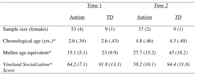
Table 1 Sample data for autism and behavioral control groups………116
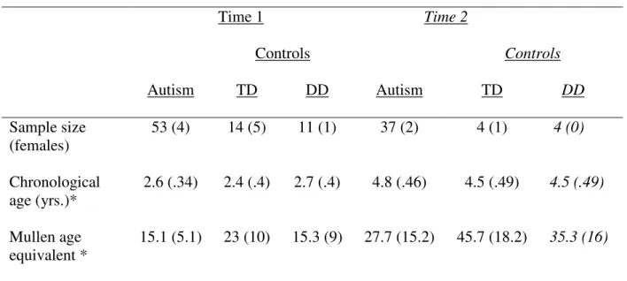
Table 2 Sample data for autism and MRI control groups……….………117
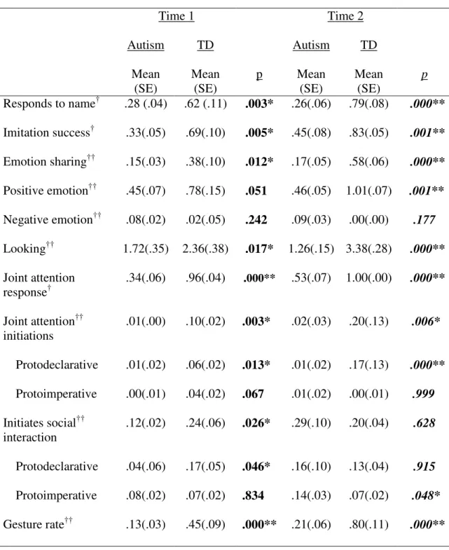
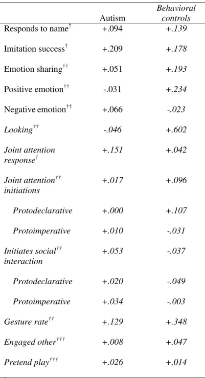
Table 3 Comparison of rates of behavior by group on ADOS-SEP………...……..…118
Table 4 Comparison of proportions of time spent in each behavioral state during the ADOS………119
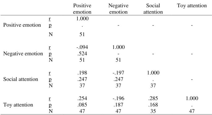
Table 5 Correlation matrix of time 1 ADOS-SEP behavioral variables ………..120
Table 6 Relationship between ADOS-SEP items and ADOS subdomains controlling for age and IQ……….………...121
Table 7 Mean behavioral change over time for autism and control groups……….……….122
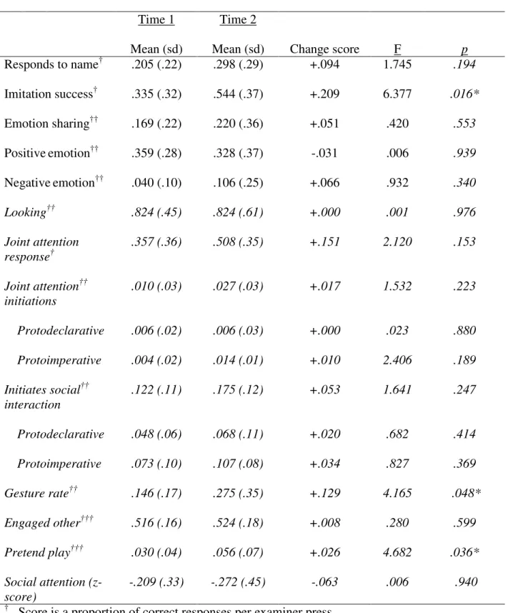
Table 8 Autism group performance at time 1 and time 2 adjusted for IQ.………...……….123
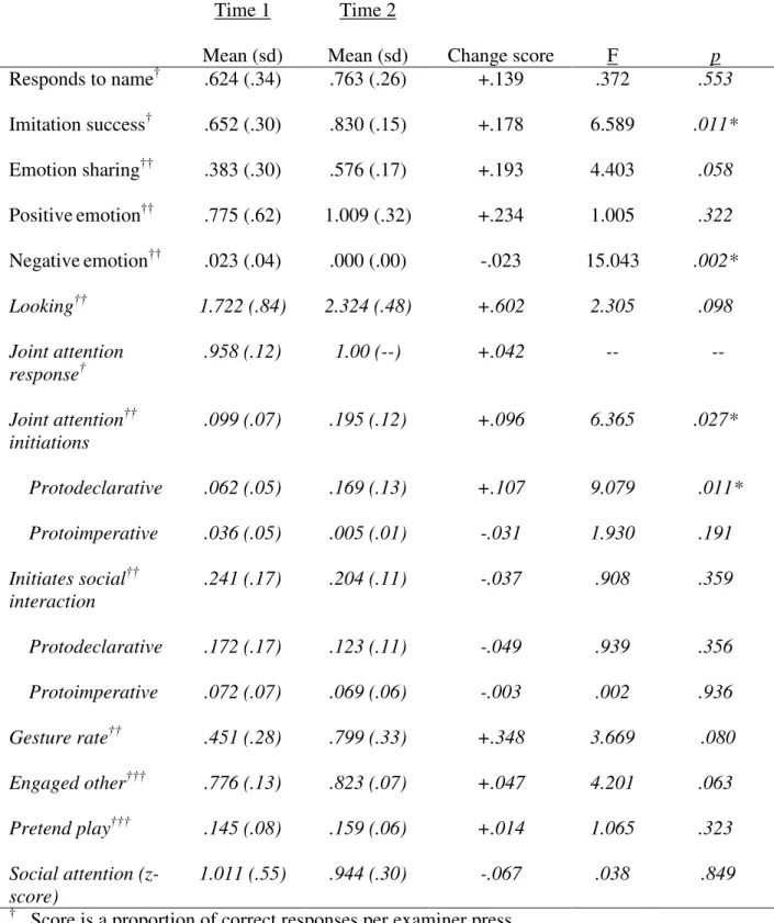
Table 9 Behavioral control group performance at time 1 and time 2 adjusted for IQ…..…124
Table 10 Autism group difference scores over time……….……125
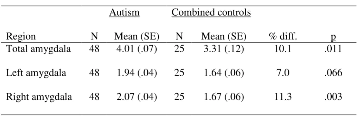
Table 11 Amygdala volumes by group at time 1 adjusted for TBV……….…127
Table 12 Amygdala volumes by group at time 2………... ………...128
Table 13 Relationship between looking rate and amygdala volume in autism group…...129
Table 14 Laterality effects of looking rate and amygdala volume in autism group……...130
Table 15 Interaction of total brain volume and looking rate predicting amygdala volume in autism group……..………..131
Table 16 Relationship between social initiation rate and amygdala volume in autism group…… ……….132
Table 18 Relationship between protodeclarative joint attention and amygdala
volume in autism group……….…134 Table 19 Relationship between protodeclarative joint attention (JA) rate and
amygdala volume over time in autism group ………...135 Table 20 Interaction of protodeclarative joint attention rate and IQ predicting
amygdala volume in autism group ………..………..136 Table 21 Relationship between overall emotion and amygdala volume in autism group ...137 Table 22 Relationship between emotion and amygdala volume over time
in autism group.. ………...138 Table 23 Relationship between positive emotion and amygdala volume in autism group..139 Table 24 Relationship between positive emotion and amygdala volume over time
LIST OF FIGURES
Figure 1 Relationship between looking rate and amygdala volume for children
Background and Significance
Autism is a severe and pervasive neurodevelopmental disorder characterized by impairments in social interaction, communication, and restricted, repetitive behaviors (APA, 1994). Since Kanner (1943) first described autism, clinicians and researchers have
emphasized the core nature of the social deficits in this disorder (Schultz, 2005; Volkmar, 1987). Indeed, social deficits appear to be the most specific features of the disorder, unlike many of the communication and repetitive abnormalities, which are evidenced in other neurodevelopmental and psychiatric disorders (e.g., severe mental retardation without autism, obsessive-compulsive disorder, nonverbal learning disorder). Still, little is known about the onset and development of these social deficits. Previous methods of assessing social behaviors in young children with autism have been limited. For example, researchers have used cross-sectional, rather than longitudinal, samples, and they have used qualitative, rather than quantitative, measurement instruments. Additionally, although autism is
recognized as involving multiple brain regions, little information is available regarding the association of social deficits and established neuroanatomical abnormalities in autism. The present investigation attempts to address each of these limitations.
Social Development in Typically Developing Children and Children with Autism
developments. These skills include eye contact, monitoring eye gaze, joint attention, social imitation, social gestures, and coordinating emotion with others. These early social skill often are deviant in children with autism as early as the first year of life (Baranek, 1999). However, little is known about the onset and development of these deficits.
Attention to others
Typically developing children. Attending to others serves a clear adaptive function, allowing infants to identify security figures in their environment. Attending to others also fosters social opportunities and contributes to social and communicative developments. For example, during the fourth and fifth months of life, infants begin to demonstrate the ability to distinguish between individuals looking at and individuals looking away from them and to coordinate their own gaze with that of others (Baron-Cohen & Cross, 1992). These behaviors are precursors to the developing capacity to monitor shifts in eye gaze, coordinate attention, and share enjoyment with others (Campbell, Walker, & Baron-Cohen, 1995; Baron-Cohen & Ring, 1994). Such skills facilitate reciprocal interaction, language development, and learning opportunities for the developing child.
From the first months of life, children recognize the importance of social stimuli. As early as three weeks of age, newborns spend more time looking at faces than shape- and size-matched non-social objects (Johnson et al., 1991; Morton & Johnson, 1991). At nine weeks, infants preferentially fixate on their caregiver’s face over a stranger’s face (Mauer, 1982), suggesting that they can identify their mothers on the basis of vision alone. Infants also may distinguish their mother’s voice in contrast to a stranger’s (Mehler & Dupoux, 1994).
Mundy, 1989; Wing, 1996). Decreased social attention is a hallmark deficit of autism (Sigman & Kim, 1999) and is one of the earliest recognizable features (Baird et al., 2000). Although the nature of the social abnormalities observed in children with autism tends to change with age, decreased attention to others is one of the most prominent problems evidenced in the first year and is present throughout life (Baranek, 1999; Sigman & Kim, 1999).
Dawson and colleagues (1994; 1999; 2000; 2004) reported that the failure of children with autism to attend to others is the most sensitive feature in distinguishing
between children with and without autism. Moreover, the authors report that this difference is evident as early as one year of age. In retrospective reports, parents rated their children with autism as being less socially engaged during the first two years of life (Wimpory, Hobson, Williams, & Nash, 2000). Retrospective videotape research found that infants who were later diagnosed with autism smiled less with their caregivers (Adrien, et al., 1991) and watched others’ faces less frequently (Kasari, Sigman, & Yirmiya, 1993; Trepagnier, 1998). Yirmiya and colleagues (1999) reported that individuals with autism looked less at the eyes of an interacting adult than did children with Down syndrome and those without
name, pointing, and joint attention. Taken together, these findings indicate that early social inattention is a key component of the social behavioral deficits in autism. Quantifying these deficits and following them over time during early childhood will be important for
understanding their impact on other developments in autism. Joint attention
Typically Developing Children. Joint attention is the coordination of attention with another person towards an object. This skill allows children to communicate with others, learn language, and share interest. Two separate forms of joint attention, protodeclarative and protoimperative, have been identified. Protodeclarative joint attention refers to acts of pointing or showing to share attention to or interest in a stimulus. In contrast,
protoimperative joint attention bids have the primary purpose of obtaining assistance, or serving a non-social, functional goal (e.g., pulling another person’s hand to attain an out-of-reach toy). Both forms of joint attention have been studied in typically developing children and children with autism.
Protodeclarative and protoimperative joint attention have been shown to be predictive of language development (Sigman & McGovern, 2005) and have been conceptualized as early precursors to a developing theory of mind (Jones, Collins & Hong, 1991). Between 9 and 12 months, typically developing children expand their ability to monitor eye gaze, share experiences with others, and develop skill in joint attention (e.g., Feinman, 1982).
Impairments in joint attention or fundamental prerequisites of joint attention may lead to a poverty of social and language opportunities and thus impede a broad range of developments.
Ungerer, & Sherman, 1986; Lewy & Dawson, 1992). Studies have shown that young children with autism are less likely than children with other developmental disabilities to follow shifts in eye gaze and thus to respond to (Sigman & Kasari, 1995) or initiate joint attention (see Mundy, 1995 for a review). These reports have indicated that children with autism show robust impairments in the use of protodeclarative (i.e., socially driven) but not protoimperative (i.e., functionally driven) joint attention (McEvoy et al., 1993; Mundy et al., 1986; Mundy, Sigman, & Kasari, 1994; Mundy & Vaughan, 2001; Mundy & Markus, 1997; Sigman & Mundy, 1989), suggesting that it is the social component of joint attention that is deficient in autism.
items as directed by others. Without being able to participate in such directed exploration, children may be deprived of core language and social learning opportunities. Identifying the onset and nature of joint attention deficits early in development may facilitate novel methods for treating this core impairment in autism.
Imitation
Typically developing children. The capacity to imitate others is important for social and cognitive development. Imitation functions as a method of communication between children and their caregiver(s) and serves to sustain social interactions (Grusec &
Abramovitch, 1982). Imitation also serves to socially connect the infant with others, offering a foundation to share experiences, emotions, and thoughts. An abundance of research has indicated that many socially important phenomena, such as gesturing and facial
communication, are acquired through observation and imitation, without direct instruction. Imitation skill is present in newborns (Meltzoff & Moore, 1989) and develops rapidly throughout the first years of life (Hanna & Meltzoff, 1993). Infants are capable of imitating facial gestures after the first few months of life (Meltzoff & Moore, 1989) and begin to imitate language sounds and actions with objects during the end of the first year (Abravanel & Gingold, 1985). Throughout development, children become better able to imitate a greater complexity of behaviors. This skill requires attention to others and serves the toddler in future interaction and social learning.
Pennington (1991) have suggested that the imitation deficit in autism has not been adequately addressed and that impoverished imitation skills may be fundamental to the social and
emotional impairments akin to this disorder. Nadel and colleagues (1999) expanded upon this hypothesis, suggesting that without properly timed imitative responsiveness, the coordination of sustained dyadic interaction is severely disrupted, impacting the likelihood that future interactions will take place.
Imitation deficits in autism are predictive of social and communication functioning. Stone et al. (1997) reported that imitation of ‘actions on objects’ was impaired in autism and predictive of later play behavior while imitation of ‘body/facial actions’ was predictive of speech development. Rapin (1996) noted that oral-facial actions were particularly difficult for children with autism to imitate. Via its strong association with speech development, emotional sharing, and social engagement (Sigman & Ungerer, 1984; Stone et al., 1997), imitation appears to be critically tied to social development and, perhaps, the social dysfunction seen in autism.
Functional and pretend play
Typically Developing Children. Studies have indicated that play behaviors offer a mechanism for early information gathering (Ruff, 1984), exploration of the classification and properties of objects (Gibson, 1988), and the development of communicative and linguistic skills (McArthur & Adamson, 1996). Research suggests that turn-taking games and social interactions around objects during infancy and the toddler years foster social,
communicative, and emotional growth (Bakeman & Adamson, 1984; Tomasello & Farrar, 1986). Several reports have suggested that the structural rules governing reciprocal
games among the infant and caregiver (Tomasello & Farrar, 1986; Tomasello & Todd, 1983). Early interactions around objects also are associated with the development of skills necessary for relating successfully to other people, including the regulation of affect and theory of mind development (Adamson and Bakeman,
1985; Hobson, 1993). Moreover, it has been proposed that early forms of play may underlie more developmentally advanced cooperative interactions (Gorlitz, 1987).
At about 3-4 months, infants begin to reach for, grasp, inspect, and manipulate novel objects (Trevarthen, 1988). Towards the end of the first year, infants begin to combine
objects in relational play. During this period, infants will put objects together in ways that are socially appropriate and which reflect the functional properties of the items (Vondra and Belsky, 1989). As infants grow older, their functional play becomes progressively elaborate, integrated, and socially directed (Fenson & Ramsay, 1980). Such play may involve face-to-face interaction with the parent, parallel or cooperative play with peers, or social games (e.g., peek-a-boo). By 12 months infants often will be observed using gestures and vocalizations to initiate social-action games with their caregivers (Platt & Coggins, 1990). This play also may begin to include forms of pretense (Leslie, 1987) and imaginative scenarios.
Children with autism. Children with autism engage in fewer face-to-face interactive games. Bernebei et al. (1998) indicated that only a small subset of children with autism engaged in active peek-a-boo when the child takes the initiative, and their involvement in other conventional social games was rare. The authors also found a relative lack of social turn-taking, occurring in less than one-third of children studied.
and Gould (1979) reported that the deficits of autism may be narrowed into three domains – socialization, communication, and imagination, and the American Psychological Association (APA) (1994) has integrated deficits in imagination into autistic preferences for routines and the prevalence of stereotyped and repetitive patterns of behavior. In other words, deficits in imagination may underlie children with autism’s repetitive behaviors while also constituting a primary feature of their early social abnormalities. Many studies have highlighted a poverty of pretend play skills in autism (Blanc et al., 2005; Charman & Baron-Cohen, 1997; Lewis & Boucher, 1988; Jarrold et al., 1993) even after matching for language skills (Baron-Cohen, 1987; Jarrold et al., 1996).
Emotion and affect
Typically Developing Children. Emotion skills, such as recognizing others’ emotions and regulating one’s own emotion, are critical to the overarching task during the 2 to 5 year age period, developing peer relationships (Howes, 1987; Parker & Gottman, 1989). Several emotion skills develop during the preschool years. “Social referencing” is the spontaneous seeking of information from another’s face when presented with a stimulus of uncertain valence (Moore & Corkum, 1994). Typically developing children often regulate their own behavior, particularly in new or uncertain situations, by means of nonverbal and emotional cues provided by adults. For example, a toddler may look towards her mother before deciding to cry after she has bumped her knee. Social referencing is firmly established by 9-12 months in normal development (Feinman, 1982; Moore & Corkum, 1994) and represents an example of early mental state awareness.
children’s ability to discern separate emotions. This skill is crucial to children’s ability to form relationships with others (Parke, 1994; Saarni, 1990) and is predictive of later social competence (Denham et al., 2003). The importance of affective attunement is evidenced by its association with quality of play (Lindsey & Colwell, 2003) and academic achievement (Birch & Ladd, 1997; Ladd, Birch, & Buhs, 1999).
Sharing positive emotions has been shown to be critical to the formation of friendships and attachment relationships (Denham, McKinley, Cochoud, & Holt, 1990; Sroufe, Shork, Motti, Lawroski & LeFreniere, 1984). Conversely, negative affect, especially anger, is problematic in relationship formation and associated with decreased rates of
reciprocal interaction (Denham et al., 1990). Preschoolers who show greater control over negative emotions (e.g., disappointment) are more resilient to stress and experience fewer behavioral and emotional problems (Endriga, Jordan, & Speltz, 2003). Moreover, parent-preschool child interactions that are characterized by decreased mutual positive emotion exchanges and more emotional mismatches predict school aged conduct problems and poor social ability (Cole, Tetti, & Zahn-Wexler, 2003).
Children with autism. Studies of social referencing suggest that children with autism rarely engage in this behavior (Dawson, Meltzoff, Osterling, Rinaldi, & Brown, 1998; Kasari, et al., 1993; Sigman, et al., 1986; Sigman & Kasari, 1995). A failure to seek out emotion information from others regarding novel stimuli or events may result in a mistaken interpretation of the social environment and ultimately, to awkward social behavior,
The coordination of emotion, communicated via changes in facial expression or communicative gestures (e.g., clapping), has been noted to be impaired in school-aged children and adolescents with autism (Buitelaar & van der Wees, 1997; Davies, Bishop, Manstead, Tantam, 1994; Fein, Lucci, Braverman, & Waterhouse, 1992; Sigman, Kasari, Kwon, 1992). Few studies have investigated emotion expression or affective tuning in children with autism younger than age 5 years. Dawson, Hill, Spencer, and Galpert (1990) indicated that 3-6 year-old children with autism were less likely than typically developing children to combine smiles with eye contact in a way that conveyed communicative intent and to smile in response to their mothers' smiles. Mothers of children with autism, in turn, were less likely to smile at their children and showed fewer smiles overall. These findings indicate that children with autism show less positive affect and positive affect sharing. Variability in social attention and emotion deficits in autism
The literature outlined above suggests that social attention and emotional behaviors are impaired in autism and may underlie many of the primary difficulties of individuals with this disorder. Decreased rates of attention to faces, responding to name called, joint attention, imitation, symbolic play, and affect attunement have been found to differentiate preschool age children with autism from their peers (Marcus, Garfinkle, Wolery, 2001; Mundy & Crowson, 1997, Stone & Lemanek, 1992). Despite these findings, a portion of children with autism evidence some early social attention and emotion skills (Dawson & Castelloe, 1993; Wing & Gould, 1979), suggesting variability in skill among children with autism.
substantial portion of children did not exhibit characteristic symptoms. Twenty-four percent of children did not “avoid looking at people in the eyes,” 50% did not “respond to affection by active withdrawal,” and 70-90% more than rarely showed normal modes of relating to others. More recent findings suggest that many children with autism show proximity-seeking behaviors and vocalizations for social attention (Sigman & Mundy, 1989). In fact, Kasari and colleagues (1993) found that when interactions were guided by a familiar adult, many children with autism make as many appropriate social responses as their non-autistic peers. However, when children with autism make social overtures in unstructured social situations, they are brief, poorly integrated, and often do not elicit a response from other children (Stone & Caro-Martinez, 1990). Contrary to the original view of the disorder, basic social
processing and interest in interaction with others are not absent in all children with autism. Behavioral patterns in autism vary not only between individuals but also across development. Experts have called for age-specific norms on the frequency of behaviors exhibited by children with autism (Freeman et al., 1979), but such reference points have not yet been established. Examining the variability in autistic symptoms among affected individuals and over time is important for separating possible phenotypes and developing more individualized treatment approaches. Although the need for developmental studies of social and emotional behavioral deficits in autism is apparent, few studies have been able to quantify these behaviors early in development. Several methodological limitations have hindered progress in this field. These limitations are reviewed below.
Measuring early social attention and emotion in autism
researchers have had difficulty developing laboratory tasks suitable for severely impaired and/or young children with autism. Laboratory tasks that require children to attend to stimuli over a prolonged period or process verbal instructions may limit the ability of young or severely impaired children with autism to participate. Researchers have utilized parent reports for assessing early behavior in autism. However, these reports are problematic because they rely on individuals to recall past episodes or periods of development.
Therefore, retrospective parent reports are subject to biased recall and may lack sensitivity to subtle behavioral abnormalities that might be noted by a trained observer. More recent studies have employed retrospective videotapes that allow expert raters to observe children’s early behavior while being blind to the child’s diagnosis. While this method is an
improvement over parent reports, it also is limited by the lack of standardization across settings in which the child is observed. The development of a structured observation assessment for measuring social attention and emotional behaviors in young children with autism would, therefore, be of significant importance.
The Autism Diagostic Observation Schedule (ADOS) (Lord et al., 2000) is one such tool and is part of the gold standard for evaluating autism. The ADOS is a semi-structured play session in which a trained examiner offers specific presses designed to elicit behaviors that are impaired in autism. The accompanying algorithm of social and communication skills has been shown to adequately differentiate children with autism from children without autism (Lord et al., 2000). The ADOS is administered to children as young as 2 years of age and offers unique opportunities for observing young children engage in a semi-structured play and social interaction session. ADOS sessions probe children’s social attention, joint
system that is not quantitative. Analyzing behavior with the ADOS is limited by a
categorical scoring system in which the differences between adjacent scores are not equally separated. Instead, ratings denote whether a behavior is present, rarely present, or absent. This scoring system is useful for clinical diagnosis only but lacks sensitivity to subtle differences in the severity of autistic children’s impairments. Codes of 2 and 3, each
indicating that a child shows impairment on a particular item, often are collapsed in order to improve reliability between raters. The ADOS and its accompanying algorithms focus on broader features of autism, and do not measure subtle, yet important social attention and emotional skills central to autistic development. The test developers warn against using the ADOS for item level and quantitative analyses. Likewise, other standardized assessments of autism are limited in their sensitivity to autistic features (e.g., Gilliam Autism Rating Scale) (South et al., 2002) or are intended solely for general screening purposes and are not useful for measuring severity (e.g., Childhood Autism Rating Scale). As a result, novel,
dimensional measures for assessing behavior in young children with autism are needed. A novel observational coding system
While the ADOS scoring system currently is not suitable for evaluating the severity of behavior in children, the structured presses do provide important tools for generating a range of social and emotional behaviors. The raw data produced from ADOS sessions may provide sufficient detail for quantitatively measuring selected autistic behavior. In addition, the ADOS is routinely videotaped in many research settings. Therefore, a tool for coding quantitative aspects of autistic behavior from ADOS tapes would be valuable.
revealed considerable variability in several social attention functions among older children with autism and linked these behaviors to the severity of other deficits (e.g., cognitive and communication domains), though results from this study have not yet been extended to early development (Meyer, 2002).
The first aim of the present study, therefore, was to develop and apply an observational coding system focused on assessing the quantity of social attention and emotion behaviors in preschool aged typically developing children and children with autism participating in an ADOS session. Briefly, rates and durations of social and emotional behaviors previously implicated in autism are tabulated. The target behaviors, as reviewed above, have been hypothesized to be central to autistic deficits, but have seldom been quantified early in development or followed over time. The ADOS-Social Emotional Perspective (SEP), is based on observations of children naturally engaged in a semi-structured play period, thereby limiting task demands and providing an opportunity to observe children as they naturally respond to a novel social environment. Moreover, the ADOS-SEP provides continuous values for each item, enabling assessment of impairment severity. The procedures used to develop the ADOS-SEP are detailed in the method section below.
Why study the neural substrates of autistic behavior?
Structural MRI studies have identified multiple regions that are abnormal in autism (for a review, see Cody Hazlett, Pelphrey, & Piven, 2003). However, the vast majority of these reports have failed to find concomitant autism related behaviors or cognitive deficits. Data on associations between MRI findings and behavioral features in autism would be helpful for clarifying which neuroanatomical abnormalities are directly relevant to autism (i.e., whether they are a pathophysiological mediator or involved in an affected neural system) and which abnormalities may reflect an epiphenomenon. Examining brain-behavior linkages and following these associations over time also could offer insight into the
substrates of the primary behavioral features of autism. Separate brain regions and structures develop at different rates. For example, while the amygdala is functional at birth (Kordower et al., 1992), more anterior regions, such as the orbitofrontal cortex, develop gradually over the postnatal period (Overman, 2004). The amygdala and orbitofrontal cortex share intricate connections and both have been implicated in autism, though they are hypothesized to be associated with separate social and emotional behaviors. If amygdala-behavior associations were evident within the first years of life, then this disruption could contribute to the
development of impairments in behaviors believed to be associated with the orbitofrontal cortex. In contrast, amygdala-behavior associations may not be present early in development and may, in contrast, be the result of later occurring orbitofrontal disruptions. Following brain-behavior associations over time could help localize dysfunctional neural systems and map the neural substrates of autistic behavior.
1967). Examining the neural substrates associated with the clinical features of autism could link behavioral characteristics to genetic mechanisms. For example, Hariri and Weinberger (2003) suggested that studying the response of brain systems involved in behavioral
impairments specific to psychiatric disorders may be more informative than studying the behavioral or cognitive sequelae alone. The authors indicated that because the biological impact of genetic variation traverses an increasingly divergent path from cells to neural systems to behavior, interceding at the most basic level is preferable. As genetic
polymorphisms begin to be linked to neural developments, identifying genetic linkages to the biological deficits in autism becomes more possible.
Understanding brain-behavior associations also will be informative for treatment efforts. Defining biological and behavioral phenotypes could facilitate the development of integrated pharmacological and behavioral intervention plans that may be more effective than either treatment method used in isolation. By increasing the number of systems that are treated simultaneously (e.g., biological and behavioral), researchers may begin to outline more potent treatment options.
Neuroanatomical abnormalities in autism
Sparks et al. (2002) noted enlargement in 3-4 year olds. Aylward et al. (2002) reported increased TBV in children up to age 12 years, and Cody-Hazlett et al. (2005) recently reported that TBV increases are present as early as 18-35 months of age. Overall, these studies suggest that patterns of cerebral enlargement in autism are present early in ontogeny, and while cerebral enlargement is not confined to early ages, it may be more robust within the first few years of life.
Novel research also has provided insight into the onset of brain enlargement in autism. Recent evidence suggests that head circumference is enlarged in children with autism beginning at 12 months (Cody-Hazlett et al., 2005). Because head circumference provides an index of overall brain volume early in development, these findings suggest that brain overgrowth begins in the latter part of the first year of life. Related to the finding that head circumference enlargement emerges within the latter part of the first year of life, two independent studies of infant siblings of autistic children have indicated that the core
behavioral features of autism are not detected at 6 months but are observable during the latter part of the first year of life (Landa & Garret-Mayer, in press; Zwaigenbaum et al., 2005). These reports suggesting that the defining features of autism may have their onset in the latter part of the first year of life, combined with findings that the onset of TBV enlargement occurs within the first year of life, suggest that the behavioral and neuroanatomical
characteristics of autism may be temporally related. Research investigating the significance of this relationship is warranted, but these recent findings do suggest that identifying the onset of neuroanatomical abnormalities in autism may help characterize the pathogenesis of this disorder.
As highlighted above, social behavior impairments, including impairments in social attention and emotional behavior, are defining features of autism. Examining the neural substrates associated with autism may be most informative if the core behavioral deficits are targeted. The role of brain regions associated with social and emotional processing has been examined in non-human primate and human lesion studies and structural and functional neuroimaging studies of adults. These investigations have begun to outline a network of brain regions associated with processing, organizing, and responding to social and emotionally salient information.
Brothers (1990) hypothesized a “social brain” inherent in neurotypical individuals, composed of the orbitofrontal cortex, superior temporal gyrus and sulcus, amygdala, and fusiform gyrus, that is sensitive to the features and movements of socially relevant information. Baron-Cohen and Ring (1994) posited a similar model, suggesting that the orbitofrontal cortex, amygdala, and superior temporal sulcus are functionally related in processing the features and movements of biological agents. Finally, McCarthy (1999) identified various nodes of a human face processing system, distinguished by their sensitivity to invariant and dynamic aspects of faces. The first two nodes, comprised of the lateral posterior fusiform gyrus and the anterior ventral temporal cortex, are involved in structural encoding and face memory. The third and fourth nodes are centered in the superior temporal sulcus region and amygdala and are sensitive to facial movements, communicative gestures (e.g., eye and mouth movements), and emotion processing.
disruption within a circumscribed component of this circuitry could grossly affect social and other behavioral and cognitive developments. For example, typical infants’ preference for faces is believed to be mediated by a subcortical visual system that passes information from the retina to the superior colliculus to the pulvinar nucleus of the thalamus and then into the amygdala (Palsey et al., 2004). Congenital abnormality of this subcortical visual system could be responsible for diminished attention to faces and socially relevant stimuli early in autistic development (Maestro et al., 2002). Disruption of this circuitry could be the first insult in a cascade of aberrant neuronal and behavioral developments affecting children with autism. For example, amygdala abnormalities could lead to a failure to orient to salient social stimuli or process the inherent emotional value of social interaction and preclude the
development of reciprocity skills. Indeed, converging animal, human lesion, and
neuroimaging studies have indicated that the amygdala is critical to the immediate processing of social information and to the formation of an appropriate emotional response, and thus may be involved in autism. Studying amygdala development in young children with autism and examining the relationship between amygdala morphometry, social attention, and emotion developments could be an important method for examining the timing of mechanisms that contribute to the social difficulties of individuals with autism. Amygdala development and its relationship to behavior in primates
other monkeys that are looking away or staring directly at them. However, by the end of the first month, infants begin to show differential and appropriate responses to gaze aversion and direct stare (Mendelson, 1982). As refinement of amygdala-visual system projections nears completion during the third month (Bachevalier, Hagger & Mishkin, 1991), infant macaques begin to initiate social interactions with behaviors such as grooming and social play (Suomi, 1990). Researchers also have shown that the timing of the myelination of projections
between the amygdala and other structures involved in motor movements (i.e., basal ganglia), autonomic responding (i.e., hypothalamus), and arousal (i.e., brainstem) coincides with increases in fearful and defensive social behaviors in infant rhesus monkeys (Amaral, 1992; Gibson, 1991). The coincident timing of significant developments in amygdala connectivity and social behaviors suggests that the amygdala plays a central role in the development of these behaviors.
amygdala of rhesus monkeys showed the highest response when animals were presented with faces, particularly faces displaying emotional expressions (Kling, Steklis, & Deutsch, 1979). These data suggest that the amygdala is involved in the evaluation of threat and
reinforcement value of social stimuli and may be involved in the animal’s emotional response. Such assessments of the social environment are likely to be important for the survival of the animal.
Amygdala development in humans
Multiple studies have examined adults with amygdala damage and have shown evidence of its involvement in human perception of facial expressions and emotions
(Adolphs & Tranel, 2003; Adolphs et al., 1994; 1995; 1999; Broks et al., 1998; Breiter et al., 1996; Calder et al., 1996; Morris et al., 1996; Whalen et al., 1998; 2001). Developmentally, the amygdala increases in size, and likely neuronal complexity, throughout childhood and into adolescence (Giedd et al., 1996; McClure et al., 2004; Thomas et al., 2001; Wang et al., 2004).
distinct from those demonstrated by individuals with amygdala damage occurring during adulthood. Specifically, this woman was severely impaired in processing fearful faces and perceiving threat, while demonstrating spared recognition of non-fear emotions.
Taken together, these findings implicate the amygdala in the processing of social stimuli. More specifically, research with non-human primates suggests that amygdala development coincides with social advancements, such as attention to faces, gaze aversion, and affiliative and fearful behaviors (Amaral, 1992; Gibson, 1991; Suomi, 1990). Research with humans suggests that amygdala damage is related to face processing and emotion recognition (Adolphs, 1999). The role of the amygdala in the processing of social stimuli suggests that it could be involved in deficits of social attention and emotionality in children with autism.
The amygdala in autism
Bauman and Kemper (1985) first reported amygdala pathology in a small sample of post-mortem brains of autistic individuals. Studying the post-mortem brains of nine
individuals with autism, the authors reported a pattern of small, immature-appearing neurons and increased neuronal packing density in the amygdala. More recently, functional MRI studies have implicated the amygdala in autistic individuals’ social and emotion processing impairments (Breiter et al., 1996; Morris et al., 1996; Whalen et al., 1998; 2001). For example, Baron-Cohen et al. (1999) reported hypoactivation of the amygdala in individuals with autism relative to controls when they were asked to make mental state judgments from viewing another person’s eyes.
(Schumann et al., 2004; Abell et al., 1999; Howard et al., 2001). Both Sparks et al. (2003) and Cody Hazlett et al. (personal communication) have reported that the amygdala was the only region of interest that was abnormal in autism after correcting for increased TBV. Sparks et al. (2003) also observed that significant volumetric differences existed between children with autism and children with less severe symptoms who were diagnosed with Pervasive Developmental Disorder-NOS. In contrast to these findings, Aylward et al. (1999) and Pierce et al. (2001) each reported reduced amygdala volumes in non-mentally retarded adolescents and young adults with autism.
Although findings from this series of studies are at first glance contradictory (i.e., enlarged in some studies, smaller in other studies), several factors are important to consider. First, the small sample sizes used in several of these studies could make the analyses
Biological heterogeneity in autism
Despite findings implicating the amygdala in autism, results have been somewhat mixed. For example, several studies have reported no volumetric difference or decreased volumes of the amygdala in individuals with autism (Abell et al., 1996; Howard et al., 1994). Inconsistencies among studies may highlight methodological limitations (e.g., small sample sizes, need for longitudinal studies), but also suggest that autism is a biologically
heterogeneous disorder. It may be that the amygdala is not affected in some children with autism, or is affected only at isolated points in development. To study these questions, it is important to examine behaviors related to amygdala volume, and to examine brain-behavior relationships over time.
The importance of a longitudinal design
Autism is a heterogeneous disorder with considerable variance observed across behavioral domains and neurobiological systems among affected individuals. The variability in neurobiology likely has confounded results from previous MRI studies. The development of brain regions and structures in typically developing children follows a non-linear course, further limiting researchers’ ability to define the neural substrates associated with autism. For example, Caviness et al. (1996) demonstrated that several neuroanatomical regions, including the amygdala, exceeded average adult volumes during childhood and reached peak volumes prior to adolescence. The majority of previous MRI studies have been
cross-sectional designs. Longitudinal studies, however, are more sensitive to intra-individual variation and non-linear growth trends.
matter cortical volume development well into the adolescent years, whereas previous cross-sectional studies had concluded that gray matter volume peaked at 4 years of age. This study demonstrated the increased sensitivity of a longitudinal design in the presence of substantial inter-individual variation and non-linear growth. The increased sensitivity of longitudinal studies is particularly important for studies of autism. McGovern and Sigman (2005) recently indicated that the majority of children (40 of 44) in their study that were diagnosed with autism between 2-5 years of age retained that diagnosis in adolescence, but there was substantial variation in the social development of these children. This inter-individual variation would not have been detectable with only cross-sectional comparisons. Summary
Autism is a disorder characterized behaviorally by a pervasive pattern of deficits, including skills involved in social reciprocity. Studies of the early development of these deficits have been limited by a reliance on retrospective parent reports, observations of non-standardized activities, and assessments that incorporate qualitative, but not quantitative, rating systems. The development of a quantitatively scored observational coding system focused on the early social and emotional deficits in autism will be important for following these core features of autism over time.
Both clinical and neuroimaging studies of autism have yielded inconsistent findings. Inconsistencies likely are, in part, due to the heterogeneous nature of autism, a disorder with variable patterns of behavioral and neuroanatomical features amongst affected individuals and over time. Cross-sectional methods often are not sensitive to heterogeneity within groups or non-linear developmental patterns. Longitudinal studies are needed to map brain and behavior developments in autism.
To date, no studies have examined closely the range of social attention deficits found in preschool-aged children with autism, explored these deficits over time, or related them to neuroanatomical markers. This study aims to 1) demonstrate the validity of a novel
observational coding system, the ADOS-SEP, focused on social and emotional behavior in young children, 2) examine the range and development of social and emotional behavior deficits measured by the ADOS-SEP in 18-35 month (time 1) and 42-59 month old (time 2) children with autism, and 3) examine the relationship between these deficits and amygdala enlargement. Such an investigation will provide insight into the nature of the social, emotional, and neuroanatomical abnormalities in autism and has potential implications for diagnostic clarity and future interventions.
Aims and Hypotheses:
1. To quantify social attention and emotional behavior in 2-4 year old children with autism, an observational coding system was developed for application to the Autism Diagnostic Observation Schedule (ADOS). The sensitivity of this coding system (Autism
behaviors, as indicated by normally distributed items. Also, the convergent validity of the coding system was assessed by examining whether item scores were significantly related to corresponding items from the previously validated ADOS. Divergent validity also was assessed by probing the relationship between items from the ADOS-SEP with the restricted, repetitive algorithm of the ADOS.
2a. Social attention and emotional behavior will be abnormal in children with autism relative to controls at both age 2 and age 4 years. Based on previous findings, it is
hypothesized that children with autism will show significant deficits in social attention skills as well as emotional behavior (i.e., decreased positive emotion, increased negative emotion), relative to age-matched typically developing children, after controlling for age, gender, and IQ.
2b. The rate of social attention and emotional behavior development will be reduced in children with autism. Although the absence of longitudinal behavioral data on typically
developing children in the present study precludes comparisons of rates of behavioral development between children with autism and typically developing children, it was hypothesized that 4 year-old typically developing children would show increased rates of social attention and emotion behaviors compared to 2 year-old typically developing children, whereas significant differences between 4- and 2-year-old children with autism would not be observed.
3a. Amygdala volume will be associated with social attention and emotional behavior, but not with non-social attention, in children with autism at both time 1 and time 2.
Method Participants
Participants included 53 children with autism. Children with autism were enrolled between 18 and 35 months of age (i.e., time 1), and 27 of these children were followed up approximately 24 months after their initial assessment (i.e., time 2). A total of 13 children were excluded from analyses due to poor ADOS videotape quality (N= 10) or failure to meet criteria for autism at time 2 (N=3). All remaining children with autism were included in the behavioral and MRI analyses. Eighteen typically developing (TD) children (9 children between 18-35 months, 9 children between 48-66 months) were included in behavioral comparisons (referred to as behavioral controls from this point forward). In addition, 25 control children (14 TD children and 11 developmentally delayed (DD) children) were included in the MRI study (referred to as MRI controls from this point forward) at time 1, and 8 of these children were followed up (4 TD children, 4 DD children) at time 2. The children with DD were included to enrich the MRI control sample with subjects who were comparable to the subjects with autism in cognitive development.
were recruited by mailing index cards to all geographically proximal parents who had newborns. The index cards requested that parents return their contact information if they were interested in their son or daughter participating in future studies of development. Parents who had returned the index card and whose child was in the target age range were contacted by phone. Children whose parents noted concern or diagnosis of any
developmental delay or neurological injury were excluded.
MRI controls were recruited separately. TD children were recruited from community advertisements. DD children were referred from selected regional state Children’s
Developmental Services Agencies in North Carolina. Subjects with DD were referred only if they had no known identifiable cause for their delay (e.g., prematurity, genetic disorder, neurological disorder) and had no diagnosis of a pervasive developmental disorder. Subjects were excluded for having evidence of a medical condition thought to be associated with autism, including fragile X syndrome, tuberous sclerosis, gross central nervous system injury, seizures, and significant motor or sensory impairments.
Subjects with autism were included if they met Autism Diagnostic Interview-Revised (ADI-R) algorithm criteria for autism and obtained ADOS scores consistent with autism. All of the cases met DSM-IV criteria for autistic disorder. Subjects with autism participated in a battery of measures, including the Mullen Scales of Early Learning, the Vineland Adaptive Behavior Scales, behavioral rating scales, and a standardized neurodevelopmental
examination, to exclude subjects with any notable dysmorphology, evidence of
Scale (Schopler et al., 1980) and were excluded if they reached cutoff score for autism (total score > 30). Medical records also were reviewed for any possible evidence of autism or pervasive developmental disorder not otherwise specified, and subjects were excluded from this group for any suggestion of these disorders. Parents of behavioral controls were asked if they had any indication that their child was developmentally delayed or had a history of neurological injury or disorder. Parents of children who did have concerns or whose children currently were receiving evaluations for developmental delays were excluded from the study. Children with a history of neurological injury or disorder also were excluded.
Measures
Cognitive Measures.
The Mullen Scales of Early Learning. (Mullen, 1995) The Mullen provides a comprehensive assessment of language, motor, and visual perceptual abilities for children from birth to 5 years, 8 months. The Mullen was used in the present investigation to obtain a single, reliable, and valid estimate of IQ for participants at both time 1 and time 2. A
limitation of the Mullen is that it has a restricted distribution of standardized scores for lower functioning individuals (i.e., subscales only provide a <50 Standard Score). Several of the lower functioning subjects in the present investigation failed to reach a basal on the Mullen. For this reason, raw scores were used to calculate mental age equivalents for participants at time 1 and time 2. Mental age equivalents were thus used as indices of cognitive
performance to allow for a better description of functioning for the lower functioning autistic and DD subjects. An average mental age equivalent across the four subscales (Visual
children scored in the mentally retarded range. Only 2 children at time 2 managed to score within the average range. In addition, few children scored in between the mentally retarded and average ranges (N=2 at time 2).
Diagnostic Assessments.
The Autism Diagnostic Interview-Revised (ADI)(Lord, Rutter & Le Couteur, 1994). The Autism Diagnostic Inventory (ADI) is a semi-structured parent interview for which items have been shown to be reliable. The accompanying algorithm adequately discriminates autistic individuals from a mental-age matched, non-autistic comparison group based on social and communication behaviors. The ADI-R is administered and scored by trained raters and also is audiotaped for random reliability checks. Administration time is approximately 2 hours.
The Autism Diagnostic Observation Schedule-Generic (ADOS; Lord et al., 2000). The ADOS is a structured observation session designed to elicit social interactions and communication in individuals suspected of having autism. During this session, the examiner engages the child in a broad array of interactions, including calling his/her name, initiating joint attention, requesting that the child imitate actions, free play, pretend play, and
for communication, reciprocal social interaction, play, and stereotyped behaviors and restricted interests. Module 2 of the ADOS is intended for children with phrase speech and contains 14 social events. Twenty-eight behavioral items are provided with four
corresponding algorithm or subdomain scores (i.e., communication, reciprocal social interaction, play, and stereotyped behaviors and restricted interests).
The ADOS is scored by trained raters and videotaped for random reliability checks. The range of scores for each item varies. Typically, children are rated as 0 if no impairments are present and up to 3 if profound abnormalities are observed. Several items are rated only as 0 or 2 (e.g., looking at others, sharing enjoyment), with 0 indicating no abnormality and 2 indicating poor quality. Administration time is approximately 30 minutes for Module 1 and 40 minutes for Module 2.
Behavioral Measures
ADOS Social and Emotional Perspective (ADOS-SEP). The ADOS-SEP was developed by reviewing research addressing the deficits characteristic of young children with autism, observing with Dr. Jerome Kagan videotaped ADOS sessions of children with autism, and conducting pilot analyses with codes hypothesized after viewing ADOS
Cases were included only if children were observable on camera for > 5 minutes. All frequency scores for target behaviors were converted to rates per minute of observable time (e.g., number of social initiations per minute). All duration scores were converted to proportion scores by dividing duration by total observable time and multiplying by 60 (to convert from rate per second to rate per minute). Groups (i.e., autism vs. behavioral controls) were compared on the frequency and duration of ADOS-SEP behaviors using two
multivariate analyses, one for each time point, with group as the predictor variable and age, IQ, gender, and all 2-way interaction terms with group (e.g., group x age, group x IQ) included in the model. Behavioral items were entered as outcome variables. Children were excluded from analyses of social attention rates if they were not presented with the
opportunity to respond to 1) their name being called, 2) joint attention bids, or 3) imitation trials during their ADOS session. Including rates of 0 for children who were not presented with the opportunity to respond to these items would have confounded results, so social attention scores were not computed for these children. A total of 3 children were excluded for this reason.
Training and reliability. Videotaped ADOS sessions were coded for each child with autism. The principal investigator (PI) trained one undergraduate student (rater 2) on coding guidelines. The PI also met with rater 2 weekly for 1.5 hours to review cases, code
individual cases simultaneously, and discuss scoring decisions. Rater 2 initially coded 4-5 cases weekly between meetings to become comfortable with the coding system. Once he was accustomed to the scoring procedures, rater 2 performed a reliability series.
were coded by both raters and examined for reliability and rater drift. For cases in which both raters completed coding, the ratings used for final analyses were randomly selected. Reliability was calculated separately for event and duration codes. For all codes, a tolerance of 3 seconds was used. Intraclass correlation coefficients (ICC’s) were computed to examine reliability between raters. Good reliability was established for both event (.83) and duration (.84) codes.
Intra-rater reliability also was established. For Rater 1, percentage agreement indices for the event codes had a mean of 81% and ranged from 76% to 84%. Kappa coefficients for event codes had a mean of .79 and ranged from .74 to .84. For the duration codes, percentage of agreement ranged between 83-98% for each case, with an average of 89%. A kappa coefficient showed an agreement index of .85.
For Rater 2, percentage agreement indices for the event codes had a mean of 80% and ranged between 74% and 86%. Kappa coefficients for event codes had a mean of .81 and ranged between .72 and .85. For the duration codes, percentage of agreement ranged between 85-93% for each case, with an average of 87%. A kappa coefficient showed an agreement of .83.
MRI acquisition
milliseconds; echo time, 5 milliseconds; flip angle 20°; thickness, 1.5 mm; number of excitations, 1; field of view, 20 cm; and matrix, 256_192.
Subjects with autism and DD subjects were scanned using moderate sedation
(combination of pentobarbital and fentanyl citrate). Physiological monitoring was conducted throughout the scan and recovery. Time 1 TD subjects were scanned without sedation in the evening while sleeping. Time 2 TD subjects received behavioral training within a ‘mock scanner’ prior to their MRI scans. The mock scanner makes use of a decommissioned General Electric MRI scanner bore and RF head coil so that its internal dimensions are the same as a real scanner. It also has a genuine GE front piece so that the simulator appears virtually identical to a real scanner. Speakers placed under the bore are used to play recorded scanner sounds to add to the realism. Children view stimuli using LCD goggles or an LCD monitor in a manner similar to that used in the real MRI scanners. The BIAC simulator scanner is equipped with a video presentation system (LCD goggles or an LCD screen) and a Polhemus FASTRAK head motion sensor. Children are trained using operant conditioning procedures with software that receives input from the head motion sensor and uses this input to direct the operation of a digital video (DVD) player. The DVD (e.g., the child’s favorite movie) is paused (for a user-defined period) whenever the child exhibits head motion above a certain (user-defined) threshold.
All of the scans were reviewed by a pediatric neuroradiologist and were screened for significant abnormalities (e.g., malformations, lesions). Seven scans were excluded from the TBV measurements for motion artifact or acquisition problems.
(Harris et al., 1999). All of the scans were registered along an anteroposterior commissure axis. The coregistered and aligned images were then processed for tissue segmentation using the Expectation Maximization Segmentation (EMS) software (originally developed at the Catholic University of Leuven, Leuven, Belgium (Van Leemput et al., 1999). Reliability and validity of the EMS software has been rigorously examined by the developers and within our laboratory (Park et al., 2001; Styner et al., 2002; Van Leemput et al., 1999). Our initial attempts to apply the existing adult-based EMS template brain atlas provided unsatisfactory results. Therefore, a new pediatric template atlas was created by our laboratory using MRI brain scans of 14 children (comprised of 9 autism, 2 DD, and 3 TD cases that were randomly selected) that first were tissue classified using BRAINS2, which provides semiautomated tissue classification procedures, and then were averaged to create a probabilistic spatial prior template. This resulted in an averaged probabilistic brain atlas that was aligned to each subject’s brain using a linear, affine transformation in a fully automated procedure. After bias estimation, inhomogeneity correction, and nonbrain stripping procedures were
conducted, subject scans were processed with EMS to produce gray matter, white matter, and cerebrospinal fluid (CSF) tissue segmented images for each subject. Total brain volume measures included total gray matter, white matter, and all of the CSF.
amygdala. All tracing was performed using IRIS software, which allows simultaneous data visualization and interaction within three planes (i.e., axial, sagittal, and coronal). Tracing guidelines are included as Appendix B. Briefly, the amygdala is first traced within the coronal view. Tracing begins at the most posterior slice in which the amygdala is visible. The dorsolateral extent of the optic tract is chosen as the starting point, and tracing proceeds counter-clockwise for the right amygdala (left side of computer screen) and clockwise for the left amygdala (right side of the computer screen). A horizontal line is drawn from the
dorsolateral extent of the optic tract to the white matter lateral and adjacent to the amygdala. The lateral border of the amygdala then is traced ventrally to the lateral ventricle or, if no CSF is present, to the dorsolateral extent of the hippocampus. Tracing proceeds medially along the dorsal border of the hippocampus, and then dorsally to the semiannular sulcus. Procedures are repeated in each successive slice moving anterior. The lateral border is re-examined within the axial plane, and the anterior border is re-re-examined within the sagittal plane.
The PI performed all bilateral amygdala segmentations, blind to diagnosis, using guidelines for tracing the amygdala that have been validated and published (Schumann et al., 2004). The PI attained good inter-rater reliability with the authors of the protocol (Right amygdala =.94, Left amygdala=.89) and also established good intra-rater reliability (Right amygdala=.98, Left amygdala=.98). The PI of the present study also established good consistency on a set of pediatric images (Right amygdala =.96, Left amygdala =.92). Procedures
ADOS was administered to children with autism who were enrolled in the MRI study at both time 1 and time 2. Each session was videotaped in order to re-examine initial coding
decisions. The development and validation procedures for the ADOS-SEP are described below.
Behavioral Coding
The ADOS-SEP was developed according to guidelines for developing new observational coding systems presented by Floyd, Baucom, Godfrey, and Palmer (1998). Floyd and colleagues suggested that three questions should be considered prior to
constructing an observational coding system: (a) “What behaviors do we want to observe?” (b) “What are our code categories?” and (c) “What is our unit of observation?” Below is a discussion regarding how each of these questions was addressed during the construction of the ADOS-SEP.
“What behaviors do we want to observe?” The present observational coding system is intended to examine the quantity and quality of social and emotion behavior among children with autism during a structured play session (i.e., the ADOS). Based on
observations of videotaped ADOS sessions, the PI first generated a list of social and emotion behaviors exhibited by TD children within the target age range (i.e., between 18-59 months). Next, a review of the relevant literature on social and emotional development in TD children and children with autism was conducted, and the list was expanded. Finally, the PI
corresponded with an expert on early behavioral development (Dr. Jerome Kagan) and the list of target social and emotional behaviors was finalized.
of possible behaviors were defined so that they could be counted for frequency of occurrence or examined for duration.
“What is our unit of observation?” Coders watched videotaped ADOS sessions while scoring the frequency of individual events or the duration of a given behavioral state (e.g., playing with a toy, engaged, disengaged). In order to operationalize each behavior that should be recorded, specific definitions were developed (see Appendix A).
After behaviors were labeled and defined, a coding manual was written. The manual contains (a) a list of all codes, (b) a descriptive definition for each code, and (c) examples of behaviors that represent each code. The PI trained one undergraduate student coder on the use of the coding system. The student viewed a series of practice videotapes both with the principal investigator and independently. All scoring was reviewed by the principal investigator and, when the student appeared to be consistent, a reliability series was
performed consisting of five cases which previously had been scored by the PI. The student scored these tapes on two independent occasions so that inter- and intra-rater reliability could be assessed.
Data analysis