© 2019 by the Serbian Biological Society How to cite this article: Zhang L, Li J, Zha X. Identification and functional 639 analysis of a testis‑biased gene encoding serine/arginine‑rich protein in silkworm, Bombyx mori. Arch Biol Sci. 2019;71(4):639‑45.
Identification and functional analysis of a testis‑biased gene encoding serine/
arginine‑rich protein in silkworm,
Bombyx mori
Li-Ying Zhang1, Juan Li1,4 and Xing-Fu Zha1,2,3,*
1State Key Laboratory of Silkworm Genome Biology, Biological Science Research Center, Southwest University, Chongqing
400715, China
2Chongqing Key Laboratory of Sericultural Science, Southwest University, Chongqing 400715, China
3Chongqing engineering and technology research center for novel silk materials, Southwest University, Chongqing 400715, China 4School of Life, Southwest University, Chongqing 400715, China
*Corresponding author: [email protected]
Received: June 16, 2019; Revised: July 24, 2019; Accepted: July 29, 2019; Published online: August 2, 2019
Abstract: Spermatogenesis is a fundamental process in sexual reproduction. In this study, we cloned a 716-bp cDNA of a testis-biased gene in Bombyx mori, named as BmRS-TS, which encodes a polypeptide of 164 amino acids, containing 26.7% arginine and serine residues. Sequence similarity analysis showed that BmRS-TS is a lepidopteran-specific gene. Results of RT-PCR and Western analysis revealed that BmRS-TS was expressed predominantly in the testis. Immunohistochemistry assay showed that the BmRS-TS protein was mostly located in primary spermatocytes. Moreover, knockdown of BmRS-TS
by RNA interference (RNAi) showed that the morphology of the mature sperm was abnormal and that sperm bundles were broken up. Our results suggest that BmRS-TS plays an important role in silkworm spermatogenesis and provide some clues for understanding the mechanism that underlies spermatogenesis, which can be used as a reference for other lepidopterans.
Keywords: silkworm; spermatogenesis; serine/arginine-rich protein; lepidopteran-specific gene; RNAi
INTRODUCTION
The silkworm, Bombyx mori (B. mori), is not only of considerable economic value, but it also serves as a model insect for Lepidoptera. Understanding the process of spermatogenesis in B. mori is important for the control of lepidopteran pests by the sterile-male technique [1]. The gonads of male B. mori consist of two testes that are connected to each other by sper-miducts. Both testes contain four compartments, each of which is surrounded by thin connective tissue. The testicular follicles have a long cavity and at the apex, germ cells develop into mature sperm [2].
Spermatogenesis is a continuous and precisely con-trolled process involving many genes that are expressed in male sperm cells [3-5]. Functional and morpho-logical changes during spermatogenesis from primary spermatocytes to mature sperm are highly complex. They include meiotic division, axoneme elongation, nebenkern formation, mitochondrial modification and
acrosome development, and have been examined ex-tensively in B. mori [6-8]. Spermatogenesis in B. mori occurs throughout larval and adult life, from spermato-gonia in newly hatched silkworms to mature sperm in the moth, which has been studied at the ultrastructural level [6,8]. However, the mechanisms that regulate cel-lular differentiation during testis development in the Ordo Lepidoptera are poorly understood [9,10]. To date, researchers have identified only a few genes that are involved in silkworm spermatogenesis; they include β2 tubulin, testis-specific tektin, Aha1, BmTGIF, BmAly, Maelstrom and two genes encoding for adenine nu-cleotide translocase [11-17]. Recently, the homolog of Drosophila Sex-lethal gene was reported to determine dimorphic sperm formation in the silkworm [18].
expres-sion levels in different tissues of B. mori. Additionally, a polyclonal antibody prepared against BmRS-TS pro-tein was used to examine the distribution of BmRS-TS in testis and germline cells. Finally, we assessed the effects of BmRS-TS knockdown on the phenotype of the mature sperm by RNAi.
MATERIALS AND METHODS Insect material
The silkworm strain Dazao was preserved at the Gene Resource Library of Domesticated Silkworm (South-west University, Chongqing, China). Larvae were raised with fresh mulberry leaves at 25±2°C under a 12 h light-dark photoperiod.
Bioinformatics analysis and cloning of BmRS-TS
The genome sequence of B. mori has been completed and published [19,20]. Xia et al. [21] reported genome-wide gene expression profiles in multiple tissues of the domesticated silkworm based on microarray analysis. Using the microarray data, we found a gene (probe ID: sw07075) that showed testis-biased expression. Inter-estingly, the predicted protein sequence of the gene was enriched in Arg and Ser amino acid content. We called the gene BmRS-TS. Some properties of BmRS-TS were analyzed using online programs (http://www.expasy. org/), including open reading frame (ORF) search, nu-cleotide sequence translation and both isoelectric point and molecular weight prediction. Signal peptide and protein domain prediction were accomplished using the SMART program (http://smart.embl-heidelberg. de). Homologs in other species were screened by BLASTX search using the BmRS-TS nucleotide se-quence as a query in the nr database of GenBank.
RT‑PCR of BmRS-TS in different tissues
On the third of the fifth instar, total RNA isolated from the head, epidermis, midgut, fat body, Malpighi-an tubule, silk glMalpighi-and, testis Malpighi-and ovary tissues of Dazao was reverse transcribed into cDNA. TransScript One-Step gDNA Removal and cDNA Synthesis SuperMix (TransGen, China) was used to prepare cDNAs ac-cording to the manufacturer’s instructions. Then, cDNAs were synthesized by reverse transcription
us-ing 2 µg total RNA as template. Total protein from various tissues of Dazao on the 3rd day of the fifth
in-star, including the fat body, midgut, gonad, head, silk gland and epidermis, was extracted and homogenized in 10 mM phosphate-buffered saline (PBS; pH 7.4) on ice. The homogenates were centrifuged at 10000×g at 4°C for 10 min and the supernatants were collected.
RT-PCR was performed to confirm the charac-teristics of gene expression in different tissues using customized primers. PCR was carried out according to the following program: one cycle at 95°C for 4 min; 30 cycles at 94°C for 40 s, 56°C for 45 s and 72°C for 30 s and one final cycle at 72°C for 10 min. Products were then analyzed by agarose gel electrophoresis. (All primers used in these experiments are listed in Sup-plementary Table S1). Relative amounts of BmRS-TS mRNA were normalized to those of B. mori actin 3, which was used as an internal control.
Western blotting
incubated with secondary antibody, horseradish per-oxidase conjugated goat anti-rabbit IgG (1:20,000; Be-yotime, China), in TBST for 1 h at room temperature. Finally, the blots were washed five times with TBST. Immune activity was visualized using the Chemi Scope series with Clarity Western electrochemilumi-nescence (ECL) substrate (Bio-Rad, Hercules, CA, USA). The measured values for anti-BmRS-TS were normalized to those of α-tubulin.
Immunohistochemistry
PBS was administered for 5 min by in vitro methods to testes excised on the 3rd day of the 5th instar larvae,
and fixed in 4% formaldehyde in PBS for 12 h at 4°C. Fixed samples were dehydrated by soaking in a series of 70%, 80%, 90% and 95% ethanol solutions at room temperature, followed by 100% xylene at room tem-perature, twice for 30 min. Samples were embedded in 50% paraffin in xylene at 60°C for 1 h, and then 60°C for 1 h in 100% xylene. Paraffin blocks were sectioned at a thickness of 5 μm using a microtome (RM2235C-CWUS, Leica, Germany), and sections were mounted on glass slides, then placed at 65°C for 1 h. Paraffin was removed by soaking in xylene. The slides were rehydrated in a series of 100%, 90%, 80% and 70% ddH2O at room temperature for 10 min per step. To remove intrinsic peroxidase activity and for antigen retrieval, mounted sections were treated with 10% H2O2 in methanol for 10 min and slices were boiled in sodium citrate buffer solution (pH 6.0) for 5 min, then allowed to cool before washing with PBS. Specimens were incubated with anti-BmRS-TS (1:800) antibody at room temperature for 1.5 h. Excess antibodies were washed with PBS 5 times, followed by incubation with cyanine-3 (Cy3) dye-conjugated goat anti-rabbit IgG (1:500; Invitrogen, Carlsbad, CA, USA) at room tem-perature with the necessary PBS washes. Nuclei were stained with 4',6-diamidino-2-phenylindole (DAPI).
RNAi and organism mashing
The BmRS-TS-specific double-stranded-RNA that was amplified by specific primers with a linker T7-TAATACGACTCACTATAGGGAGAT (Supplementa-ry Table S1) was synthesized using a Ribo MAX Large Scale System-T7 (Promega, Madison, WI, USA). A total of 30 silkworm prepupa were injected with 40
μg BmRS-TS-specific double-stranded-RNA, DsRed and double distilled H2O per 10 individuals at each prepupal stage. DsRed is the gene encoding for Disco-soma sp. red fluorescent protein. DsRed dsRNA was used as the control group. The expression of BmRS-TS in the testes of injected prepupa was estimated using quantitative real-time PCR (qPCR) 48 h later. Additionally, the wild-type group and DsRed group were tested. The rest of the experimental and control group insects were mashed until the organisms split, after which tissues were immediately observed using a microscope (Olympus U-HGLGPS, Japan). For qPCR, total RNA was extracted using TRIzol® reagent (In-trovigen, USA) following the manufacturer’s instruc-tions, and the first strand of the cDNA was synthe-sized using Moloney murine leukemia virus (MMLV) reverse transcriptase according to the manufacturer’s instructions (Promega, USA). The qPCR reactions were run on an ABI7500 Real-Time PCR machine (Applied Biosystems, USA) with SYBR® Premix Ex Taq™II (TaKaRa, Japan). The eukaryotic translation initiation factor 4A (silkworm microarray probe ID sw22934) was used as the internal control [22]. (Prim-ers used for qPCR are listed in Supplementary Table S1). We used the 2−ΔΔCt method to analyze data.
RESULTS
Sequence analysis of BmRS-TS
The cDNA of BmRS-TS was cloned and sequenced. The full-length sequence was 716 bp, consisting of an ORF of 498 bp, a 5’-untranslated region (UTR) of 182 bp and a 36 bp 3’-UTR sequence (Fig. 1). The se-quence of the B. mori BmRS-TS gene was submitted to GenBank (Acc. No.: KM884956). The ORF encoded a polypeptide of 164 amino acids with a calculated mo-lecular weight of ~18.3 kDa. The predicted amino acid sequence of BmRS-TS showed that it was enriched in arginine and serine (26.8%). Interestingly, the protein contained a RSRSRS di-peptide repeat that is charac-teristic of RS protein family members.
and the farthest with Heliothis virescens hypothetical protein B5V51_4589 (E value 4.00E-7) (Table 1). All species belong to Lepidoptera, indicating that BmRS-TS is a lepidopteran-specific gene.
Expression profiles of BmRS-TS
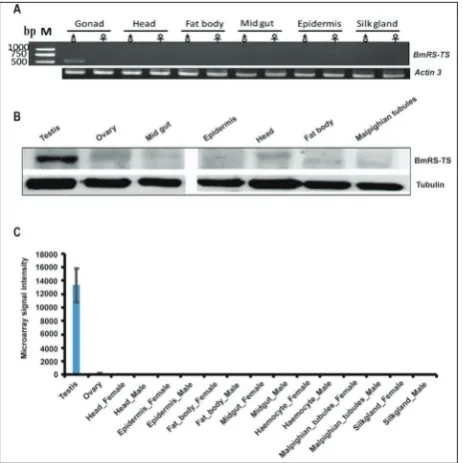
We analyzed the expression profile of BmRS-TS in different silkworm tissues on the 3rd day of the 5th
in-star by RT-PCR. A PCR product with the expected
498-bp fragment size was detected in testis, but not amplified from the other tissues examined, including the epidermis, silk gland, head, fat body, midgut and female gonad (Fig. 2A).
Additionally, Western blotting revealed that testis extracts reacted with rabbit anti-BmRS-TS serum and formed a single band of about 18.3 kDa (Fig. 2B), which corresponded to the molecular mass predicted for BmRS-TS CDS. These findings sug-gested that the protein encoded by BmRS-TS was expressed predomi-nantly in the testis of B. mori. Based on genome-wide microarray expres-sion data of tissues [21], BmRS-TS was also highly and significantly expressed in the testis of B. mori (Fig. 2C).
Cellular localization of BmRS‑TS
Testes of the 5th instar are about 2.5
mm in length, with a middle section of 1.5 mm and a thickness of 0.8-1.0 mm. Spermatocytes are located at the apical part of each compartment of the testes (Fig. 3). Cysts of primary spermatocytes are located at the most peripheral region of the middle zone. After division of primary spermato-cytes by meiosis, each round sper-matid of cyst began to elongate, af-ter which the period of deformation began. Immunofluorescence analysis showed that BmRS-TS protein was most abundant in developing sper-matogonia, as well as evenly abundant in the other types of cells. The BmRS-TS signal reached a peak in spermatogonia and primary spermatocytes, while its decrease was detected during meiosis (Fig. 3).
Fertilization rate after RNAi
To investigate the role of BmRS-TS in sperm develop-ment, RNAi assay was carried out to reduce BmRS-TS expression in male individuals. We found that
BmRS-Fig. 1. Nucleotide and predicted amino acid sequence of BmRS-TS. The arginine and serine (RS) residues are marked with a triangle. The numerical positions of the nucleotide and amino acid sequences are shown on the left. ORF of BmRS-TS encodes a polypeptide of 164 amino acids, of which there are 44 serine/arginine residues (26.8%). The protein contains a RSRSRS triple di-peptide repeat that is characteristic of the RS protein family members.
Table 1. All homologs of the BmRS-TS gene in other species determined by BLASTX search in GenBank.
Acc. Number E valuea Annotation Organism Order
XP_028036720.1 6.00E-51 uncharacterized protein
LOC114247851 mandarinaBombyx Lepidoptera XP_026750535.1 2.00E-15 uncharacterized protein
LOC113511134 mellonellaGalleria Lepidoptera XP_028165190.1 9.00E-13 uncharacterized protein
LOC114356302 furnacalisOstrinia Lepidoptera XP_022818713.1 2.00E-11 uncharacterized protein
LOC111351155 Spodoptera litura Lepidoptera
PZC74609.1 4.00E-11 hypothetical protein
B5X24_HaOG207627 Helicoverpa armigera Lepidoptera
RVE41883.1 7.00E-10 hypothetical protein
evm_013457 suppressalisChilo Lepidoptera XP_026728482.1 8.00E-08 uncharacterized protein
LOC113494369 Trichoplusia ni Lepidoptera
PCG69041.1 4.00E-07 hypothetical protein
B5V51_4589 Heliothis virescens Lepidoptera a E value is the number of distinct alignments that are expected to occur in a database search
Fig. 3. Immunofluorescent images of BmRS-TS expression in testis at day 3 of the 5th instar. Immunostaining was performed
with rabbit anti-BmRS-TS antibody, followed by treatment with Cy3-conjugated goat anti-rabbit IgG. Nuclei of testis were treated with DAPI (blue) and examined under an inverted fluorescence microscope. From top to bottom, DAPI-treated nuclei, red fluo-rescence for Cy3-treated BmRS-TS protein, and the merge images are shown. Primary spermatocytes showed prominent BmRS-TS expression with strong DAPI labeling, overlaid with Cy3 staining. The red arrow indicates the location of BmRS-TS.
Fig. 2. (A) Semi-quantitative RT-PCR analyses were performed us-ing cDNA from testis, ovary, head♂, head♀, fatbody♂, fatbody♀, midgut♂, midgut♀, epidermis♂, epidermis♀, silk gland♂, and silk gland♀ tissues of Dazao at the 3rd day of the 5th instar. (B)
Detec-tion of the protein levels of BmRS-TS. Western blot was performed to detect BmRS-TS with anti-BmRS-TS antibody (1:2500), with α-tubulin used for the internal control. (C) Microarray-based ex-pression analysis of BmRS-TS in different tissues of the silkworm at the 3rd day of the fifth instar. Microarray data were downloaded
from the previous study. The probe ID for BmRS-TS is sw07075.
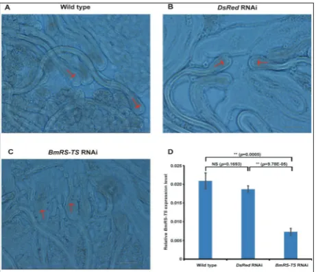
TS expression level was dramatically reduced in the BmRS-TS RNAi group which was transfected with dsRNAs targeting BmRS-TS (Fig. 4D). After RNAi, testes of the silkworm were squashed on a slide and observed under a microscope. We noticed that the morphology of the sperms in the BmRS-TS RNAi group was different compared with that of the wild-type group and the DsRed RNAi group (Fig. 4A, B, C). The sperms were abnormal, and each sperm bun-dle seemed fragmentary (Fig. 4C). Additionally, we observed that there was an indication of expansion of the sperm acrosome. Also, autolysis of the acro-some membrane was observed (Fig. 4C). Because of the breakdown of the membrane of the sperm, we suspected that this gene might affect fertilization in silkworms. We carried out a survey of the fertilization rate. The results showed that in the control group, the fertilization rate of eggs reached 93.0%, while the fertilization rate of the BmRS-TS RNAi group was only 30.8% (Supplementary Table S2).
DISCUSSION
RS-rich domain plays an important role in mRNA export and translation initiation [26]. We found that BmRS-TS protein was mainly expressed in spermato-gonia and primary spermatocytes, suggesting that it might regulate splicing or translation and thus affect sperm development.
Spermatogenesis for fertilizing sperm begins in the larval period and stops after pupation, whereas non-fertilizing sperm undergoes deformation. RNAi led to the abnormality in sperm morphology, includ-ing the intumescence and dissolution of the acrosome membrane and the breaking up of the sperm bundles. The break-up of the paired sperm of the opossum (Didelphis virginiana) in vitro was associated with the loss of motility by one of the participants, which is a critical indicator of the ability of a sperm to fertilize an egg [27]. In species where interactions on the cell surface are important for the formation or
stabiliza-tion of conjugates, there is a great probability that death of participating sperm would lead to conjugate break-up [28]. Membrane disruption caused by con-jugate break-up has been proposed as fitness cost as-sociated with sperm conjugation [29]. However, the mechanism underlying conjugate dissociation remains poorly defined for many species [28]. In most diving beetles, the sperm remain conjugated until they are positioned for fertilization [30].
The aim of this study was to identify BmRS-TS and to characterize its transcript levels and predicted protein distribution in order to investigate how the protein might function. RNAi resulted in a reduction of the fertilization rate, mainly as a consequence of the abnormal morphology of the sperm and break up of sperm bundles. We propose that this sperm activation defect occurred because of failure in the breakdown of the sperm plasma membrane, i.e. at a step that nor-mally occurs immediately after sperms enter the eggs. We believe that our study provides some important clues for understanding of the reproductive biology of the silkworm.
Funding: This work was funded by Fundamental Research Funds for Central Universities (Grant No. XDJK2019B044) and grants from the National Natural Science Foundation of China (Grant Nos. 31272502 and 31530071).
Author contributions: Data curation, L.-Y.Z.; formal analysis, L.-Y.Z., J.L.; funding acquisition, X.-F.Z.; investigation, X.-F.Z.; methodology, L.-Y.Z. and X.-F.Z.; software, L.-Y.Z.; validation, L.-Y.Z.; writing of the original draft, L.-Y.Z., J.L.; writing, review and editing, J.L., X.-F.Z.
Conflict of interest disclosure: The authors declare no conflict of interest.
REFERENCES
1. Alphey N, Bonsall MB. Genetics-based methods for agri-cultural insect pest management. Agric For Entomol. 2018;20(2):131-40.
2. Toshimori K, Iwashita T, Oura C. Cell junctions in the cyst envelope in the silkworm testis, Bombyx mori Linné. Cell Tissue Res. 1979;202(1):63-73.
3. Nyberg KG, Carthew RW. Out of the testis: biological impacts of new genes. Genes Dev. 2017;31(18):1825-6.
4. Li X, Yu H, Wang Y, Liu X, Liu Y, Qu J, Wang X. Roles of two Sox9 genes during gonadal development in Japanese Floun-der: sex differentiation, spermatogenesis and gonadal func-tion maintenance. Int J Mol Sci. 2018;19(2):E512.
Fig. 4. Observation of the morphology after RNAi. A – Wild-type group. Normal sperm bundles were shown. The red arrow indicates the sperm head. B – Negative control group. This group was injected with DsRed dsRNA and showed normal sperm head.
C – Experimental group. Injection of BmRS-TS dsRNA showed that the sperm bundle was disorganized and that the sperm had an acrosome that was not intact. The red arrow indicates a broken acrosome membrane. D – Expression levels of BmRS-TS were reduced after using dsRNA of BmRS-TS. The male silkworms on day 3 of the 5th instar larva were injected with dsRNA specific
for DsRed (control) and BmRS-TS, respectively. After 48 h, the expression of BmRS-TS was detected by qPCR. Results showed that the expression of BmRS-TS was significantly reduced in the
BmRS-TS RNAi group than that of the wild-type group and the
5. Kazarian E, Son H, Sapao P, Li W, Zhang Z, Strauss JF, Teves ME. SPAG17 is required for male germ cell differentiation and fertility. Int J Mol Sci. 2018;19(4):E1252.
6. Kawamura N, Yamashiki N, Saitoh H, Sahara K. Peristal-tic squeezing of sperm bundles at the late stage of sper-matogenesis in the silkworm, Bombyx mori. J Morphol. 2000;246(2):53-8.
7. Osanai M, Nagaoka S. Adenine compounds in the male repro-ductive tract and the spermatophore of the silkmoth, Bombyx mori. Comp Biochem Phys B. 1992;102(1):49-55.
8. Yamashiki N, Kawamura N. Behaviors of nucleus, basal bod-ies and microtubules during eupyrene and apyrene spermio-genesis in the silkworm, Bombyx mori (Lepidoptera). Dev Growth Differ. 1997;39(6):715-22.
9. Polanska MA, Ciuk MA, Cymborowski B, Bebas P. Germ cell death in the testis and its relation to spermatogenesis in the wax moth, Galleria mellonella (Lepidoptera: Pyrali-dae), effects of facultative diapause. J Exp Zool Part A. 2005;303(11):1013-29.
10. Shimoda M, Kubo-Irie M, Ohta K, Irie M, Mohri H. Sper-matogenesis in the testes of diapause and non-diapause pupae of the sweet potato hornworm, Agrius convolvuli (L.) (Lepi-doptera: Sphingidae). Zool Sci. 2007;24(10):1036-44. 11. Mita K, Nenoi M, Morimyo M, Tsuji H, Ichimura S, Sawai
M, Hamana K. Expression of the Bombyx mori beta-tubulin-encoding gene in testis. Gene. 1995;162(2):329-30.
12. Ota A, Kusakabe T, Sugimoto Y, Takahashi M, Nakajima Y, Kawaguchi Y, Koga K. Cloning and characterization of tes-tis-specific tektin in Bombyx mori. Comp Biochem Phys B. 2002;133(3):371-82.
13. Miyagawa Y, Lee JM, Maeda T, Koga K, Kawaguchi Y, Kusakabe T. Differential expression of a Bombyx mori AHA1 homologue during spermatogenesis. Insect Mol Biol. 2005;14(3):245-53.
14. Zhang PJ, Cao GL, Sheng J, Xue RY, Gong CL. BmTGIF, a Bombyx mori homolog of Drosophila DmTGIF, regulates progression of spermatogenesis. PloS One. 2012;7(11):e47861. 15. Zhang PJ, Zhong JF, Cao GL, Xue RY, Gong CL. BmAly is
an important factor in meiotic progression and spermatid differentiation in Bombyx mori (Lepidoptera: Bombycidae). J Insect Sci. 2014;14:188.
16. Chen K, Chen S, Xu J, Yu Y, Liu Z, Tan A, Huang Y. Mael-strom regulates spermatogenesis of the silkworm, Bombyx mori. Insect Biochem Mol Biol. 2019;109:43-51.
17. Sugahara R, Jouraku A, Nakakura T, Kusakabe T, Yamamoto T, Shinohara Y, Miyoshi H, Shiotsuki T. Two adenine nucleo-tide translocase paralogues involved in cell proliferation and spermatogenesis in the silkworm Bombyx mori. PloS One. 2015;10(3):e0119429.
18. Sakai H, Oshima H, Yuri K, Gotoh H, Daimon T, Yaginuma T, Sahara K, Niimi T. Dimorphic sperm formation by Sex-lethal. Proc Natl Acad Sci U S A. 2019;116(21):10412-7.
19. Kawamoto M, Jouraku A, Toyoda A, Yokoi K, Minakuchi Y, Katsuma S, Fujiyama A, Kiuchi T, Yamamoto K, Shimada T. High-quality genome assembly of the silkworm, Bombyx mori. Insect Biochem Mol Biol. 2019;107:53-62.
20. Xia QY, Zhou ZY, Lu C, Cheng DJ, Dai FY, Li B, Zhao P, Zha X, Cheng T, Chai C, Pan G, Xu J, Liu C, Lin Y, Qian J, Hou Y, Wu Z, Li G, Pan M, Li C, Shen Y, Lan X, Yuan L, Li T, Xu H, Yang G, Wan Y, Zhu Y, Yu M, Shen W, Wu D, Xiang Z, Yu J, Wang J, Li R, Shi J, Li H, Li G, Su J, Wang X, Li G, Zhang Z, Wu Q, Li J, Zhang Q, Wei N, Xu J, Sun H, Dong L, Liu D, Zhao S, Zhao X, Meng Q, Lan F, Huang X, Li Y, Fang L, Li C, Li D, Sun Y, Zhang Z, Yang Z, Huang Y, Xi Y, Qi Q, He D, Huang H, Zhang X, Wang Z, Li W, Cao Y, Yu Y, Yu H, Li J, Ye J, Chen H, Zhou Y, Liu B, Wang J, Ye J, Ji H, Li S, Ni P, Zhang J, Zhang Y, Zheng H, Mao B, Wang W, Ye C, Li S, Wang J, Wong GK, Yang H, Biology Analysis Group. A draft sequence for the genome of the domesticated silkworm (Bombyx mori). Science. 2004;306(5703):1937-40.
21. Xia Q, Cheng D, Duan J, Wang G, Cheng T, Zha X, Liu C, Zhao P, Dai F, Zhang Z, He N, Zhang L, Xiang Z. Micro-array-based gene expression profiles in multiple tissues of the domesticated silkworm, Bombyx mori. Genome Biol. 2007;8(8):R162.
22. Wang GH, Xia QY, Cheng DJ, Duan J, Zhao P, Chen J, Zhu L. Reference genes identified in the silkworm Bombyx mori during metamorphism based on oligonucleotide microarray and confirmed by qRT-PCR. Insect Sci. 2008;15(5):405-13. 23. Black DL. Mechanisms of alternative pre-messenger RNA
splicing. Annu Rev Biochem. 2003;72:291-336.
24. Graveley BR. Sorting out the complexity of RS protein func-tions. RNA. 2000;6(9):1197-211.
25. Hastings ML, Krainer AR. Pre-mRNA splicing in the new millennium. Curr Opin Cell Biol. 2001;13(3):302-9. 26. Shepard PJ, Hertel KJ. The RS protein family. Genome Biol.
2009;10(10):242.
27. Rodger JC, Bedford JM. Separation of sperm pairs and sperm-egg interaction in the opossum, Didelphis virginiana. J Reprod Fertil. 1982;64(1):171-9.
28. Higginson DM, Pitnick S. Evolution of intra-ejaculate sperm interactions: do sperm cooperate? Biol Rev Camb Philos Soc. 2011;86(1):249-70.
29. Moore H, Dvorakova K, Jenkins N, Breed W. Excep-tional sperm cooperation in the wood mouse. Nature. 2002;418(6894):174-7.
30. Higginson DM, Miller KB, Segraves KA, Pitnick S. Female reproductive tract form drives the evolution of complex sperm morphology. Proc Natl Acad Sci U S A. 2012;109(12):4538-43.
Supplementary Material