223
© 2018 by the Serbian Biological Society
Differences between α-linolenic and linoleic acid supplementation on the redox status
and cardiodynamic parameters of male and female
Wistar albino
rats
Kristina Radoman1, Vladimir Živković2, Tamara Nikolić3, Isidora Stojić3, Danijela Raičević4, Jovana Jeremić3,
Ivan Srejović2 and Vladimir Jakovljević2,5,*
1College of Health Studies, Podgorica, Montenegro
2 University of Kragujevac, Serbia, Faculty of Medical Sciences, Department of Physiology, Kragujevac, Serbia 3University of Kragujevac, Serbia, Faculty of Medical Sciences, Department of Pharmacy, Kragujevac, Serbia 4University of Montenegro, Biotechnical Faculty, Department for Viticulture and Enology, Podgorica, Montenegro 5IM Sechenov First Moscow State Medical University, Moscow, Russia
*Corresponding author: [email protected]
Received: August 10, 2017; Revised: September 24, 2017; Accepted: October 6, 2017; Published online: October 16, 2017
Abstract: The aim of present study was to investigate the difference between α-linolenic acid (ALA, omega-3) and linoleic acid (LA, n-6) on the redox status and cardiac function of the isolated rat heart. ALA or LA were administered for 6 weeks by gavage to all animals, which were randomly divided into 4 groups: male rats treated with a linoleic acid (M-LA), dose of 7.3 mg/kg/day; female rats treated with a linoleic acid (F-LA), dose of 7.3 mg/kg/day; male rats treated with an α-linolenic acid (M-ALA), dose of 165 mg/kg/day; female rats treated with α-linolenic acid (F-ALA), dose of 165 mg/kg/day. Using the Langendorff technique, markers of heart function were evaluated: the maximum and minimum rates of pressure development in the left ventricle (LV; dp/dt max, dp/dt min), systolic and diastolic left ventricle pressure (SLVP, DLVP, respectively), heart rate (HR) and coronary flow (CF). We measured the concentrations of prooxidative markers: nitrites (NO2-), superoxide anion radicals (O
2-) and hydrogen peroxide (H2O2), as well as the index of lipid peroxidation (TBARS)
in the plasma and effluent. In the lysate, we measured the concentrations of reduced glutathione (GSH), catalase (CAT) and superoxide dismutase (SOD). ALA more negatively influenced the isolated rat heart, especially in females. In contrast, the administration of LA was linked to more prominent oxidative stress, while the application of ALA was associated with improved activity of the antioxidative defense system (with better values in males).
Key words: α-linolenic acid; linoleic acid; redox status; cardiac contractility; rat
How to cite this article: Radoman K, Živković V, Nikolić T, Stojić I, Raičević D, Jeremić J, Srejović I, Jakovljević V. Differences between α-linolenic and linoleic acid supplementation on the redox status and cardiodynamic parameters of male and female Wistar albino rats. Arch Bio Sci. 2018;70(2):223-31.
INTRODUCTION
Polyunsaturated fatty acids (PUFAs) are fatty acids (FAs) that contain more than one double bond in their carbon backbone [1]. Polyunsaturated FAs can be classified into different groups according to their chemical structure; essential FAs are all omega-3 and omega-6 methylene-interrupted fatty acids. Among omega-6 FA, linoleic acid (LA; 18:2) is the most com-mon PUFA, whereas α-linolenic acid (ALA; 18:3), a cis-omega-3 PUFA, is the most prevalent omega-3 FA in the diet [2]. Alpha-linolenic acid (ALA), or all-cis-9,12,15-octadecatrienoic acid, is an 18-carbon omega-3 essential FA and is the precursor of both eicosapentaenoic acid (EPA) and docosahexaenoic
acid (DHA). Linoleic acid (LA), or all-cis -9,12-octa-decadienoic acid, is an 18-carbon omega-6 essential FA used in the biosynthesis of arachidonic acid (AA) and, thus, some prostaglandins, leukotrienes (LTA, LTB, LTC), and thromboxanes (TXA) [3].
effects of omega-3 PUFAs against atherosclerosis and cardiac arrhythmia [4-8], and cardiologists and other physicians have begun to prescribe them routinely [9]. There has been an enormous discontinuity in the intake of LA during the last century as the availability of vegetable oils containing LA has increased 20-fold, yet at the same time, the generally accepted benefit of LA in slowing the development of atherosclerosis has come into question [9-10]. While some studies showed that the highest category of LA intake corre-sponded to a 15% lower risk of coronary heart disease, others have observed no benefit of LA supplementa-tion on coronary disease [9-12].
Since LA and ALA, as well as other PUFAs, are highly oxidizable molecules, there is concern about their use at high concentrations for the prevention of chronic diseases that have oxidative stress as an underlying mechanism [13]. Some in vitro and in vivo studies have demonstrated the protective effects of ALA and LA against oxidative stress and lipid peroxi-dation. However, only a few, conflicting studies have been conducted showing the antioxidant potential of these essential FAs [14-16]. Controversy still exists over the precise mechanisms by which essential fatty acids could modulate cardiovascular function.
Omega-3 PUFAs have been shown to upregulate the antioxidant system in various cells, counteracting oxidative stress as antiinflammatory and cardiopro-tective FAs, while the omega-6 PUFAs were shown to be proinflammatory [7,9,17]. The intake of omega 3, omega 6 and trans-FAs may influence testicular function and compromise cardiovascular function in males [17]. Data have suggested that the omega-3 PUFA status is inversely associated with type 2 dia-betes in women but not in men, suggesting a sex-dependent effect of PUFAs [18]. Therefore, LA and ALA likely differentially modulate the redox status and cardiac function in different genders.
Using an ex vivo model of the isolated rat heart and biochemical analyses, we investigated the dif-ference between the effects of ALA omega-3 and LA omega-6, on the redox status and cardiac function of rat heart. Additionally, this study also examined the controversy over the use of these FAs as preventive/ curative tools for cardiovascular disease and pays spe-cial attention to the role of gender in this.
MATERIALS AND METHODS
Ethics statement
All research procedures were carried out in accor-dance with the European Directive for the Welfare of Laboratory Animals, No. 86/609/EEC and the prin-ciples of good laboratory practice (GLP). The proto-col of the current study was approved by the Ethics Committee for experimental animal well-being of the Faculty of Medical Sciences at the University of Kra-gujevac, Serbia.
Animals and experimental design
This study was carried out using 48 adult male and fe-male Wistar albino rats (24 weeks old; b.w. 550±50 g). Animals were housed under controlled environmental conditions, at 25ºC and a 12-h light/dark cycle with ad libitum access to food and tap water. For 6 weeks, the FA ALA or LAwere administered by gavage to all animals [19], which were randomly divided into 4 groups as follows: (i) male rats treated with linoleic acid (M-LA) at a dose of 7.3 mg/kg/day [20]; (ii) fe-male rats treated with linoleic acid (F-LA) at a dose of 7.3 mg/kg/day; (iii) male rats treated with α-linolenic acid (M-ALA) at a dose of 165 mg/kg/day [21]; (iv) female rats treated with α-linolenic acid (F-ALA) at a dose of 165 mg/kg/day.
Isolated heart perfusion
On the 43rd day, after 6 weeks of treatment with the
within 30 min with a basal coronary perfusion pres-sure of 70 cmH2O. Following the stabilization period, the perfusion pressure was reduced to 50 and then to 40 cmH2O; the pressure was then gradually increased to 60, 80, 100 and 120 cmH2O to establish coronary autoregulation. Testing started immediately after the control experiment to avoid unwanted, time-depen-dent consequences. With a sensor placed in the left ventricle, software was used to measure the cardio-dynamic markers at each perfusion pressure (40, 60, 80, 100 and 120 cmH2O). Markers of heart function included maximum and minimum rates of pressure development in the LV (dp/dt max, dp/dt min), sys-tolic and diassys-tolic left ventricle pressure (SLVP, DLVP, respectively), heart rate (HR) and coronary flow (CF). We evaluated all of the mentioned parameters during hypoxic (40 cmH2O), normoxic (60-80 cmH2O) and hyperoxic conditions (100-120 cmH2O) according to coronary perfusion pressure (CPP).
Biochemical analyses
Blood samples were collected after 42 days of PUFA treatment and as the rats were killed by exsanguina-tion. Samples of the coronary venous effluent were collected after stabilization of the coronary flow and for each perfusion pressure.
Preparation of plasma and erythrocyte lysate
Blood was collected into vacutainer tubes with antico-agulant (sodium citrate) and centrifuged for 10 min at 2000 x g to separate the plasma. The obtained plasma was used for determination of prooxidative markers: nitrites (the amount of NO released), superoxide an-ion radicals (O2-), and hydrogen peroxide (H
2O2),
for the indirect quantification of the index of lipid peroxidation with reactive thiobarbituric substances (TBARS), which were measured by spectrophoto-metric quantification in the plasma. These param-eters were also measured in the effluent samples. The remaining packed red blood cells (RBC) were washed three times with normal saline to remove the buffy coat. Hemolysis was performed by pipetting out 1 mL of washed RBC suspension in ice cold distilled water. RBC ghosts were sedimented in a high-speed refrig-erated centrifuge at 10000 x g for 40 min. The cell
content was separated carefully and used for reduced glutathione (GSH), catalase (CAT) and superoxide dismutase (SOD) estimation. For hemoglobin (Hb) estimation we used the lysate and an equal volume of Drabkin’s reagent, which were mixed and left for 3 min to determine the hemoglobin concentration. Ab-sorbance was read at 540 nm against a reagent blank. Hemolysates containing about 50 g Hb/L and CAT, SOD and GSH were calculated as U/Hbx103 according
to McCord and Fridovich [22].
Determination of prooxidative parameters:
superoxide anion radicals (O2-), nitrites (NO2-),
hydrogen peroxide (H2O2), and the index of lipid
peroxidation measured as TBARS
The concentration of the O2- radical was measured
us-ing the nitro blue tetrazolium (NBT) reagent in TRIS buffer (assay mixture) with coronary venous perfusate or plasma. The measurement was performed at 530 nm. The Krebs-Henseleit solvent served as the blank for effluent samples, and distilled water was the blank for plasma samples [23].
Nitric oxide (NO) rapidly decomposes into stable nitrite and nitrate metabolites. Nitrites can therefore be used as an index of NO production via a spec-trophotometric method that uses the Griess reagent. Briefly, 0.5 mL of the perfusate or plasma was precipi-tated with 200 µL of 30% sulfosalicylic acid, mixed for 30 min and centrifuged at 30000 x g. Equal volumes of the supernatant and Griess reagent were mixed and stabilized for 10 min in the dark and the sample was measured spectrophotometrically at 543 nm. Nitrite concentrations were determined using sodium nitrite as the standard [24].
Hydrogen peroxide (H2O2) measurement was based on the oxidation of phenol red by H2O2 in a re-action catalyzed by horseradish peroxidase (HRP). A total of 200 µL of perfusate or plasma was precipitated with 800 mL of freshly prepared phenol red solution, followed by the addition of 10 µL of (1:20) HRP (made ex tempore). For the blank, Krebs-Henseleit solution or distilled water was used. The concentration of H2O2 was measured at 610 nm [25].
with thiobarbituric acid (TBA) or reactive substances (RS). Briefly, 1% TBA in 0.05 M NaOH was incubated with coronary venous perfusate or plasma at 100ºC for 15 min and then spectrophotometrically measured at 530 nm. The Krebs-Henseleit solvent or distilled water served as the blanks [26].
Determination of antioxidant parameters
In the RBC lysate, we determined the concentrations of nonenzymatic antioxidants, such as reduced glutathione (GSH), and the activities of the antioxidant enzymes, catalase (CAT) and superoxide dismutase (SOD).
CAT activity was determined according to Beutler [27]. The lysates were diluted with distilled water (1:7 v/v) and treated with chloroform-ethanol (0.6:1 v/v) to remove Hb. Fifty µL of CAT buffer, 100 µL of sample and 1 mL of 10 mM H2O2 were mixed. Detection was performed at 360 nm.
SOD activity was determined by the McCord method [22]. The lysate (100 µL) and 1 mL of carbon-ate buffer were mixed together and 100 µL of epineph-rine was added. Detection was performed at 470 nm. The concentration of reduced glutathione (GSH) was determined based on GSH oxidation with 5,5’-dithiobis(2-nitrobenzoic acid), using the method by Beutler [28].
Drugs
α-Linolenic acid (product number L2376) and linoleic acid (product number L1376), both of ≥99% purity, were purchased from Sigma-Aldrich Chemie GmbH, Eschenstr. 5, Taufkirchen, Germany.
Statistics
We used traditional parameters of descriptive statis-tics: average value±standard deviation (SD), and mini-mal and maximini-mal values. Normini-mality of the parameter distribution was evaluated with the Kolmogorov-Smirnov test. Dependent variables were compared using an analysis of variance (ANOVA) for repeated measurements (post hoc Bonferroni test). For inde-pendent groups, we used ANOVA in one direction (post hoc Tukey test). Statistical significance was based
on p<0.05. Complete statistical evaluation was per-formed with SPSS Statistics 22 (SPSS, Chicago, IL).
RESULTS
Differences between chronic treatments with LA and ALA on the cardiodynamic parameters of isolated hearts of male and female rats
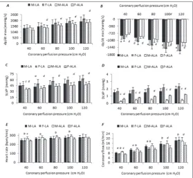
The values of dp/dt min (at all CPPs) and SLVP (60 cm CPP) after chronic administration of LA were significantly lower in male than in female rats (Fig. 1A-D). The values of CF were higher in the male group when compared with the female group under hypoxic (40-60 cm CPP) and normoxic conditions (80 cm CPP) (Fig. 1F). There were no differences in HR between the groups (Fig. 1E). Females treated with ALA had lower values of dp/dt max than males only under hypoxic conditions (Fig. 1A). In addition, CF and HR were also lower at all CPPs (Fig. 1 E and F).
In males, the values of SLVP (40, 60 cm CPP) and DLVP (at all CPPs) were lower in the ALA group as compared to the LA group (Fig. 1C and D). In contrast, ALA enhanced the HR and CF only in hypoxic condi-tions as compared with LA (Fig. 1E and F). In females, the values of all examined cardiodynamic parame-ters (except HR) were lower in the ALA group when compared to the LA group at all CPPs (Fig. 1A-D). CF was also lower under hyperoxic conditions (100 and 120 cm CPP) (Fig. 1E and F).
Differences between chronic treatments with LA and ALA on the biomarkers of oxidative stress in isolated heart (male and female rats)
Male rats treated with LA had higher values of H2O2 and NO2 (in hypoxic conditions) when compared to female rats (Fig. 2 B and D). In contrast, under normoxic and hyperoxic conditions, the females had higher values of O2- and TBARS (Fig. 2A and C). Male
rats treated with ALA showed a larger release of all examined biomarkers of oxidative stress (Fig. 2A-D).
Male groups treated with ALA had higher values of measured cardiac oxidative markers as compared to the LA groups (Fig. 2A-D). The values of NO2- were
O2- in normoxic and hyperoxic conditions
(Fig. 2A). Female groups treated with ALA exhibited a stronger release of H2O2 (nor-moxic conditions) and TBARS (hypoxic and normoxic conditions) (Fig. 2B and C), and a lower release of O2- and NO
2-
(nor-moxic and hyperoxic conditions) in com-parison to the LA groups (Fig. 2A and D).
Differences between chronic treatments with LA and ALA on the biomarkers of oxidative stress in blood samples of male and female rats
Males treated with LA showed higher release of O2- and NO
2- (Fig. 3A and D).
There were no statistically significant dif-ferences in the production of H2O2 and TBARS between the sexes (Fig. 3B and C). Males treated with ALA also had higher values of O2- (Fig. 3A). There were no
statistically significant differences in the production of other examined biomarkers between the sexes (Fig. 3B-D).
The male ALA group had lower val-ues of TBARS and NO2- when compared
to the LA group (Fig. 3C and D). There were no statistically significant differences in the production of O2- and H
2O2 between
the compared groups (Fig. 3A and B). In females, there were no statistically signifi-cant differences in the production of all of the examined biomarkers between groups (Fig. 3A-D).
Differences between chronic treatments with LA and ALA on the levels of antioxidant markers in blood samples of male and female rats
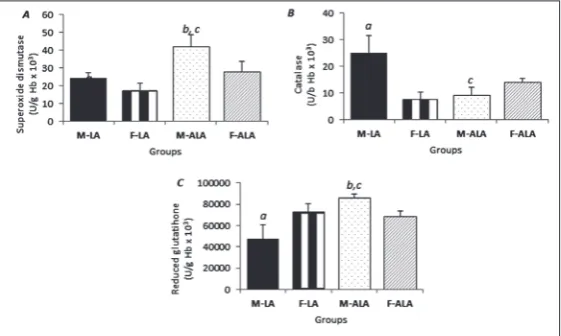
Males treated with LA had higher activities of SOD and CAT (Fig. 4A and B), while the concentration of GSH was lower (Fig. 4C). In contrast, males treated with ALA had a higher activity of SOD and concentration of GSH (Fig. 4A and C), whereas CAT activity was unchanged in both sexes (Fig. 4B).
Fig.1. The effects of chronic administration of LA and ALA on the cardio-dynamic parameters of isolated rat heart. A – The maximum rate of pressure development in the left ventricle (dp/dt max); B – the minimum rate of pressure development in the left ventricle (dp/dt mix); C – SLVP; D – DLVP; E – HR;
F – CF. All values are expressed as the means±standard deviation. Statistically significant differences (p<0.05) between the groups at the same coronary perfu-sion pressure are marked as follows: a – differences between the sexes treated with LA, b – differences between the sexes treated with ALA, c – differences between male rats treated with LA or ALA, d – differences between female rats treated with LA or ALA.
Fig. 2. The effects of the chronic administration of LA and ALAon the bio-markers of oxidative stress in isolated rat heart. A – O2- concentration; B –H
The ALA group had higher values of SOD activity and GSH concentrations (Fig. 4A and C) and lower values of CAT activity when compared with the LA group (Fig. 4B). In females, there were no statistically significant differences in antioxidant markers between the compared groups (Fig. 4A-C).
DISCUSSION
The aim of the current study was to investigate the differencesbetween the two FAs, α-linolenic acid (ALA, omega-3) and linoleic acid (LA, omega-6), on the redox status and cardiac function of rat heart. Ad-ditionally, this study aimed to evaluate the potential
gender-related differences in response to ALA and LA treatment.
The two major types of long-chain PUFAs, omega-3 and omega-6, were shown to have antagonistic effects [1-7]. While omega-3 PUFAs emerged as anti-inflammatory and cardioprotective, ome-ga-6 PUFAs were shown to be proinflam-matory. To test these assumptions and the differencesbetween these FAs on cardiac contractility, we evaluated the changes in cardiac function after 6 weeks of FA treat-ment using the following markers of heart function: maximum and minimum rates of pressure development in the LV (dp/dt max, dp/dt min), SLVP, DLVP, HR and CF. Myocardial contractile dysfunction, a ma-jor manifestation of heart failure, can be revealed through the mechanical defects of left ventricular pressure development (+dp/dt max) and the rate of relaxation, as measured by LV negative (-dp/dt min).
The contractility parameters were lower in the F-ALA group, while the same parameters were not affected in male groups. The other important indicators used to assess LV (SLVP and DLVP) were higher in the F-LA group (compared with the F-ALA). HR had the most drastic de-crease in the M-ALA group. Interestingly, CF was lower in male animals treated with LA in contrast to female rats. When com-paring the effects of LA between the male and female rats, lusitropic force and cardiac perfusion was lower in the female group. When comparing the effects of ALA between male and female rats, the only differences were observed in HR and CF.
A likely explanation for the cardiac effects of PUFA is that application of FA has profound effects on calcium management. PUFA can also produce a dose-dependent reduction in sodium and calcium currents by inhibiting the ryanodine receptor (RyR). These findings support the hypothesis that a lower omega-3 index may also be a marker of an increased propensity for the hypertensive rat heart towards malignant arrhythmias [29]. Other studies have
pre-Fig. 3. The effects of the chronic administration of LA and ALAon the levels of prooxidant markers in blood samples. A – O2- concentration; B – H
2O2 con-centration; C – the index of lipid peroxidation, measured as TBARS; D – the concentration of nitrites NO2-.
sented similar results [28]. In accordance with their results, our findings suggest that these two FAs induce changes in cardiac function of isolated rat heart and act in opposite manners between genders. Generally, compared with ALA, LA significantly affected cardiac function and strongly disturbed heart muscle func-tion, especially in female animals. It is probable that the duration of FA treatment is one of the crucial fac-tors in the development of cardiac dysfunction and consequently of heart hypertrophy.
LA intake may compromise the omega-3 PUFA status of the tissue since its conversion to omega-6 LC-PUFA shares a common enzymatic pathway with the n-3 family [30]. The ALA diet led to higher levels of omega-3 LC-PUFAs, including DHA in the brain and heart [30]. One investigator described the dif-ferent distribution of omega-6 and omega-3 PUFAs throughout the whole rat body, and 18:2 omega-6 was the most concentrated in the heart (13 wt%); this may be one of the reasons for the diverse effects of LA and ALA on heart function in our study [31].
One more mechanism whereby LA and ALA can modulate cardiac function was demonstrated by Berecki et al [31]. They stated that membrane incorporation of PUFAs reduces the response to norepinephrine. In particular, incorporated PUFAs blunted the increase in the sarcoplasmic reticulum (SR) calcium content produced by norepinephrine and therefore attenuated the increase in transient calcium produced by norepinephrine.
To determine the mechanism by which these FAs act on cardiac function in vitro, we evaluated the ef-fects of LA and ALA on the prooxidative markers and parameters of systemic redox balance in the heart. In our study, female animals treated with LA exhib-ited a higher release of O2-, while the male groups
treated with ALA had higher values of H2O2. On the other hand, TBARS levels were significantly changed in almost all groups, with a higher release of both TBARS and NO observed in all female groups after treatment with either LA or ALA. It important to note that in the M-ALA group, we observed a higher rate of coronary flow and all the prooxidative parameters measured from the effluent had a positive correlation with coronary flow. Most likely, FA-induced cardiac dysfunction is a consequence of myocardial disorders
but not of coronary circulation pathologies or endo-thelial dysfunction.
In the third part of this study we decided to ex-clude the effects of coronary flow rate by measuring all prooxidative and antioxidant markers from rat blood. We found higher values of O2- after treatment
with both FAs in the male group, and higher levels of TBARS and nitrites in the M-LA group. These results are in accordance with the results of other relevant studies. Di Nunzio et al. [16] investigated the effect of LA and ALA on oxidative stress and confirmed the as-sumption that LA strongly induces lipid peroxidation. It is known that omega-3 PUFAs are important com-ponents of cell membranes that affect their function, and an omega-3 deficiency is deleterious to health. They are, however, prone to lipid peroxidation due to their many double bonds [4-10]. The metabolic reactions within the omega-3 and omega-6 PUFA families take place in the cell endoplasmic reticulum, apart from the last reaction of β-oxidation, which takes place in peroxisomes, thereby requiring trans-location of adequate substrates into this cell compart-ment. PUFA residues of membrane phospholipids are very sensitive to oxidation and the action of reactive oxygen species (ROS).
Omega-3 PUFAs are known to have antiinflam-matory effects. Excess production of NO is associated with inflammation [4,5]. NO is synthesized from L-arginine by NO synthase (NOS) with NADPH and oxygen as cosubstrates [32]. Inducible NOS (iNOS) is the key enzyme that produces large amounts of NO from macrophages stimulated by the bacterial endo-toxin lipopolysaccharide (LPS) and by proinflamma-tory cytokines such as interferon-γ (IFN-γ) and tumor necrosis factor (TNF) [33]. In the present study, NO was significantly higher after LA supplementation, but the elevated levels of this parameter were decreased after ALA treatment.
activity of antioxidant enzymes in different manners and they have different impacts on cell oxidative sta-tus. Popovic et al. [34] suggested that CAT activity and nitrite concentrations in liver were significantly decreased after the EPA/DHA supplementation. On the other hand, sex-related differences in this inves-tigation indicate that the cardiodepressive effects of LA were more obvious in female rats during hypoxic conditions. Generally, males had higher values of car-diac and systemic oxidative parameters which was accompanied by a compensatory higher mobility of antioxidative protection.
Differences in the effects of LA and ALA on male and female animals may be related to the character-istics of male and female organisms. First, a higher content of body fat is characterized by a favorable fatty acid composition [35]. Second, the intake of omega-3 PUFAs is positively related to testicular volume, while the intake of omega-6 PUFAs is inversely related to testicular volume.
Finally, both fatty acids achieved their effects in hypoxic and hyperoxic conditions, suggesting that the level of oxygenation can also be an important factor in the response of the heart to the applied PUFAs. Thus, it seems that during normoxic conditions the heart is more resistant to their supplementation. ALA more negatively influenced the isolated rat heart in females. The administration of LA was connected to more prominent oxidative stress, while the application of ALA was also associated with improved activity of the antioxidative defense system (displaying higher values in males).
CONCLUSION
The findings of present study may help to elucidate the difference between the effect of ALA and LA on the heart and on local and systemic oxidative stress. The obtained results demonstrate that these PUFAs had a cardiodepressive impact on the heart, especially ALA. ALA apparently possesses antioxidative prop-erties. Moreover, the effects of both FA were gender-specific in terms of exhibiting more negative effects in females. Lastly, ALA can be considered a beneficial supplement in all conditions and pathophysiological processes characterized by increased ROS production.
Acknowledgments: This research did not receive any specific grant from funding agencies in the public, commercial, or not-for-profit sectors.
Author contributions: KR and VJ designed the study, IS, DR, JJ and TN collected and analyzed all data, IS and VZ performed sta-tistical analyses. All authors approved final version of manuscript.
Conflict of interest disclosure: All authors of the present paper confirm no actual or potential conflicts of interest, including any financial, personal, or other relationships with people or orga-nizations.
REFERENCES
1. Spector A, Kim H. Discovery of essential fatty acids. J Lipid Res. 2015;56:11-21.
2. Das U. Biological significance of essential fatty acids. J Assoc Physicians India. 2006;54:309-19.
3. Mele M, Cannelli G, Carta G, Cordeddu L, Melis M, Murru E, Stanton C, Banni S. Metabolism of c9,t11-conjugated lin-oleic acid (CLA) in humans. Prostaglandins Leukot Essent Fatty Acids. 2013;89:115-9.
4. Lunn J, Theobald H. The health effects of dietary unsatu-rated fatty acids. Nutr Bull. 2006;31:178-224.
5. Simopoulos A. The importance of the omega-6/omega-3 fatty acid ratio in cardiovascular disease and other chronic diseases. Exp Biol Med. 2008;233:674-88.
6. Patterson E, Wall R, Fitzgerald G, Ross R, Stanton C. Health implications of high dietary omega-6 polyunsaturated fatty acids. J Nutr Metab. 2012;53:942-6.
7. Arsić A, Prekajski N, Vučić V, Tepšić J, Popović T, Vrvić M, Glibetić M. Milk in human nutrition: comparison of fatty acid profiles. Acta Vet (Beogr). 2009;59:569-78.
8. Du R, Zhong T, Zhang W, Song P, Song W, Zhao Y, Wang C, Tang Y, Zhang X, Zhang Q. Antitumor effect of iRGD-modi-fied liposomes containing conjugated linoleic acid-paclitaxel (CLA-PTX) on B16-F10 melanoma. Int J Nanomedicine. 2014;9:3091-105.
9. Farvid M, Ding M, Pan A, Sun Q, Chiuve S, Steffen L, Willett W, Hu F. Dietary linoleic acid and risk of coronary heart dis-ease: A systematic review and meta-analysis of prospective cohort studies. Circulation. 2014;130:1568-78.
10. Chowdhury R, Warnakula S, Kunutsor S, Crowe F, Ward H, Johnson L, Franco O, Butterworth A, Forouhi N, Thomp-son S, Khaw K, Mozaffarian D, Danesh J, Di Angelantonio E. Association of dietary, circulating, and supplement fatty acids with coronary risk: A systematic review and meta-analysis. Ann Intern Med. 2014;160:398-06.
11. Blasbalg T, Hibbeln J, Ramsden C, Majchrzak S, Rawlings R. Changes in consumption of omega-3 and omega-6 fatty acids in the United States during the 20th century. Am J Clin Nutr. 2011;93:950-62.
13. Miyashita K. Paradox of omega-3 PUFA oxidation. Eur J Lipid Sci Technol. 2014;116:1268-79.
14. Di Nunzio M, Valli V, Bordoni A. PUFA and oxidative stress. Differential modulation of the cell response by DHA. Int J Food Sci Nutr. 2016;67:834-43.
15. Espinosa-Diez C, Miguel V, Mennerich D, Kietzmann T, Sánchez-Pérez P, Cadenas S, Lamas S. Antioxidant responses and cellular adjustments to oxidative stress. Redox Biol. 2015;6:183-97.
16. Chinnadurai K, Kanwal K, Tyagi A, Stanton C, Ross P. High conjugated linoleic acid enriched ghee (clarified butter) increases the antioxidant and antiatherogenic potency in female Wistar rats. Lipids Health Dis. 2016;12:121-25. 17. Kukoba T, Shysh A, Moĭbenko O, Kotsiuruba A, Kharchenko
O. The effects of alpha-linolenic acid on the functioning of the isolated heart during acute myocardial ischemia/reperfu-sion. Fiziol Zh. 2006;52:12-20.
18. Hassan A, Ibrahim A, Mbodji K, Coëffier M, Ziegler F, Bou-noure F, Chardigny M, Skiba M, Savoye G, Déchelotte P, Marion-Letellier R. An α-linolenic acid-rich formula reduces oxidative stress and inflammation by regulating NF-κB in rats with TNBS-induced colitis. J Nutr. 2010;140:1714-21. 19. Sikorska-Wiśniewska M, Mika A, Śledziński T, Małgorzewicz
S, Stepnowski P, Rutkowski B, Chmielewski M. Disorders of serum omega-3 fatty acid composition in dialyzed patients, and their associations with fat mass. Ren Fail. 2017;39:406-12. 20. Folino A, Sprio E, Di Scipio F, Berta N, Rastaldo R. Alpha-linolenic acid protects against cardiac injury and remod-elling induced by beta-adrenergic overstimulation. Food Funct. 2015;6:2231-9.
21. Mitchell L, Grant F, Melchert B, Petty M, Kennedy R. Lin-oleic acid metabolites act to increase contractility in isolated rat heart. Cardiovasc Toxicol. 2002;2:219-30.
22. McCord M, Fridovich I. SOD enzyme function for erythro-cuprein. J Biol Chem. 1969;224:6049-55.
23. Auclair C, Voisin E. Nitroblue tetrazolium reduction. In: Greenwald RA, editor. CRC handbook of methods for oxy-gen radical research. CRC Press Boca Raton; 1985. p. 123-32. 24. Green L, Wagnwr D, Glogowski J, Skipper P, Wishnok J, Tan-nenbaum S. Analysis of nitrate, nitrite and (15 N) nitrate in biological fluids. Anal Biochem. 1982;126:131-8.
25. Pick E, Keisari Y. A simple colorimetric method for the mea-surement of hydrogen peroxide produced by cells in culture. J Immunol Methods.1980;38:161-70.
26. Ohkawa H, Ohishi N. Assay for lipid peroxides in animal tissues by thiobarbituric acid reaction. Anal Biochem. 1979;95:351-8.
27. Beutler E. Catalase. In: Beutler E, editor. Red cell metabo-lism: A manual of biochemical methods. New York: Grune and Stratton; 1982. p. 105-6.
28. Beutler E, Duron O, Kelly B. Improved method for the deter-mination of blood glutathione. J Lab Clin Med. 1963;61:882-8. 29. Bačová B, Seč P, Radošinská J, Certík M, Vachulová A, Trib-ulová N. Lower omega-3 index is a marker of increased pro-pensity of hypertensive rat heart to malignant arrhythmias. Physiol Res. 2013;62:201-8.
30. Ristic-Medic D, Suzic S, Vucic V, Takic M, Tepsic J, Glibetic M. Serum and erythrocyte membrane phospholipids fatty acid composition in hyperlipidemia: effects of dietary inter-vention and combined diet and fibrate therapy. Gen Physiol Biophys. 2009;28:190-9.
31. Berecki G, Den Ruijter H, Verkerk A, Schumacher C, Baartscheer A, Bakker D. Dietary fish oil reduces the inci-dence of triggered arrhythmias in pig ventricular myocytes. Heart Rhythm. 2007;4:1452-60.
32. Blanchard H, Pédrono F, Boulier-Monthéan N, Catheline D, Rioux V, Legrand P. Comparative effects of well-balanced diets enriched in α-linolenic or linoleic acids on LC-PUFA metabolism in rat tissues. Prostaglandins Leukot Essent Fatty Acids. 2013;88:383-9.
33. Salem N, Lin Y, Moriguchi T, Lim S, Salem Jr N, Hibbeln J. Distribution of omega-6 and omega-3 polyunsaturated fatty acids in the whole rat body and 25 compartments. Prosta-glandins Leukot Essent Fatty Acids. 2010;100:13-20. 34. Popović T, Borozan S, Arsić A, Martačić J, Vučić V, Trbović
A, Mandić L, Glibetić M. Fish oil supplementation improved liver phospholipids fatty acid composition and parameters of oxidative stress in male Wistar rats. J Anim Phys Anim Nutr. 2012;96:1020-9.