Couples who are carriers of genetic disorders, including recessive or dominant single-gene defects, sex-linked conditions, or chromosome re-arrangements, face a reproductive risk: affected pregnancies may result in miscarriage or in the birth of a child with significant phenotypic abnormality, sometimes resulting in early death. Such couples have the option of undergoing prenatal diagnosis once a pregnancy is established, either by amniocentesis or chorionic villus sampling (CVS), to allow the detection of the genetic disorder in the fetus. However, if the fetus is found to be carrying a genetic abnormality, the only options available to couples are to have a child with a genetic disease or to terminate the affected pregnancy. This is a difficult and often traumatic decision, as termination, especially in advanced pregnancies, can have substantial psychological and even physical morbidity. Some couples may also experience repeated pregnancy terminations in attempts to conceive a healthy child and might feel unable to accept further affected pregnancies. The prospect of repeating the process of pregnancy and termination one or more times in an attempt to achieve an unaffected pregnancy will be unacceptable to many. Other couples may not contemplate termination because of religious or moral principles.
Such couples have other reproductive choices such as gamete donation or adoption, or to remain childless, but each of these alternatives carries drawbacks. Pre-implantation genetic diagnosis (PGD) has been introduced as an alternative to prenatal diagnosis in order to increase the options available for fertile couples who have a known genetically transmittable disease. PGD is a very early form of prenatal diagnosis. Its intended goal is to reduce significantly a couple’s risk of transmitting a genetic disorder by diagnosing a specific genetic disease in oocytes or early human embryos that have been cultured in vitro, before a clinical pregnancy has been established. After Francesco Florentino
1 Francesco Florentino
EmbryoGen, Centre for Pre-implantation Genetic Diagnosis, via Po 102, 00198 Rome, Italy E-mail: [email protected]
m8
Pre-implantation genetic
diagnosis: current status
and future prospects
diagnosis, only embryos diagnosed as unaffected are selected for transfer to the woman’s uterus.1,2The great advantage of PGD over prenatal diagnosis is that a potential termination of pregnancy is avoided. This gives couples the opportunity to start a pregnancy with the knowledge that their child will be unaffected by the genetic disorder. Consequently, PGD does not require a decision regarding possible pregnancy termination.
Following its first application in 1990,3 PGD has become an important complement to the presently available approaches for prevention of genetic disorders and an established clinical option in reproductive medicine. The number of centres performing PGD has risen steadily, along with the number of diseases that can be tested,4and new applications and methodologies are introduced regularly. The range of genetic defects which can be diagnosed includes structural chromosomal abnormalities, such as reciprocal or Robertsonian translocations, in which it has proven to decrease the number of spontaneous abortions while preventing the conception of affected babies,5and most of the common single gene disorders (SGDs).6–8The scope of PGD has also been extended to improve IVF success for infertile couples, by screening embryos for common or age-related chromosomal aneuploidies in patients at increased risk, including advanced maternal age and repeated miscarriage.9,10
More recently, PGD has been used not only to diagnose and avoid genetic disorders, but also to screen out embryos carrying a mutation predisposing to cancer or to a late-onset disease, or to select for certain characteristics, such as matching for tissue type with the aim of recovering compatible stem cells from cord blood at birth for transplantation to an existing sick child.11–14
There are several stages during pre-implantation development at which genetic testing can be performed. PGD is usually performed by testing single blastomeres removed from cleavage stage embryos (6–8 cells). An alternative approach is represented by testing the first polar body (1PB) before oocyte fertilisation (so-called ‘pre-conception genetic diagnosis’, PCGD)15 or sequential analysis of both first and second (2PB) polar bodies,16which are by-products of female meiosis as oocytes undergo maturation and fertilisation. In women who are carriers for a genetic disease, genetic analysis of 1PB and 2PB allows the identification of oocytes that contain the maternal unaffected gene. Analysis of PBs might be considered an ethically preferable way to perform PGD for couples with moral objections to any micromanipulation and potential discarding of abnormal embryos (so-called ‘pre-embryonic genetic diagnosis’).17It may also be an acceptable alternative in those countries where genetic testing of the embryos is prohibited.15,18Polar body analysis may be also preferred to blastomere biopsy as it is minimally invasive and damaging since the integrity of the embryo is not compromised. However, this technique is labour intensive, because all oocytes must be tested despite the fact that a significant number will not fertilise or will fail to form normal embryos suitable for IVF. Furthermore, it cannot be used for conditions where the male partner carries the genetic disorder, because only the maternal genetic contribution can be studied.
Single cells for genetic analysis may also be obtained from the embryo at blastocyst stage, on day 5 or 6 after fertilisation.19Biopsy at this stage has the advantage of allowing more cells to be sampled (5–10 cells), making genetic tests more robust. It also removes trophectoderm cells, leaving the integrity of
R e ce n t A d v a n ce s in O b st e tr ic ss a n d G y n a e co lo g y 2 4 2
3 P re -im p la n ta tio n g e n e tic d ia g n o sis : cu rr e n t st a tu s a n d fu tu re p ro sp e ct s
the inner cell mass intact. However, due to the difficulty in extended culturing of human embryos, together with the fact that same day testing is required, blastocyst biopsy is currently used routinely in only a few centres.
MAIN INDICATIONS
Currently, there are mainly three groups of patients who may benefit from PGD. The first group consists of couples having a reproductive risk, i.e. risk of conceiving a child with a genetic disease. This can be a monogenic disorder (autosomal recessive, autosomal dominant or X-linked disorders) or a chromosomal structural aberration (such as a balanced translocation).
The second group consists of couples that undergo IVF treatment and whose embryos are screened for chromosome aneuploidies to increase the chances of an on-going pregnancy. The main indications for pre-implantation genetic screening (PGS) are an advanced maternal age, a history of recurrent miscarriages or repeated unsuccessful implantation. It has also been proposed for patients with obstructive azoospermia (OA) and non-obstructive azoospermia (NOA), in cases of unexplained infertility, or for patients with a previous child or pregnancy with a chromosomal abnormality.
A third group of indications can be defined that include ethically difficult cases. These include situations such as HLA typing of the embryo, so that the child born out of this treatment could be a cord-blood, stem-cell donor for a sick sibling, or PGD to diagnose late-onset diseases and cancer predisposition syndromes.
SINGLE GENE DISORDERS
Many genetic disorders are a consequence of mutations in single genes. PGD is then indicated for couples at risk for transmitting a monogenic disease to their offspring.
Although it is more than a decade since the first PGD for a SGD was performed,20 the complexity of the approach has so far limited its clinical application. PGD is a multidisciplinary procedure that requires combined expertise in reproductive medicine and molecular genetics. Additionally, genetic diagnosis of single cells is technically demanding, and protocols have to be stringently standardised before clinical application.
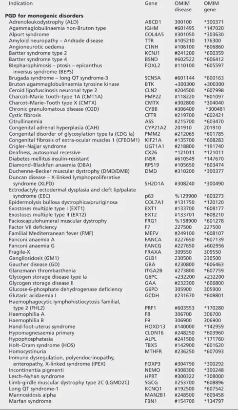
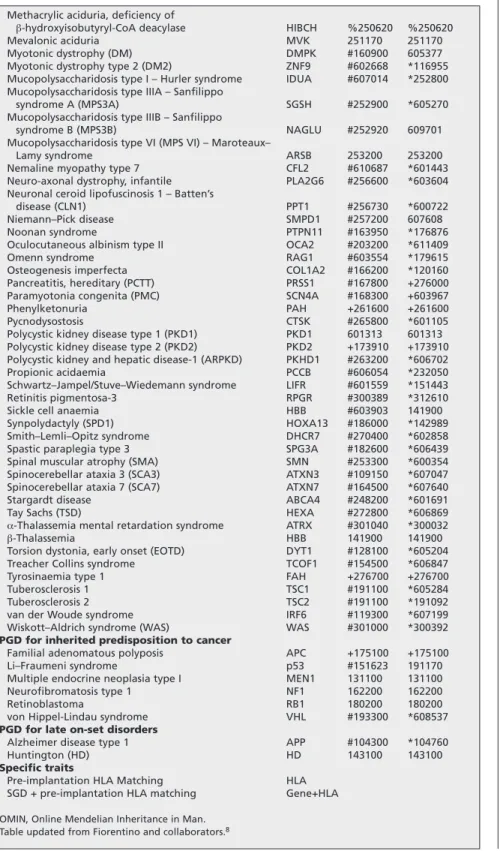
Thus, only a few centres world-wide are offering PGD for SGDs as a clinical service. Nowadays, PGD is available for a large number of monogenic disorders. It is estimated that PGD has been applied for more than 60 different SGDs in over 1800 cycles, resulting in the birth of more than 300 unaffected children.4 The monogenic diseases for which PGD protocols have been developed generally reflect those for which prenatal diagnosis is already offered and includes a continuously growing list of autosomal recessive, dominant, and X-linked diseases. The most frequently diagnosed autosomal recessive disorders are cystic fibrosis,β-thalassemia, sickle cell disease and spinal muscular atrophy. For autosomal dominant disorders, the most common indications are myotonic dystrophy and Charcot–Marie–Tooth disease type 1A. PGD for X-linked diseases has been performed mostly for fragile X syndrome, haemophilia A and Duchenne muscular dystrophy.4,8
4 R e ce n t A d v a n ce s in O b st e tr ic ss a n d G y n a e co lo g y 2 4
Indication Gene OMIM OMIM
disease gene PGD for monogenic disorders
Adrenoleukodystrophy (ALD) ABCD1 300100 *300371
Agammaglobulinaemia non-Bruton type IGHM #601495 *147020
Alport syndrome COL4A5 #301050 *303630
Amyloid neuropathy – Andrade disease TTR #105210 176300
Angioneurotic oedema C1NH #106100 *606860
Bartter syndrome type 2 KCNJ1 #241200 *600359
Bartter syndrome type 4 BSND #602522 *606412
Blepharophimosis – ptosis – epicanthus FOXL2 #110100 *605597 inversus syndrome (BEPS)
Brugada syndrome – long QT syndrome-3 SCN5A #601144 *600163 Bruton agammaglobulinaemia tyrosine kinase BTK +300300 +300300 Ceroid lipofuscinosis neuronal type 2 CLN2 #204500 *607998 Charcot–Marie Tooth–type 1A (CMT1A) PMP22 #118220 *601097
Charcot–Marie–Tooth type X (CMTX) CMTX #302800 *304040
Chronic granulomatous disease (CGD) CYBB #306400 *300481
Cystic fibrosis CFTR #219700 *602421
Citrullinaemia ASS #215700 *603470
Congenital adrenal hyperplasia (CAH) CYP21A2 201910 201910 Congenital disorder of glycosylation type Ia (CDG Ia) PMM2 #212065 *601785 Congenital fibrosis of extra-ocular muscles 1 (CFEOM1) KIF21A #135700 *608283
Crigler–Najjar syndrome UGT1A1 #218800 *191740
Deafness, autosomal recessive CX26 *121011 *121011
Diabetes mellitus insulin-resistant INSR #610549 *147670
Diamond–Blackfan anaemia (DBA) RPS19 #105650 *603474
Duchenne–Becker muscular dystrophy (DMD/DMB) DMD #310200 *300377 Duncan disease – X-linked lymphoproliferative
syndrome (XLPD) SH2D1A #308240 *300490
Ectrodactyly ectodermal dysplasia and cleft lip/palate
syndrome (EEC) p63 %129900 *603273
Epidermolysis bullosa dystrophica/pruriginosa COL7A1 #131750 *120120
Exostoses multiple type I (EXT1) EXT1 #133700 *608177
Exostoses multiple type II (EXT2) EXT2 #133701 *608210
Facioscapulohumeral muscular dystrophy FRG1 %158900 *601278
Factor VII deficiency F7 227500 227500
Familial Mediterranean fever (FMF) MEFV #249100 *608107
Fanconi anaemia A FANCA #227650 *607139
Fanconi anaemia G FANCG #227650 +602956
Fragile-X FRAXA 309550 309550
Gangliosidosis (GM1) GLB1 230500 230500
Gaucher disease (GD) GBA #230800 *606463
Glanzmann thrombasthenia ITGA2B #273800 *607759
Glycogen storage disease type Ia G6PC +232200 +232200
Glycogen storage disease II GAA #232300 *606800
Glucose-6-phosphate dehydrogenase deficiency G6PD 305900 305900
Glutaric acidaemia I GCDH #231670 *608801
Haemophagocytic lymphohistiocytosis familial,
type 2 (FHL2) PRF1 #603553 *170280
Haemophilia A F8 306700 306700
Haemophilia B F9 306900 306900
Hand-foot-uterus syndrome HOXD13 #140000 *142959
Hypomagnesaemia primary CLDN16 #248250 *603960
Hypophosphatasia ALPL #241500 *171760
Holt–Oram syndrome (HOS) TBX5 #142900 *601620
Homocystinuria MTHFR #236250 *607093
Immune dysregulation, polyendocrinopathy,
enteropathy, X-linked syndrome (IPEX) FOXP3 #304790 *300292
Incontinentia pigmenti NEMO #308300 *300248
Lesch–Nyhan syndrome HPRT #300322 *308000
Limb-girdle muscular dystrophy type 2C (LGMD2C) SGCG #253700 *608896
Long QT syndrome-1 KCNQ1 #192500 *607542
Mannosidosis alpha MAN2B1 #248500 *609458
Marfan syndrome FBN1 #154700 *134797
Table 1 Summary of the common monogenic disorders for which a PGD protocol has been established
5 P re -im p la n ta tio n g e n e tic d ia g n o sis : cu rr e n t st a tu s a n d fu tu re p ro sp e ct s
Methacrylic aciduria, deficiency of
β-hydroxyisobutyryl-CoA deacylase HIBCH %250620 %250620
Mevalonic aciduria MVK 251170 251170
Myotonic dystrophy (DM) DMPK #160900 605377
Myotonic dystrophy type 2 (DM2) ZNF9 #602668 *116955
Mucopolysaccharidosis type I – Hurler syndrome IDUA #607014 *252800 Mucopolysaccharidosis type IIIA – Sanfilippo
syndrome A (MPS3A) SGSH #252900 *605270
Mucopolysaccharidosis type IIIB – Sanfilippo
syndrome B (MPS3B) NAGLU #252920 609701
Mucopolysaccharidosis type VI (MPS VI) – Maroteaux–
Lamy syndrome ARSB 253200 253200
Nemaline myopathy type 7 CFL2 #610687 *601443
Neuro-axonal dystrophy, infantile PLA2G6 #256600 *603604 Neuronal ceroid lipofuscinosis 1 – Batten’s
disease (CLN1) PPT1 #256730 *600722
Niemann–Pick disease SMPD1 #257200 607608
Noonan syndrome PTPN11 #163950 *176876
Oculocutaneous albinism type II OCA2 #203200 *611409
Omenn syndrome RAG1 #603554 *179615
Osteogenesis imperfecta COL1A2 #166200 *120160
Pancreatitis, hereditary (PCTT) PRSS1 #167800 +276000
Paramyotonia congenita (PMC) SCN4A #168300 +603967
Phenylketonuria PAH +261600 +261600
Pycnodysostosis CTSK #265800 *601105
Polycystic kidney disease type 1 (PKD1) PKD1 601313 601313 Polycystic kidney disease type 2 (PKD2) PKD2 +173910 +173910 Polycystic kidney and hepatic disease-1 (ARPKD) PKHD1 #263200 *606702
Propionic acidaemia PCCB #606054 *232050
Schwartz–Jampel/Stuve–Wiedemann syndrome LIFR #601559 *151443
Retinitis pigmentosa-3 RPGR #300389 *312610
Sickle cell anaemia HBB #603903 141900
Synpolydactyly (SPD1) HOXA13 #186000 *142989
Smith–Lemli–Opitz syndrome DHCR7 #270400 *602858
Spastic paraplegia type 3 SPG3A #182600 *606439
Spinal muscular atrophy (SMA) SMN #253300 *600354
Spinocerebellar ataxia 3 (SCA3) ATXN3 #109150 *607047
Spinocerebellar ataxia 7 (SCA7) ATXN7 #164500 *607640
Stargardt disease ABCA4 #248200 *601691
Tay Sachs (TSD) HEXA #272800 *606869
α-Thalassemia mental retardation syndrome ATRX #301040 *300032
β-Thalassemia HBB 141900 141900
Torsion dystonia, early onset (EOTD) DYT1 #128100 *605204
Treacher Collins syndrome TCOF1 #154500 *606847
Tyrosinaemia type 1 FAH +276700 +276700
Tuberosclerosis 1 TSC1 #191100 *605284
Tuberosclerosis 2 TSC2 #191100 *191092
van der Woude syndrome IRF6 #119300 *607199
Wiskott–Aldrich syndrome (WAS) WAS #301000 *300392
PGD for inherited predisposition to cancer
Familial adenomatous polyposis APC +175100 +175100
Li–Fraumeni syndrome p53 #151623 191170
Multiple endocrine neoplasia type I MEN1 131100 131100
Neurofibromatosis type 1 NF1 162200 162200
Retinoblastoma RB1 180200 180200
von Hippel-Lindau syndrome VHL #193300 *608537
PGD for late on-set disorders
Alzheimer disease type 1 APP #104300 *104760
Huntington (HD) HD 143100 143100
Specific traits
Pre-implantation HLA Matching HLA
SGD + pre-implantation HLA matching Gene+HLA
OMIN, Online Mendelian Inheritance in Man. Table updated from Fiorentino and collaborators.8
Table 1 (continued)Summary of the common monogenic disorders for which a PGD protocol has been established
6 R e ce n t A d v a n ce s in O b st e tr ic ss a n d G y n a e co lo g y 2
4 Almost all genetically inherited conditions that are diagnosed prenatally
can also be detected by PGD. It can theoretically be performed for any genetic disease with an identifiable gene. Diagnostic protocols now exist for more than 100 monogenic disorders (Table 1).
To establish a diagnostic PGD protocol, extensive preclinical experiments are carried out on single cells (lymphocytes, fibroblasts, cheek cells or spare blastomeres from research embryos) in order to evaluate the efficiency and reliability of the procedure.
Protocols for genotyping single cells for monogenic disorders are based on polymerase chain reaction (PCR). Amplified fragments can be then analysed according to the specific requirements of the test; procedures such as restriction enzyme digestion, single-strand conformation polymorphism (SSCP), denaturing gradient gel electrophoresis (DGGE), allele-specific amplification (ARMS), and recently minisequencing21 have been used for mutation detection. The introduction of fluorescence multiplex PCR22allowed the incorporation of linked polymorphic markers, to improve the robustness of the PGD protocol or to be used as a tool for indirect mutation analysis in linkage-based protocols.
Originally, X-linked diseases were avoided by selection of female embryos, by using the fluorescencein situhybridisation (FISH) procedure to identify embryos with two X chromosomes.23A disadvantage of this approach is that half of the discarded male embryos will be healthy, a fact that gives rise to ethical criticism and reduces the chances of pregnancy by depleting the number of embryos suitable for transfer. In addition, half of the female embryos transferred are carriers of the condition. In several X-linked dominant disorders (e.g. fragile X syndrome), there is also the possibility that, to a varying degree, carrier females may manifest the disease. For many X-linked diseases, the specific genetic defect has now been identified allowing a specific DNA diagnosis. Therefore, there is now a consensus that it is preferable to use PCR-based tests for sex-linked disorders for which the causative gene is known, instead of performing sex selection.24
Many genetic disorders can now be diagnosed using DNA from single cells. However, when using PCR in PGD, one is faced with a problem that only minute amounts of genomic DNA are available for routine genetic analysis. In fact, since PGD is performed on single cells, PCR has to be adapted and pushed to its physical limits. This implies a long process of fine-tuning of the PCR conditions in order to optimise and validate the PGD protocol before clinical application.
There are three main inherent difficulties associated with single-cell DNA amplification. The limited amount of template makes single-cell PCR very sensitive to contamination. The presence of extraneous DNA can easily lead to a misdiagnosis in clinical PGD. Cellular DNA from excess sperm or maternal cumulus cells that surround the oocyte are a potential source of contamination. These cells can be sampled accidentally during the biopsy procedure. For these reasons, oocytes used for PGD of single gene defects should always be stripped of their cumulus cells and fertilised by the use of intracytoplasmic sperm injection (ICSI) in which only a single sperm is inserted into the oocyte. Furthermore, biopsied cells should be washed through a series of droplets of medium before transfer to the PCR tube, and the wash drop should be tested for contamination.
7 P re -im p la n ta tio n g e n e tic d ia g n o sis : cu rr e n t st a tu s a n d fu tu re p ro sp e ct s
Other sources of contamination include skin cells from the operators performing the IVF/PGD procedure or ‘carry over’ contamination of previous PCR products.
Another problem specific to single-cell PCR is the allele drop out (ADO) phenomenon.25It consists of the random non-amplification of one of the alleles present in a heterozygous sample. ADO seriously compromises the reliability of PGD for single-gene disorders as a heterozygous embryo could potentially be diagnosed as either homozygous affected (in which case it would be lost from the cohort of available embryos) or homozygous normal (and, therefore, as suitable for replacement) depending on which allele would fail to amplify. This is particularly concerning in PGD for autosomal dominant disorders, where ADO of the affected allele could lead to the transfer of an affected embryo.
To obviate to the above-mentioned problems, PGD protocols now involve direct mutation(s) detection in combination with analysis of a panel of polymorphic short tandem repeat (STR) markers that are closely linked to the gene region containing the disease causing the mutation(s) (Fig. 1). This approach substantially increases the robustness of the diagnostic procedure and decreases the possibility of misdiagnosis, providing the added assurance Fig. 1 Pre-implantation genetic diagnosis forβ-thalassemia. Pedigree of a couple carrying β-thalassemia mutations and examples of different results of the HBB gene mutation analysis. Informative STR markers are ordered from telomere (top) to centromere (bottom). The numbers in STR markers represent the size of PCR products in base pair. STR alleles linked to the paternal and maternal mutations are represented in bold. Embryo 1 is carrier for IVSI-6 T/C mutation, embryo 2 is compound heterozygote for the mutations IVSI-110 G-A and IVSI-6 T/C, embryo 3 is normal. Embryo 4 is also affected, although mutation analysis result shows a heterozygosity for mutation IVSI-110 G-A. In fact, linked STR markers highlight an allele drop-out (ADO) of the affected allele.
8 R e ce n t A d v a n ce s in O b st e tr ic ss a n d G y n a e co lo g y 2
4 of a partial ‘fingerprint’ of the embryo and confirming that the amplified
fragment is of embryonic origin. In fact, determination of the specific STR haplotype associated with the mutation acts both as a diagnostic tool for indirect mutation analysis, providing an additional confirmation of the results obtained with the direct genotyping procedure, and as a control of misdiagnosis due to undetected ADO. Diagnosis is assigned only when haplotype profiles, obtained from linked STR markers, and mutation analysis profiles are concordant. The multiplex STR marker system also provides an additional control for contamination with exogenous DNA, as other alleles, differing in size from those of the parents, would be detected. The experience of a large series of PGD cycles8strongly suggests that PGD protocols for SGD are not appropriate for clinical practice without including a set of linked STR markers; consequently, this strategy is currently used by most PGD laboratories.
CHROMOSOME TRANSLOCATION
Individuals who carry a balanced chromosomal translocation (reciprocal or Robertsonian), inversion, or other structural chromosomal re-arrangements, typically have no medical issues although some have reduced fertility. However, although the individual is healthy, there is increased risk that the egg or sperm of that individual can have an unbalanced chromosome make-up due to excess or missing genetic material. An embryo derived from the union of such an unbalanced gamete with a partner’s normal gamete also will have an unbalanced genetic composition.26 The presence of an unbalanced translocation can lead to an embryo not implanting, a pregnancy being lost or a child being born with mental and physical problems. Individuals with a translocation may, therefore, experience multiple pregnancy losses or have a child with a chromosomal abnormality.
The primary aim of PGD for translocation is to improve live-birth rates by either reducing the risk of recurrent spontaneous abortions or to improve pregnancy rate in infertile couples (e.g. after failed IVF attempts).27
Reciprocal translocations, characterised by the exchange of two terminal segments between different chromosomes, is the commonest form of chromosome abnormality, which occurs in about 1 in 500 live-births. Robertsonian translocations, in which a whole chromosome is translocated to another through centromeric fusion, are less common and occur in only about 1 in 1000 individuals.
FISH is the method of choice for diagnosing chromosome re-arrangements. This technique uses DNA probes labelled with distinctly coloured fluorochromes that bind to specific DNA sequences unique to each chromosome. Imaging systems enable the fluorescent probe signals to be identified and counted to detect missing or excess chromosomal material.
Analysis of reciprocal translocations for PGD is difficult since each translocation is effectively unique to the family or person within which it occurs, and the break-point may have arisen at any point on any chromosome; thus, different combinations of FISH probes are usually required for each couple. FISH strategies for assessment of reciprocal translocations use three differentially labelled probes, two probes that are specific for the subtelomeric
9 P re -im p la n ta tio n g e n e tic d ia g n o sis : cu rr e n t st a tu s a n d fu tu re p ro sp e ct s
regions of the translocated segments, combined with a centromeric probe.5,26 Analysis of Robertsonian translocations is simpler, involving the use of specific probes chosen to bind at any point on the long arm of each chromosome that is involved in the translocation. The above combination of probes allows embryos that carry an unbalanced chromosome complement to be distinguished from healthy ones. However, both strategies do not discriminate between non-carrier embryos and those that carry the balanced form of the translocation.
A potential pitfall of PGD for chromosomal translocations is that, in some cases, FISH probes might cross-hybridise with other loci in the genome or polymorphisms might occur in some individuals, potentially leading to misdiagnosis. FISH probes should, therefore, always be tested on blood samples from both partners before being used for PGD.
By selecting and transferring embryos with unbalanced chromosome composition, it has been possible for translocation carriers to achieve normal pregnancies, and miscarriage rates in this patient group have been reduced.28
GENETIC SCREENING FOR CHROMOSOME ANEUPLOIDY
Selection of the most competent embryo(s) for transfer is generally based on morphological criteria. However, many women fail to achieve a pregnancy after transfer of good-quality embryos. One of the presumed causes is that such morphologically normal embryos show an abnormal number of chromosomes (aneuploidies).29Aneuploid embryos have a lower survival rate than normal embryos and the majority fail to implant.
Aneuploidy screening of embryos derived from subfertile patients undergoing IVF is probably the most frequent indication for PGD.4Pre-implantation genetic diagnosis for aneuploidy screening, also named pre-implantation genetic screening (PGS), enables the assessment of the numerical chromosomal constitution of cleavage stage embryos. It aims to identify and select for transfer only chromosomally normal (euploid) embryos so to increase the implantation and pregnancy rate for IVF patients, lower the risk for miscarriage and reduce the risk of having a baby with an aneuploidy condition.30
PGS and PGD are often presented as similar treatments, although they have completely different indications: PGS aims to improve pregnancy rates in subfertile couples undergoing IVF/ICSI treatment; PGD aims to prevent the birth of affected children in fertile couples with a high risk of transmitting genetic disorders.
The detection of chromosomes in a single biopsied blastomere is usually achieved using FISH. Essentially, FISH probes to detect those aneuploidies most commonly observed after birth or in miscarriages (involving detection of chromosomes X, Y, 13, 16, 18, 21, and 22) are used. This panel of probes has the potential of detecting over 70% of the aneuploidies found in spontaneous abortions.31Aneuploidy conditions (involving chromosomes 8, 9, 15, and 17) that cause lack of implantation or can results in a miscarriage early in pregnancy are also tested.32,33
The main indications suggested for PGS are repeated implantation failure,34 advanced maternal age,34,35,37 repeated miscarriage in patients with normal karyotypes,10,33,36,37and severe male factor infertility.38,39
10 R e ce n t A d v a n ce s in O b st e tr ic ss a n d G y n a e co lo g y 2
4 Repeated implantation failure
Since the beginning of IVF, many efforts have been made to enhance success rates – the optimisation of embryo selection being one of the most evaluated strategies. Some couples, even after numerous IVF attempts, are unable to become pregnant. Recurrent implantation failure (RIF) has been defined as three or more unsuccessful embryo transfer procedures. Extensive evidence34,40 has revealed a high incidence of numerical chromosomal abnormalities (60–70%) in embryos of patients with a poor outcome after IVF. This has been observed despite good embryo morphology, which offers a possible explanation for their low implantation potential. In women with repeated implantation failure, PGS has been proposed to minimise the likelihood of a chromosome abnormality in a future pregnancy and increase the chance of achieving an ongoing successful pregnancy.
Advanced maternal age
It is well known that the chance of live birth after IVF treatment decreases dramatically with maternal age. It is widely accepted that reduced fertility with age is mostly egg related. Women of advanced maternal age (AMA; usually defined as maternal age ≥ 37 years) are at a higher risk of producing aneuploid embryos, resulting in implantation failure, a higher risk of miscarriage or the birth of a child with a chromosome abnormality (e.g. Down syndrome). Aneuploidy is also believed to be a major reason for the decrease of fertility with age. Several studies have determined that approximately 70% of embryos from women of advanced maternal age may be aneuploid,34,40,41 providing an explanation for their low implantation and high miscarriage rates after IVF treatment. The poor IVF success rate in this category of patients has been well documented and it was suggested that PGS could be used as a screening technique to detect the most common aneuploid syndromes (those involving chromosomes X, Y, 13, 18, 21 and also 16, 22, 15, 17, involved in spontaneous abortions) in an attempt to improve the on-going pregnancy rate, reduce miscarriage rates, and decrease the chance of a chromosomally abnormal pregnancies in these patients.
Repeated miscarriage
Repeated miscarriage (RM), classically defined as three or more spontaneous pregnancy losses, affects about 1% of the population.42Approximately 5–8% of couples with a history of RM have an abnormal karyotype, usually a balanced translocation. Many couples have no identifiable cause or predisposing factor for their miscarriages and, therefore, no standard treatment options. Because women with a history of unexplained RM are more likely to have a high incidence (50–60%) of numerical chromosomal abnormalities in embryos,10,36,37,43PGS has been proposed as a means of reducing the risk of first-trimester miscarriage and of decreasing the risk of conceiving an aneuploid pregnancy.
Severe male infertility
Meiotic disorders are reported to occur frequently in infertile males. Multiple studies have documented an increased aneuploidy rate in both testicular and epididymal spermatozoa from NOA and OA compared with normozoospermic men.44,45
11 P re -im p la n ta tio n g e n e tic d ia g n o sis : cu rr e n t st a tu s a n d fu tu re p ro sp e ct s
Increased aneuploidy and mosaicism rates have also been demonstrated for embryos derived from azoospermic men (NOA and OA) compared with embryos derived from fertile men.38,39
As the chromosomal defects in gametes can be transmitted to the resulting embryos, PGS has also been proposed for such categories of patients.
Limitations of PGS
PGS currently has several disadvantages that limit its clinical value. The main concern is the elevated mosaicism rate observed at the human pre-implantation stage. Mosaicism is defined as the embryo having cells with different chromosome make-up and it has been found in up to 57% of day-3 biopsied embryos.46,47 Mosaicism may represent a major source of misdiagnosis (60%) because of both false-positive and false-negative results.
Besides mosaicism, several technical limitations inherent to the FISH technique have been described. FISH is considered to have an error rate of 5–10%.30,32Overlapping signals may be a source of misdiagnosis resulting in false diagnosis of monosomies. Signal splitting has also been described, resulting in the detection of false trisomies. Finally, evidence suggests that up to half of all embryos identified as aneuploid at the cleavage stage and that survive to the blastocyst stage will ‘self-correct’;48,49 therefore, an abnormal result may not necessarily indicate that the embryo is abnormal and ill-fated.
As a consequence of the above limitations, it has been questioned whether some normal embryos might be excluded from the cohort that is considered suitable for embryo transfer, which, especially in older women who might have small embryo cohorts, could result in the failure to reach embryo transfer. The debate on PGS usefulness
In the last few years, there has been a steady increase in the number of PGS cycles reported to the ESHRE PGD Consortium, from 116 cycles in the data collection from 1997–1998 to 1722 cycles in 2003.4The rapid increase in the use of this procedure has raised questions about its efficacy for routine use.
There have been a number of non-randomised, comparative studies of IVF/ICSI with or without PGS, for AMA or RIF. Most of these studies report that PGS increases the implantation rate,9,10,34,35decreases the abortion rate and reduces trisomic conceptions.33,50,51
Three randomised, controlled trials have been performed for AMA,40,52,53 indicating that PGS does not improve on-going pregnancy or live-birth rates. In contrast, one of these studies showed that PGS decreased the chance of achieving an on-going pregnancy or live birth.52Furthermore, a randomised, controlled trial evaluating the effectiveness of PGS in women under the age of 35 years undergoing IVF treatment with single embryo transfer did not show a benefit for PGS.54
The two largest randomised trials for AMA have been criticised.55–58 Staessen and collaborators40were criticised for biopsying two cells instead of one cell, which may have a negative impact on embryo survival.59The study by Mastenbroek and collaborators52was criticised for its high percentage of embryos without a diagnosis (20%), not including probes for chromosomes 15 and 22 and the low on-going pregnancy rate in the control group (39 pregnancies from 195 oocyte retrievals; 20%).
12 R e ce n t A d v a n ce s in O b st e tr ic ss a n d G y n a e co lo g y 2
4 The debate on the usefulness of PGS continues. Further data are required to
establish whether PGS results in enhanced live-birth rate and, if this is the case, to identify which patients may benefit. The only effective way to resolve this debate is to perform well-designed and well-executed randomised, controlled, clinical trials. The design of further studies should include an adequate randomisation protocol with a clear stratification concerning the indications to perform PGS, the replacement of the same number of embryos in both study and control groups and the healthy live-birth rate per treatment cycle as the main outcome measure. Additional points of interest for future research are which chromosomes should be evaluated (as well as the added value of using more probes) and the implementation of new technologies for chromosome analysis; for example, comparative genomic hybridisation (CGH) and array-CGH, that enable a complete assessment of the numerical chromosomal constitution of pre-implantational embryos.
ETHICALLY DIFFICULT INDICATIONS
The proposed indications for use of PGD are being extended. New uses include PGD to detect mutations for susceptibility to cancer and for late-onset disorders. In addition, parents with children needing haematopoietic stem cell transplants have used PGD to ensure that their next child is free of disease and also to provide a good tissue match for an existing sick child.
Late-onset disorders
One of the proposed uses of PGD is the identification of embryos at risk for late-onset or adult-onset diseases.60,61Many serious genetic disorders, such as Alzheimer’s disease or Huntington disease, have their onset later in adult life, with the result that those affected remain healthy for years or even decades, until the onset of the disease, living a normal healthy life. However, some of these disorders are progressive and disabling, or even lethal, and account for serious ill health. On the other hand, the high probability or certainty of developing the disorders, and their incurable nature, can lead to a stressful life as the patient waits for the first symptoms to occur, and anticipates premature death.
Thus, late-onset disorders present a dilemma: at birth a child will be healthy and free of disease, but will carry the potential to develop ill health in later life. Ethically, the question is whether the burden of carrying susceptibility genes is so great for the child and parents that the burdens of IVF and PGD to screen embryos to avoid the affected children are justified. Many believe PGD is ethically acceptable for these indications because of the heavy burden imposed on patients who are carriers of one of these diseases.
The use of PGD for asymptomatic individuals with the Huntington mutation is one of the most common application of PGD for late-onset disorders.4‘Non-disclosure’ PGD for Huntington disease is applied in those cases in which the prospective parent at risk does not wish to be informed about his or her own carrier status but wants to have offspring free of the disease.62 Embryos can be tested for the presence of the mutation without revealing any of the details of the cycle or diagnosis to the prospective parents. Non-disclosure testing is controversial and not generally approved by professionals,61,63because it puts practitioners in an ethically difficult position,
13 P re -im p la n ta tio n g e n e tic d ia g n o sis : cu rr e n t st a tu s a n d fu tu re p ro sp e ct s
i.e. when no embryos are available for transfer and a mock transfer has to be carried out to avoid the patient suspecting that he/she is a carrier or having to undertake PGD cycles even when the results of previous cycles preclude the patient being a carrier.
The ESHRE Ethics Task Force24 currently discourages non-disclosure testing, recommending the use of exclusion testing instead. Exclusion testing is based on a linkage analysis with polymorphic markers, in which the parental and grandparental origin of the chromosomes can be established. In this way, only embryos are replaced that do not contain the chromosome derived from the affected grandparent, avoiding the need to detect the mutation itself.61
However, exclusion testing PGD is also considered as ethically dubious by some because embryos with an allele from an affected grandparent will be excluded for transfer, although only in half of the cases will the allele be affected.
Inherited cancer predisposition syndromes
Inherited cancer predisposition has become one of the emerging indications for PGD.64 The use of PGD to screen out embryos carrying a mutation predisposing to cancer (e.g. breast/ovarian cancer – BRCA1, BRCA2 genes; Li–Fraumeni syndrome – p53 gene; neurofibromatosis – NF1 and NF2 genes; retinoblastoma – RB1 gene; familial adenomatous polyposis – APC gene; hereditary non-polyposys colon cancer – MSH2 and MLH1 genes; von Hippel–Lindau syndrome – VHL gene) prevents the birth of children who would face a greatly increased life-time risk of cancer, and hence require close monitoring, prophylactic surgery, or other preventive measures. Through PGD, couples with a familial history of cancer, where one partner has the high-risk gene, now have an opportunity to start a pregnancy knowing that their offspring will not carry the cancer-predisposing mutation.
Contrary to the monogenic late-onset diseases, which have full penetrance (i.e. a person found to have a mutation will inevitably get the symptoms in the future), having a predisposing mutation for a susceptibility gene only increases the risk of developing the disease (e.g. inheritance of a familial breast cancer mutation in the BrCa1gene is associated with an 80% life-time risk of breast cancer and otherBrCa1-related cancers), since its effect is modified by other genes and other factors. For the hereditary forms of breast cancer (due to mutations in BRCA1 and BRCA2 genes) and for some hereditary forms of colorectal cancer (e.g. FAP and HNPCC), the absolute risk figures are relatively high but, unlike late-onset diseases, the above cancer predisposition syndromes can usually be treated or preventive measures can be adopted.
As couples present for PGD for heritable cancer genes, there is likely to be wide-spread debate in the community about the associated ethical issues, including disposal of embryos carrying the cancer-predisposition gene mutation and the practice of eugenics.65Although these indications do not involve diseases that manifest themselves in infancy or childhood, the conditions in question lead to substantial health problems for offspring in their thirties or forties. However, owing to the adult onset of hereditary cancer, prenatal diagnosis raises ethical issues surrounding the acceptability of terminating an affected pregnancy.
14 R e ce n t A d v a n ce s in O b st e tr ic ss a n d G y n a e co lo g y 2
4 Human leukocyte antigen matching
PGD of single gene disorders, combined with human leukocyte antigen (HLA) matching, represents one of the most recent applications of the technique in reproductive medicine.11–14This strategy has emerged as a tool for couples at risk of transmitting a genetic disease to allow them to select unaffected embryos of a HLA tissue type compatible with those of an existing affected child. In such cases, PGD is used not only to avoid the birth of affected children, but also to conceive healthy children who may also be potential HLA-identical donors of haematopoietic stem cells (HSC) for transplantation in siblings with a life-threatening disorder. At delivery, HSCs from the newborn umbilical cord blood are collected and used for the haematopoietic reconstruction of the affected sibling.
At present, allogenic HSC transplantation represents the only curative treatment for restoring normal haematopoiesis in severe cases of neoplastic (e.g. leukaemia) or congenital (e.g. β-thalassemia) disorders affecting the haematopoietic and/or the immune system. A critical factor associated with favourable outcome in stem cell transplantation is the use of HLA-genotype identical donors, and HLA-identical siblings provide the best chance to the recipient in the achievement of a successful transplantation. Unfortunately, because of the limited availability of HLA-matched sibling donors, most patients face the option of transplantation using a volunteer unrelated matched donor, identified from national or international registers. In these cases, the results are less favourable compared to the matched-sibling transplant. HLA mismatches are increased using unrelated donors, with a consequent higher incidence of both transplant-related mortality and graft-versus-host disease.
Therefore, if no HLA-identical donor is available in the family, an increasing number of couples with a child affected by an haematopoietic disorder are considering the use of IVF and PGD techniques for therapeutic intent. Before the existence of PGD, natural conception followed by prenatal diagnosis and possibly termination of pregnancy was the only option when trying to find a HLA matching future sibling.66
Verlinsky and collaborators11 described the first pre-implantation HLA matching case in 2001. Since then, HLA typing has become an important PGD indication in four countries – the US,11Italy,12,13Turkey,67,68Belgium.14
The procedure is particularly indicated for patients with children affected by Fanconi anaemia, β-thalassemia, sickle cell anaemia, Wiscott–Aldrich syndrome (WAS), X-linked adrenoleukodystrophy (X-ALD), X-linked hyper-IgM syndrome (HIGM), X-linked hypohidrotic ectodermal dysplasia with immune deficiency (HED-ID) and other similar disorders, that require a HLA-compatible HSC or bone marrow donation to be treated effectively. The great difficulty in finding a HLA-matched donor, even among family members, led to the application of pre-implantation HLA matching also for diseases such as acute lymphoid leukaemia (ALL), acute myeloid leukaemia (AML), or sporadic Diamond–Blackfan anaemia (DBA).13 For these conditions, not involving testing of a causative gene, PGD for HLA matching becomes the primary indication.
Technically, PGD for HLA typing is a difficult procedure due to the extreme polymorphism of the HLA region. Taking into account also the complexity of
15 P re -im p la n ta tio n g e n e tic d ia g n o sis : cu rr e n t st a tu s a n d fu tu re p ro sp e ct s
the region (presence of a large number of loci and alleles), the use of a direct HLA typing approach would require standardisation of a PCR protocol specific for each family, presenting different HLA allele combinations, making it time consuming and unfeasible. The use of a pre-implantation HLA matching protocol irrespective of the specific genotypes involved makes the procedure more straight-forward. Currently, PGD laboratories use a strategy based on a flexible indirect HLA typing protocol applicable to a wide spectrum of possible HLA genotypes.11–14 The approach involves testing of single blastomeres by fluorescent multiplex PCR analysis of polymorphic STR markers, scattered throughout the HLA complex, obtaining a ‘fingerprint’ of the entire HLA region (Fig. 2).
Fig. 2 Pre-implantation HLA matching in combination with PGD forβ-thalassemia, result-ing in the birth of two twins, HLA matched with the affected siblresult-ing. Specific haplotypes were determined by genomic DNA analysis of HLA STR markers andβ-globin (HBB) linked markers from father, mother (upper panel) and affected child (lower panel-left side, black square). Informative STR markers are ordered from telomere (top) to centromere (bottom). The numbers in STR markers represent the size of PCR products in base pair. Paternally and maternally derived HLA haplotypes matched to the affected child are shown in boldface. STR alleles linked to the paternal and maternal mutations are also represented in bold. Examples of different results of HBB mutation analysis and HLA haplotyping from biopsied blastomeres are shown in the lower panel. Paternally and maternally derived haplotypes from each embryo are shown on the left and the right, respectively. The HLA identity of the embryos with the affected sibling has been ascertained evaluating the inheritance of the matching haplotypes. Embryos 1, 2 (carriers) and 8 (affected) represent HLA non-identical embryos. Embryos 3 and 6 were diagnosed as normal, and embryo 7 as carrier, HLA matched with the affected sibling, and were transferred resulting in a HLA matched unaffected birth of two twins (babies have been originated from embryos 3 and 6). ET, embryo transfer.
16 R e ce n t A d v a n ce s in O b st e tr ic ss a n d G y n a e co lo g y 2 4 D is e a se N o . o f N o . o f N o . o f cl in ic a l N o . o f p re g n a n ci e s N o . o f p re g n a n ci e s N o . o f b a b ie s N o . o f P G D cy cl e s co u p le s p re g n a n ci e s a(% ) b st il l g o in g o n d e li v e re d b o rn C B T H L A ty p in g c o m b in e d w it h P G D β -T h a la ss e m ia 1 6 4 8 4 3 9 (3 6 .1 ) 6 2 5 2 7 5 G a u ch e r d is e a se 4 1 1 (3 3 .3 ) 0 0 0 A d re n o le u k o d y st ro p h y 3 2 1 (5 0 .0 ) 1 0 0 D u n ca n sy n d ro m e 2 1 0 (0 .0 ) 0 0 0 H u rl e r sy n d ro m e 2 2 0 (0 .0 ) 0 0 0 S ic k le ce ll d is e a se 2 2 0 (0 .0 ) 0 0 0 M a n n o si d o si s a lp h a 2 1 0 (0 .0 ) 0 0 0 C h ro n ic g ra n u lo m a to u s d is e a se 1 1 1 (1 0 0 .0 ) 0 1 2 W is k o tt – A ld ri ch sy n d ro m e 1 1 1 (1 0 0 .0 ) 0 1 1 1 B ru to n a g a m m a g lo b u li n a e m ia 1 1 1 (1 0 0 .0 ) 1 0 0 G la n z m a n n th ro m b a st h e n ia 1 1 0 (0 .0 ) 0 0 0 F a n co n i a n a e m ia – G 1 1 0 (0 .0 ) 0 0 0 H L A -o n ly ty p in g A cu te ly m p h o b la st ic le u k a e m ia 2 2 1 6 6 (3 7 .5 ) 3 2 2 1 D ia m o n d – B la ck fa n a n a e m ia 1 6 3 3 (2 7 .3 ) 1 0 0 Is ti o ci to si s 3 1 1 (3 3 .3 ) 0 1 1 T o ta l 2 2 5 1 1 8 5 4 (3 5 .5 ) 1 2 3 0 3 3 aN u m b e r o f p re g n a n ci e s e v id e n ce d b y cl in ic a l o r u lt ra so u n d p a ra m e te rs (u lt ra so u n d v is u a li sa ti o n o f a g e st a ti o n a l sa c) . bP re g n a n cy ra te p e r e m b ry o tr a n sf e r. U p d a te d fr o m F io re n ti n o a n d co ll a b o ra to rs . 1 3 Ta b le 2 D if fe re n t in d ic a ti o n s fo r p re -i m p la n ta ti o n H L A m a tc h in g a n d cl in ic a l o u tc o m e
17 P re -im p la n ta tio n g e n e tic d ia g n o sis : cu rr e n t st a tu s a n d fu tu re p ro sp e ct s
Although several publications11–14on the argument have demonstrated the success and the usefulness of the PGD/HLA approach (Table 2), some limitations have to be considered. First, advanced maternal age as well as a poor ovarian response to hormonal hyperstimulation are known to have a major impact on the number of the retrievable oocytes and, consequently, on the number of embryos available for analysis, reducing the likelihood of finding transferable embryos. Thus, several IVF cycles may be necessary to obtain a pregnancy and a live birth. Second, the selection of a donor embryo for HSC transplantation before implantation is restricted by the intrinsic genetic constitution of the embryos: only a quarter or 25% (HLA typing), 3 of 16 or 19% (HLA typing and mutation analysis) of embryos will be transferable. Third, the clinical success rate of PGD/HLA typing is low. This is a common phenomenon in all centres offering HLA/PGD and couples should be counselled for this because they have put all their hope in this approach to cure their sick child.
Ethical discussions concerning PGD in combination with HLA are on-going, mainly because the procedure involves embryo selection.66In case of embryo selection based on HLA typing, the ethical discussion is even stronger since the selection here is based on a non-disease trait and opponents claim that the child to be born is instrumentalised, although some authors maintain that the Kantian imperative is not breached since the future donor child will not only be a donor but also become a loved individual within the family. However, if there would be an existing HLA-matched sibling, we would find it acceptable to use that child as a donor of haematopoietic stem cells. Unless the use as a donor would be the sole reason for creating the child, there would be no violation of its autonomy.
The question of the motivation of the couples also may raise concerns. They could be tempted to have an additional child solely for the purpose of furthering the interest of the existing sibling and not because they desire another baby. This difficult ethical issue can be partially addressed by careful genetic and psychological counselling of the couples to ascertain their real motivation. However, considering the efforts by the parents to save their sick child, it is very unlikely that they would not treat the saviour child as equal to the existing child.
Finally, considerable numbers of embryos are also needed to identify one closely matched with the sick sibling, although all resultant embryos not transferred to the patients are usually frozen for future possible use, in case the couples wish to have more unaffected children.
However, most consider the above criticisms a minor concern when compared with the possibility of saving a child’s life from a devastating disease.
FUTURE PERSPECTIVES
The most remarkable impact on PGD is likely to come from emerging new technologies. Using current FISH methods, less than half of the chromosome complement can be analysed in a single cell. New molecular cytogenetic techniques might allow a full analysis of all chromosomes, improving accuracy and reliability of chromosomal analysis.
18 R e ce n t A d v a n ce s in O b st e tr ic ss a n d G y n a e co lo g y 2
4 The most promising new technique is array-CGH, in which CGH is
performed using microarrays as the target DNA.69These microarrays are made up of DNA sequences specific to human chromosomes spotted onto a platform, usually a glass slide. Array-CGH enables the assessment of all the chromosomes by comparing the studied DNA with a normal sample and is appropriate for analysis of minute quantities of DNA and single cells, as for PGD.70This method has a high resolution and is amenable to automation. The array-CGH procedure detects imbalance across the genome allowing identification of whole-chromosome aneuploidies and even small structural aberrations. However, the technique is limited to detecting relative imbalance and, therefore, changes in whole ploidy cannot be identified.
The analysis of mutations in specific genes is also evolving. The principal advance has been the wider use of multiplex PCR. Other techniques aiming at maximising the data obtained from a single cell are also becoming increasingly important. One of the most exciting developments in single-cell analysis has been the introduction of protocols designed to amplify the entire genome from a single cell. A recent important evolution of these whole genome amplification (WGA) protocols is multiple displacement amplification (MDA), a technique that may offer greater accuracy and more rapid through-put in the future.71 Aliquots of MDA products can be taken and used as a source of templates for subsequent locus-specific PCRs, allowing many individual DNA sequences or genes to be analysed in the same cell. The main limitations of using this method on single cells are the high ADO and preferential amplification rates compared with direct PCR on DNA from a single cell.71,72 Moreover, WGA provides a sufficient supply of sample DNA available for other applications, such as aneuploidy screening by using array-CGH technique. It could be envisaged that, in the future, every embryo tested for a monogenic disorder is also routinely screened for aneuploidy, investigating all chromosomes.
CONCLUSIONS
PGD is an effective clinical tool for assisted reproduction and genetic screening. From the patients’ perspective, PGD is an important alternative to standard prenatal diagnosis. Low pregnancy and birth rates, and the high cost of the procedure, however, make it unlikely that PGD will supersede, completely, the more conventional methods of prenatal testing. PGD remains a complex combination of different technologies, which involve reproductive medicine as well as clinical and molecular genetics and require the close collaboration of a team of specialists. Rapid advances in molecular genetics are likely to stimulate further use of PGD and to encourage a substantial increase in the range of genetic conditions for which PGD is offered. The accuracy of procedures will be improved and its clinical application will be simplified. In the future, PGD will play an increasing role as a specialised clinical procedure, becoming a useful option for many more couples with a high risk of transmitting a genetic disease, to prevent the birth of affected children, and infertile couples to improve IVF success.
19 P re -im p la n ta tio n g e n e tic d ia g n o sis : cu rr e n t st a tu s a n d fu tu re p ro sp e ct s Abbreviations
1PB, first polar body; 2PB, second polar body; ADO, allele drop out; ALL, acute lymphoid leukaemia; AMA, advanced maternal age; AML, acute myeloid leukaemia; ARMS, allele-specific amplification; CGH, comparative genomic hybridisation; CVS, chorionic villus sampling; DBA, sporadic Diamond–Blackfan anaemia; DGGE, denaturing gradient gel electrophoresis; FISH, fluorescencein situhybridisation; HED-ID, X-linked hypohidrotic ectodermal dysplasia with immune deficiency; HIGM, X-linked hyper-IgM syndrome; HLA, human leukocyte antigen; HSCs, haematopoietic stem cells; ICSI, intracytoplasmic sperm injection; IVF,in vitrofertilisation; MDA, multiple displacement amplification; NOA, non-obstructive azoospermia; OA, obstructive azoospermia; PB, polar body; PCGD, pre-conception genetic diagnosis; PCR, polymerase chain reaction; PGD, pre-implantation genetic diagnosis; PGS, pre-implantation genetic screening; RIF, recurrent implantation failure; RM, repeated miscarriage; SGD, single gene disorder; SSCP, single-strand conformation polymorphism; STR, short tandem repeat; WAS, Wiscott–Aldrich syndrome; WGA, whole genome amplification; X-ALD, X-linked adrenoleukodystrophy
References
1. Braude P, Pickering S, Flinter F, Ogilvie CM. Preimplantation genetic diagnosis.Nat Rev Genet2002;3: 941–953.
2. Sermon K, Van Steirteghem A, Liebaers I. Preimplantation genetic diagnosis.Lancet2004;
363: 1633–1641.
3. Handyside AH, Kontogianni EH, Hardy K, Winston RM. Pregnancies from biopsied human preimplantation embryos sexed by Y-specific DNA amplification.Nature1990;
344: 769–770.
4. Sermon KD, Michiels A, Harton Get al. ESHRE PGD Consortium data collection VI: Cycles from January to December 2003 with pregnancy follow-up to October 2004.Hum Reprod2007;22: 323–336.
5. Munne S, Sandalinas M, Escudero Tet al. Outcome of preimplantation genetic diagnosis of translocations.Fertil Steril2000;73: 1209–1218.
6. Vandervorts M, Staessen C, Sermon Ket al. The Brussels’ experience of more than 5 years of clinical preimplantation genetic diagnosis.Hum Reprod Update2000;6: 364–373. 7. Pickering S, Polidoropoulos N, Caller Jet al. Strategies and outcomes of the first 100
cycles of preimplantation genetic diagnosis at the Guy’s and St Thomas’ Center.Fertil Steril2003;79: 81–90.
8. Fiorentino F, Biricik A, Nuccitelli Aet al. Strategies and clinical outcome of 250 cycles of preimplantation genetic diagnosis for single gene disordersHum Reprod2006;21: 670–684.
9. Munne S, Cohen J, Sable D. Preimplantation genetic diagnosis for advanced maternal age and other indications.Fertil Steril2002;78: 234–236.
10. Rubio C, Simon C, Vidal Fet al. Chromosomal abnormalities and embryo development in recurrent miscarriage couples.Hum Reprod2003;18: 182–188.
11. Verlinsky Y, Rechitsky S, Schoolcraft W, Strom C, Kuliev A. Preimplantation diagnosis for Fanconi anemia combined with HLA matching.JAMA2001;285: 3130–3133. 12. Fiorentino F, Biricik A, Karadayi Het al. Development and clinical application of a
strategy for preimplantation genetic diagnosis of single gene disorders combined with HLA matching.Mol Hum Reprod2004;10: 445–460.
13. Fiorentino F, Kahraman S, Karadayi Het al. Short tandem repeats haplotyping of the HLA region in preimplantation HLA matching.Eur J Hum Genet2005;13: 953–958. 14. Van de Velde H, Georgiou I, De Rycke Met al. Novel universal approach for
preimplantation genetic diagnosis of b-thalassemia in combination with HLA matching of embryos.Hum Reprod2004;19: 700–708.
15. Fiorentino F, Biricik A, Nuccitelli Aet al. Rapid protocol for pre-conception genetic diagnosis of single gene mutations by first polar body analysis: a possible solution for the Italian patients.Prenat Diagn2008; In press.
16. Verlinsky Y, Rechitsky S, Cieslak Jet al. Preimplantation diagnosis of single gene disorders by two-step oocyte genetic analysis using first and second polar body.Biochem Mol Med1997;62: 182–187.
20 R e ce n t A d v a n ce s in O b st e tr ic ss a n d G y n a e co lo g y 2
4 18. Tomi D, Griesinger G, Schultze-Mosgau AReprod Biomed Online2006;12: 328–333. et al. Polar body diagnosis for hemophilia a
using multiplex PCR for linked polymorphic markers.J Histochem Cytochem2005;53: 277–280.
19. Veiga A, Sandalinas M, Benkhalifa Met al. Laser blastocyst biopsy for preimplantation diagnosis in the human.Zygote1997;5: 351–354.
20. Handyside AH, Lesko JG, Tarin JJ, Winston RM, Hughes MR. Birth of a normal girl after
in vitrofertilization and preimplantation diagnostic testing for cystic fibrosis.N Engl J Med1992;327: 905–909.
21. Fiorentino F, Magli MC, Podini Det al. The minisequencing method: an alternative strategy for preimplantation genetic diagnosis of single gene disorders.Mol Hum Reprod
2003;9: 399–410.
22. Findlay I, Quirke P, Hall J, Rutherford A. Fluorescent PCR: a new technique for PGD of sex and single-gene defects.J Assist Reprod Genet1996;13: 96–103.
23. Harper JC, Coonen E, Ramaekers FCet al. Identification of the sex of human preimplantation embryos in two hours using an improved spreading method and fluorescentin-situhybridization (FISH) using directly labelled probes.Hum Reprod1994;
9: 721–724.
24. Shenfield F, Pennings G, Devroey P, Sureau C, Tarlatzis B, Cohen J. Taskforce 5: preimplantation genetic diagnosis.Hum Reprod2003;18: 649–651.
25. Findlay I, Ray P, Quirke P, Rutherford A, Lilford R. Allelic drop-out and preferential amplification in single cells and human blastomeres: implications for preimplantation diagnosis of sex and cystic fibrosis.Hum Reprod1995;10: 1609–1618.
26. Scriven PN, Handyside AH, Ogilvie CM. Chromosome translocations: segregation modes and strategies for preimplantation genetic diagnosis.Prenat Diagn1998;18: 1437–1449.
27. Scriven PN, Flinter F, Bickerstaff H, Braude P, Ogilvie MC. Robertsonian translocations – reproductive risks and indications for preimplantation genetic diagnosis.Hum Reprod
2001;16: 2267–2273.
28. Munne S, Morrison L, Fung Jet al. Spontaneous abortions are reduced after preconception diagnosis of translocations.J Assist Reprod Genet1998;15: 290–296. 29. Marquez C, Sandalinas M, Bahçe M, Alikani M, Munné S. Chromosome abnormalities in
1255 cleavage-stage human embryos.Reprod BioMed Online2000;1: 17–26.
30. Wilton L. Preimplantation genetic diagnosis for aneuploidy screening in early human embryos: a review.Prenat Diagn2002;22: 512–518.
31. Simpson JL, Bombard AT. Chromosomal abnormalities in spontaneous abortion: frequency, pathology and genetic counselling. In: Edmonds K, Bennett MJ. (eds)
Spontaneous Abortion. London: Blackwell, 1987; 51.
32. Munné S, Magli C, Bahçe Met al. Preimplantation diagnosis of the aneuploidies most commonly found in spontaneous abortions and live births: XY, 13, 14, 15, 16, 18, 21, 22.
Prenat Diagn1998;18: 1459–1466.
33. Munné S, Chen S, Fischer Jet al. Preimplantation genetic diagnosis reduces pregnancy loss in women aged 35 years and older with a history of recurrent miscarriages.Fertil Steril2005;84: 331–335.
34. Gianaroli L, Magli MC, Ferraretti AP, Munne S. Preimplantation diagnosis for aneuploidies in patients undergoingin vitrofertilization with a poor prognosis: identification of the categories for which it should be proposed.Fertil Steril1999;72: 837–838.
35. Munné S, Magli MC, Cohen Jet al. Positive outcome after preimplantation diagnosis of aneuploidy in human embryos.Hum Reprod1999;14: 2191–2199.
36. Pellicer A, Rubio C, Vidal Fet al.In vitrofertilization plus preimplantation genetic diagnosis in patients with recurrent miscarriage: an analysis of chromosome abnormalities in human preimplantation embryos.Fertil Steril1999;71: 1033–1039. 37. Munné S, Chen S, Fischer Jet al. Preimplantation genetic diagnosis reduces pregnancy
loss in women aged 35 years and older with a history of recurrent miscarriages.Fertil Steril2005;84: 331–335.
38. Platteau P, Staessen C, Michiels Aet al. Comparison of the aneuploidy frequency in embryos derived from testicular sperm extraction in obstructive azoospermic men.Hum
21 P re -im p la n ta tio n g e n e tic d ia g n o sis : cu rr e n t st a tu s a n d fu tu re p ro sp e ct s Reprod2004;19: 1570–1574.
39. Silber S, Escudero T, Lenahan K. Chromosomal abnormalities in embryos derived from testicular sperm extraction.Fertil Steril2003;79: 30–38.
40. Staessen C, Platteau P, Van Assche Eet al. Comparison of blastocyst transfer with or without preimplantation genetic diagnosis for aneuploidy screening in couples with advanced maternal age: a prospective randomized controlled trial.Hum Reprod2004;19: 2849–2858.
41. Platteau P, Staessen C, Michiels Aet al. Preimplantation genetic diagnosis for aneuploidy screening in women older than 37 years.Fertil Steril2005;84: 319–324.
42. Li TC, Makris M, Tomsu M, Tuckerman E, Laird S. Recurrent miscarriage: aetiology management and prognosis.Hum Reprod Update2002;8: 463–481.
43. Platteau P, Staessen C, Michiels Aet al. Preimplantation genetic diagnosis for aneuploidy screening in patients with unexplained recurrent miscarriages.Fertil Steril2005;83: 393–397.
44. Egozcue S, Blanco J, Vendrell JMet al. Human male infertility: chromosome anomalies, meiotic disorders, abnormal spermatozoa and recurrent abortion.Hum Reprod Update
2000;6: 93–105.
45. Calogero A, Burello N, De Palma Aet al. Sperm aneuploidy in infertile men.Reprod Biomed Online2003;6: 310–317.
46. Baart E, Van Opstal D, Los F, Fauser B, Martini E. Fluorescencein situhybridization analysis of two blastomeres from day 3 frozen-thawed embryos followed by analysis of the remaining embryo on day 5.Hum Reprod2004;19: 685–693.
47. Coonen E, Derhaag JG, Dumoulin JCet al. Anaphase lagging mainly explains chromosomal mosaicism in human preimplantation embryo.Hum Reprod2004;19: 316–324.
48. Li M, DeUgarte C, Surrey Met al. Fluorescencein situhybridization reanalysis of day-6 human blastocysts diagnosed with aneuploidy on day 3.Fertil Steril2005;84: 1395–1400. 49. Munne S, Velilla E, Colls Pet al. Self-correction of chromosomally abnormal embryos in
culture and implications for stem cell production.Fertil Steril2005;84: 1328–1334. 50. Munné S, Fischer J, Warner Aet al. Preimplantation genetic diagnosis significantly
reduces pregnancy loss in infertile couples: a multi-center study.Fertil Steril2006;85: 326–332.
51. Colls P, Escudero T, Cekleniak Net al. Increased efficiency of preimplantation genetic diagnosis for infertility using ‘no result rescue’.Fertil Steril2007;88: 53–61.
52. Mastenbroek S, Twisk M, Van Echten-Arends Jet al.In vitrofertilization with preimplantation genetic screening.N Engl J Med2007;357: 9–17.
53. Stevens J, Wale P, Surrey ES, Schoolcraft WB. Is aneuploidy screening for patients aged 35 or over beneficial? A prospective randomized trial.Fertil Steril2004;82 (Suppl 2): 249. 54. Staessen C, Michiels A, Verpoest Wet al. Does PGS improve pregnancy rates in young
patients with single embryo transfer?Hum Reprod2007;22 (Suppl): i32. 55. Handyside AH, Thornhill AR.In vitrofertilization with preimplantation genetic
screening.N Engl J Med2007;357: 1770.
56. Wilton LJ.In vitrofertilization with preimplantation genetic screening.N Engl J Med
2007;357: 1770.
57. Munné S, Cohen J, Simpson JL.In vitrofertilization with preimplantation genetic screening.N Engl J Med2007;357: 1769–1770.
58. Munné S, Gianaroli L, Tur-Kaspa Iet al. Substandard application of preimplantation genetic screening may interfere with its clinical success.Fertil Steril2007;88: 781–784. 59. Cohen J, Wells D, Munné S. Removal of 2 cells from cleavage stage embryos is likely to
reduce the efficacy of chromosomal tests that are used to enhance implantation rates.
Fertil Steril2007;87: 496–503.
60. Verlinsky Y, Rechitsky S, Verlinsky Oet al. Preimplantation diagnosis for early-onset Alzheimer disease caused by V717L mutation.JAMA2002;287: 1018–1021.
61. Sermon K, De Rijcke M, Lissens Wet al. Preimplantation genetic diagnosis for Huntington’s disease with exclusion testing.Eur J Hum Genet2002;10: 591–598. 62. Stern H, Harton G, Sisson Met al. Non-disclosing preimplantation genetic diagnosis for
Huntington disease.Prenat Diagn2002;22: 303–307.
Non-22 R e ce n t A d v a n ce s in O b st e tr ic ss a n d G y n a e co lo g y 2
4 disclosure preimplantation genetic diagnosis for Huntington’s disease: practical andethical dilemma.Prenat Diagn1998;18: 1422–1426.
64. Rechitsky S, Verlinsky O, Chistokhina Aet al. Preimplantation genetic diagnosis for cancer predisposition.Reprod BioMed Online2002;4: 148–155.
65. Robertson J. Extending preimplantation genetic diagnosis: the ethical debate – ethical issues in new uses of preimplantation genetic diagnosis.Hum Reprod2003;18: 465–471. 66. Pennings G, Schots R, Liebaers I. Ethical considerations on preimplantation genetic
diagnosis for HLA typing to match a future child as a donor of haematopoietic stem cells to a sibling.Hum Reprod2002;17: 534–538.
67. Kahraman S, Findikli N, Karliklaya Get al. Medical and social perspectives of PGD for single gene disorders and human leukocyte antigen typing.Reprod BioMed Online2007;
14 (Suppl 1): 104–108.
68. Kahraman S, Karlikaya G, Serteyl Set al. Clinical aspects of preimplantation genetic diagnosis for single gene disorders combined with HLA matching.Reprod BioMed Online
2004;9: 529–532.
69. Pinkel D, Segraves R, Sudar Det al. High resolution analysis of DNA copy number variation using comparative genomic hybridization to microarrays.Nat Genet1998;20: 207–211.
70. Le Caignec C, Spits C, Sermon Ket al. Single-cell chromosomal imbalances detection by array CGH.Nucleic Acids Res2006;34: e68.
71. Handyside AH, Robinson MD, Simpson RJet al. Isothermal whole genome amplification from single and small numbers of cells: a new era for preimplantation genetic diagnosis of inherited disease.Mol Hum Reprod2004;10: 767–772.
72. Spits C, Le Caignec C, De Rycke Met al. Optimization and evaluation of single-cell whole-genome multiple displacement amplification.Hum Mutat2006;27: 496–503.
Author Query