REVIEW
The Responses of Cells to
Electrical Fields: A Review
KENNETH R. ROBINSON
Department of Biological Sciences, Purdue University, West Lafayette, Indiana 47907
The application of DC electric fields to cells has a long and contentious history. The interpretation of the response of cells to such fields was hampered by lack of adequate recording technique, contamination of cultures by electrode products, uncertainty about the magnitude of fields, and sometimes by the complexity of the biological system under study. Never- theless, when Jaffe and Nuccitelli (12) reviewed the literature in 1977, they found eight reliable reports of the response of plant cells to applied fields and four of animal cells. Since then, several laboratories have applied electrical fields to isolated cells in culture and recorded the responses on film or video tape so that responses could be characterized carefully and the threshold field strengths established. In these later studies, the size and direction of the fields are known unam- biguously and possible artifacts such as electrode products, nutrient gradients, flow effects, and temperature changes are controlled.
The significance of these recent results is increased by the finding that several cell types that normally migrate or grow long distances in embryos respond directionally to surpris- ingly small fields (Table I), and by the concurrent finding that developing embryos produce substantial endogenous currents. These two facts raise the possibility that endogenous electrical fields are involved in long-range signalling during embryonic development and during certain responses to injury. I review here the recent literature on the responses of isolated cells to small electrical fields, discuss possible mechanisms by which cells might sense these small fields and, finally, consider the physiological relevance of these responses.
RESPONSES OF CELLS TO APPLIED FIELDS Nerve Cells
The question of whether, and how, growing nerve cell processes (neurites) respond to electrical fields, long contro- versial, has been resolved. Jaffe and Poo (13) reopened the issue by exposing explanted fragments of embryonic chick dorsal root ganglia to electrical fields. They found that the neurites grew faster toward the cathode (negative electrode) than toward the anode (positive electrode); however, they did not observe neurites turning toward the cathode, as others had. Hinkle et al. (10), using primary cultures of dissociated THE JOURNAL OF CELL BIOLOGY • VOLUME 101 DECEMBER 1985 2 0 2 3 - 2 0 2 7 © The Rockefeller University Press • 0021-9525/85/12/2023/05 $ 1.00
spherical neuroblasts from the neural tubes of 1-d-old Xeno- pus embryos, found that the neurites produced by these cells grew toward the cathode and turned through 180 ° in some cases in order to do so. By measuring the angle between the initial and final directions of growth ofa neurite and averaging the difference between the two angles over many neurites, it was found that the minimum detectable response occurred at a field strength of about 7 m V / m m , which represents a voltage drop of 0.35 mV across a 50-urn growth cone. The response was not due to electrode products, since long agar bridges were used to isolate the culture dish from the silver-silver chloride electrodes. To establish that these results were not caused by a field-induced gradient of proteins or other charged molecules in the medium, experiments were done in which culture medium was perfused across the chamber at right angles to the electric field; under these conditions the neurites still turned toward the cathode.
Somewhat unexpectedly, it was found that the points of origin of the neurites on the cell bodies were not affected by the field but were randomly distributed. Another unexpected finding was that in the presence of the electric current, a larger fraction of the neuroblasts sprouted neurites and differen- tiated into neurons than in the absence of the current.
Many of these results have been confirmed and extended by Patel and Poo (24), who studied the same cells by similar methods. In agreement with Hinkle et al. (10), they found that neurites curved toward the cathode; in addition they showed that neurites grew faster toward the cathode than the anode. They reported that total neurite production was in- creased by the presence of a field; however, they reported that more neurites were initiated on the cathode-facing side of the cell body than on the anodal side. The basis of the difference is unclear, but a possible explanation can be offered. Patel and Poo (24) determined the asymmetry in site of origin of neurites after the cells had been exposed to a field for 24 h, while Hinkle et al. (10) did this determination after 2-4 h. Since both groups agree that cells actively retract anodally directed neurites (10, 24), the reported asymmetry after 24 h may be misleading.
Despite the disagreement about effects on the site of origin, it is clear from these two studies that growing neurites do respond to applied electrical fields by growing toward the 2023
TAaLE I. Summary of Cellular Responses to Electrical Fields
Cell type Response Threshold (mV/cell diameter) Reference
Neurite (Xenopus) Turn toward cathode 0.3 - 1 (uniform field) 10, 24
<0.3 (non-uniform field) 25
Accelerated growth toward cathode Not determined 24
Increased differentiation 0.3 10, 24
Myoblasts (Xenopus) Perpendicular alignment to field 0.3 10
Neural crest cells
Xenopus Migrate toward cathode <1 33
Xenopus and Ambys- Migrate toward cathode, perpendicular Not determined 6
toma alignment
Quail (Coturnix cotur- Migrate toward cathode ~1 8
nix)
Fibroblasts (Quail) Migrate toward cathode 0.1-1 9, 22
Epithelial cells
Xenopus embryo Perpendicular alignment, actin localization 17
on cathodal side Not determined
Xenopus tadpole tail Migrate toward cathode 0.2 Muncy, L., and K.R. Robinson, unpublished results
Fish scale Migrate toward cathode ~1 7
cathode. Further, it is agreed that an electrical field somehow stimulates neuronal cells to differentiate overtly into neurons and to send out neurites. It is as though the cells need an external directional signal in order to begin the highly polar- ized process of neurite formation. Earlier disagreement about the responsiveness of nerves to electrical fields, which lasted 50 years, may have been due largely to the use of intact ganglia instead of isolated cells, which made the analysis of the response of individual neurites difficult or impossible.
Patel and Poo (25) have also applied focal electric fields to growing neurites by positioning micropipettes near the growth cones. They found that fields of 0.3-3 m V / m m were required at the center of the growth cone to produce a detectable response in 15 min. It is difficult to compare this value to the threshold determined for uniform fields (7 m V / m m ) since these focal fields vary with the inverse square of the distance from the pipette tip. Regions of the growth cone nearer the tip would have experienced fields fourfold larger than the center of the growth cone (25), so the threshold determined by this method may be an underestimate.
Muscle
CellsAn unexpected additional finding of Hinkle et al. (10) was that the initially spherical myoblasts formed their bipolar axes at right angles to an applied electrical field. These
Xenopus
embryonic cells do not migrate and do not fuse with other cells; each cell forms a spindle-shaped, striated unit that can form a functional neuromuscular junction. The threshold for the perpendicular alignment was also ~0.3 mV/cell diameter, similar to that for neurite turning. This response was interest- ing both because it did not fit any theories of how cells sense electrical fields and because it produces an approximation of the geometrical relationship between neurite and muscle that occurs in vivo. As will be discussed later, this perpendicular response has been observed in other cell types, so it will be important to understand the mechanism underlying this phe- nomenon.
Neural Crest Cells
The neural crest is a transient structure that consists of a population of cells that first accumulates on the dorsal side of
2024 THE JOURNAL OF CELt B,OLOG¥ • VOLUME 101, 1985
the vertebrate neural tube, and then disperses as these cells follow characteristic pathways to form a remarkable number of derivatives, including the autonomic nervous system, pig- ment cells, and facial cartilage (15). The question of how these cells are guided to their targets has been under intense inves- tigation. Three laboratories have shown recently that the cells of the neural crest respond to an applied electrical field by migrating toward the cathode (6, 8, 33); two of these studies used amphibian material (6, 33) while the third used quail (8). It was found that the cells responded directionally to a transcellular voltage difference of 0.7 mV and that the direc- tion of migration reversed when the field direction reversed (33). An interesting feature of this phenomenon was the finding of a lag as long as 2 h before the cells responded to a newly applied field; however, when the field was reversed, the cells reversed their direction with no measurable lag (33). No information about the cause of the lag in the initial response is available.
At higher field strengths (_>100 mV/mm), amphibian neural crest cells have been shown to withdraw protrusions on both the anode- and cathode-facing sides; simultaneously they extend protrusions at right angles to the field with the result that the cells begin to assume a perpendicular orienta- tion within a few minutes (6). These cells then migrate toward the cathode, a direction that is perpendicular to their long axes. Similar perpendicular alignment to an applied field has been observed in quail neural crest cells (8). As with the muscle cells, which form their long axes perpendicular to the field, the mechanism of this perpendicular alignment is un- known since any asymmetries induced by a field will be parallel to the field. Cooper and Keller (6) have pointed out that the perpendicular alignment minimizes the perturbing effect of the field on the membrane potential and suggest that cells respond in such a way as to achieve this minimization; however, it is not obvious how such a feedback loop might work.
Fibroblasts and Epithelial Cells
Erickson and Nuccitelli (9, 22) have studied the behavior of fibroblasts from embryonic quail in electrical fields. These cells migrated toward the cathode at field strengths of 1-10
m V / m m , which corresponded to a voltage difference of 0.2 mV/cell diameter. This is the lowest threshold yet reported for a uniform field effect and is well below the magnitude of endogenous fields known to exist in animals under some circumstances, as discussed below. At somewhat higher fields (50-100 mV/mm), the fibroblasts aligned perpendicularly to the field as well as migrating toward the cathode.
Using much larger fields (500 mV/mm), Luther et al. (17) found that cells, identified by them as epithelial, elongated perpendicularly and that the cathodal edge began to ruffle, although actual migration was rarely observed. Using anti- bodies against actin, they detected arrays of stress fibers run- ning the length of the cells, perpendicular to the field; actin was also found in cathode-facing lamellae. L. Muncy and K. R. Robinson (unpublished results) have isolated epidermal cells from the tail fins of Xenopus larvae. These cells migrated toward the cathode in fields as small as 8 m V / m m , which corresponds to - 0 . 2 mV across these small cells. This response is important since these are the cells which are involved in skin wound healing (29). There also may be a subpopulation of these cells which migrate toward the anode; if so, this would be the first example of such a response by an animal cell. Cooper and Schliwa (7) have isolated fish epidermal cells and exposed them to fields of 50 m V / m m and larger. They report that single cells, cell clusters, and cell sheets migrated toward the cathode. The locomotion of the cells was blocked by a variety of calcium channel antagonists.
MECHANISM OF INTERACTION W I T H SMALL FIELDS
It is not clear at this point how cells sense and respond to voltage differences as small as 0.1 mV across themselves. In one case, it has been shown to be likely that a response to a larger voltage gradient is due to the difference in the mem- brane potential that is set up by the field, and the conse- quences of that difference on passive calcium entry into the cathode- and anode-facing sides of the cells (5). The sym- metrical zygotes of the brown algae, Fucus and Pelvetia, will polarize and usually develop their rhizoids on the anodal side in an electric field; one-tenth maximal response occurs at 6 mV/cell diameter (16, 26). During normal light-induced po- larization, these zygotes drive an endogenous current through themselves, with current entering the future rhizoidal pole (21), and it has been shown that a substantial part of this current is calcium (21, 31). Chen and Jaffe (5) found that if the membrane potential was varied in a known way, calcium influx (as measured with 45Ca) usually varied in the manner expected if only passive (non-voltage-gated) entry sites were involved: that is, depolarizing the membrane potential de- creased calcium influx while hyperpolarizing it increased cal- cium influx. (It should be noted that in a minority of cases, the Pelvetia zygotes formed their rhizoids on the cathodal side; anomalous batches of zygotes were also found in which calcium influx was increased by depolarization [26].) There- fore, they argued, the imposed electrical field polarized the zygotes by driving in calcium on the andoal (hyperpolarized) side, mimicking the normal calcium-driven process (Fig. I c). This straight-forward mechanism cannot explain the gal- vanotropic and galvanotactic processes discussed above. First, the cells all respond toward the cathode, and second, they respond at fields too small to be effective in this simple way. One obvious way to get around the problem is to invoke
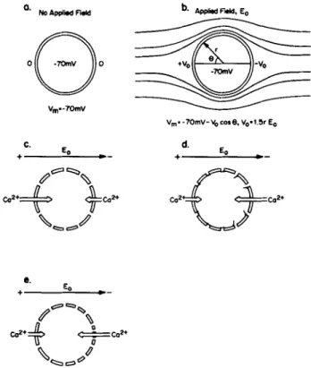
O. NO Applied Field
o@o
V m- - 7 0 m Y C. Eo ÷ , r ' - -c.'*+ ~co~.
b. Applied F'.dcl, ( o Vmo - 7 0 m V - V 0 c o l e , Vo, 1.5r E o d. Eo e° E, ÷ I P - ° C02 '¢" ~ - ,~a2, oFIGURE 1 (a) A spherical cell with a membrane potential of - 7 0 mV. (b) The effects of a uniform applied electrical field. The field is distorted by the highly resistive cell as shown. The potential just outside the cell will vary sinusoidally and thus the transmembrane potential, Vm, will also vary so that the anodal-facing side is hyper- polarized and the cathode-facing side depolarized. In addition, there will be an electric field parallel to the membrane. (c) The effects of a field on calcium fluxes through non-voltage-gated channels. Since the anode-facing side is relatively hyperpolarized (that is, the cytoplasm is more negative), the inward driving force on calcium is increased, while the inward driving force on the other side is decreased. These mechanisms seem to be involved in the response of fucoid eggs to applied fields. (d) The effects of a field on calcium fluxes through voltage-gated channels. Channels would be opened on the depolarized {cathode-facing) side. {e) Electro- phoretic redistribution of calcium channels. Other membrane pro- teins that have been studied migrate toward the cathode-facing side; this mechanism may be involved in cellular galvanotaxis. Calcium fluxes are considered here only for illustrative purposes, similar consideration apply to other ion channels.
voltage-gated channels which might be opened on the depo- larized (cathode-facing) side, which has the additional attrac- tion of providing an amplification mechanism (Fig. 1 d). The available evidence argues against this; a perturbation of the membrane potential by 0.1 mV probably isn't enough to change significantly the probability that a Ca channel is open
(30).
An alternate mechanism whereby an applied electrical field might produce an asymmetic distribution of ion channels is by electrophoresis or electroosmosis. It has been proposed (11) that an external electrical field might redistribute charged, mobile components in the plane of the plasma membrane. The degree of redistribution of a component achieved in a given field was shown to be dependent on the ratio of the diffusion coefficient (D) to the electrophoretic motility (rn), and the voltage drop per cell required to produce an asym- metry of 0.1 (0 representing a uniform distribution and + 1 or - 1 representing the complete redistribution of the corn-
ponent to one pole or the other) is between 0.1 and 1 mV, using realistic assumptions for m and D. Poo and Robinson (28) showed that Con A receptors could be moved to the cathodal (negative) side of muscle cells by applied fields. Subsequently, it has been shown that a variety of membrane receptors can be redistributed by electrical fields, including acetylcholine receptors (23) and Fc~ receptors (19). These receptors (indeed, most that have been studied) move to the cathodal side, a surprising result considering that most gly- coproteins have a net negative charge at physiological pH. This observation has led to the suggestion that the mechanism of electric field redistribution of receptors is due to electroos- motic water flow near the surface of the cell, produced by the immobile negative charge on the surface. Evidence to support this has come from experiments which show that the direction of migration of Con A receptors is reversed following treat- ment of cells with neuraminidase, which would be expected to remove much of the fixed negative charge, allowing a direct electrophoretic response by the receptors (20). Whatever the mechanism, the data indicate that a voltage drop of ~ 1 mV is required to produce an assymetry index of 0.1 in Con A receptors (27). Since the fields used in these studies may have been overestimated (9), it is clear that the thresholds for lateral electrophoresis and for galvanotaxis fall within the same order of magnitude. We have no idea what receptors or channels might be responsible for electrotactic responses, but it remains a reasonable possibility that they may be electrophoretically (or electroosmotically) redistributed by the field and in that way transduce an electrical gradient into directional growth or movement, perhaps through an interaction with the cyto- skeleton (Fig. 1 e).
There is little direct evidence that local calcium entry is involved in galvanotactic responses; however, there are rea- sons for suspecting the involvement of calcium in this process. As mentioned earlier, fucoid zygotes drive an endogenous current through themselves, part of which consists of localized calcium entry at the future growth point. It is thought that the effects of applied electrical fields on these cells mimics or modulates the endogenous current and hence local calcium entry, with the result that a transcellular gradient of calcium is formed. Recent evidence indicates that local polymerization of actin occurs at the future growth point (4), perhaps as a result of local calcium entry, and this f-actin seems to be involved in the positive feedback mechanism that increases the current and eventually leads to germination.
By analogy, it seems reasonable to hypothesize that the highly polarized processes of neurite extension and cell mi- gration may involve endogenous currents and calcium entry. Indeed, it has been shown that voltage-gated calcium channels are more abundant in the growth cones than elsewhere in neurites of neuroblastoma cells, and it was suggested "that Ca 2. entry might trigger neurite extension" (1). An applied field may direct neurite growth by biasing calcium entry to one side of the growth cone. The finding that calcium channel antagonists block epidermal cell motility and response to electrical fields (7) is another indication that calcium may be involved; however, the report that Xenopus embryonic neu- rons can grow neurites in culture in the absence of calcium (3) argues against a ubiquitous involvement of external cal- cium entry in growth. Regardless of whether calcium is in- volved, the applied current does bias whatever mechanism drives neurite extension; and electrophoresis of membrane 2026 ~.E JOURNAL OF CELL B,OLOGY • VOLUME 101, 1985
components seems to be the most reasonable mechanism for such rearrangements. One indication that this may be so is the observation that Con A, which inhibits the electric field- induced redistribution of Con A receptors, also inhibits the
field-induced growth asymmetries in Xenopus neurites with-
out interfering with growth itself (24). This suggests that the redistribution of receptors in the membrane is essential for the electrotropic response.
PHYSIOLOGICAL RELEVANCE
Is the response of cells to applied electrical fields simply a laboratory curiosity or is it an indication of a natural mecha- nism of guidance? In the case of the fucoid eggs, it is obvious that these cells would never see an exogenous field; they develop as isolated cells in the ocean. This is not true of the other cell types discussed above. They develop, grow, and move in an intact animal or embryo, and there is growing evidence that electrical fields of sufficient magnitude do exist in embryos, and in certain cases, adults. Among the sources of these currents are the various electrically polarized epithe- lia; currents will flow whenever there is a macroscopic heter- ogeneity in the electrogenic or resistive properties of an epi- thelium. Consider, for example, the epithelium that covers the early Xenopus embryo. After 1 d of development, this epithelium is already polarized, with the interior of the animal being tens of millivolts positive with respect to the outside pond water, due to sodium uptake (18). Further, it has been shown that the blastopore continues to be a low-resistance leak in this epithelium with the result that current pumped in over the surface leaks out the blastopore; thus, there are currents through the embryo (32). A similar circumstance seems to exist in the chick embryo (14). Unfortunately, in neither of these cases has it been possible to measure directly either the magnitude or direction of electrical gradient in the interior of these embryos, and the pathways of the currents through the embryos aren't known. Various calculations sug- gest that fields sufficiently large to have effects on developing cells may exist, but this situation is clearly unsatisfactory.
Currents will arise in any circumstance where the integrity of an electrically polarized layer of cells is compromised. One obvious example is a wound to the skin of an adult animal. Barker et al. (2) have studied the current produced by wound- ing guinea pig skin and found that the current flows out of the wound. Furthermore, they were able to measure the lateral voltage gradient (that is, the gradient parallel to the skin) in the epidermis near the wound, and it was quite steep, more than 100 m V / m m . This gradient is 10-100 times larger than the in vitro-measured threshold for other cells; it would be surprising if the cells involved in the healing response were not responding to such large fields. A study of the wounding healing response, either in mammals or amphibians, may provide the most direct way to answer the central question: do endogenous electrical currents guide cellular movement in vivo? Radice (29) has shown that it is possible to make minute wounds in the transparent tail of the Xenopus tadpole and monitor the migration of the individual cells from the margins of the wound as they move in to fill the gap. If, simultaneously, one could measure and modulate the wound current and correlate the rate of migration of the cells with the magnitude and direction of the current, clear-cut evidence for or against in vivo galvanotaxis could be developed. If it turns out that such electrical guidance does exist, there are obvious clinical
implications, both for diagnosis and treatment.
In broader terms, the widespread occurrence of endogenous currents during development and the in vitro responses of embryonic cells to electrical fields (Table I) suggest an impor- tant role for currents in directing the emergence of spatial pattern during development. Undoubtedly, other determi- nants of order also exist, such as diffusible chemical gradients and substrate gradients, but electrical currents uniquely offer the possibility of long-range communication between parts of an embryo that can be transmitted rapidly and, with the appropriate circuitry, undiminished.
I am grateful for the helpful comments of several colleagues, especially David Asai and Joseph Vanable (Department of Biological Sciences, Purdue University).
The research in my laboratory on electrical fields has been sup- ported by grants from the National Science Foundation and the
American Heart Association.
Received for publication 13 February 1985, and in revised form 8
A u g u s t 1 9 8 5 .
REFERENCES
1. Anglister. L.. I. C. Farber, A. Shahar, and A. Grinvald. 1982. Localization of voltage- sensitive calcium channels along developing neurites: their possible role in regulating neurite elongation. Dev. Biol. 94:351-365.
2. Barker, A. T., L. F. Jaffe, and J. W. Vanable, Jr. 1982. The glabrous epidermis of cavies contains a powerful battery. Am. J. PhysioL 242:R358-R366.
3. Bixby, J. L., and N. C. Spitzer. 1984. Early differentiation of vertebrate spinal neurons in the absence of voltage-dependent Ca 2+ and Na + influx. Dev. Biol. 106: 89-96. 4. Brawiey, S. H., and K. R. Robinson. 1985. Cytochalasin treatment disrupts the endog-
enous currents associated with cell polarization in fucoid zygotes: studies on the role of f-actin in embryogenesis..L Cell BioL 100:1173-1184.
5. Chen, T. H., and L. F. Jaffe. 1978. Effects of membrane potential on calcium fluxes of
Pelvetia eggs. Planta (Berl.). 140:63-67.
6. Cooper, M. S., and R. E. Keller. 1984. Perpendicular orientation and directional migration of amphibian neural crest ceils in DC electrical fields. Proc. NatL Acad. Sci. USA. 81:1(30-164.
7. Cooper, M. S., and M. Sehliwa. 1985. Electrical and ionic control of tissue cell locomotion in IX? electric fields. 3". Neurosci. Res. 13:223-244.
8. Erickson, C. A., and R. Nuccitelli. 1982. Embryonic cell motility can be guided by weak
electric fields. Z Cell BioL 95(2, Pt.2):314a. (Abstr.)
9. Erickson, C. A., and R. Nuccitelli. 1984. Embryonic fibroblast motility and orientation can be influenced by physiological electric fields..L Cell Biol. 98:296-307.
10. Hinkle, L., C. D. McCaig, and K. R. Robinson. 1981. The direction of growth of differentiating neurones and myoblasts from frog embryos in an applied electric field.
J. Physiol. (Lond). 314:121-135.
11. Jaffe, L. F. 1977. Electrophoresis along cell membranes. Nature (Lond). 265:600-602. 12. Jaffe, L. F., and R. Nucciteni. 1977. Electrical controls of development. Annu. Rev.
Biophys. Bioeng. 6:445--475.
13. Jaffe, L. F., and M.-M. Pop. 1979. Neuritcs grow faster towards the cathode than the anode in a steady field. J. Exp. ZooL 209:115-128.
14. Jaffe, L. F., and C. D. Stern. 1979. Strong electrical currents leave the primitive streak of chick embryos. Science ( Wash. DC). 206:569-571.
15. LeDourain, N. 1980. Migration and differentiation of neural crest cells. Curr. Top. Dev. Biol. 16:31-85.
16. Lurid, E. J. 1923. Electrical control of organic polarity in the egg ofFucus. Bot. Gaz.
76:288-301.
17. Luther, P. W., H. B. Peng, and J.J-C. Lin. 1983. Changes in cell shape and actin distribution induced by constant electric fields. Nature (Lond.). 303:61-64. 18. MeCaig, C. D., and K. R. Robinson. 1982. The ontogeny of the transepidermal potential
difference in frog embryos. Dev. BioL 90:335-339.
19. McCloskey, M. A., Z.-Y. Liu, and M.-M. Pop. 1984. Lateral electromigration and diffusion of Fc~ receptors on rat basophilic leukemia cells: effects of lsE binding. J. Cell Biol. 99:778-787.
20. McLaughlin, S. and M.-M. Pop. 1981. The role of electro-osmosis in the electric field- induced movement of charged macromolecules on the surfaces of cells. Biophys. J.
34:85-93.
21. Nuccitelli, R. 1978. Ooplasmic segregation and secretion in the Pelvetia egg is accom- panied by a membrane-generated electrical current. Dev. Biol. 62:13-33.
22. Nuccitelli, R., and C. A. Erickson. 1983. Embryonic cell motility can be guided by physiological electric fields. Exp. Cell Res. 147:195-201.
23. Orida, N., and M.-M. Pop. 1978. Electrophoretic movement and localisation of acetyl- choline receptors in the embryonic muscle cell membrane. Nature (Land.). 275:31-35. 24. Patel, N., and M.-M. Pop. 1982. Orientation of neuritc growth by extracellular electric
fields. J. Neurosci. 2:483--496.
25. Patel, N. B., and M.-M. Pop. 1984. Perturbation of the direction of neurite growth by pulsed and focal electric fields. ,L Neurosci. 4:2939-2947.
26. Peng, H. B., and L. F. Jaffe. 1976. Polarization of fucoid eggs by steady electrical fields.
DeF. BioL 53:277-284.
27. Poo, M.-M. 1981.1n situ electrophoresis of membranc components.Annu. R ev. Biophys. Bioeng. 10:245-276.
28. Pop, M.-M., and K. R. Robinson. 1977. Electrophoresis of concanavalin A receptors along embryonic muscle cell membrane. Nature (Lond). 265:602-605.
29. Radice, G. P. 1980. The spreading of epithelial cells during wound closure in Xenopus
larvae. Dev. Biol. 76:26-46.
30. Router, H., C. F. Stevens, R. W. Tsien, and G. Yellen. 1982. Properties of single calcium channels in cardiac cell culture. Nature (Lond.). 297:501-504.
31. Robinson, K. R., and L. F. Jaffc. 1975. Polarizing fucoid eggs drive a calcium current through themselves. Science (Wash. DC). 187:70-72.
32. Robinson, K. R., and F. R. Stump. 1984. Self-generated electrical currents through
Xenopus neurulac. J. Physiol. 352:339-352.
33. Stump, R. F., and K. R. Robinson. 1983. Xenopus neural crest cell migration in an applied electrical field..L Cell BioL 97:1226-1233.