Guidelines on
Urolithiasis
C. Türk (chair), T. Knoll (vice-chair), A. Petrik,
K. Sarica, A. Skolarikos, M. Straub, C. Seitz
TABLE OF CONTENTS
PAGE
1. METHODOLOGY 7
1.1 Introduction 7
1.2 Data identification 7
1.3 Evidence sources 7
1.4 Level of evidence and grade of recommendation 7
1.5 Publication history 8
1.5.2 Potential conflict of interest statement 8
1.6 References 8
2. CLASSIFICATION OF STONES 9
2.1 Stone size 9
2.2 Stone location 9
2.3 X-ray characteristics 9
2.4 Aetiology of stone formation 9
2.5 Stone composition 9
2.6 Risk groups for stone formation 10
2.7 References 11
3. DIAGNOSIS 12
3.1 Diagnostic imaging 12
3.1.1 Evaluation of patients with acute flank pain 12 3.1.2 Evaluation of patients for whom further treatment of renal stones is planned 13
3.1.3 References 13
3.2 Diagnostics - metabolism-related 14
3.2.1 Basic laboratory analysis - non-emergency urolithiasis patients 15
3.2.2 Analysis of stone composition 15
3.3 References 16
4. TREATMENT OF PATIENTS WITH RENAL COLIC 16
4.1 Renal colic 16
4.1.1 Pain relief 16
4.1.2 Prevention of recurrent renal colic 16
4.1.3 Recommendations for analgesia during renal colic 17
4.1.4 References 17
4.2 Management of sepsis in obstructed kidney 18
4.2.1 Decompression 18
4.2.2 Further measures 18
4.2.3 References 18
5. STONE RELIEF 19
5.1 Observation of ureteral stones 19
5.1.1 Stone-passage rates 19
5.2 Observation of kidney stones 19
5.3 Medical expulsive therapy (MET) 20
5.3.1 Medical agents 20
5.3.2 Factors affecting success of medical expulsive therapy (tamsulosin) 20
5.3.2.1 Stone size 20
5.3.2.2 Stone location 20
5.3.2.3 Medical expulsive therapy after extracorporeal shock wave
lithotripsy (SWL) 20
5.3.2.4 Medical expulsive therapy after ureteroscopy 21 5.3.2.5 Medical expulsive therapy and ureteral stents 21 5.3.2.6 Duration of medical expulsive therapy treatment 21 5.3.2.7 Possible side-effects include retrograde ejaculation and hypotension 21
5.3.3 References 21
5.4 Chemolytic dissolution of stones 24
5.4.1 Percutaneous irrigation chemolysis 24
5.4.3 References 25
5.5 Extracorporeal shock wave lithotripsy (SWL) 25
5.5.1 Contraindications of extracorporeal shock wave lithotripsy 25 5.5.2 Stenting before carrying out extracorporeal shock wave lithotripsy 25
5.5.2.1 Stenting in kidney stones 25
5.5.2.2 Stenting in ureteral stones 25
5.5.3 Best clinical practice 26
5.5.3.1 Pacemaker 26
5.5.3.2 Shock wave rate 26
5.5.3.3 Number of shock waves, energy setting and repeat treatment sessions 26 5.5.3.4 Improvement of acoustic coupling 26
5.5.3.5 Procedural control 26
5.5.3.6 Pain control 26
5.5.3.7 Antibiotic prophylaxis 26
5.5.3.8 Medical expulsive therapy after extracorporeal shock wave lithotripsy 27 5.5.4 Complications of extracorporeal shock wave lithotripsy 27
5.5.5 References 27
5.6 Endourology techniques 31
5.6.1 Percutaneous nephrolithotomy (PNL) 31
5.6.1.1 Intracorporeal lithotripsy 31
5.6.1.2 Extraction tools 31
5.6.1.3 Best clinical practice 31
5.6.1.3.1 Contraindications 31
5.6.1.3.2 Preoperative imaging 31
5.6.1.3.3 Positioning of the patient 32
5.6.1.3.4 Puncture 32
5.6.1.3.5 Dilatation 32
5.6.1.3.6 Nephrostomy and stents 32
5.6.1.4 Management of complications 32
5.6.2 Ureterorenoscopy (URS) (including retrograde access to renal collecting system) 33
5.6.2.1 Best clinical practice in URS 33
5.6.2.1.1 Preoperative work-up and preparations 33
5.6.2.1.2 Contraindications 33
5.6.2.1.3 Access to the upper urinary tract 33
5.6.2.1.4 Safety aspects 34
5.6.2.1.5 Ureteral access sheaths 34
5.6.2.1.6 Stone extraction 34
5.6.2.1.7 Intracorporeal lithotripsy 34
5.6.2.1.8 Stenting before and after URS 34
5.6.2.2 Complications 35
5.6.3 References 35
5.7 Open and laparoscopic surgery for removal of renal stones 38
5.7.1 Open surgery 38
5.7.1.1 Indications for open surgery 38
5.7.2 Laparoscopic surgery 39
5.7.2.1 Indications for laparoscopic stone surgery 39
5.7.3 References 40
6. INDICATION FOR ACTIVE STONE REMOVAL AND SELECTION OF PROCEDURE 42 6.1 Indications for active removal of ureteral stones 42 6.2 Indications for active removal of kidney stones 42
6.2.1 Natural history of caliceal stones 42
6.2.2 References 43
6.3 General recommendations and precautions for stone removal 43
6.3.1 Infections 43
6.3.2 Antithrombotic therapy and stone treatment 44
6.3.3 Obesity 44
6.3.4 Hard stones 44
6.3.5 Radiolucent stones 44
6.3.7 References 45 6.4 Selection of procedure for active removal of kidney stones 46 6.4.1 Stones in renal pelvis or upper/middle calices 46
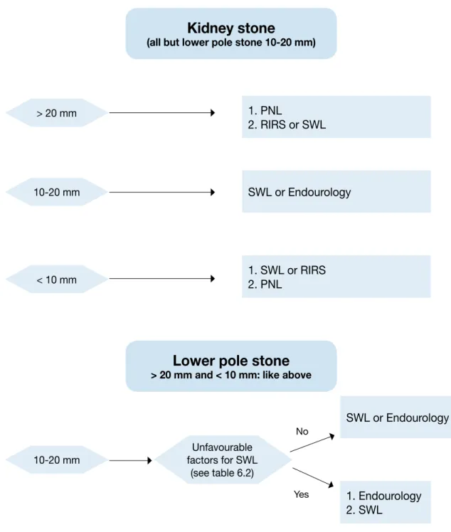
6.4.2 Stones in the lower renal pole 46
6.4.3 References 48
6.5 Selection of procedure for active removal of ureteral stones 50
6.5.1 Methodology 50
6.5.2 Extracorporeal shock wave lithotripsy and ureteroscopy 50
6.5.2.1 Stone free rates (SFRs) 50
6.5.2.2 Complications 51
6.5.3 Percutaneous antegrade ureteroscopy 51
6.5.4 Other methods for ureteral stone removal 51
6.5.5 References 51
7. RESIDUAL STONES 52
7.1 Clinical evidence 52
7.2 Therapy 53
7.3 References 53
8. MANAGEMENT OF URINARY STONES AND RELATED PROBLEMS DURING PREGNANCY 54
8.1 Diagnostic imaging 54
8.2 Management 54
8.3 References 55
9. MANAGEMENT OF STONE PROBLEMS IN CHILDREN 56
9.1 Aetiology 56
9.2 Diagnostic imaging 56
9.2.1 Ultrasound 56
9.2.2 Plain films (KUB radiography) 57
9.2.3 Intravenous urography (IVU) 57
9.2.4 Helical computed tomography (CT) 57
9.2.5 Magnetic resonance urography (MRU) 57
9.2.6 Nuclear imaging 57
9.3 Stone removal 57
9.3.1 Medical expulsive therapy (MET) in children 57
9.3.2 Extracorporeal shock wave lithotripsy 57
9.3.3 Endourological procedures 58
9.3.3.1 Percutaneous nephrolithotripsy (PNL) 58
9.3.3.2 Ureteroscopy 58
9.3.4 Open or laparoscopic surgery 58
9.4 Special considerations on recurrence prevention 59
9.5 References 59
10. STONES IN URINARY DIVERSION AND OTHER VOIDING PROBLEMS 62 10.1 Management of stones in patients with urinary diversion 62
10.1.1 Aetiology 62
10.1.2 Management 62
10.1.3 Prevention 63
10.1.4 References 63
10.2 Management of stones in patients with neurogenic bladder 64 10.2.1 Aetiology, clinical presentation and diagnosis 64
10.2.2 Management 64
10.2.3 References 64
10.3 Management of stones in transplanted kidneys 64
10.3.1 Aetiology and clinical presentation 64
10.3.2 Management 65
10.3.3 References 65
10.4 Special problems in stone removal 66
11. METABOLIC EVALUATION AND RECURRENCE PREVENTION 67 11.1 General metabolic considerations for patient work-up 67
11.1.1 Evaluation of patient risk 67
11.1.2 Urine sampling 68
11.1.3 Timing of specific metabolic work-up 69
11.1.4 Reference ranges of laboratory values 69 11.1.5 Risk indices and additional diagnostic tools 69
11.1.6 References 71
11.2 General considerations for recurrence prevention 71
11.2.1 Fluid intake 72
11.2.2 Diet 72
11.2.3 Lifestyle 72
11.2.4 Recommendations for recurrence prevention 73
11.2.5 References 73
11.3 Stone-specific metabolic evaluation and pharmacological recurrence prevention 74
11.3.1 Introduction 74
11.3.2 References 75
11.4 Calcium oxalate stones 77
11.4.1 Diagnosis 77
11.4.2 Interpretation of results and aetiology 77
11.4.3 Specific treatment 79
11.4.4 Recommendations for pharmacological treatment of patients with specific
abnormalities in urine composition 79
11.4.5 References 79
11.5 Calcium phosphate stones 81
11.5.1 Diagnosis 81
11.5.2 Interpretation of results and aetiology 81
11.5.3 Pharmacological therapy 82
11.5.4 Recommendations for the treatment of calcium phosphate stones 82
11.5.5 References 82
11.6 Disorders and diseases related to calcium stones 83
11.6.1 Hyperparathyroidism 83
11.6.2 Granulomatous diseases 83
11.6.3 Primary hyperoxaluria 83
11.6.4 Enteric hyperoxaluria 83
11.6.5 Renal tubular acidosis 84
11.6.6 Nephrocalcinosis 85
11.6.6.1 Diagnosis 85
11.6.7 References 85
11.7 Uric acid and ammonium urate stones 86
11.7.1 Diagnosis 86
11.7.2 Interpretation of results 86
11.7.3 Specific treatment 86
11.7.4 References 87
11.8 Struvite and infection stones 88
11.8.1 Diagnosis 88
11.8.2 Specific treatment 88
11.8.3 Recommendations for therapeutic measures of infection stones 89
11.8.4 References 89
11.9 Cystine stones 91
11.9.1 Diagnosis 91
11.9.2 Specific treatment 92
11.9.2.1 Pharmacological treatment of cystine stones 92 11.9.3 Recommendations for the treatment of cystine stones 93
11.9.4 References 93
11.10 2,8-dihydroyadenine stones and xanthine stones 94
11.10.1 2,8-dihydroxyadenine stones 95
11.10.2 Xanthine stones 95
11.10.3 Fluid intake and diet 95
11.12 Unknown stone composition 95
11.13 References 96
1. METHODOLOGY
1.1
Introduction
The European Association of Urology (EAU) Urolithiasis Guidelines Panel have prepared these guidelines to help urologists assess evidence-based management of stones/calculi and incorporate recommendations into clinical practice.
The document covers most aspects of the disease, which is still a cause of significant morbidity despite technological and scientific advances. The Panel is aware of the geographical variations in healthcare provision.
1.2
Data identification
For this 2014 print of the Urolithiasis guidelines, a scoping search, covering all content, was performed. Time frame of the search was October 16th 2012 through July 2013. This search was limited to level 1 evidence (systematic reviews [SRs] and meta-analyses of randomised controlled trials [RCTs]) and English language publications in peer-reviewed journals. Animal studies were excluded. The search identified 237 unique records.
Additionally, lower level searches were performed for each chapter of the Urolithiasis guidelines, covering the past two years, with a cut-off date of November 25th, 2013. Selection of the papers was done through a consensus meeting of the Panel held in December 2013.
Annual scoping searches will be repeated as a standard procedure.
1.3
Evidence sources
Searches were carried out in the Cochrane Library Database of Systematic Reviews, Cochrane Library of Controlled Clinical Trials, and Medline and Embase on the Dialog-Datastar platform. The searches used the controlled terminology and the use of free text ensured search sensitivity.
Randomised controlled trial strategies were based on Scottish Intercollegiate Guidelines Network (SIGN) and Modified McMaster/Health Information Research Unit (HIRU) filters for RCTs, systematic reviews and practice guidelines on the OVID platform and then translated into Datastar syntax.
There is a need for ongoing re-evaluation of the current guidelines by an expert panel. It must be emphasised that clinical guidelines present the best evidence available but following the recommendations will not necessarily result in the best outcome. Guidelines can never replace clinical expertise when making treatment decisions for individual patients - also taking personal values and preferences/individual circumstances of patients into account.
1.4
Level of evidence and grade of recommendation
References in the text have been assessed according to their level of scientific evidence (Table 1.1), and guideline recommendations have been graded (Table 1.2) according to the Oxford Centre for Evidence-based Medicine Levels of Evidence (1). Grading aims to provide transparency between the underlying evidence and the recommendation given.
Table 1.1: Level of evidence (LE)*
Level Type of evidence
1a Evidence obtained from meta-analysis of randomised controlled trials. 1b Evidence obtained from at least one randomised trial.
2a Evidence obtained from one well-designed controlled study without randomisation. 2b Evidence obtained from at least one other type of well-designed quasi-experimental study. 3 Evidence obtained from well-designed non-experimental studies, such as comparative studies,
correlation studies and case reports.
4 Evidence obtained from expert committee reports or opinions or clinical experience of respected authorities.
* Modified (1).
When recommendations are graded, the link between the level of evidence and grade of recommendation is not directly linear. Availability of RCTs may not translate into a grade A recommendation when there are methodological limitations or disparity in published results.
Absence of high-level evidence does not necessarily preclude a grade A recommendation, if there is overwhelming clinical experience and consensus. There may be exceptions where corroborating studies cannot be performed, perhaps for ethical or other reasons, and unequivocal recommendations are considered helpful. Whenever this occurs, it is indicated in the text as “upgraded based on panel consensus”. The quality of the underlying scientific evidence must be balanced against benefits and burdens, values and preferences and cost when a grade is assigned (2-4).
The EAU Guidelines Office does not perform cost assessments, nor can it address local/national preferences systematically. The expert panels include this information whenever it is available.
Table 1.2: Grade of recommendation (GR)* Grade Nature of recommendations
A Based on clinical studies of good quality and consistency addressing the specific recommendations and including at least one randomised trial.
B Based on well-conducted clinical studies, but without RCTs.
C Made despite the absence of directly applicable clinical studies of good quality. *Modified from. (1).
1.5
Publication history
The current 2014 print presents a limited update of the 2013 publication of the EAU Urolithiasis Guidelines. Four sections of the text have been replaced (3.1 Diagnostic Imaging, 5.5 Extracorporeal Shockwave Lithotripsy, 6.3.2 Anticoagulation and 6.3.6 Steinstrasse). The flowcharts included in Chapter 11 (Metabolic evaluation and recurrence prevention) have been amended, with a revisit of all references.
Recommendations have not changed, with the exception of section 6.3.2 Anticoagulation.
The first EAU Guidelines on Urolithiasis were published in 2000. Subsequent updates were presented in 2001 (limited update), 2005 (comprehensive update), 2008 (comprehensive update), 2009, 2010, 2011 (limited update), 2012 (comprehensive update) and 2013 (limited update).
A quick reference document presenting the main findings of the urolithiasis guidelines is also available alongside several scientific publications in European Urology and the Journal of Urology (5-7). All texts can be viewed and downloaded for personal use at the EAU website:
http://www.uroweb.org/guidelines/online-guidelines/.
1.5.2 Potential conflict of interest statement
The expert panel have submitted potential conflict of interest statements which can be viewed on the EAU website: http://www.uroweb.org/guidelines/online-guidelines/.
1.6
References
1. Oxford Centre for Evidence-based Medicine Levels of Evidence. Produced by Bob Phillips, Chris Ball, Dave Sackett, Doug Badenoch, Sharon Straus, Brian Haynes, Martin Dawes since November 1998. Updated by Jeremy Howick March 2009.
http://www.cebm.net/index.aspx?o=1025 [Access date March 2014]
2. Atkins D, Best D, Briss PA, et al; GRADE Working Group. Grading quality of evidence and strength of recommendations. BMJ 2004 Jun;328(7454):1490.
http://www.ncbi.nlm.nih.gov/pubmed/15205295
3. Guyatt GH, Oxman AD, Vist GE, et al. GRADE: an emerging consensus on rating quality of evidence and strength of recommendations. BMJ 2008;336(7650):924-6.
http://www.ncbi.nlm.nih.gov/pubmed/18436948
4. Guyatt GH, Oxman AD, Kunz R, et al; GRADE Working Group. Going from evidence to recommendations. BMJ 2008 May;336(7652):1049-51.
http://www.ncbi.nlm.nih.gov/pmc/articles/PMC2376019/?
http://www.gradeworkinggroup.org/publications/Grading_evidence_and_recommendations_BMJ.pdf 5. Tiselius HG, Ackermann D, Alken P, et al; Working Party on Lithiasis, European Association of Urology.
Guidelines on Urolithiasis. Eur Urol 2001 Oct;40(4):362-71. http://www.ncbi.nlm.nih.gov/pubmed/11713390
6. Preminger GM, Tiselius HG, Assimos DG, et al; American Urological Association Education and Research, Inc; European Association of Urology. 2007 Guideline for the management of ureteral calculi. Eur Urol 2007 Dec;52(6):1610-31.
7. Preminger GM, Tiselius HG, Assimos DG, et al; EAU/AUA Nephrolithiasis Guideline Panel. Guidelines on urolithiasis. J Urol 2007 Dec;178(6):2418-34.
http://www.ncbi.nlm.nih.gov/pubmed/17993340
2. CLASSIFICATION OF STONES
Urinary stones can be classified according to size, location, X-ray characteristics, aetiology of formation, composition, and risk of recurrence (1-4).
2.1
Stone size
Stone size is usually given in one or two dimensions, and stratified into those measuring up to 5, 5-10, 10-20, and > 20 mm in largest diameter.
2.2
Stone location
Stones can be classified according to anatomical position: upper, middle or lower calyx; renal pelvis; upper, middle or distal ureter; and urinary bladder. Treatment of bladder stones is not discussed here.
2.3
X-ray characteristics
Stones can be classified according to plain X-ray appearance [kidney-ureter-bladder (KUB) radiography] (Table 2.1), which varies according to mineral composition (3). Non-contrast-enhanced computer tomography (NCCT) can be used to classify stones according to density, inner structure and composition, which can affect treatment decisions (Section 6.3.4) (2,3).
Table 2.1: X-ray characteristics
Radiopaque Poor radiopacity Radiolucent
Calcium oxalate dihydrate Magnesium ammonium phosphate Uric acid Calcium oxalate monohydrate Apatite Ammonium urate
Calcium phosphates Cystine Xanthine
2,8-dihydroxyadenine Drug-stones (Section 11.11)
2.4
Aetiology of stone formation
Stones can be classified into those caused by: infection, or non-infectious causes (infection and non-infection stones); genetic defects (5); or adverse drug effects (drug stones) (Table 2.2).
Table 2.2: Stones classified by aetiology* Non-infection stones
• Calcium oxalate
• Calcium phosphate (including brushite and carbonate apatite) • Uric acid
Infection stones
• Magnesium ammonium phosphate • Carbonate apatite • Ammonium urate Genetic causes • Cystine • Xanthine • 2,8-dihydroxyadenine Drug stones *See section 11.4.2
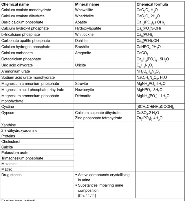
2.5
Stone composition
Metabolic aspects are important in stone formation, and metabolic evaluation is required to rule out any disorders. Analysis in relation to metabolic disorders is the basis for further diagnostic and management
decisions. Stones are often formed from a mixture of substances. Table 2.3 lists the clinically most relevant substances and their mineral components.
Table 2.3: Stone composition
Chemical name Mineral name Chemical formula
Calcium oxalate monohydrate Whewellite CaC2O4.H2O Calcium oxalate dihydrate Wheddelite CaC2O4.2H2O Basic calcium phosphate Apatite Ca10(PO4)6.( OH)2 Calcium hydroxyl phosphate Hydroxylapatite Ca5(PO3)3(OH)
b-tricalcium phosphate Whitlockite Ca3(PO4)2
Carbonate apatite phosphate Dahllite Ca5(PO4)3OH
Calcium hydrogen phosphate Brushite CaHPO4.2H2O
Calcium carbonate Aragonite CaCO3
Octacalcium phosphate Ca8H2(PO4)6 . 5H2O
Uric acid dihydrate Uricite C5H4N4O3
Ammonium urate NH4C5H3N4O3
Sodium acid urate monohydrate NaC5H3N4O3. H2O Magnesium ammonium phosphate Struvite MgNH4PO4.6H2O Magnesium acid phosphate trihydrate Newberyite MgHPO4. 3H2O Magnesium ammonium phosphate
monohydrate
Dittmarite MgNH4(PO4) . 1H2O
Cystine [SCH2CH(NH2)COOH]2
Gypsum Calcium sulphate dihydrate
Zinc phosphate tetrahydrate
CaSO4.2 H2O Zn3(PO4)2.4H2O Xanthine 2,8-dihydroxyadenine Proteins Cholesterol Calcite Potassium urate Trimagnesium phosphate Melamine Matrix
Drug stones • Active compounds crystallising in urine
• Substances impairing urine composition
(Ch. 11.11) Foreign body calculi
2.6
Risk groups for stone formation
The risk status of stone formers is of particular interest because it defines the probability of recurrence or regrowth, and is imperative for pharmacological treatment.
About 50% of recurrent stone formers have just one lifetime recurrence (4,6). Highly recurrent disease is observed in slightly more than 10% of patients. Stone type and disease severity determine low or high risk of recurrence (Table 2.4) (7,8).
Table 2.4: High-risk stone formers (7-13) General factors
Early onset of urolithiasis (especially children and teenagers) Familial stone formation
Brushite-containing stones (CaHPO4. 2H2O) Uric acid and urate-containing stones Infection stones
Solitary kidney (the kidney itself does not particularly increase risk of stone formation, but prevention of stone recurrence is of more importance)
Diseases associated with stone formation Hyperparathyroidism
Nephrocalcinosis
Gastrointestinal diseases (i.e., jejuno-ileal bypass, intestinal resection, Crohn’s disease, malabsorptive conditions, enteric hyperoxaluria after urinary diversion) and bariatric surgery
Sarcoidosis
Genetically determined stone formation Cystinuria (type A, B and AB)
Primary hyperoxaluria (PH) Renal tubular acidosis (RTA) type I 2,8-dihydroxyadenine
Xanthinuria
Lesch-Nyhan syndrome Cystic fibrosis
Drugs associated with stone formation
Anatomical abnormalities associated with stone formation Medullary sponge kidney (tubular ectasia)
Ureteropelvic junction (UPJ) obstruction Calyceal diverticulum, calyceal cyst Ureteral stricture
Vesico-uretero-renal reflux Horseshoe kidney Ureterocele
2.7
References
1. Leusmann DB. Results of 5035 stone analyses: A contribution to epidemiology of urinary stone disease. Scand J Urol Nephrol 1990;24:205-210.
http://www.ncbi.nlm.nih.gov/pubmed/2237297
2. Leusmann DB. Whewellite, weddellite and company: where do all the strange names originate? BJU Int 2000 Sep;86(4):411-13.
http://www.ncbi.nlm.nih.gov/pubmed/10971263
3. Kim SC, Burns EK, Lingeman JE, et al. Cystine calculi: correlation of CT-visible structure, CT number, and stone morphology with fragmentation by shock wave lithotripsy. Urol Res 2007 Dec;35(6):319-24. http://www.ncbi.nlm.nih.gov/pubmed/17965956
4. Hesse A, Brandle E, Wilbert D, et al. Study on the prevalence and incidence of urolithiasis in Germany comparing the years 1979 vs. 2000. Eur Urol 2003 Dec;44(6):709-13.
http://www.ncbi.nlm.nih.gov/pubmed/14644124
5. Yasui T, Okada A, Usami M, et al. Association of the loci 5q35.3, 7q14.3, and 13.q14.1 with urolithiasis: A case-control study in the Japanese population. J Urol 2013 Apr;189(4 Suppl):e854. 6. Strohmaier WL. Course of calcium stone disease without treatment. What can we expect? Eur Urol
2000 Mar;37(3):339-44.
http://www.ncbi.nlm.nih.gov/pubmed/10720863
7. Keoghane S, Walmsley B, Hodgson D. The natural history of untreated renal tract calculi. BJU Int 2010 Jun;105(12):1627-9.
8. Straub M, Strohmaier WL, Berg W, et al. Diagnosis and metaphylaxis of stone disease Consensus concept of the National Working Committee on Stone Disease for the Upcoming German Urolithiasis Guideline. World J Urol 2005 Nov;23(5):309-23.
http://www.ncbi.nlm.nih.gov/pubmed/16315051
9. Hesse AT, Tiselius H-G, Siener R, et al. (Eds). Urinary Stones, Diagnosis, Treatment and Prevention of Recurrence. 3rd edn. Basel, S. Karger AG, 2009. ISBN 978-3-8055-9149-2.
10. Basiri A, Shakhssalim N, Khoshdel AR, et al. Familial relations and recurrence pattern in nephrolithiasis: new words about old subjects. Urol J 2010 Jun;7(2):81-6.
http://www.ncbi.nlm.nih.gov/pubmed/20535692
11. Goldfarb DS, Fischer ME, Keich Y, et al. A twin study of genetic and dietary influences on
nephrolithiasis: a report from the Vietnam Era Twin (VET) Registry. Kidney Int 2005 Mar;67(3):1053-61. http://www.ncbi.nlm.nih.gov/pubmed/15698445
12. Durrani O, Morrisroe S, Jackman S, et al. Analysis of stone disease in morbidly obese patients undergoing gastric bypass surgery. J Endourol 2006 Oct;20(10):749-52.
http://www.ncbi.nlm.nih.gov/pubmed/17094749
13. Asplin JR, Coe FL. Hyperoxaluria in kidney stone formers treated with modern bariatric surgery. J Urol 2007 Feb;177(2):565-9.
http://www.ncbi.nlm.nih.gov/pubmed/17222634
3. DIAGNOSIS
3.1
Diagnostic imaging
Patients with urinary stones usually present with loin pain, vomiting, and sometimes fever, but may also be asymptomatic. Standard evaluation includes a detailed medical history and physical examination. The clinical diagnosis should be supported by appropriate imaging.
If available, ultrasound (US) should be used as the primary diagnostic imaging tool, although pain relief, or any other emergency measures should not be delayed by imaging assessments. US is safe (no risk of radiation), reproducible and inexpensive. It can identify stones located in the calices, pelvis, and pyeloureteric and vesicoureteric junctions, as well as in patients with upper urinary tract dilatation. For all stones, US has a sensitivity of 19-93% and specificity of 84-100% (1).
The sensitivity and specificity of KUB radiography is 44-77% and 80-87%, respectively (2). KUB radiography should not be performed if NCCT is considered (3), however, it is helpful in differentiating between radiolucent and radiopaque stones and for comparison during follow-up.
Recommendation LE GR
With fever or solitary kidney, and when diagnosis is doubtful, immediate imaging is indicated 4 A* *Upgraded following panel consensus.
3.1.1 Evaluation of patients with acute flank pain
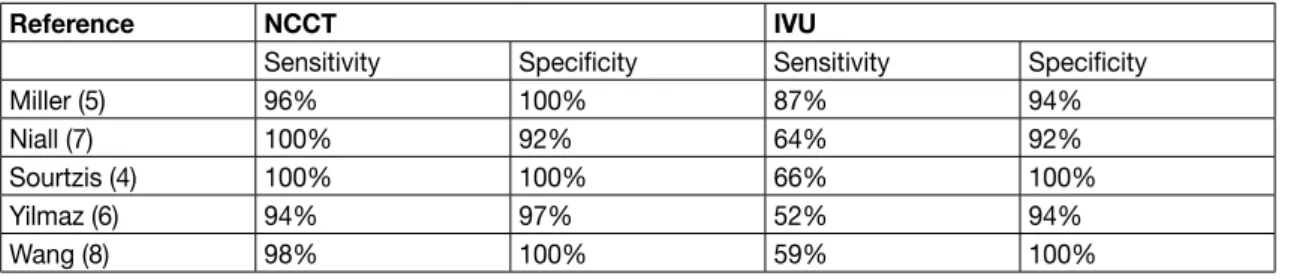
NCCT has become the standard for diagnosing acute flank pain, and has replaced intravenous urography (IVU), which was the gold standard for many years. NCCT can determine stone diameter and density. When stones are absent, the cause of abdominal pain should be identified.
Compared to IVU, NCCT shows higher sensitivity and specificity for identifying urinary stones (Table 3.1) (4-9). Table 3.1: Comparison of NCCT and IVU*
Reference NCCT IVU
Sensitivity Specificity Sensitivity Specificity
Miller (5) 96% 100% 87% 94%
Niall (7) 100% 92% 64% 92%
Sourtzis (4) 100% 100% 66% 100%
Yilmaz (6) 94% 97% 52% 94%
Wang (8) 98% 100% 59% 100%
Recommendation LE GR NCCT should be used to confirm stone diagnosis in patients with acute flank pain, because it
is superior to IVU (10).
1a A
NCCT can detect uric acid and xanthine stones, which are radiolucent on plain films, but not indinavir stones (11).
NCCT can determine stone density, inner structure of the stone and skin-to-stone distance; all of which affect extracorporeal shock wave lithotripsy (SWL) outcome (12-15). The advantage of non-contrast imaging must be balanced against loss of information about renal function and urinary collecting system anatomy, as well as higher radiation dose (Table 3.2).
Radiation risk can be reduced by dose CT (16). In patients with body mass index (BMI) < 30, low-dose CT has been shown to have sensitivity of 86% for detecting ureteric stones < 3 mm and 100% for calculi > 3 mm (17). A meta-analysis of prospective studies (18) has shown that low-dose CT diagnosed urolithiasis with a pooled sensitivity of 96.6% (95% CI: 95.0-97.8) and specificity of 94.9% (95% CI: 92.0-97.0).
Table 3.2: Radiation exposure of imaging modalities (19-22)
Method Radiation exposure (mSv)
KUB radiography 0.5-1 IVU 1.3-3.5 Regular-dose NCCT 4.5-5 Low-dose NCCT 0.97-1.9 Enhanced CT 25-35 Recommendation LE GR
If NCCT is indicated in patients with BMI < 30, use a low-dose technique. 1b A
3.1.2 Evaluation of patients for whom further treatment of renal stones is planned
Recommendation LE GR
A contrast study is recommended if stone removal is planned and the anatomy of the renal collecting system needs to be assessed.
3 A*
Enhanced CT is preferable because it enables 3D reconstruction of the collecting system, as well as measurement of stone density and skin-to-stone distance. IVU may also be used. * Upgraded based on panel consensus.
3.1.3 References
1. Ray AA, Ghiculete D, Pace KT, et al. Limitations to ultrasound in the detection and measurement of urinary tract calculi. Urology 2010 Aug;76(2):295-300.
http://www.ncbi.nlm.nih.gov/pubmed/20206970
2. Heidenreich A, Desgrandschamps F, Terrier F. Modern approach of diagnosis and management of acute flank pain: review of all imaging modalities. Eur Urol 2002 Apr;41(4):351-62.
http://www.ncbi.nlm.nih.gov/pubmed/12074804
3. Kennish SJ, Bhatnagar P, Wah TM, et al. Is the KUB radiograph redundant for investigating acute ureteric colic in the non-contrast enhanced computed tomography era? Clin Radiol 2008 Oct;63(10):1131-5.
http://www.ncbi.nlm.nih.gov/pubmed/18774360
4. Sourtzis S, Thibeau JF, Damry N, et al. Radiologic investigation of renal colic: unenhanced helical CT compared with excretory urography. AJR Am J Roentgenol 1999 Jun;172(6):1491-4.
http://www.ncbi.nlm.nih.gov/pubmed/10350278
5. Miller OF, Rineer SK, Reichard SR, et al. Prospective comparison of unenhanced spiral computed tomography and intravenous urogram in the evaluation of acute flank pain. Urology 1998 Dec;52(6):982-7.
http://www.ncbi.nlm.nih.gov/pubmed/9836541
6. Yilmaz S, Sindel T, Arslan G, et al. Renal colic: comparison of spiral CT, US and IVU in the detection of ureteral calculi. Eur Radiol 1998;8(2):212-7.
http://www.ncbi.nlm.nih.gov/pubmed/9477267
7. Niall O, Russell J, MacGregor R, et al. A comparison of noncontrast computerized tomography with excretory urography in the assessment of acute flank pain. J Urol 1999 Feb;161(2):534-7.
8. Wang JH, Shen SH, Huang SS, et al. Prospective comparison of unenhanced spiral computed tomography and intravenous urography in the evaluation of acute renal colic. J Chin Med Assoc 2008 Jan;71(1):30-6.
http://www.ncbi.nlm.nih.gov/pubmed/18218557
9. Shine S. Urinary calculus: IVU vs. CT renal stone? A critically appraised topic. Abdom Imaging 2008 Jan-Feb;33(1):41-3.
http://www.ncbi.nlm.nih.gov/pubmed/17786506
10. Worster A, Preyra I, Weaver B, et al. The accuracy of noncontrast helical computed tomography versus intravenous pyelography in the diagnosis of suspected acute urolithiasis: a meta-analysis. Ann Emerg Med 2002 Sep;40(3):280-6.
http://www.ncbi.nlm.nih.gov/pubmed/12192351
11. Wu DS, Stoller ML. Indinavir urolithiasis. Curr Opin Urol 2000 Nov;10(6):557-61. http://www.ncbi.nlm.nih.gov/pubmed/11148725
12. El-Nahas AR, El-Assmy AM, Mansour O, et al. A prospective multivariate analysis of factors predicting stone disintegration by extracorporeal shock wave lithotripsy: the value of high-resolution noncontrast computed tomography. Eur Urol 2007 Jun;51(6):1688-93;discussion 93-4.
http://www.ncbi.nlm.nih.gov/pubmed/17161522
13. Patel T, Kozakowski K, Hruby G, et al. Skin to stone distance is an independent predictor of stone-free status following shockwave lithotripsy. J Endourol 2009 Sep;23(9):1383-5.
http://www.ncbi.nlm.nih.gov/pubmed/19694526
14. Kim SC, Burns EK, Lingeman JE, et al. Cystine calculi: correlation of CT-visible structure, CT number, and stone morphology with fragmentation by shock wave lithotripsy. Urol Res 2007 Dec;35(6):319-24. http://www.ncbi.nlm.nih.gov/pubmed/17965956
15. Zarse CA, Hameed TA, Jackson ME, et al. CT visible internal stone structure, but not Hounsfield unit value, of calcium oxalate monohydrate (COM) calculi predicts lithotripsy fragility in vitro. Urol Res 2007 Aug;35(4):201-6.
http://www.ncbi.nlm.nih.gov/pubmed/17565491
16. Jellison FC, Smith JC, Heldt JP, et al. Effect of low dose radiation computerized tomography protocols on distal ureteral calculus detection. J Urol 2009 Dec;182(6):2762-7.
http://www.ncbi.nlm.nih.gov/pubmed/19837431
17. Poletti PA, Platon A, Rutschmann OT, et al. Low-dose versus standard-dose CT protocol in patients with clinically suspected renal colic. AJR Am J Roentgenol 2007 Apr;188(4):927-33.
http://www.ncbi.nlm.nih.gov/pubmed/17377025
18. Niemann T, Kollmann T, Bongartz G. Diagnostic performance of low-dose CT for the detection of urolithiasis: a meta-analysis. AJR Am J Roentgenol 2008 Aug;191(2):396-401.
http://www.ncbi.nlm.nih.gov/pubmed/18647908
19. Kluner C, Hein PA, Gralla O, et al. Does ultra-low-dose CT with a radiation dose equivalent to that of KUB suffice to detect renal and ureteral calculi? J Comput Assist Tomogr 2006 Jan-Feb;30(1):44-50. http://www.ncbi.nlm.nih.gov/pubmed/16365571
20. Caoili EM, Cohan RH, Korobkin M, et al. Urinary tract abnormalities: initial experience with multi-detector row CT urography. Radiology 2002 Feb;222(2):353-60.
http://www.ncbi.nlm.nih.gov/pubmed/11818599
21. Van Der Molen AJ, Cowan NC, Mueller-Lisse UG, et al. CT urography: definition, indications and techniques. A guideline for clinical practice. Eur Radiol 2008 Jan;18(1):4-17.
http://www.ncbi.nlm.nih.gov/pubmed/17973110
22. Thomson JM, Glocer J, Abbott C, et al. Computed tomography versus intravenous urography in diagnosis of acute flank pain from urolithiasis: a randomized study comparing imaging costs and radiation dose. Australas Radiol 2001 Aug;45(3):291-7.
http://www.ncbi.nlm.nih.gov/pubmed/11531751
3.2
Diagnostics - metabolism-related
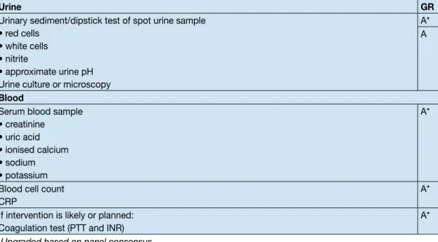
Each emergency patient with urolithiasis needs a succinct biochemical work-up of urine and blood besides imaging. At that point, no distinction is made between high- and low-risk patients.
Table 3.3: Recommendations: basic laboratory analysis - emergency urolithiasis patients (1-4)
Urine GR
Urinary sediment/dipstick test of spot urine sample • red cells
• white cells • nitrite
• approximate urine pH Urine culture or microscopy
A* A
Blood
Serum blood sample • creatinine • uric acid • ionised calcium • sodium • potassium A*
Blood cell count CRP
A* If intervention is likely or planned:
Coagulation test (PTT and INR)
A* * Upgraded based on panel consensus.
CPR = C-reactive protein; INR = international normalised ratio; PTT = partial thromboplastin time.
3.2.1 Basic laboratory analysis - non-emergency urolithiasis patients
Biochemical work-up is similar for all stone patients. However, if no intervention is planned, examination of sodium, potassium, CRP, and blood coagulation time can be omitted.
Only patients at high risk for stone recurrence should undergo a more specific analytical programme (4). Stone-specific metabolic evaluation is described in Chapter 11.
The easiest means to achieve correct diagnosis is by analysis of a passed stone using a valid method as listed below (see 3.2.2). Once mineral composition is known, the potential metabolic disorders can be identified.
3.2.2 Analysis of stone composition
Stone analysis should be performed in all first-time stone formers. In clinical practice, repeat stone analysis is needed in case of: • recurrence under pharmacological prevention;
• early recurrence after interventional therapy with complete stone clearance; • late recurrence after a prolonged stone-free period (6).
Patients should be instructed to filter their urine to retrieve a concrement for analysis. Stone passage and restoration of normal renal function should be confirmed.
The preferred analytical procedures are infrared spectroscopy (IRS) or X-ray diffraction (XRD) (5,7,8). Equivalent results can be obtained by polarisation microscopy, but only in centres with expertise. Chemical analysis (wet chemistry) is generally deemed to be obsolete (5).
Recommendations LE GR
Always perform stone analysis in first-time formers using a valid procedure (XRD or IRS). 2 A Repeat stone analysis in patients:
• presenting with reccurent stones despite drug therapy; • with early recurrence after complete stone clearance;
• with late recurrence after a long stone-free period because stone composition may change (3).
3.3
References
1. S-3 Guideline AWMF-Register-Nr. 043/044 Urinary Tract Infections. Epidemiology, diagnostics, therapy and management of uncomplicated bacterial community acquired urinary tract infections in adults. http://www.awmf.org/leitlinien/detail/ll/043-044.html
2. Hesse AT, Tiselius H-G. Siener R, et al. (Eds). Urinary Stones, Diagnosis, Treatment and Prevention of Recurrence. 3rd edn. Basel, S.Karger AG, 2009. ISBN 978-3-8055-9149-2.
3. Pearle MS, Asplin JR, Coe FL, et al (Committee 3). Medical management of urolithiasis. In: 2nd
International consultation on Stone Disease. Denstedt J, Khoury S. eds. pp. 57-84. Health Publications
2008, ISBN 0-9546956-7-4.
http://www.icud.info/publications.html
4. Straub M, Strohmaier WL, Berg W, et al. Diagnosis and metaphylaxis of stone disease. Consensus concept of the National Working Committee on Stone Disease for the upcoming German Urolithiasis Guideline. World J Urol 2005 Nov;23(5):309-23.
http://www.ncbi.nlm.nih.gov/pubmed/16315051
5. Hesse A, Kruse R, Geilenkeuser WJ, et al. Quality control in urinary stone analysis: results of 44 ring trials (1980-2001). Clin Chem Lab Med 2005;43(3):298-303.
http://www.ncbi.nlm.nih.gov/pubmed/15843235
6. Mandel N, Mandel I, Fryjoff K, et al. Conversion of calcium oxalate to calcium phosphate with recurrent stone episodes. J Urol 2003 Jun;169(6):2026-9.
http://www.ncbi.nlm.nih.gov/pubmed/12771710
7. Suror DJ, Scheidt S. Identification standards for human urinary calculus components, using crystallographic methods. Br J Urol 1968 Feb;40(1):22-8.
http://www.ncbi.nlm.nih.gov/pubmed/5642759
8. Abdel-Halim RE, Abdel-Halim MR. A review of urinary stone analysis techniques. Saudi Med J 2006 Oct;27(10):1462-7.
http://www.ncbi.nlm.nih.gov/pubmed/17013464
4. TREATMENT OF PATIENTS WITH RENAL COLIC
4.1
Renal colic
4.1.1 Pain relief
Pain relief is the first therapeutic step in patients with an acute stone episode (1,2).
Non-steroidal anti-inflammatory drugs (NSAIDs) are effective in patients with acute stone colic (3-6), and have better analgesic efficacy than opioids. Patients receiving NSAIDs are less likely to require further analgesia in the short term.
Opioids, particularly pethidine, are associated with a high rate of vomiting compared to NSAIDs, and carry a greater likelihood of further analgesia being needed (7,8) (Section 4.1.3). If an opioid is used, it is recommended that it is not pethidine.
Statement LE
For symptomatic ureteral stones, urgent SWL as first-line treatment is a feasible option (9). 1b
Recommendations GR
In acute stone episodes, pain relief should be initiated immediately. A Whenever possible, an NSAID should be the first drug of choice. A
4.1.2 Prevention of recurrent renal colic
Facilitation of passage of ureteral stones is discussed in Section 5.3.
For patients with ureteral stones that are expected to pass spontaneously, NSAID tablets or suppositories (e.g., diclofenac sodium, 100-150 mg/day, 3-10 days) may help reduce inflammation and risk of recurrent pain (8,10,11). Although diclofenac can affect renal function in patients with already reduced function, it has no effect in patients with normal kidney function (LE: 1b) (12).
In a double-blind, placebo-controlled trial, recurrent pain episodes of stone colic were significantly fewer in patients treated with NSAIDs (as compared to no NSAIDs) during the first 7 days of treatment (11).
Daily α-blockers reduce recurrent colic (LE: 1a) (Section 5.3) (13,14).
If analgesia cannot be achieved medically, drainage, using stenting or percutaneous nephrostomy, or stone removal, should be performed.
4.1.3 Recommendations for analgesia during renal colic
LE GR
First choice: start with an NSAID, e.g. diclofenac*, indomethacin or ibuprofen**. 1b A Second choice: hydromorphine, pentazocine or tramadol. 4 C
Use α-blockers to reduce recurrent colics. 1a A
*Affects glomerular filtration rate (GFR) in patients with reduced renal function (15) (LE: 2a). **Recommended to counteract recurrent pain after ureteral colic.
4.1.4 References
1. Phillips E, Kieley S, Johnson EB, et al. Emergency room management of ureteral calculi: current practices. J Endourol 2009 Jun;23(6):1021-4.
http://www.ncbi.nlm.nih.gov/pubmed/19445640
2. Micali S, Grande M, Sighinolfi MC, et al. Medical therapy of urolithiasis. J Endourol 2006 Nov;20(11):841-7.
http://www.ncbi.nlm.nih.gov/pubmed/17144848
3. Ramos-Fernández M, Serrano LA. Evaluation and management of renal colic in the emergency department. Bol Asoc Med P R 2009 Jul-Sep;101(3):29-32.
http://www.ncbi.nlm.nih.gov/pubmed/20120983
4. Engeler DS, Schmid S, Schmid HP. The ideal analgesic treatment for acute renal colic--theory and practice. Scand J Urol Nephrol 2008;42(2):137-42.
http://www.ncbi.nlm.nih.gov/pubmed/17899475
5. Cohen E, Hafner R, Rotenberg Z, et al. Comparison of ketorolac and diclofenac in the treatment of renal colic. Eur J Clin Pharmacol 1998 Aug;54(6):455-8.
http://www.ncbi.nlm.nih.gov/pubmed/9776434
6. Shokeir AA, Abdulmaaboud M, Farage Y, et al. Resistive index in renal colic: the effect of nonsteroidal anti-inflammatory drugs. BJU Int 1999 Aug;84(3):249-51.
http://www.ncbi.nlm.nih.gov/pubmed/10468715
7. Holdgate A, Pollock T. Nonsteroidal anti-inflammatory drugs (NSAIDs) versus opioids for acute renal colic. Cochrane Database Syst Rev 2005 Apr;(2):CD004137.
http://www.ncbi.nlm.nih.gov/pubmed/15846699
8. Ebell MH. NSAIDs vs. opiates for pain in acute renal colic. Am Fam Physician 2004 Nov;70(9):1682. http://www.ncbi.nlm.nih.gov/pubmed/15554485
9. Picozzi SC, Ricci C, Gaeta M, et al. Urgent shock wave lithotripsy as first-line treatment for ureteral stones: a meta-analysis of 570 patients. Urol Res 2012 Dec;40(6):725-31.
http://www.ncbi.nlm.nih.gov/pubmed/22699356
10. Holdgate A, Pollock T. Systematic review of the relative efficacy of non-steroidal anti-inflammatory drugs and opioids in the treatment of acute renal colic. BMJ 2004 Jun;328(7453):1401.
http://www.ncbi.nlm.nih.gov/pubmed/15178585
11. Laerum E, Ommundsen OE, Gronseth JE, et al. Oral diclofenac in the prophylactic treatment of recurrent renal colic. A double-blind comparison with placebo. Eur Urol 1995;28(2):108-11. http://www.ncbi.nlm.nih.gov/pubmed/8529732
12. Lee A, Cooper MG, Craig JC, et al. Effects of nonsteroidal anti-inflammatory drugs on postoperative renal function in adults with normal renal function. Cochrane Database Syst Rev 2007;18(2):CD002765.
http://www.ncbi.nlm.nih.gov/pubmed/17443518
13. Dellabella M, Milanese G, Muzzonigro G. Randomized trial of the efficacy of tamsulosin, nifedipine and phloroglucinol in medical expulsive therapy for distal ureteral calculi. J Urol 2005 Jul;174(1):167-72. http://www.ncbi.nlm.nih.gov/pubmed/15947613
14. Resim S, Ekerbicer H, Ciftci A. Effect of tamsulosin on the number and intensity of ureteral colic in patients with lower ureteral calculus. Int J Urol 2005 Jul;12(7):615- 20.
http://www.ncbi.nlm.nih.gov/pubmed/16045553
15. Walden M, Lahtinen J, Elvander E. Analgesic effect and tolerance of ketoprofen and diclofenac in acute ureteral colic. Scand J Urol Nephrol 1993;27(3):323-5.
4.2
Management of sepsis in obstructed kidney
The obstructed kidney with all signs of urinary tract infection (UTI) is a urological emergency. Urgent decompression is often necessary to prevent further complications in infected hydronephrosis secondary to stone-induced, unilateral or bilateral renal obstruction.
The optimal method of decompression has yet to be established (1-3). However, it is known that compromised delivery of antibiotics into the obstructed kidney means that the collecting system must be drained to encourage resolution of infection.
4.2.1 Decompression
Currently, there are two options for urgent decompression of obstructed collecting systems: • placement of an indwelling ureteral catheter;
• percutaneous placement of a nephrostomy catheter.
There is little evidence to support the superiority of percutaneous nephrostomy over retrograde stenting for primary treatment of infected hydronephrosis. There is no good-quality evidence to suggest that ureteric stenting has more complications than percutaneous nephrostomy (1,4,5).
Only two RCTs (2,5) have assessed decompression of acute infected hydronephrosis. The
complications of percutaneous nephrostomy insertion have been reported consistently, but those of ureteric stent insertion are less well described (1). Definitive stone removal should be delayed until the infection is cleared following a complete course of antimicrobial therapy (6,7).
Emergency nephrectomy may become necessary in highly complicated cases to eliminate further complications.
Statement LE
For decompression of the renal collecting system, ureteral stents and percutaneous nephrostomy catheters are equally effective.
1b
Recommendations LE GR
For sepsis with obstructing stones, the collecting system should be urgently decompressed, using percutaneous drainage or ureteral stenting.
1b A Definitive treatment of the stone should be delayed until sepsis is resolved. 1b A
4.2.2 Further measures
Following urgent decompression of the obstructed and infected urinary collecting system, both urine- and blood samples should be sent for culture-antibiogram sensitivity testing, and antibiotics should be initiated immediately thereafter. The regimen should be re-evaluated in the light of the culture-antibiogram test. Intensive care might become necessary.
Recommendations GR
Collect urine for antibiogram test following decompression. A* Start antibiotics immediately thereafter (+ intensive care if necessary).
Re-evaluate antibiotic regimen following antibiogram findings. * Upgraded based on panel consensus.
4.2.3 References
1. Ramsey S, Robertson A, Ablett MJ, et al. Evidence-based drainage of infected hydronephrosis secondary to ureteric calculi. J Endourol 2010 Feb;24(2):185-9.
http://www.ncbi.nlm.nih.gov/pubmed/20063999
2. Pearle MS, Pierce HL, Miller GL, et al. Optimal method of urgent decompression of the collecting system for obstruction and infection due to ureteral calculi. J Urol 1998 Oct;160(4):1260-4. http://www.ncbi.nlm.nih.gov/pubmed/9751331
3. Uppot RN. Emergent nephrostomy tube placement for acute urinary obstruction. Tech Vasc Interv Radiol 2009 Jun;12(2):154-61.
http://www.ncbi.nlm.nih.gov/pubmed/19853233
4. Lynch MF, Anson KM, Patel U. Percutaneous nephrostomy and ureteric stent insertion for acute renal deobstruction. Consensus based guidelines. Br J Med Surg Urol 2008 Nov;1(3);120-5.
5. Mokhmalji H, Braun PM, Portillo FJ, et al. Percutaneous nephrostomy versus ureteral stents for diversion of hydronephrosis caused by stones: A prospective, randomized clinical trial. J Urol 2001 Apr;165(4):1088-92.
http://www.ncbi.nlm.nih.gov/pubmed/11257644
6. Klein LA, Koyle M, Berg S. The emergency management of patients with ureteral calculi and fever. J Urol 1983 May;129(5):938-40.
http://www.ncbi.nlm.nih.gov/pubmed/6854761
7. Camúñez F, Echenagusia A, Prieto ML, et al. Percutaneous nephrostomy in pyonephrosis. Urol Radiol 1989;11(2):77-81.
http://www.ncbi.nlm.nih.gov/pubmed/2667249
5. STONE RELIEF
When deciding between active stone removal and conservative treatment with medical expulsive therapy (MET), it is important to consider all the patients’ circumstances that may affect treatment decisions (1).
5.1
Observation of ureteral stones
5.1.1 Stone-passage rates
There are only limited data about spontaneous stone passage according to size (2,3). A meta-analysis of 328 patients harbouring ureteral stones < 10 mm investigated the likelihood of ureteral stone passage (Table 5.1) (2). These studies had limitations including non-standardisation of stone size measurement, and lack of analysis of stone position, stone-passage history, and time to stone passage.
Table 5.1: Likelihood of ureteral stone passage of ureteral stones (2)
Stone size Average time to pass Percentage of passages (95% CI)
< 5 mm (n = 224) 68% (46-85%)
> 5 mm (n = 104) 47% (36-58%)
< 2 mm 31 days
2-4 mm 40 days
4-6 mm 39 days
95% of stones up to 4 mm pass within 40 days (3).
Recommendations LE GR
In patients with newly diagnosed ureteral stones < 10 mm, and if active removal is not indicated (Chapter 6), observation with periodic evaluation is an optional initial treatment.
1a A
Such patients may be offered appropriate medical therapy to facilitate stone passage during observation.*
*see Section 5.3, Medical expusive therapy (MET).
5.2
Observation of kidney stones
Observation of kidney stones, especially in calices, depends on their natural history (Section 6.2.1).
Statement LE
It is still debatable whether kidney stones should be treated, or whether annual follow-up is sufficient for asymptomatic caliceal stones that have remained stable for 6 months.
4
Recommendations GR
Kidney stones should be treated in case of growth, formation of de novo obstruction, associated infection, and acute or chronic pain.
A* Comorbidity and patient preference need to be taken into consideration when making treatment
decisions.
C If kidney stones are not treated, periodic evaluation is needed. A* * Upgraded based on panel consensus.
5.3
Medical expulsive therapy (MET)
Drugs that expel stones might act by relaxing ureteral smooth muscle through inhibition of calcium channel pumps or α-1 receptor blockade (4,5).
MET should only be used in patients who are comfortable with this approach and when there is no obvious advantage from immediate active stone removal.
Meta-analyses have shown that patients with ureteral stones treated with α-blockers or nifedipine are more likely to pass stones with fewer episodes of colic than those not receiving such therapy (4,5).
Statement LE
There is good evidence that MET accelerates spontaneous passage of ureteral stones and fragments generated with SWL, and limits pain (4-16).
1a
5.3.1 Medical agents
Tamsulosin is one of the most commonly used α-blockers (4,6,17-20). However, one small study has suggested that tamsulosin, terazosin and doxazosin are equally effective, indicating a possible class effect (21). This is also indicated by several trials demonstrating increased stone expulsion using doxazosin (4,21,22), terazosin (21,23), alfuzosin (24-27) naftopidil (28,29), and silodosin (30,31).
Statement LE
Several trials have demonstrated an α-blocker class effect on stone expulsion rates. 1b With regard to the class effect of calcium-channel blockers, only nifedipine has been investigated (LE = 1a) (4,9-11,32,33).
Administration of tamsulosin and nifedipine is safe and effective in patients with distal ureteral stones with renal colic. However, tamsulosin is significantly better than nifedipine in relieving renal colic and facilitating and accelerating ureteral stone expulsion (11,32,33).
Based on studies with a limited number of patients (34,35) (LE: 1b), no recommendation for the use of corticosteroids in combination with α-blockers in MET can be made.
Statement LE
There is no evidence to support the use of corticosteroids as monotherapy for MET. Insufficient data exist to support the use of corticosteroids in combination with α-blockers as an accelerating adjunct (3,21,34,35).
1b
Recommendations for MET LE GR
For MET, α-blockers are recommended. 1a A
Patients should be counseled about the attendant risks of MET, including associated drug side effects, and should be informed that it is administered off-label†**.
A* Patients, who elect for an attempt at spontaneous passage or MET, should have
well-controlled pain, no clinical evidence of sepsis, and adequate renal functional reserve.
A Patients should be followed once between 1 and 14 days to monitor stone position and be
assessed for hydronephrosis.
4 A*
† It is not known if tamsulosin harms the human foetus or if it is found in breast milk.
* Upgraded based on panel consensus.
**MET in children cannot be recommended due to the limited data in this specific population.
5.3.2 Factors affecting success of medical expulsive therapy (tamsulosin)
5.3.2.1 Stone size
Due to the high likelihood of spontaneous passage of stones up to ~5 mm, MET is less likely to increase the stone-free rate (SFR) (5,36-39) (LE: 1b). However, MET does reduce the need for analgesics (4,6) (LE: 1a). 5.3.2.2 Stone location
The vast majority of trials have investigated distal ureteral stones (4). One RCT has assessed the effect of tamsulosin on spontaneous passage of proximal ureteral calculi 5-10 mm. The main effect was to encourage stone migration to a more distal part of the ureter (40) (LE: 1b).
5.3.2.3 Medical expulsive therapy after extracorporeal shock wave lithotripsy (SWL)
expedite expulsion and increase SFRs and reduce analgesic requirements (7,12,41-49) (LE: 1a). 5.3.2.4 Medical expulsive therapy after ureteroscopy
MET following holmium:YAG laser lithotripsy increases SFRs and reduces colic episodes (50) (LE: 1b). 5.3.2.5 Medical expulsive therapy and ureteral stents (Section 5.6.2.1.8)
5.3.2.6 Duration of medical expulsive therapy treatment
Most studies have had a duration of 1 month or 30 days. No data are currently available to support other time-intervals.
5.3.2.7 Possible side-effects include retrograde ejaculation and hypotension (4).
5.3.3 References
1. Skolarikos A, Laguna MP, Alivizatos G, et al. The role for active monitoring in urinary stones: a systematic review. J Endourol 2010 Jun;24(6):923-30.
http://www.ncbi.nlm.nih.gov/pubmed/20482232
2. Preminger GM, Tiselius HG, Assimos DG, et al. American Urological Association Education and Research, Inc; European Association of Urology. 2007 Guideline for the management of ureteral calculi. Eur Urol 2007 Dec;52(6):1610-31.
http://www.ncbi.nlm.nih.gov/pubmed/18074433
3. Miller OF, Kane CJ. Time to stone passage for observed ureteral calculi: a guide for patient education. J Urol 1999 Sep;162(3 Pt 1):688-90;discussion 690-1.
http://www.ncbi.nlm.nih.gov/pubmed/10458343
4. Seitz C, Liatsikos E, Porpiglia F, et al. Medical Therapy to Facilitate the Passage of Stones: What Is the Evidence? Eur Urol 2009 Sep;56(3):455-71.
http://www.ncbi.nlm.nih.gov/pubmed/19560860
5. Liatsikos EN, Katsakiori PF, Assimakopoulos K, et al. Doxazosin for the management of distal-ureteral stones. J Endourol 2007 May;21(5):538-41.
http://www.ncbi.nlm.nih.gov/pubmed/17523910
6. Hollingsworth JM, Rogers MA, Kaufman SR, et al. Medical therapy to facilitate urinary stone passage: a meta-analysis. Lancet 2006 Sep;368(9542):1171-9.
http://www.ncbi.nlm.nih.gov/pubmed/17011944
7. Gravina GL, Costa AM, Ronchi P, et al. Tamsulosin treatment increases clinical success rate of single extracorporeal shock wave lithotripsy of renal stones. Urology 2005 Jul;66(1):24-8.
http://www.ncbi.nlm.nih.gov/pubmed/15992885
8. Resim S, Ekerbicer HC, Ciftci A. Role of tamsulosin in treatment of patients with steinstrasse developing after extracorporeal shock wave lithotripsy. Urology 2005 Nov;66(5):945-8. http://www.ncbi.nlm.nih.gov/pubmed/16286100
9. Borghi L, Meschi T, Amato F, et al. Nifedipine and methylprednisolone in facilitating ureteral stone passage: a randomized, double-blind, placebo-controlled study. J Urol 1994 Oct;152(4):1095-8. http://www.ncbi.nlm.nih.gov/pubmed/8072071
10. Porpiglia F, Destefanis P, Fiori C, et al. Effectiveness of nifedipine and deflazacort in the management of distal ureter stones. Urology 2000 Oct;56(4):579-82.
http://www.ncbi.nlm.nih.gov/pubmed/11018608
11. Dellabella M, Milanese G, Muzzonigro G. Randomized trial of the efficacy of tamsulosin, nifedipine and phloroglucinol in medical expulsive therapy for distal ureteral calculi. J Urol 2005 Jul;174(1):167-72. http://www.ncbi.nlm.nih.gov/pubmed/15947613
12. Naja V, Agarwal MM, Mandal AK, et al. Tamsulosin facilitates earlier clearance of stone fragments and reduces pain after shockwave lithotripsy for renal calculi; results from an open-label randomized study. Urology 2008 Nov;72(5):1006-11.
http://www.ncbi.nlm.nih.gov/pubmed/18799202
13. Schuler TD, Shahani R, Honey RJ, et al. Medical expulsive therapy as an adjunct to improve shockwave lithotripsy outcomes: a systematic review and meta-analysis. J Endourol 2009 Mar;23(3):387-93.
http://www.ncbi.nlm.nih.gov/pubmed/19245302
14. Parsons JK, Hergan LA, Sakamoto K, et al. Efficacy of alpha blockers for the treatment of ureteral stones. J Urol 2007 Mar;177(3):983-7.
15. Singh A, Alter HJ, Littlepage A. A systematic review of medical therapy to facilitate passage of ureteral calculi. Ann Emerg Med 2007 Nov;50(5):552-63.
http://www.ncbi.nlm.nih.gov/pubmed/17681643
16. Arrabal-Martin M, Valle-Diaz de la Guardia F, Arrabal-Polo MA, et al. Treatment of ureteral lithiasis with tamsulosin: literature review and meta-analysis. Urol Int 2010;84(3):254-9.
http://www.ncbi.nlm.nih.gov/pubmed/20389151
17. Lojanapiwat B, Kochakarn W, Suparatchatpan N, et al. Effectiveness of low-dose and standard-dose tamsulosin in the treatment of distal ureteric stones: A randomized controlled study. J Int Med Res 2008 May-Jun;36(3):529-36.
http://www.ncbi.nlm.nih.gov/pubmed/18534135
18. Wang CJ, Huang SW, Chang CH. Efficacy of an alpha1 blocker in expulsive therapy of lower ureteral stones. J Endourol 2008 Jan;22(1):41-6.
http://www.ncbi.nlm.nih.gov/pubmed/18315472
19. Kaneko T, Matsushima H, Morimoto H, et al. Efficacy of low dose tamsulosin medical expulsive therapy for ureteral stones in Japanese male patients: a randomized controlled study. Int J Urol 2010 May;17(5):462-5.
http://www.ncbi.nlm.nih.gov/pubmed/20202002
20. Al-Ansari A, Al-Naimi A, Alobaidy A, et al. Efficacy of tamsulosin in the management of lower ureteral stones: a randomized double-blind placebo-controlled study of 100 patients. Urology 2010 Jan;75(1):4-7.
http://www.ncbi.nlm.nih.gov/pubmed/20109697
21. Yilmaz E, Batislam E, Basar MM, et al. The comparison and efficacy of 3 different alpha1-adrenergic blockers for distal ureteral stones. J Urol 2005 Jun;173(6):2010-2.
http://www.ncbi.nlm.nih.gov/pubmed/15879806
22. Zehri AA, Ather MH, Abbas F, et al. Preliminary study of efficacy of doxazosin as a medical expulsive therapy of distal ureteric stones in a randomized clinical trial. Urology 2010 Jun;75(6):1285-8. http://www.ncbi.nlm.nih.gov/pubmed/20189226
23. Mohseni MG, Hosseini SR, Alizadeh F. Efficacy of terazosin as a facilitator agent for expulsion of the lower ureteral stones. Saudi Med J 2006 Jun;27(6):838-40.
http://www.ncbi.nlm.nih.gov/pubmed/16758046
24. Agrawal M, Gupta M, Gupta A, et al. Prospective Randomized Trial Comparing Efficacy of Alfuzosin and Tamsulosin in Management of Lower Ureteral Stones. Urology 2009 Apr;73(4):706-9.
http://www.ncbi.nlm.nih.gov/pubmed/19193417
25. Pedro RN, Hinck B, Hendlin K, et al. Alfuzosin stone expulsion therapy for distal ureteral calculi: a double-blind, placebo controlled study. J Urol 2008 Jun;179(6):2244-7, discussion 2247.
http:/www.ncbi.nlm.nih.gov/pubmed/18423747
26. Ahmed AF, Al-Sayed AY. Tamsulosin versus Alfuzosin in the Treatment of Patients with Distal Ureteral Stones: Prospective, Randomized, Comparative Study. Korean J Urol 2010 Mar;51(3):193-7.
http://www.ncbi.nlm.nih.gov/pubmed/20414396
27. Chau LH, Tai DC, Fung BT, et al. Medical expulsive therapy using alfuzosin for patient presenting with ureteral stone less than 10mm: a prospective randomized controlled trial. Int J Urol 2011 Jul;18(7): 510-4.
http://www.ncbi.nlm.nih.gov/pubmed/21592234
28. Sun X, He L, Ge W, et al. Efficacy of selective alpha1D-Blocker Naftopidil as medical expulsive therapy for distal ureteral stones. J Urol 2009 Apr;181(4):1716-20.
http://www.ncbi.nlm.nih.gov/pubmed/19233432
29. Zhou SG, Lu JL, Hui JH. Comparing efficacy of < (1)D-receptor antagonist naftopidil and < 1A/ Dreceptor antagonist tamsulosin in management of distal ureteral stones. World J Urol 2011 Dec;29(6):767-71.
http://www.ncbi.nlm.nih.gov/pubmed/21845472
30. Tsuzaka Y, Matsushima H, Kaneko T, et al. Naftopidil vs silodosin in medical expulsive therapy for ureteral stones: a randomized controlled study in Japanese male patients. Int J Urol 2011 Nov;18(11):792-5.
http://www.ncbi.nlm.nih.gov/pubmed/21917021
31. Itoh Y, Okada A, Yasui T, et al. Efficacy of selective α1A adrenoceptor antagonist silodosin in the medical expulsive therapy for ureteral stones. Int J Urol 2011 Sep;18(9):672-4.
http://www.ncbi.nlm.nih.gov/pubmed/21707766
32. Porpiglia F, Ghignone G, Fiori C, et al. Nifedipine versus tamsulosin for the management of lower ureteral stones. J Urol 2004 Aug;172(2):568-71.
33. Ye Z, Yang H, Li H, et al. A multicentre, prospective, randomized trial: comparative efficacy of tamsulosin and nifedipine in medical expulsive therapy for distal ureteric stones with renal colic. BJU Int 2011 Jul;108(2):276-9.
http://www.ncbi.nlm.nih.gov/pubmed/21083640
34. Porpiglia F, Vaccino D, Billia M, et al. Corticosteroids and tamsulosin in the medical expulsive therapy for symptomatic distal ureter stones: single drug or association? Eur Urol 2006 Aug;50(2):339-44. http://www.ncbi.nlm.nih.gov/pubmed/16574310
35. Dellabella M, Milanese G, Muzzonigro G. Medical-expulsive therapy for distal ureterolithiasis: randomized prospective study on role of corticosteroids used in combination with tamsulosin simplified treatment regimen and health-related quality of life. Urology 2005 Oct;66(4):712-5. http://www.ncbi.nlm.nih.gov/pubmed/16230122
36. Ferre RM, Wasielewski JN, Strout TD, et al. Tamsulosin for ureteral stones in the emergency department: a Randomized controlled trial. Ann Emerg Med 2009 Sep;54(3):432-9.
http://www.ncbi.nlm.nih.gov/pubmed/19200622
37. Hermanns T, Sauermann P, Rufibach K, et al. Is there a role for tamsulosin in the treatment of distal ureteral stones of 7 mm or less? Results of a randomised, double-blind, placebo-controlled trial. Eur Urol 2009 Sep;56(3):407-12.
http://www.ncbi.nlm.nih.gov/pubmed/19375849
38. Vincendeau S, Bellissant E, Houlgatte A, et al; Tamsulosin Study Group. Tamsulosin hydrochloride vs placebo for management of distal ureteral stones: a multicentric, randomized, double-blind trial. Arch Intern Med 2010 Dec;170(22):2021-7.
http://www.ncbi.nlm.nih.gov/pubmed/21149761
39. Ochoa-Gómez R, Prieto-Díaz-Chávez E, Trujillo-Hernández B, et al. Tamsulosin does not have greater efficacy than conventional treatment for distal ureteral stone expulsion in Mexican patients. Urol Res 2011 Dec;39(6)491-5.
http://www.ncbi.nlm.nih.gov/pubmed/21516496
40. Yencilek F, Erturhan S, Canguven O, et al. Does tamsulosin change the management of proximally located ureteral stones? Urol Res 2010 Jun;38(3):195-9.
http://www.ncbi.nlm.nih.gov/pubmed/20182703
41. Bhagat SK, Chacko NK, Kekre NS, et al. Is there a role for tamsulosin in shock wave lithotripsy for renal and ureteral calculi? J Urol 2007 Jun;177(6):2185-8.
http://www.ncbi.nlm.nih.gov/pubmed/17509314
42. Küpeli B, Irkilata L, Gürocak S, et al. Does tamsulosin enhance lower ureteral stone clearance with or without shock wave lithotripsy? Urology 2004 Dec;64(6):1111-5.
http://www.ncbi.nlm.nih.gov/pubmed/15596181
43. Wang H, Liu K, Ji Z, et al. Effect of alpha1-adrenergic antagonists on lower ureteral stones with extracorporeal shock wave lithotripsy. Asian J Surg 2010 Jan;33(1):37-41.
http://www.ncbi.nlm.nih.gov/pubmed/20497881
44. Zhu Y, Duijvesz D, Rovers MM, et al. alpha-Blockers to assist stone clearance after extracorporeal shock wave lithotripsy: a meta-analysis. BJU Int 2010 Jul;106(2):256-61.
http://www.ncbi.nlm.nih.gov/pubmed/19889063
45. Hussein MM. Does tamsulosin increase stone clearance after shockwave lithotripsy of renal stones? A prospective, randomized controlled study. Scand J Urol Nephrol 2010 Feb;44(1):27-31.
http://www.ncbi.nlm.nih.gov/pubmed/19947900
46. Singh SK, Pawar DS, Griwan MS, et al. Role of tamsulosin in clearance of upper ureteral calculi after extracorporeal shock wave lithotripsy: a randomized controlled trial. Urol J 2011 Winter;8(1):14-20. http://www.ncbi.nlm.nih.gov/pubmed/21404197
47. Zheng S, Liu LR, Yuan HC, et al. Tamsulosin as adjunctive treatment after shockwave lithotripsy in patients with upper urinary tract stones: a systematic review and meta-analysis. Scand J Urol Nephrol 2010 Dec;44(6):425-32.
http://www.ncbi.nlm.nih.gov/pubmed/21080841
48. Falahatkar S, Khosropanah I, Vajary AD, et al. Is there a role for tamsulosin after shock wave lithotripsy in the treatment of renal and ureteral calculi? J Endourol 2011 Mar;25(3):495-8.
http://www.ncbi.nlm.nih.gov/pubmed/21166579
49. Singh SK, Pawar DS, Griwan MS, et al. Role of tamsulosin in clearance of upper ureteral calculi after extracorporeal shock wave lithotripsy: a randomized controlled trial. Urol J 2011 Winter;8(1):14-20. http://www.ncbi.nlm.nih.gov/pubmed/21404197
50. John TT, Razdan S. Adjunctive tamsulosin improves stone free rate after ureteroscopic lithotripsy of large renal and ureteric calculi: a prospective randomized study. Urology 2010 May;75(5):1040-2. http://www.ncbi.nlm.nih.gov/pubmed/19819530
5.4
Chemolytic dissolution of stones
Oral or percutaneous irrigation chemolysis of stones or their fragments can be useful first-line therapy. It may also be an adjunct to SWL, percutaneous nephrolithotomy (PNL), ureterorenoscopy (URS) or open surgery to support elimination of small residual fragments, considering that its use as first-line therapy may take several weeks to be effective.
Combined treatment with SWL and chemolysis is a minimally invasive option for patients with partial or complete infection staghorn stones who are not eligible for PNL. Stone fragmentation leads to increased stone surface area and improved efficacy of chemolitholysis.
Chemolysis is possible only for the stone compositions listed below, therefore, knowledge of stone composition is mandatory before treatment.
5.4.1 Percutaneous irrigation chemolysis
Percutaneous irrigation chemolysis may be an option for infection- and uric acid stones (1,2).
Recommendations GR
In percutaneous chemolysis, at least two nephrostomy catheters should be used to allow irrigation of the renal collecting system, while preventing chemolytic fluid draining into the bladder and reducing the risk of increased intrarenal pressure*.
A
Pressure- and flow-controlled systems should be used if available.
* Alternatively, one nephrostomy catheter with a JJ stent and bladder catheter can serve as a through-flow system preventing high pressure.
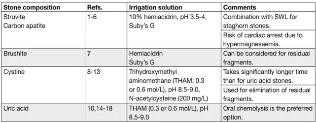
Table 5.2: Methods of percutaneous irrigation chemolysis
Stone composition Refs. Irrigation solution Comments
Struvite Carbon apatite
1-6 10% hemiacidrin, pH 3.5-4, Suby’s G
Combination with SWL for staghorn stones.
Risk of cardiac arrest due to hypermagnesaemia.
Brushite 7 Hemiacidrin
Suby’s G
Can be considered for residual fragments.
Cystine 8-13 Trihydroxymethyl
aminomethane (THAM; 0.3 or 0.6 mol/L), pH 8.5-9.0, N-acetylcysteine (200 mg/L)
Takes significantly longer time than for uric acid stones. Used for elimination of residual fragments.
Uric acid 10,14-18 THAM (0.3 or 0.6 mol/L), pH 8.5-9.0
Oral chemolysis is the preferred option.
Irrigation chemolysis appears to the panel to be used rarely, probably because of the complexity of the technique and the possible side effects.
5.4.2 Oral chemolysis
Oral chemolitholysis is efficient only for uric acid calculi, and is based on alkalinisation of urine by application of alkaline citrate or sodium bicarbonate (3-6).
When chemolitholysis is planned, the pH should be adjusted to 6.5-7.2. Within this range chemolysis is more effective at a higher pH, which, however, might lead to calcium phosphate stone formation.
In case of uric acid obstruction of the collecting system, oral chemolysis in combination with urinary drainage is indicated (7). A combination of alkalinisation with tamsulosin seems to achieve the highest SFRs for distal ureteral stones (8).
Recommendations GR
The dosage of alkalising medication must be modified by the patient according to urine pH, which is a direct consequence of such medication.
A Dipstick monitoring of urine pH by the patient is required at regular intervals during the day. Morning urine must be included.
A The physician should clearly inform the patient of the significance of compliance. A