Gene expression and digit homology in the chicken embryo wing
Monique C. M. Welten, Fons J. Verbeek, Annemarie H. Meijer, and Michael K. Richardson Institute for Biology, Leiden University, Kaiserstraat 63, 2311 GP Leiden, The Netherlands
Author for correspondence (email: [email protected])
SUMMARY The bird wing is of special interest to students of homology and avian evolution. Fossil and developmental data give conflicting indications of digit homology if a pentadactyl ‘‘archetype’’ is assumed. Morphological signs of a vestigial digit I are seen in bird embryos, but no digit-like structure develops in wild-type embryos. To examine the developmental mechanisms of digit loss, we studied the expression of the high-mobility group box containingSox9gene, and bone morphogenetic protein receptor 1b (bmpR-1b)Fmarkers for precondensation and prechondrogenic cells, respectively. We find an elongated domain ofSox9expression, but nobmpR-1b expression, anterior to digit II. We interpret this as a digit I domain that reaches precondensation, but not condensation or
precartilage stages. It develops late, when the tissue in which it is lodged is being remodeled. We consider these findings in the light of previous Hoxd-11 misexpression studies. Together, they suggest that there is a digit I vestige in the wing that can be rescued and undergo development if posterior patterning cues are enhanced. We observed Sox9 expression in the elusive ‘‘element X’’ that is sometimes stated to represent a sixth digit. Indeed, incongruity between digit domains and identities in theropods disappears if birds and other archosaurs are considered primitively polydactyl. Our study provides the first gene expression evidence for at least five digital domains in the chick wing. The failure of the first to develop may be plausibly linked to attenuation of posterior signals.
INTRODUCTION
The homology of avian wing digits is of interest to paleon-tologists and evolutionary biologists because it bears on the important question of the evolution of birds. Digit homology is also important for experimental developmental biologists who use the phenotypes (‘‘identities’’) of digits as markers of position along the anteroposterior axis of the limb. Further-more, because the wing is a well-studied experimental model, it provides opportunities to explore the mechanistic basis of developmental homology. Finally, the history of ideas about the avian wing presents us with an unrivaled catalog of ar-chetypes and recapitulation.
Our aim in this article is not to overturn current hypotheses of avian phylogeny, but to ask how developmental mecha-nisms were modified in the transition from the presumed an-cestral state. We also wish to ask whether alternative hypotheses of avian digit homology have been overlooked. We begin by reviewing some of the relevant evolutionary and developmental issues. (Note: all references to ‘‘digits’’ in this article are to those of the forelimb, unless otherwise stated.)
Avian phylogeny
Archaeopteryx lithographica(Fig. 1) has always been central to the debate about avian origins because of its historical fame as a ‘‘missing link,’’ and its possession of a mosaic of avian and more inclusive theropod characters (Ostrom 1976;
Christiansen and Bonde 2004). Cladistic analyses support the hypothesis that birds belong to the ‘‘Coelurosauria,’’ a clade of theropod dinosaurs (Gauthier 1986; Sereno 1999).
Recent discoveries of nonavian maniraptorans with feath-ers or feather-like coverings (e.g.,Protarchaeopteryx robusta andCaudipteryx zoui; Ji et al. 1998, 2001; Zhou et al. 2003) support the inclusion of birds in Theropoda. The fossil Ra-hona ostromi also shows a mosaic of avian and theropod features, and phylogenetic analysis places it as a sister species to Archaeopteryx (Forster et al. 1998). Although other an-cestries for birds have been proposed (notably the thecodont and crocodilian hypotheses; Tarsitano and Hecht 1980; Hecht and Tarsitano 1982; Thulborn and Hamley 1982; Hecht 1985; Moinar 1985; Walker 1985), we will adopt here the consensus view that birds are theropods (Fig. 1).
Digit position and digit phenotype
Digits are traditionally assigned Roman numerals I–V, with reference to a pentadactyl archetype (Fig. 2). These designa-tions have two different meanings: as positional references, the terms I–V describe the spatial relations of digits along the anteroposterior axis of the limb. By contrast, the phenotypes or identities I–V apply to different complexes of morpholog-ical characters that relate to the skeleton of a digit.
The danger of having one numbering system with two meanings is that circular arguments may be developed about digit homologies that are not independent. Furthermore, it
&BLACKWELL PUBLISHING, INC.
has been questioned whether characters such as digit shape and phalangeal formula can in fact be used to ‘‘identify’’ ho-mologous digits in different species (Goodwin and Trainor 1983). In birds, we are faced with uncertainty both about the positional homologies of the wing digits (because only four distinct digits are seen in the wing), and also their phenotypes (because phalanges and other structures may have been lost or remodeled during evolution).
Positional homologies and the problem of vestigial digits
The homologies of cartilaginous and precartilaginous ele-ments at the postaxial margin of the chicken embryo wing are
a puzzle, but a plausible vestige of metacarpal V can be iden-tified in alcian blue whole mounts (Montagna 1945; Burke and Feduccia 1997). Hinchliffe and Hecht (1984) identified an intriguing triad of postaxial elements: (a) the vestigial meta-carpal V, lying laterally along the proximal part of metameta-carpal IV, and becoming reduced or disappearing completely; (b) the elongated pisiform, lying at the lateral border of the wing, near the palmar aspect of the carpus; and (c) an element ‘‘X,’’ possibly an avian apomorphy, that lies near the proximal end of metacarpal IV and near the palmar aspect of the carpus, and that may persist to adulthood (see also Montagna 1945). Element X was formerly identified as an extension or process of the pisiform (Montagna 1945; Hinchliffe 1977). Part of element X was described by Montagna (1945) as fusing with his ‘‘centrale IV,’’ whereas another part was said to persist in the adult wing as a tuberosity on metacarpal III.
Histogenesis at the pre-axial margin is less clear, despite numerous studies (the classical literature is reviewed by Holm-gren 1933; Montagna 1945). Labeling for early cartilage ma-trix with35SO4(Hinchliffe 1977) fails to find a vestigial digit I.
However, a condensation for distal carpal I was claimed by Montagna (1945). Recently, a small avascular zone (Kundra´t et al. 2002), cell condensation (Kundra´t et al. 2002; Larsson and Wagner 2002), or small cartilage nodule (Feduccia and Nowicki 2002), next to digit II, has been cited as evidence of a digit I vestige in Gallus gallus andStruthio camelus (the os-trich). There is a tuberosity for attachment of extensor carpi alualae radialis longusand ligamentum elasticum prepatagiale at the base of metacarpal II (Yasuda 2002). This tuberosity has been interpreted as evidence of a vestigial digit I, although this interpretation has been contested (Montagna 1945).
A finding of great significance comes from studies of chicken wings experimentally infected withHoxd-13-encoding retroviral constructs. A supernumerary digit, resembling wild-type digit II, develops in the vestigial digit I position in some cases (Fig. 4F in Morgan et al. 1992). This suggests that a digit I domain exists in that position, and can be rescued if the tissue is experimentally posteriorized.
Naming a vestigial anterior digit in the chick as ‘‘digit I’’ assumes that ancestral digits have been correctly identified. Basal archosaurs are thought to show a trend toward reduc-tion of digits IV and V (Romer 1956; Wagner and Gauthier 1999). Thus the manus of the dinosaur Herrerasaurus is-chigualastensis, possibly a basal theropod, shows reduced dig-its IV and V (Sereno 1993). The vestigial digdig-its are positioned toward the palmar surface of the manus (Fig. 1). This and other evidence can be used to reconstruct a scenario in which the reduction of digits IV and V in basal archosaurs is con-tinued in the lineage leading to birds (Fig. 1; see also Gauthier 1986; Wagner and Gauthier 1999).
Debates about the taxonomic position of Herrerasaurus ischigualastensis (Sereno 1993; Padian et al. 1999; Sereno 1999; Galis et al. 2003; Larsson and Wagner 2003) do not
Fig. 1. Schematic illustration of some hand elements in archosaurs (anterior is to the top; some images were inverted to make com-parison easier). The phylogeny is modified from Gauthier (1986) and Sereno (1999). The anterior digit is labeled with I or II ac-cording to current consensus on digit homologies. Schematic illus-trations of the manus are shown and the sources for the drawings are: Gallus gallus (Yasuda 2002); Archaeopteryx lithographica, ‘‘Maxberg’’ specimen (Wellnhofer 1985);Deinonychus antirrhopus
(Ostrom 1969);Struthiomimus altus(Osborn 1916);Herrerasaurus ischigualastensis (Sereno 1993); Heterodontosaurus tucki (Santa Luca 1980);Alligator mississippiensis(Gegenbaur 1864). Names of clades are indicated on the left. The value of the infra-order ‘‘Co-elurosauria’’ is questioned by Carroll (1987). There is uncertainty, indicated in this figure by the question mark (?), about the tax-onomic position ofHerrerasaurus(Padian et al. 1999; Sereno 1999).
completely overturn these arguments because crocodilians also show reduction of digits IV and V. For example, in the extant Crocodylus porosus (estuarine crocodile), digits I–III are prominently clawed, whereas IV and V are much smaller (Ku¨kenthal 1893).
Phenotype homologies (identity) of avian digits
What are the phenotypic homologies of the three avian wing digits? Considerations of phalangeal formula, and the pres-ence of a semilunate carpal (assumed to represent fused distal carpals I and II) have been used among other characters to assign the phenotypes I–II–III to avian digits (Wagner and Gauthier 1999; Chatterjee 2004). Adult birds commonly have an ossified phalangeal formula of 1–2–1 (our unpublished observation on specimens in the National Museum of Natural History/Naturalis, Leiden, The Netherlands), although it is always possible that tiny distal phalanges have been lost in preparation.
A phalangeal formula of 2–3–3 is reported for goose em-bryos (Ansersp.; Schestakowa 1927) and 2–3–1 for adult os-triches (S. camelus; Kundra´t et al. 2002). A formula of 2–3–2 is reported forG. gallusby some authors (Chamberlain 1943; Yasuda 2002). In summary, modern birds have a lower phalangeal count for at least the third digit than nonavian theropods, where the formula is typically 2–3–4 (Wagner and Gauthier 1999).
Two models have recently been proposed to account for these data. Both models assume that birds retain digit posi-tions II–IV of nonavian theropods, and both acknowledge the incongruity that the phenotypes correspond to those of digits I–III (Fig. 2). The Frame Shift model solves the problem by proposing that embryonic digit domains II–IV have under-gone a homeotic transformation so as to adopt the more anterior phenotypes of I–III (Wagner and Gauthier 1999). The solution presented by the Pyramid Reduction hypothesis (Kundra´t et al. 2002) is that avian wing evolution involved bilateral loss of digits, and that the remaining central digits (II–IV) have simply been remodeled during evolution, with attendant loss of phalanges. They have thereby converged on the I–II–III phenotype (Fig. 2).
The primary axis
One further line of evidence for the homology of chicken embryo wing digits comes from the time-sequence in which cartilage elements differentiate in the embryo, and their spa-tial relations with each other as they develop (Holmgren 1933; Shubin and Alberch 1986).
In tetrapod embryos, alcian blue preparations show char-acteristic arrangements and staining intensities of limb carti-lage elements. At certain stages, a chain of precociously differentiated elements may be seen running proximodistally along the limbFthe primary axis of Burke and Alberch
Fig. 2. Schematic representation of some models that attempt to corre-late digit position with digit identity in birds. The adult situation is shown, and gray shading indicates that a particular digit is not visible in the adult (but may be present as a vestigial embryonic domain, as is likely the case for positions I and V for birds). (A) Frame Shift model, in which the digital domains take on new phenotypes (Wagner and Gaut-hier 1999). (B) Bilateral reduction of digits I and V, and convergence of the remaining central digits on a I– III phenotype (Kundra´t et al. 2002). (C) A model that rejects the pen-tadactyl archetype in favor of hex-adactyly. The attraction of this model is that there is continuity of digit position and phenotype across the phylogeny. The disadvantage is that there is no direct evidence in its support.
(1985). The primary axis is presumably an expression of het-erochronies in the underlying patterning mechanisms active at earlier stages. It is unlikely that the axis itself dictates the course of digit development because it only becomes visible after positional values have been encoded in a particular me-senchymal cell population (Wolpert and Hornbruch 1990; Cohn et al. 2002).
Extensive surveys (Holmgren 1933; Shubin and Alberch 1986) show that the primary axis in the forelimb passes through the humerus, ulna, ulnare, distal carpal IV (if present), and digit IV. This pattern is seen in all five-fingered tetrapods except urodeles, where the axis passes through digit II. Birds show a primary axis passing through digit IV, so as to make the three bird digits II–III–IV (Burke and Feduccia 1997).
Developmental mechanisms and digit homology
Many recent studies have expanded our knowledge of the molecular mechanisms of limb patterning (reviewed by Sanz-Ezquerro and Tickle 2003). Studies on sonic hedgehog (Shh) cast doubt on previous ideas about digit identity being a sim-ple readout of a morphogen gradient (Yang et al. 1997; Ahn and Joyner 2004). In fact, patterning appears to be much more complex, with distinct phases. The same genes may have different roles at early stages, when broad domains are es-tablished in the limb (Dudley et al. 2002; Richardson et al. 2004) and later stages, when digits differentiate according to particular identities. Members of the bone morphogenetic protein (BMP) family of growth factors are involved both in the formation of prechondrogenic condensations and their later differentiation (Pizette and Niswander 2000). The later actions of Bmp2 could include the specification of digit iden-tity itself (Yang et al. 1997).
The complexity of interactions, and lack of true inde-pendence between patterning mechanisms, are among possi-ble objections (Feduccia 1999; Kundra´t et al. 2002) to the Frame Shift hypothesis (Wagner and Gauthier 1999); thus, the Frame Shift might require a whole suite of mechanisms to be modified in concert to yield a phenocopy of the nonavian theropod digits.
Lack of independence between mechanisms has been shown using loss-of-function studies in mice (Za´ka´ny et al. 1997). These suggest that posteriorHoxgenes regulate both the number of initial primordia formed, and the subsequent shapes of the digits. Further studies suggest that there are two critical phases of posteriorHoxgene expression in the mouse, the first establishing anteroposterior polarity and the second involved in the readout of digit identities; in this model, pos-teriorly expressed Shh acts as a relay between the two phases (Za´ka´ny et al. 2004).
The time of action of Shh has been analyzed inshh ho-mozygous mutant mice, leading to the suggestion that the
proximal limb elements are already specified at early stages, and have normal polarity; but successively more distal ele-ments require Shh both for their initial specification, and for the establishment of normal phenotype (Chiang et al. 2001).
In support of the Frame Shift hypothesis, digit primordia can undergo anterior transformation when BMP signaling is attenuated by local implantation of Noggin (Dahn and Fallon 2000). These studies also support the idea that digit phenotype is not irreversibly fixed at the early condensation stage. Time of exposure to Shh may also be important (Harfe et al. 2004), raising the possibility that a Frame Shift could be based on heterochrony (for more on heterochrony in limb diversifica-tion, see Blanco et al. 1998; Blanco and Alberch 1992).
Digit II specification in particular is thought to be de-pendent on a low threshold of Shh exposure (Harfe et al. 2004), and digits can undergo transformations of identity when Hoxgenes are misexpressed (e.g., the posterior trans-formation seen in birds injected with the RCAS-Hoxd-11 retroviral construct; Morgan et al. 1992).
The Pyramid Reduction model (Kundra´t et al. 2002), and its attendant loss of phalanges, can be considered in the light of studies on phalanx development. The number of phalanges on each digit may be controlled in part by the duration of limb outgrowth. In the dolphin flipper, there is evidence that prolonged outgrowth on digits II and III selectively leads to hyperphalangy (Richardson and Oelschla¨ger 2002). Termina-tion of outgrowth, and therefore formaTermina-tion of the distal pha-lanx, may be signaled by disappearance ofFgf8expression in the apical ectodermal ridge (Merino et al. 1998).
Aims and objectives
We have investigated these issues using whole-mount in situ hybridization, and alcian blue staining for hyaline cartilage matrix. Markers of early digit formation includedSox 9, and BMP receptor 1b(Merino et al. 1998; Pizette and Niswander 2000; Karsenty and Wagner 2002; Chimal-Monroy et al. 2003). The Sox9 gene belongs to the high-mobility group (HMG) box superfamily of DNA-binding proteins, and is one of the earliest markers of limb mesoderm destined to form cartilage (Chimal-Monroy et al. 2003). It is probably a differ-entiation factor, and not a patterning molecule, being ex-pressed in cells that have already been patterned with respect to the limb axes, but have not yet started to condense and differentiate (Akiyama et al. 2002). Expression has been shown to be detectable in the stage 22 chick limb (Healy et al. 1999). BmpR-1b, a secreted protein receptor, follows the expres-sion ofSox9closely (Healy et al. 1999; Chimal-Monroy et al. 2003). It is expressed in prechondrogenic aggregates, imma-ture chondrocytes, and perichodrium (Pizette and Niswander 2000). BmpR-1b expression has been reported in the chick limb at stage 24 (Merino et al. 1998; Healy et al. 1999). We also examined the expression of Wnt-14, a relatively late
marker that shows hybridization to interdigital mesenchyme and future joints (Hartmann and Tabin 2001).
MATERIALS AND METHODS
Chicken embryos (G. gallus) of stages 26–33 (Hamburger and Hamilton 1951) were used. These stages cover the principal phases of digit formation (Sanz-Ezquerro and Tickle 2003). Limbs were dissected from embryos ranging from 4.5 to 8 days and processed for mRNA in situ hybridization. cDNA clones for Wnt14, bmpR-1b, andSox9were kindly provided, respectively, by C. Tabin, L. Niswander, and J. M. Hurle. Anti-sense and sense RNA probes were synthesized and labeled with digoxigenin (Roche Diagnostics GmbH, Penzberg, Germany). Sense RNA probes were used as negative controls. In situ hybridization was performed on the dis-sected chick limbs according to standard protocols (Wilkinson 1998). Samples were treated with concentrations of proteinase K ranging from 10mg/ml for stages 25–26 to 50mg/ml for stages 31–34 for 10–15 min at room temperature. Hybridization was performed at 65–681C in 50% formamide. Color reactions were developed
with NBT/BCIP substrate (Roche).
To further characterize patterns ofSox9andbmpR-1b expres-sion, stage 30 and 31 wings from the embryos hybridized as above were embedded in Technovit 7100 (Heraeus Kulzer Gmbh, Wehrheim, Germany). Transverse sections of 10mm were cut on tungsten knives, and counterstained with Neutral Red. Three-di-mensional computer reconstructions were made as previously de-scribed (Verbeek 2000).
The onset of alcian blue staining (whole limbs were stained with 0.3% in acid alcohol), and of gene expression, were recorded for each skeletal element in the chick fore- and hindlimbs. The se-quences were then compared.
Homology of gene expression domains
A major problem in comparing patterns of developmental gene expression is that early expression domains may initially represent primordia of several adult bones, and then segregate into individual bone primordia. Thus there is no direct mapping of domains onto single named organs. We therefore have had to designate each domain in terms of its daughters. To make this designation, we mapped the expression domains in processed whole mounts that were then stained with alcian blue, dehydrated in ethanol, and cleared in methyl salicylate.
RESULTS
Spatial patterns of gene expression
General points
Sox9andbmpR-1bwere only expressed transiently in the de-veloping cartilage elements, and so they were not enduring markers (as alcian blue is). Thus, we only saw a primary axis in certain early stages (Fig. 3A). Later, however, the proximal elements no longer expressed the molecular markers we used, and so no continuous primary axis was seen along the full
proximodistal extent of the limb. No evidence was seen of budding or branching of elements, only the segregation of discrete subdomains from within a common domain. (Note: square brackets indicate a common domain for the elements enclosed.)
Sox9
expression in the wing
Sox9 was expressed in the elements of the stylopodium, zeugopodium, and digital arch at stages 25 and 26. The digital arch appeared to consist of two separate domains: one for [digit V1IV], and one for the anterior digits (Fig. 3A; al-though we use the term ‘‘digit,’’ we cannot say without cell marking experiments whether the domain encompasses the entire presumptive digit, or only its metacarpal).
At stages 27 and 28, strong expression patterns ofSox9 could be observed in the three separate digit III, IV, and V domains. Anterior to digit III, another expression pattern was visible, which might be interpreted as a common do-main for digit I and II anlagen (Fig. 3B). This dodo-main appeared to divide into two at stage 29 (Fig. 3D). One daughter, which showed strong expression, was for the digit II metacarpal. The other was weaker, and we identify it as a presumptive digit I metacarpal domain; at no stage was it resolvable into separate bones. It was lodged in the flange of tissue along the anterior margin of the wing that becomes thinned during formation of the prepatagium (Murray and Wilson 1994). A putative pisiform showed hybridization at stage 29 (Fig. 3D).
Wings from stage 30 onward showed strongSox9 expres-sion patterns for the posterior four digits. The weak Sox9 domain for digit I was now very distinct, and quite elongated, and lay anterior to digit II, separated from it by a clear in-terdigital space (Fig. 3E). It bore a striking resemblance to the vestigial digit V domain of Sox9expression in the chicken foot (Fig. 3G, see below).
Transverse sections through the wing (Fig. 4, B and C) showed that the Sox9 domain for digit I was close to the ectoderm, on the palmar side of the autopodium, and in alignment with the other four digits, like the avascular zone described previously (Kundra´t et al. 2002). This expression pattern was consistent in the eight embryos studied at this stage. At stage 31, hybridization became weaker and at stage 32 was no longer visible. From stage 29,Sox9was expressed strongly in the interzones of future joints as previously re-ported (Hartmann and Tabin 2001; Karsenty and Wagner 2002). At stage 30, the putative pisiform was seen to lie lateral to the ulnare; element ‘‘X’’ was visible between metacarpal V distally, and the pisiform proximally, but lying in a more ventral plane (Fig. 4, E and G).
Sox9
expression in the foot
In the foot (Fig. 3, F–H), expression ofSox9showed a pat-tern similar to that in the wing. A significant difference was
that we saw no common domain for digits I and II, as we had seen in the wing. Instead, the foot digit I and II domains appeared to develop as separate domains from the outset. Interestingly, the vestigial foot digit (V) showed a weak, elon-gated domain of expression ofSox9 at stage 27 (Fig. 3G). This domain therefore bore a close resemblance to that of the vestigial wing digit I (Fig. 3E). We saw no evidence for more than five digit primordia in the chick foot.
BmpR-1b
expression in the wing
The hybridization patterns followed the patterns ofSox9 ex-pression quite closely and appeared slightly later. At stage 30, no significant expression could be observed in the putative digit I region (Fig. 4F).BmpR-1bexpression showed a clear
primary axis at stage 26, consisting of hybridization extending from the humerus, and through the ulna to a single common domain for digits IV and V (data not shown).
BmpR-1b
expression in the foot
The notable feature of bmpR-1b expression was strong ex-pression in the vestigial digit V, in contrast to the weakSox9 expression in this same digit. Wnt14 (data not shown) ex-pression was visible initially in the interdigital mesenchyme and along the distal margin of the digital plate. From stage 29 on, Wnt14 was expressed in the interzones of future joints (Hartmann and Tabin 2001). Wnt14 expression confirmed that no late digital structures, such as joints, were formed in the digit I anlage.
Fig. 3. (A–E) Left chicken wings stage 26–30, after in situ hybridi-zation withSox9probe (anterior is to the top, ventral aspect). (F–H) Left chicken hindlimbs stage 26, 27, and 30, respectively, Sox9
probe. Anterior is to the top, ven-tral aspect. Roman numerals, digit or metacarpal number; mc, meta-carpal; R, radius; U, ulna; Ue, ulnare; ph, phalanx; p?, pisiform?; [mc IV1V], common expression domain for metacarpals IV and V. Some images were inverted to make the orientation consistent.
Temporal analysis of gene expression during skeletal patterning and development
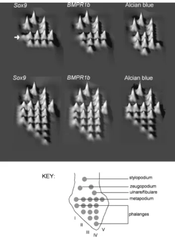
We made landscape maps showing the relative sequence in which the different cartilage elements were first distinct ac-cording to various markers (Fig. 5). As can be seen, there was the expected proximodistal gradient in the appearance of el-ements, and also some evidence of earlier differentiation along the primary axis.
The most notable feature of these maps is that the pre-sumptiveSox9digit I domain in the wing (Fig. 5,Sox9, wing, arrow) appeared relatively late in the developmental sequence. Thus the temporal homology of the anterior Sox9 domain
was consistent with predictions from the primary axis model, namely that digit I should develop late in the sequence.
DISCUSSION
We have shown evidence of aSox9-expressing domain in the mesenchyme anterior to digit II, and separated from it by a nonexpressing zone. ThisSox9expression domain was locat-ed at the palmar side of the hand, close to the ectoderm of the anterior margin. We suggest that it is reasonable to interpret this domain as being a vestige of digit I, and further argue, on
Fig. 4. (A) Schematic interpreta-tion of the Sox9 gene expression patterns superimposed on cartilage pattern (alcian blueFin situ dou-ble stains). Roman numerals, dig-its; vertical green line, plane of section in B; vertical blue line, plane of section in C; dark red box, area reconstructed in D, E. (B and C) Transverse sections of wings (stage 30, hybridized with
Sox9probe), neutral red counter-stain. Sections from the same spec-imen, C more proximal than B. (C) Detail from boxed area in B, showing expression ofSox9in the noncondensed mesenchyme ante-rior to digit II. (D) Three-dimen-sional (3D) reconstruction of the same specimen. Yellow, cartilage, dark blue, gene expression digits II–V, light blue,Sox9 expression, presumptive digit I. Anterior is to the top. Ventral view. (E) Proximal view of the 3D reconstruction. An-terior to the top. The element at the level ‘‘V’’ may consist of mc V1element X. (F) Left chicken wing, stage 30, bmpR-1b probe. Anterior to the top, ventral view. Distinct prechondrogenic domains are seen in digits II–IV, but not anterior to digit II (labeled II). p?, pisiform. (G) Wing, stage 30, ob-lique posterior–ventral view,Sox9
probe. X, element ‘‘X’’; p?, pisi-form. (H) Wing, stage 30, over-stained in NBT/BCIP substrate after Sox9 hybridization, then counterstained with alcian blue and cleared in methyl salicylate. p?, pisiform; ue, ulnare.
the basis of serial expression patterns, that it splits away from a common digit I–II anlage around stage 29. This pattern broadly conforms to the ‘‘digit I’’ condensation observed in tissue sections at this stage (Larsson and Wagner 2002). Fur-thermore, the avascular digit I zone (AZ-1 of Kundra´t et al. 2002) resembles in location the digit ISox9domain (compare
their Fig. 1C with our Fig. 4B). Finally, the putative Sox9 digit I domain develops last in the digit sequence, in agree-ment with the late developagree-ment of digit I in pentadactyl am-niotes (Shubin and Alberch 1986).
Mechanisms of digit reduction and vestige formation
Our study provides evidence that an extensive digit I domain exists in the precondensation limb mesenchyme, but fails to reach condensation and precartilage stages, as shown by lack of bmpR-1bandWnt14 expression. This lack of differentia-tion could explain why no precartilage matrix was found in the putative digit I position by35SO4labeling (Hinchliffe 1985
book), and supports the idea that the digit I domain shows developmental arrest (Galis et al. 2003).
The expression ofSox9in this domain is significant, be-cause the gene is thought to be expressed after the initial patterning events have taken place (Akiyama et al. 2002). This supports the idea that in developmental terms, the wing is initially pentadactyl (Kundra´t et al. 2002; Larsson and Wag-ner 2002; Galis et al. 2003). Some block must exist at later stages when digit morphogenesis normally takes place. It is significant that misexpression ofHoxd-11leads to formation of a supernumerary digit I in the chick wing digit I (Morgan et al. 1992). Our interpretation of these findings is that the digit I domain fails to develop because it does not receive adequate posterior signals during development; misexpression ofHoxd-11, a posterior Hox gene, provides those signals and rescues the vestigial digit.
We find that other vestigial digits in the chicken show arrest at different points in the sequence of cartilage forma-tion and differentiaforma-tion (Table 1). Thus wing digit I arrests at theSox9-expressing stage. Foot digit V develops further, ex-pressing Sox9weakly, but then showing moderatebmpR-1b expression and some cartilage differentiation. Wing digit V shows strongSox9and bmpR-1bexpression, and some car-tilage differentiation (summarized in Table 1).
We saw no evidence in gene expression patterns for what other workers have described as budding or branching (Shubin and Alberch 1986; Garner and Thomas 2004). What we did observe was the establishment of discrete domains
Fig. 5. Developmental timing landscapes showing the relative se-quence in which various markers of skeletal formation appear. Schematic palmar views of right limb; proximal is to top, anterior to left (see key for orientation and position of elements). Each element was assigned a rank according to its place in the devel-opmental sequence. The ranks were then inverted so that the ear-liest elements have a tall peak, and the latest elements have a low peak. Arrow, presumptive wingSox9digit I domain.
Table 1. Summary of gene expression and development in normal and vestigial chicken digits Sox9 bmpR-1b Alcian blue Final form
Formed digits 111 111 111 Fully developed
Vestigial digits
Wing digit I 1 1/ Absent
Wing digit V 111 11 11(small) Greatly reduced metacarpal Foot digit V 11 111 11(small) Greatly reduced metatarsal
from within a common domain. We do acknowledge, though, that domains remain connected by bridges of less-differentiated tissue, and this may create the impression of a branch.
From approximately stage 28 onward, the interdigital re-gion in the wing becomes greatly thinned, as does a flange of tissue along its anterior border, which becomes the prep-atagium (see Figs. 3 and 4 in Murray and Wilson 1994). The Sox9domain for digit I is embedded in the flange, where it lies in close proximity to the ventral ectoderm (Fig. 4).
Frame shift and bilateral (‘‘pyramid’’) reduction hypotheses
Our findings cannot distinguish between the Frame Shift and Pyramid Reduction hypotheses because both of those models accept a vestigial anterior digit in the chick. We are impressed, however, by the rescue of the digit I domain in the chick by Hoxd-11misexpression (Morgan et al. 1992). This is consis-tent with the idea that a shift in anteroposterior positional signaling has occurred in the evolution of birds, such that digit I no longer receives an adequate threshold of posteriorizing signals. One could of course argue that the ‘‘rescued’’ digit I in those experiments was in fact a reduplicated digit II produced by localized mimicking of polarizing activity. However, because expression of the Hoxd-11-RCAS construct was ubiquitous in the limb bud, and not localized to the anterior border, we think this objection is unlikely.
Alternative models
Alternatives to the Frame Shift and bilateral reduction models can be considered:
The anterior vestige in the chicken embryo wing is not a digit but some other structure or primordium;
Birds are not a clade within the theropods;
Digit identities are not meaningful units of homology; rather, they are emergent patterns generated nonspecifi-cally by interactions between developmental mechanisms (Goodwin and Trainor 1983); and
The pentadactyl ‘‘archetype’’ is false and the archosaur limb may in fact be primitively polydactylous.
We will discuss the polydactyly model in some detailFnot because we consider it the most parsimonious explanation, but because it has scarcely been discussed in the context of avian evolution for many decades. It also has the unique vir-tue of providing continuity between digit position and digit identity across archosaur phylogeny.
If the vestigial digit I domain of chicks is primitive for archosaurs, then the ‘‘vestigial digits IV and V’’ of Her-rerasaurusand other archosaurs are in fact digits V and VI (Fig. 2). This would mean that birds could also have a vestigial digit VI as Schestakowa (1927) suggested. Element X or the
pisiform are potential candidates for such a vestige. Bardele-ben (1889) considered the pisiform of mammals to be a vestige of a sixth digit. This opinion was also held by Holmgren (1952), who viewed the tetrapod limb as primitively seven-fingered, on the basis of his extensive developmental studies.
Studies in other taxa predict that digital loss should be bilateral, affecting digit I as well as posterior digits (Alberch and Gale 1983). This has always made the asymmetric re-duction in archosaurs (affecting digits IV and V) seem anom-alous. However, if archosaurs are polydactyl, and have a vestigial digit I domain in their embryos, then there is no anomaly (Fig. 2).
Polydactyly is not robustly supported at this time. Most evidence for digit I in birds, and for extra digits generally, is of the ‘‘nodules and shadows’’ type, where morphological vestiges in adults, or histological traces in embryos, are in-terpreted as recapitulated digits. Other difficulties with a polydactyly theory are: embryos from nonavian theropods are not available for study; no adult archosaur has six dis-tinct digits; there is no evidence for a vestigial digit I in archosaurs outside birds; and we saw no evidence of more than five digital domains ofSox9expression in the chick foot in this study.
Examples of supposed extra digital elements are seen in other Eutetrapoda, and include the claimed ‘‘postminimus’’ in the pes of some salamanders (e.g.,Hynobius lichenatus; Ha-sumi and Iwsawa 2004), and polydactyly in humans (Biesecker 2002). Late Devonian tetrapods were certainly po-lydactylous (Coates and Clack 1990) and the Early Carbon-iferous tetrapodPederpes finneyaeis speculated to have had a hexadactylous manus (Clack 2002). However,Casineria kiddi, possibly an early amniote, has a pentadactyl manus (Paton et al. 1999).
In summary, we have found molecular evidence of a digit I domain in the chicken wing that is specified by early pattern-ing mechanisms, but fails to undergo terminal differentiation. In the light of previous studies where Hoxd-11 was misex-pressed, we suggest that the digit I domain can be rescued by increasing the strength of posterior patterning signals. Con-flicts between fossil and developmental data can be eliminated by a Frame Shift, by bilateral reduction, or by assuming that archosaurs are primitively polydactyl. On the basis of current data, no one model of digit homology is more parsimonious than others.
Acknowledgments
We thank M. A. G. De Bakker for helpful advice, and C. J. Tabin, J. M. Hurle, and L. Niswander for kindly providing us with the
Wnt14,Sox9, andbmpR-1bcDNA clones, respectively. R. Tibbits and M. Brittijn helped with the figures. We are especially grateful to F. Zitman for her valuable help with this project. We have had valuable discussions with M. Coates, A. Feduccia, R. Hinchliffe, M. Kundra´t, C. Tickle, and G. Wagner, among many others; but this article does not necessarily reflect their views.
REFERENCES
Ahn, S., and Joyner, A. L. 2004. Dynamic changes in the response of cells to positive hedgehog signaling during mouse limb patterning.Cell118: 505–516.
Akiyama, H., Chaboissier, M. C., Martin, J. F., Schedl, A., and de Crombrugghe, B. 2002. The transcription factor Sox9 has essential roles in successive steps of the chondrocyte differentiation pathway and is required for expression of Sox5 and Sox6.Genes Dev.16: 2813–2828. Alberch, P., and Gale, E. A. 1983. Size dependence during the development
of the amphibian foot. Colchicine-induced digital loss and reduction. J. Embryol. Exp. Morphol.76: 177–197.
Bardeleben, K. 1889. On the præpollex and præhallux, with observations on the carpus ofTheriodesmus phylarchus.Proc. Zool. Soc. Lon.259–262. Biesecker, L. G. 2002. Polydactyly: how many disorders and how many
genes?Am. J. Med. Genet.112: 279–283.
Blanco, M. J., and Alberch, P. 1992. Caenogenesis, developmental varia-bility, and evolution in the carpus and tarsus of the marbled newt Triturus marmoratus.Evolution46: 677–687.
Blanco, M. J., Misof, B. Y., and Wagner, G. P. 1998. Heterochronic dif-ferences of Hoxa-11 expression inXenopusfore- and hind limb devel-opment: evidence for lower limb identity of the anuran ankle bones.Dev. Genes Evol.208: 175–187.
Burke, A. C., and Alberch, P. 1985. The development and homology of the chelonian carpus.J. Morphol.186: 119–131.
Burke, A. C., and Feduccia, A. 1997. Developmental patterns and the identification of homologies in the avian hand.Science278: 666–668. Carroll, R. L. 1987.Vertebrate Paleontology and Evolution. W. H. Freeman
and Company, New York.
Chamberlain, F. W. 1943.Atlas of Avian Anatomy. Michigan State College Agricultural Experiment Station, East Lansing, Michigan.
Chatterjee, S. 2004. Counting the fingers of birds and dinosaurs.Science 280: 355a.
Chiang, C., et al. 2001. Manifestation of the limb prepattern: limb devel-opment in the absence of Sonic hedgehog function. Dev. Biol. 236: 421–435.
Chimal-Monroy, J., et al. 2003. Analysis of the molecular cascade respon-sible for mesodermal limb chondrogenesis: Sox genes and BMP sig-naling.Dev. Biol.257: 292–301.
Christiansen, P., and Bonde, N. 2004. Body plumage in Archaeopteryx: a review, and new evidence from the Berlin specimen.Comptes Rendus Palevol3: 99–118.
Clack, J. A. 2002. An early tetrapod from ‘Romer’s Gap.’Nature418: 72–76.
Coates, M. I., and Clack, J. A. 1990. Polydactyly in the earliest tetrapod limbs.Nature347: 66–69.
Cohn, M. J., Lovejoy, C. O., Wolpert, L., and Coates, M. I. 2002. Branch-ing, segmentation and the metapterygial axis: pattern versus process in the vertebrate limb.Bioessays24: 460–465.
Dahn, R. D., and Fallon, J. F. 2000. Interdigital regulation of digit identity and homeotic transformation by modulated BMP signalling. Science 289: 438–441.
Dudley, A. T., Ros, M. A., and Tabin, C. J. 2002. A re-examination of proximodistal patterning during vertebrate limb development. Nature 418: 539–544.
Feduccia, A. 1999. 1,2,352,3,4: accommodating the cladogram.Proc. Natl. Acad. Sci. USA96: 4740–4742.
Feduccia, A., and Nowicki, J. 2002. The hand of birds revealed by early ostrich embryos.Naturwissenschaften89: 391–393.
Forster, C. A., Sampson, S. D., Chiappe, L. M., and Krause, D. W. 1998. The theropod ancestry of birds: new evidence from the late cretaceous of Madagascar.Science279: 1915–1919.
Galis, F., Kundra´t, M., and Sinervo, B. 2003. An old controversy solved: bird embryos have five fingers.Trends Ecol. Evol.18: 7–9.
Garner, J. P., and Thomas, A. L. R. 2004. Counting the fingers of birds and dinosaurs.Science280: 355a.
Gauthier, J. A. 1986. Saurischian monophyly and the origin of birds.Mem. Calif. Acad. Sci.8: 1–55.
Gegenbaur, C. 1864. Untersuchungen zur vergleichenden Anatomie der Wirbelthiere: Erstes heft. Carpus und Tarsus. Engelmann, Leipzig.
Goodwin, B. C., and Trainor, L. E. H. 1983. The ontogeny and phylogeny of the pentadactyl limb. In B. C. Goodwin, N. Holder, and C. C. Wylie (eds.).Development and Evolution. Cambridge University Press, Cam-bridge, UK, pp. 75–98.
Hamburger, V., and Hamilton, H. L. 1951. A series of normal stages in the development of the chick embryo.J. Morphol.88: 49–92.
Harfe, B. D., Scherz, P. J., Nissim, S., Tian, H., McMahon, A. P., and Tabin, C. J. 2004. Evidence for an expansion-based temporal Shh gra-dient in specifying vertebrate digit identities.Cell118: 517–528. Hartmann, C., and Tabin, C. J. 2001. Wnt-14 plays a pivotal role in
in-ducing synovial joint formation in the developing appendicular skeleton. Cell104: 341–351.
Hasumi, M., and Iwsawa, H. 2004. Geographic variation in the pes of the salamander Hynobius lichenatus: a comparison with tetradactyl Hynobius hidamontanusand pentadactylHynobius nigrescens.Zool. Sci. 10: 1027.
Healy, C., Uwanogho, D., and Sharpe, P. T. 1999. Regulation and role of Sox9 in cartilage formation.Dev. Dyn.215: 69–78.
Hecht, M. K. 1985. The biological significance ofArchaeopteryx. In M. K. Hecht, J. H. Ostrom, G. Viohl, and P. Wellnhofer (eds.).The Beginnings of Birds: Proceedings of the International Archaeopteryx Conference, Eichsta¨tt. Freunde des Jura-Museums Eichsta¨tt, Willibaldsburg, Eich-sta¨tt, pp. 149–160.
Hecht, M. K., and Tarsitano, S. 1982. The paleobiology and phylogenetic position ofArchaeopteryx.Gebios Mem. Spec.6: 141–149.
Hinchliffe, J. R. 1977. The chondrogenic pattern in chick limb morpho-genesis: a problem of developmentandevolution. In D. A. Ede, J. R. Hinchliffe, and M. Balls (eds.).Vertebrate Limb and Somite Morpho-genesis. Cambridge University Press, Cambridge, UK, pp. 293–309. Hinchliffe, J. R. 1985. ‘One, two, three’ or ‘two, three, four’: an
embryologist’s view of the homologies of the digits and carpus of modern birds. In M. K. Hecht, J. H. Ostrom, G. Viohl, and P. Well-nhofer (eds.).The Beginnings of Birds: Proceedings of the International Archaeopteryx Conference, Eichsta¨tt. Freunde des Jura-Museums Eichsta¨tt, Willibaldsburg, Eichsta¨tt, pp. 141–147.
Hinchliffe, J. R., and Hecht, M. K. 1984. Homology of the bird wing skeleton: embryological versus paleontological evidence.Evol. Biol.18: 21–39.
Holmgren, N. 1933. On the origin of the origin of the tetrapod limb.Acta Zool.14: 185–295.
Holmgren, N. 1952. An embryological analysis of the mammalian carpus and its bearing upon the question of the origin of the tetrapod limb.Acta Zoologica33: 1–115.
Ji, Q., Currie, P. J., Norell, M. A., and Ji, S. A. 1998. Two feathered dinosaurs from north eastern China.Nature393: 753–761.
Ji, Q., Norell, M. A., Gao, K. Q., Ji, S. A., and Ren, D. 2001. The dis-tribution of integumentary structures in a feathered dinosaur.Nature 410: 1084–1088.
Karsenty, G., and Wagner, E. F. 2002. Reaching a genetic and molecular understanding of skeletal development.Dev. Cell2: 389–406.
Ku¨kenthal, W. 1893. Zur Entwicklung des Handskelettes des Krokodils. Morph. Jahrb.19: 42–55.
Kundra´t, M., Seichert, V., Russell, A. P., and Smetana, K. 2002. Pen-tadactyl pattern of the avian wing autopodium and pyramid reduction hypothesis.J. Exp. Zool. (Mol. Dev. Evol.)294: 152–159.
Larsson, H. C., and Wagner, G. P. 2002. Pentadactyl ground state of the avian wing.J. Exp. Zool.294: 146–151.
Larsson, H. C. E., and Wagner, G. P. 2003. Old morphologies misinter-preted.Trends Ecol. Evol.18: 10.
Merino, R., Ganan, Y., Macias, D., Economides, A. N., Sampath, K. T., and Hurle, J. M. 1998. Morphogenesis of digits in the avian limb is controlled by FGFs, TGFbetas, and noggin through BMP signaling. Dev. Biol.200: 35–45.
Moinar, R. E. 1985. Alternatives toArchaeopteryx: a survey of proposed early or ancestral birds. In M. K. Hecht, J. H. Ostrom, G. Viohl, and P. Wellnhofer (eds.).The Beginnings of Birds: Proceedings of the Inter-national Archaeopteryx Conference, Eichsta¨tt. Freunde des Jura-Muse-ums Eichsta¨tt, Willibaldsburg, Eichsta¨tt, pp. 209–217.
Montagna, W. 1945. A re-investigation of the development of the wing of the fowl.J. Morphol.76: 87–113.
Morgan, B. A., Izpisua-Belmonte, J. C., Duboule, D., and Tabin, C. J. 1992. Targeted misexpression ofHox-4.6in the avian limb bud causes apparent homeotic transformations.Nature358: 236–239.
Murray, B. M., and Wilson, D. J. 1994. A scanning electron microscopic study of the normal development of the chick wing from stages 19 to 36. Anat. Embryol.189: 147–155.
Osborn, H. F. 1916. Skeletal adaptations ofOrnitholestes, Struthiomimus, Tyrannosaurus.Bull. Am. Mus. Nat. Hist.35: 733–771.
Ostrom, J. H. 1969. Osteology ofDeinonychus antirrhopus, an unusual the-ropod from the Lower Creataceous of Montana.Bull. Peabody Mus.30: 1–165.
Ostrom, J. H. 1976.Archaeopteryxand the origin of birds.Biol. J. Linn. Soc.8: 91–182.
Padian, K., Hutchinson, J. R., and Holtz, T. R. 1999. Phyloge-netic definitions and nomenclature of the major taxonomic categories of the carnivorous Dinosauria (Theropoda).J. Vertebr. Paleontol.19: 69–80.
Paton, R. L., Smithson, T. R., and Clack, J. A. 1999. An amniote-like skeleton from the Early Carboniferous of Scotland.Nature398: 508–513. Pizette, S., and Niswander, L. 2000. BMPs are required at two steps of limb chondrogenesis: formation of prechondrogenic condensations and their differentiation into chondrocytes.Dev. Biol.219: 237–249.
Richardson, M. K., and Oelschla¨ger, H. A. 2002. Time, pattern, and het-erochrony: a study of hyperphalangy in the dolphin embryo flipper.Evol. Dev.4: 435–444.
Richardson, M. K., Jeffery, J. E., and Tabin, C. J. 2004. Proximodistal patterning of the limb: insights from evolutionary morphology.Evol. Dev.6: 1–5.
Romer, A. S. 1956.Osteology of the Reptiles. University of Chicago Press, Chicago.
Santa Luca, A. P. 1980. The postcranial skeleton ofHeterodontosaurus tucki (Reptilia, Ornithischia) from the Stormberg of South Africa.Ann. S. Afr. Mus.7: 159–211.
Sanz-Ezquerro, J. J., and Tickle, C. 2003. Digital development and morphogenesis.J. Anat.202: 51–58.
Schestakowa, G. S. 1927. Die Entwicklung des Vlogelfu¨gels.Bull. Soc. Nat. Moscou. (Biol.)36: 163–210.
Sereno, P. C. 1993. The pectoral girdle and forelimb of the basal theropod Herrerasaurus ischigualastensis.J. Vertebr. Paleontol.13: 425–450. Sereno, P. C. 1999. The evolution of dinosaurs.Science284: 2137–2147.
Shubin, N. H., and Alberch, P. 1986. A morphogenetic approach to the origin and basic organisation of the tetrapod limb. In M. K. Hecht, B. Wallace, and G. I. Prance (eds.).Evolutionary Biology. Plenum Press, New York, pp. 319–387.
Tarsitano, S., and Hecht, M. K. 1980. A reconsideration of the reptilian relationships of Archaeopteryx.Zool. J. Linn. Soc.69: 149–182. Thulborn, R. A., and Hamley, T. L. 1982. The reptilian relationships of
Archaeopteryx.Aust. J. Zool.30: 611–634.
Verbeek, F. J. 2000. Theory & practice of 3D-reconstructions from serial sections. In Anon. Image Processing, A Practical Approach. Oxford University Press, Oxford, pp. 153–195.
Wagner, G. P., and Gauthier, J. A. 1999. 1,2,352,3,4: a solution to the problem of the homology of the digits in the avian hand.Proc. Natl. Acad. Sci. USA96: 5111–5116.
Walker, A. 1985. The braincase ofArchaeopteryx. In M. K. Hecht, J. H. Ostrom, G. Viohl, and P. Wellnhofer (eds.).The Beginnings of Birds: Pro-ceedings of the International Archaeopteryx Conference, Eichsta¨tt. Freunde des Jura-Museums Eichsta¨tt, Willibaldsburg, Eichsta¨tt, pp. 123–134. Wellnhofer, P. 1985. Remarks of the digit and pubis problems of
Arch-aeopteryx. In M. K. Hecht, J. H. Ostrom, G. Viohl, and P. Wellnhofer (eds.).The Beginnings of Birds: Proceedings of the International Arch-aeopteryx Conference, Eichsta¨tt. Freunde des Jura-Museums Eichsta¨tt, Willibaldsburg, Eichsta¨tt, pp. 113–122.
Wilkinson, D. G. 1998. In Situ Hybridization. Oxford University Press, Oxford.
Wolpert, L., and Hornbruch, A. 1990. Double anterior chick limb buds and models for cartilage rudiment specification.Development109: 961–966. Yang, Y., et al. 1997. Relationship between dose, distance and time in
Sonic-Hedgehog-mediated regulation of anteroposterior polarity in the chick limb.Development124: 4393–4404.
Yasuda, M. 2002.The Anatomical Atlas of Gallus (English Edition). Uni-versity of Tokyo Press, Tokyo.
Za´ka´ny, J., Fromental-Ramain, C., Warot, X., and Duboule, D. 1997. Regulation of digit number and size of digits by posteriorHoxgenes: a dose-dependent mechanism with potential evolutionary implications. Proc. Natl. Acad. Sci. USA94: 13695–13700.
Za´ka´ny, J., Kmita, M., and Duboule, D. 2004. A dual role for Hox genes in limb anterior-posterior asymmetry.Science304: 1669–1672.
Zhou, Z., Barrett, P. M., and Hilton, J. 2003. An exceptionally preserved Lower Cretaceous ecosystem.Nature421: 807–814.