703
Introduction
The term morphogen is used rigorously to describe a particular type of signaling molecule that acts on cells directly to induce distinct cellular responses in a concentration-dependent manner. Although there is abundant evidence for concentration-dependent activity of secreted signaling molecules, evidence for their direct action on cells has been lacking in many cases and, so far, only a few such molecules fulfill the criteria of morphogens. Nevertheless, the roles of morphogens during the development of Drosophila appendages have been extensively studied, and a few examples of morphogens have recently been identified in vertebrate development. These include members of the Hedgehog (Hh) family, for example, Hh in Drosophila appendage development (Mullor et al., 1997; Strigini and Cohen, 1997) and Sonic hedgehog (Shh) in chick neural tube development (Briscoe et al., 2001); the Wnt family member Wingless (Wg), which acts during Drosophila appendage development (Neumann and Cohen, 1997; Zecca et al., 1996); and some members of the TGFβfamily, including Decapentaplegic (Dpp) in Drosophila appendage development (Lecuit et al., 1996; Nellen et al., 1996) and Squint in Zebrafish early embryonic development (Chen and Schier, 2001). Activin, another member of TGFβ family, has been well studied in the Xenopus model system, in which the exogenously supplied signaling molecule activates target genes in a dose-dependent manner, and this has already been well described in a comprehensive review (Gurdon and Bourillot, 2001). In this primer, we will give a brief history of strategies adopted for identifying secreted morphogens, taking the development of Drosophila appendages as a model system, and we discuss how these strategies could be applied to the study in vertebrates.
However, even for those morphogens that have been identified, we still do not understand crucial issues such as how morphogens are moved through a tissue, how a gradient is maintained, and how morphogens coordinate growth and patterning. In addition, the morphogen gradient must be invariable despite genetic or environmental fluctuations.
Recent studies have revealed significant roles for cell surface molecules in shaping morphogen gradients, and these include morphogen receptors and heparan sulfate proteoglycans (HSPGs). Several reports have suggested the involvement of transcytosis, cytonemes and argosomes in morphogen transport, whereas numerical analyses have favored restricted diffusion as a mechanism for the formation of morphogen gradients. More cell biological and biochemical studies will be needed to evaluate the role of these activities and structures in gradient formation.
Morphogens in Drosophila wing development Hh: a short range morphogen in wing development
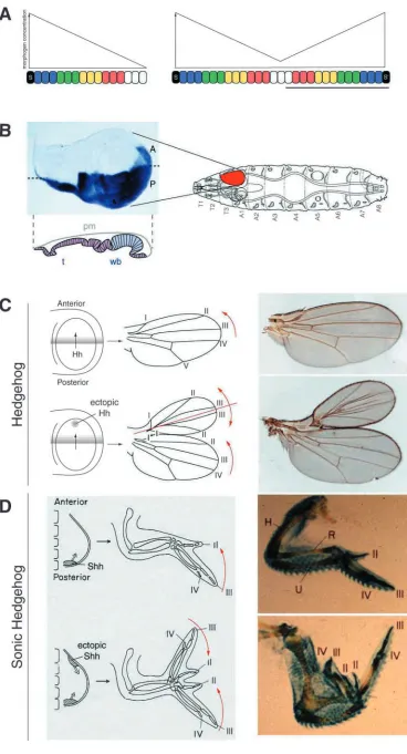
The first step towards identifying and analyzing a morphogen is to determine whether a signaling molecule fulfills the criteria required to qualify as a morphogen, i.e. whether the molecule induces distinct cellular responses in a concentration-dependent manner and whether it acts directly on cells at a distance from its source. The Drosophila adult wing arises from the larval imaginal wing disc. An imaginal disc is a two-sided sac comprising a columnar cell layer (which gives rise to eye, antennae, wing or leg) and overlying squamous epithelium, known as the peripodial membrane (Fig. 1B). The wing imaginal disc is subdivided into non-intermingling anterior (A) and posterior (P) compartments along the anteroposterior axis. The identity of cells in the P compartment is imparted by the expression of the gene engrailed (en) (Guillen et al., 1995; Simmonds et al., 1995; Tabata et al., 1995). Under the control of En, cells of the posterior compartment synthesize Hh, which is secreted into the A compartment (Tabata and Kornberg, 1994) (Fig. 1C). There, Hh induces several target genes, including patched, en and dpp, and patterns the central domain of the wing (Fig. 2).
Does Hh act as a morphogen? The morphogen model predicts that an ectopic source of morphogen can induce a mirror image duplication of the developing field that is patterned by that morphogen (Fig. 1A). Ectopic production of Hh in the Drosophila wing (Fig. 1C) can induce mirror image
During the course of development, cells of many tissues differentiate according to the positional information that is set by the concentration gradients of morphogens. Morphogens are signaling molecules that emanate from a restricted region of a tissue and spread away from their source to form a concentration gradient. As the fate of each
cell in the field depends on the concentration of the morphogen signal, the gradient prefigures the pattern of development. In this article, we describe how morphogens and their functions have been identified and analyzed, focusing on model systems that have been extensively studied.
Summary
Morphogens, their identification and regulation
Tetsuya Tabata* and Yuki Takei
Institute of Molecular and Cellular Biosciences, University of Tokyo, Yayoi 1-1-1, Bunkyo-ku, Tokyo 113-0032, Japan *Author for correspondence (e-mail: [email protected])
Development 131, 703-712
Published by The Company of Biologists 2004 doi:10.1242/dev.01043
instar Drosophila larva. The imaginal disc is a two-sided sac comprising a columnar cell layer that contains presumptive wing blade (wb) and thorax (t) regions, and an overlying squamous peripodial membrane (pm); it is set aside from the embryonic epidermis and develops at the larval stage. The imaginal disc is subdivided into anterior (A) and posterior (P) compartments along the anteroposterior axis. hedgehog (hh) is expressed in the posterior
compartment; hh mRNA is visualized with in situ hybridization (left). Schematic on right modified with permission from Bryant and Levinson (Bryant and Levinson, 1985). (C) Ectopic expression of hh, by making a clone of cells expressing
hh, induces a mirror image
duplication of the anterior wing structure. Hh produced in the P compartment is secreted into the A compartment (top). A clone of cells ectopically expressing hh in the A compartment induces a complete mirror image duplication of the A compartment (bottom). Wing veins I-V are indicated. Reproduced with permission from Tabata (Tabata, 2001). (D) Ectopic production of Shh, induced by implanting shh-expressing cells into the anterior limb bud, induces a mirror image duplication of the wing structure. shh is expressed in the region
[image:2.612.190.558.71.752.2]duplications of the relevant tissues of the wing (Basler and Struhl, 1994). By making use of a temperature-sensitive hh allele, it has been shown that dpp expression can be activated by levels of Hh activity that are below the minimal levels required to activate en (Strigini and Cohen, 1997). This suggests that the different spatial domains of dpp and en expression are defined by the same local concentration gradient of Hh. Evidence that Hh acts directly on cells has come from experiments comparing the activities of the wild-type secreted form of Hh with a membrane-tethered form of the protein (Strigini and Cohen, 1997). Secreted Hh protein activates target genes in nearby cells over a range of 10 cells, whereas the membrane-tethered Hh only activates target genes in cells immediately adjacent to the Hh source. This demonstrates that Hh activates target genes directly and, together, these experiments show that Hh functions as a morphogen.
Dpp: a long range morphogen in wing development
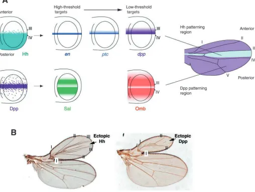
[image:3.612.56.564.80.463.2]One of the targets of Hh regulation, Dpp, then functions as a second morphogen that patterns the wing beyond the central domain (Lecuit et al., 1996; Nellen et al., 1996) (Fig. 2A). Thus, the wing is patterned by two different types of morphogens, and this has been well illustrated in experiments that ectopically express these proteins: Hh induces the mirror image duplication of the entire A compartment, whereas the duplication caused by an ectopic source of Dpp lacks the central domain (Zecca et al., 1995) (Fig. 2B). Dpp is expressed along the border between the A and P compartments, and induces several target genes including sal and omb (bifid – FlyBase), with omb being expressed in a wider domain than sal (Figs 2, 3). Dpp distribution in the columnar cell layer can be monitored using an antibody against Dpp (Gibson et al., 2002) or by fusing Dpp with green fluorescent protein (Dpp-GFP) (Entchev et al., 2000;
Fig. 2. The wing is patterned by two morphogens, Hh and Decapentaplegic (Dpp). (A) Hh produced in the posterior (P) compartment generates a short range gradient of Hh in the anterior (A) compartment. Hh both patterns the central domain of the wing and induces the expression of en,
ptc and dpp, at high, middle and low thresholds, respectively, in a stripe of cells adjacent to the AP compartment boundary. Note that en is
Dpp is also detected uniformly in the disc lumen and is thought to be required mainly for cell survival (Gibson et al., 2002).
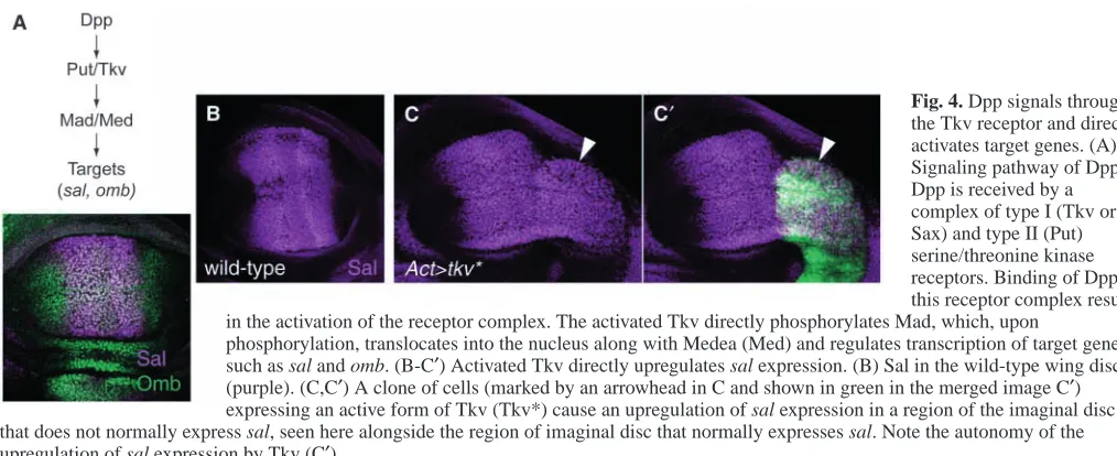
Evidence showing that Dpp acts in a concentration-dependent manner and acts directly on cells, rather than by acting through a signal relay mechanism, comes from experiments that used a constitutively active form of the Dpp receptor (Lecuit et al., 1996; Nellen et al., 1996). The pathway that transduces the Dpp signal involves a combination of two types of serine/threonine kinase receptors, type I and type II. The activated type I receptor phosphorylates cytoplasmic transducers, so-called receptor-regulated Smads (named after the first-identified members of this family: Sma in C. elegans and Mad in Drosophila), which, upon phosphorylation, translocate into the nucleus and regulate the expression of target genes (Fig. 4A). In Drosophila wing development, Thickveins (Tkv) acts as a type I receptor; its constitutively active form (Tkv*), when ectopically expressed, can induce the expression of the target genes sal and omb (Fig. 4). The key to showing whether Dpp acts directly on cells lies in determining whether the effect of expressing activated Tkv is cell-autonomous. If Dpp functions as a morphogen, the effects of Tkv* should be strictly cell-autonomous, because a second signal would not be secreted from the Tkv*-expressing cells.
[image:4.612.44.296.61.167.2](Diaz-Benjumea and Cohen, 1993), inducing expression of the gene fringe (Irvine and Wieschaus, 1994), which results in activation of the Notch receptor pathway at the DV border (Kim et al., 1995). Activated Notch induces Wg synthesis at the DV border (Doherty et al., 1996; Neumann and Cohen, 1996) (Fig. 4) where it functions as a morphogen to induce the expression of target genes, such as achaete (ac) Distal-less (Dll) and vestigial (vg), and organizes wing patterning (Fig. 5). Wg has been shown to function as a morphogen by experiments similar to those that provided evidence that Hh functions as a morphogen; although the wild-type secreted Wg activates target genes over a distance, a membrane-tethered form upregulates Wg-target genes only in its immediate neighbors (Zecca et al., 1996). In addition, the expression of Wg-target genes is cell-autonomously disrupted in clones of cells mutant either for dishevelled or armadillo, each of which encode a component of the Wg signal transduction pathway (Neumann and Cohen, 1997; Zecca et al., 1996). Furthermore, an experiment using a temperature-sensitive allele of wg has shown that the level of Wg activity minimally required to activate synthesis of Dll is higher than that required to activate synthesis of Vg (Neumann and Cohen, 1997). Together, these experiments indicate that Wg functions as a morphogen.
Fig. 4. Dpp signals through the Tkv receptor and directly activates target genes. (A) Signaling pathway of Dpp. Dpp is received by a complex of type I (Tkv or Sax) and type II (Put) serine/threonine kinase receptors. Binding of Dpp to this receptor complex results in the activation of the receptor complex. The activated Tkv directly phosphorylates Mad, which, upon
[image:4.612.43.550.530.737.2]Morphogens in vertebrate development Shh during limb bud development
Anterior/posterior polarity of the vertebrate limb is regulated by a posteriorly localized signaling center called the zone of polarizing activity (ZPA). Shh mirrors the properties of the ZPA; ectopic Shh activity induces digit duplications, with higher concentrations specifying increasingly more posterior digits (Riddle et al., 1993; Yang et al., 1997) (Fig. 1D). Furthermore, an ectopic source of Shh induces a mirror image duplication of the limb (Fig. 1C). Shh induces dose-dependent production of a Dpp ortholog, BMP2 (Yang et al., 1997), and misexpressed BMP2 can induce ectopic formation of the most anterior digit (Duprez et al., 1996). These observations readily prompt us to see the analogy between patterning by Shh of the chick limb bud and the role of Hh in Drosophila wing (Fig. 1C,D). However, no evidence for the direct action of Shh in regulating different target genes has yet been demonstrated, suspending the conclusion that Shh functions as a morphogen in the limb development.
Shh in neural tube development
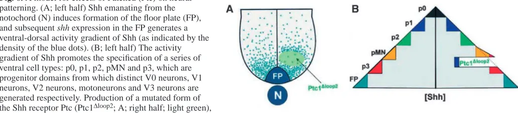
During development of the chick neural tube, however, Shh does function as a morphogen. Shh emanates from the
notochord to induce formation of the floor plate. Subsequent Shh expression in the floor plate generates a ventral-dorsal activity gradient of Shh that promotes the specification of a series of ventral cell types (Ericson et al., 1997). Furthermore, ectopic expression of a mutated form of the Shh receptor, Patched (Ptc), which does not bind Shh but does antagonize its signaling, causes cell-autonomous ventral-to-dorsal switches in neural progenitor identity (Briscoe et al., 2001) (Fig. 6), clearly indicating that Shh functions by acting on cells directly.
Squint in early embryogenesis
[image:5.612.83.540.74.252.2]Evidence for another morphogen in vertebrate development has come from a study in Zebrafish. Squint is a member of the TGFβfamily of signaling molecules, which induces formation of mesoderm and endoderm in embryos, and regulates different target genes in a concentration-dependent manner. Its direct action was revealed using a mutant embryo in which the Squint signal is not transduced because it lacks the activity of the protein One-eyed pinhead (which is required cell-autonomously for the reception of Squint). When squint RNA is injected into a single cell of the mutant embryo, the target genes are induced in wild-type cells implanted distantly from the squint-injected cell. Thus, although the mutant cells cannot
Fig. 5. Wg and its target genes. (A-D) Wg (A; shown in green) is produced along the DV border and induces the expression of target genes, such as Ac (B; yellow, and expressed only in the A compartment), Dll (C; purple) and Vg (D; red), at high, middle and low thresholds,
respectively. Anterior is to the left. (E) Schematics showing the domains of target gene expression. Reproduced with permission from Briscoe et al. (Briscoe et al., 2001).
Fig. 6. A model for effects of Patched (Ptc) on neural patterning. (A; left half) Shh emanating from the notochord (N) induces formation of the floor plate (FP), and subsequent shh expression in the FP generates a ventral-dorsal activity gradient of Shh (as indicated by the density of the blue dots). (B; left half) The activity gradient of Shh promotes the specification of a series of ventral cell types: p0, p1, p2, pMN and p3, which are progenitor domains from which distinct V0 neurons, V1 neurons, V2 neurons, motoneurons and V3 neurons are generated respectively. Production of a mutated form of the Shh receptor Ptc (Ptc1∆loop2; A; right half; light green),
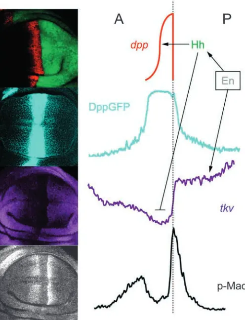
[image:5.612.50.566.609.722.2]phosphorylated form of Mad (p-Mad) can be used as an intracellular marker to monitor Dpp morphogen activity. To this end, p-Mad levels are visualized using an antibody that specifically recognizes p-Mad (Tanimoto et al., 2000). In the wing imaginal disc, the amount of p-Mad is high in cells near the AP border, where Dpp concentration is high, but is severely reduced in cells along the AP border that express dpp, where the level of Dpp is also very high (Fig. 7). This reduction in p-Mad levels along the AP border, despite the high concentration of Dpp, is a result of the direct repressive action of Hh. Hh also directly organizes patterning in the region where it attenuates Dpp signaling, and it is possible that Hh downregulates Dpp signaling in this region to prevent it from interfering with patterning by Hh.
Morphogen receptors
It is possible that the concentration gradient of Dpp is modulated by the distribution of the Dpp receptor itself. Dpp preferentially signals through the Tkv receptor in the wing disc and also negatively regulates tkv expression (Lecuit and Cohen, 1998). The level of tkv expression is higher in cells located at the periphery of the disc and is lower in the central region (Tanimoto et al., 2000). In addition, tkv expression is lowest at the AP border (Fig. 7). We refer to the level of tkv expression in the area between the peripheral regions and the AP border as ‘basal’ and, interestingly, the basal level of tkv expression in the P compartment is higher than it is in the A compartment (Fig. 7). This might account for a steeper gradient of p-Mad in the P compartment compared with in the A compartment. The Dpp gradient is expected to be steeper in tissue with more receptors, because high levels of Tkv limit the distribution of Dpp. Thus, the distribution of Tkv modulates the distribution of Dpp itself.
The distribution of Tkv may also regulate Dpp activity, rather than just Dpp concentration. Hh-dependent reduction of p-Mad levels at the AP border occurs largely by repressing transcription of the tkv gene. Conversely, the higher Tkv level in the P compartment than in the A compartment is maintained by the activity of the transcription factor Engrailed. Both the Hh and En activities that regulate tkv levels are mediated by the gene mtv (master of thickveins), which encodes a putative nuclear protein (Funakoshi et al., 2001).
The ability of receptor levels to regulate the distribution of receptor ligands is not restricted to the Dpp morphogen, but is also seen for Hh. The Hh receptor Ptc is expressed in the A compartment at low levels and is strongly induced by Hh at the AP border. Here, Hh induces a high level of Ptc to limit the range of the Hh distribution gradient (Chen and Struhl, 1996).
Heparan sulfate proteoglycans
[image:6.612.313.561.72.396.2]Recently, several reports have suggested that heparan sulfate proteoglycans (HSPGs) play a key role in morphogen transport and/or signaling. HSPGs are abundant cell surface molecules and are a part of the extracellular matrix. HSPGs consist of a protein core (such as syndecan and glypican) to which heparan sulfate glycosaminoglycan (HS GAG) chains are attached. GAG chains are long unbranched polymers consisting of many sulfated disaccharides. Genetic screens for mutations that affect morphogen signaling pathways in Drosophila have identified genes that have sequence homologies to genes that encode vertebrate GAG biosynthetic enzymes. These putative enzymes are encoded by sugarless (sgl), sulfateless (sfl), and members of the Drosophila EXT gene family consisting of
Fig. 7. Ligand and activity gradient of Dpp. Confocal microscopy images (left) and schematic drawings (right) of the part of the wing imaginal disc that gives rise to the adult wing. Hh (green), the synthesis of which is maintained by Engrailed (En) in P-compartment cells, induces Dpp expression (red) along the AP border. Dpp diffuses in both A and P directions and forms a gradient, which can be visualized by the distribution of the chimeric Dpp-GFP protein (blue). The level of Tkv, the Dpp receptor (purple), is very low along the AP border because Hh downregulates its expression. In the middle of the wing disc, abutting the AP border, the level of Tkv in the P compartment is higher than it is in the A compartment, which causes a steeper Dpp gradient to be present in the P
tout-velu (ttv), brother of ttv (botv) and sister of ttv (sotv; Ext2 – FlyBase), which encode proteins with homology to UDP-glucose dehydrogenase, N-deacetylase/N-sulfotransferase and HS copolymerase, respectively. Mutations in sgl compromise signaling mediated by Wg (Binari et al., 1997; Hacker et al., 1997; Haerry et al., 1997) and Dpp (Haerry et al., 1997). Similarly, the sfl mutation affects Wg and Hh signaling (Lin et al., 1999; The et al., 1999), and in somatic sfl mutant clones Wg protein levels are diminished (Baeg et al., 2001). ttv, botv and sotv mutants affect Hh, Dpp and Wg signaling (Bellaiche et al., 1998; The et al., 1999; Takei et al., 2004). In addition, Notum, a gene that encodes a member of the α/β-hydrolase superfamily, influences Wg protein distribution by destabilizing the HSPGs (Gerlitz and Basler, 2002; Giraldez et al., 2002). Lastly, dally is proposed to encode a HSPG protein core, and is required for Wg and Dpp activity. Dally, and the related Dally-like protein (Dlp), bind and stabilize Wg at the cell surface (Baeg et al., 2001; Strigini and Cohen, 2000), and may provide a pool of Wg protein that can become available for receptor binding upon its
release from HSPGs. Both dally and tkv expression are regulated by Hh and Engrailed. In addition, elevated levels of Dally increase the sensitivity of cells to Dpp, and alterations in the levels of Dally affect formation of both Dpp ligand and activity gradients (Fujise et al., 2003). Together, these findings indicate that HSPGs are major regulators of morphogen gradients.
Hh protein levels are significantly decreased in clones of cells mutant for the EXT genes ttv, botv and sotv (for Hh; Fig. 8C) when these clones are generated in the Hh-expressing cells of the wing. This indicates that HSPGs are required for stable retention of Hh on the cell surface. In wild-type imaginal discs, Hh protein synthesized in the P compartment appears to flow into the A compartment, with a moderate concentration gradient starting from the middle of the posterior compartment (Fig. 8A). However, when the EXT mutant clone is created in the A compartment along the AP boundary, Hh accumulates abnormally in the P compartment (Fig. 8B). This indicates that, because of a lack of appropriate HSPG, Hh fails to move into the mutant cells and, as a consequence, accumulates in posterior cells instead. Dpp-GFP and Wg also accumulate
abnormally in the cells near EXT mutant clones, probably because these proteins cannot move into the cells mutant for EXT genes (Takei et al., 2004). These observations indicate that the HSPG-dependent diffusion is a common mechanism for the distribution of the three morphogens, Hh, Dpp and Wg.
Mechanisms by which morphogens traffic through tissues
[image:7.612.196.568.304.676.2]We have witnessed significant progress in understanding the mechanisms by which morphogen signals regulate pattern formation in various contexts of development. Nevertheless, we still do not know the answer to the simple and fundamental question of how morphogens are propagated through tissues. Movement by free diffusion alone cannot explain the graded pattern of a morphogen because a secreted GFP fusion protein composed of GFP and the secretory transport domains of Dpp (i.e. lacking the mature Dpp peptide) fails to form a gradient (Entchev et al., 2000). We are beginning to see cell-biological findings that postulate mechanisms by which the morphogen
Fig. 8. Hh requires HSPGs in order to move. (A,D,G) Hh distribution; (B,E,H) merged images of Hh distribution (purple), with cells of the imaginal disc marked uniformly with GFP (green). The clone of cells mutant for EXT originated from a single cell lacking EXT activity, created by mitotic recombination and marked by the absence of GFP. Thus, cells homozygous for the EXT mutant are marked by an absence of GFP and cells heterozygous for the EXT mutant are pale green. The staining intensity of Hh in the selected area (white boxes in B and E) was integrated along AP axis, plotted using NIH Image software and presented schematically (C,F). (A-C) Hh protein synthesized in the posterior compartment appears to flow into the anterior compartment, with a moderate
endocytosis and resecretion – known as planer transcytosis. The requirement for endocytosis in Dpp function was indicated by an experiment using Dpp-GFP and a mutation in the gene shibire (shi), which encodes Dynamin, a GTPase required for clathrin-dependent endocytosis. When a shi clone is made shortly after a short burst of Dpp-GFP expression, Dpp-GFP-positive endosomes are not present in the area behind the shi clone (Entchev et al., 2000). Furthermore, the activity of the small GTPase Rab5 is required for the fusion between endocytotic vesicles and early endosomes, and is thought to be rate limiting in the early endocytic pathway. When a dominant-negative mutant of Rab5 is expressed in the wing imaginal disc of wild-type flies, target gene expression is restricted to the cells adjacent to the Dpp source (Entchev et al., 2000). By contrast, overexpression of Rab5 broadens the expression domain of target cells. In addition, another small GTPase, Rab7, targets endocytic cargo from the early to the late endosome, and then to the lysosome for degradation. Expression of a dominant gain-of-function mutant of Rab7 decreases the levels of GFP-Dpp that are internalized and reduces the range of Dpp signaling (Entchev et al., 2000).
Nevertheless, Dpp could also be propagated in part by its diffusion in the extracellular space; the digestion of the extracellular proteins of the intact wing disc with proteinase K demonstrates that most of the Dpp-GFP signal appears to be in the extracellular space (Teleman and Cohen, 2000). Thus, more careful genetic and cell biological studies will be required to determine how much of the Dpp trafficking can be ascribed to the endocytotic mechanism, extracellular movement or other mechanisms.
In contrast to the role of planer transcytosis in the transport of Dpp, a report has argued against transcytosis as a mechanism of Wg trafficking in wing imaginal disc development (Strigini and Cohen, 2000). By devising a new antibody-staining protocol to detect extracellular Wg protein, the authors revealed that the Wg protein makes a shallow extracellular gradient (Strigini and Cohen, 2000). Wg does not localize to punctate structures in the shi mutant clones (as is the case for Dpp), and, in contrast with Dpp, Wg is internalized by wild-type cells behind the shi clone. This shows that Wg can move across the shi mutant tissue and is internalized by the adjacent wild-type cells. In fact, higher levels of extracellular Wg protein are present in shi mutant clones than in wild-type cells. (Strigini and Cohen, 2000). Recent reviews discuss the problems of morphogen transport that we have not been able
in the peripodial cells nevertheless disrupts growth and patterning of the wing (Gibson et al., 2002), suggesting that mechanisms that govern the growth and patterning of peripodial cells coordinate with those of columnar cells. The peripodial cells extend long cellular processes that traverse the acellular space between these layers and terminate on the surface of the columnar cells (Gibson and Schubiger, 2000). These processes may function to transmit the signal between the two layers of cells.
Argosomes
When parts of the membranes of cells of the imaginal disc are labeled with GFP linked to glycophosphatidylinositol (gpi), GFP-gpi localizes predominantly to the basolateral membrane. However, strikingly, GFP-gpi is also rapidly detected in nearby cells, suggesting that the basolateral membranes of disc cells can vesiculate and travel thoughout the disc epithelium (Greco et al., 2001) (Fig. 9). These membrane fragments, named argosomes, are produced by wg-expressing cells and co-localize with the Wg protein, suggesting that argosomes may provide a vehicle for the movement of Wg protein. The existence of argosomes may also have implications for the transport of other morphogens that have high membrane affinity.
Restricted diffusion and mathematical studies
hours of the onset of Dpp expression, transcytosis would have to occur at implausibly fast rates (Lander et al., 2002).
If morphogens are transported by restricted diffusion, another numerical analysis also predicts the significance of morphogen receptors in establishing robust gradients. It has been proposed that morphogens are decayed rapidly close to the source and more gradually further away from the source in order to ensure their robustness and long-range action (Eldar et al., 2003). These authors proposed two models: (1) morphogen signaling represses receptor expression, while the receptor stabilizes the free morphogen; and (2) morphogen signaling activates receptor expression, while the receptor enhances the degradation of the free morphogen. They suggested that the former model represents the Wg system, and the latter the Hh system, as Wg downregulates its own recepter levels whereas Hh upregulates them, and they also showed that the Wg receptor Dfz2 stabilizes free Wg (Eldar et al., 2003). The models will be tested by experimental characterization of the receptor-dependent regulation of degradation of free morphogens.
Future perspectives
In 1924, Hans Spemann and Hilde Mangold carried out a spectacular experiment in which they transplanted the dorsal rip from an early gastrula of a newt into another early gastrula in the region that would become ventral epidermis. This resulted in a complete mirror image duplication of the whole body (Spemann and Mangold, 1924) (Fig. 10). We now know that many signaling molecules and their antagonists are involved in this process. When the whole process driven by the organizer is analyzed at the molecular level, it is probable that we will see successive rounds of regulation by both morphogens and signal relay mechanisms.
The potency of morphogens is impressive; when applied, they can organize the whole pattern of adult structures and
sometimes induce complete mirror image duplications. Although there are many secreted signaling molecules that seem to organize these patterns, only a small number of them are known to function as morphogens. Other candidates for morphogen molecules include members of the Fgf (Fibroblast growth factor) family of secreted signaling molecules, which have long been known to regulate many aspects of development. For example, Fgf8 is expressed at the junction between the midbrain and the hindbrain, known as the isthmic organizer, and it is thought to mediate, at least in part, the activity of the isthmic organizer that patterns the midbrain and rostral hindbrain (Martinez et al., 1999; Irving and Mason, 2000). The finding that varying amounts of Fgfs can induce different classes of spinal motoneurons indicates that Fgfs may function as morphogens (Liu et al., 2001).
Much of the knowledge about morphogens has come from the studies of imaginal disc development, mainly because of the simple structure of the disc and the powerful tools of analysis now available, especially mosaic clones (Blair, 2003). Advances in similar techniques will reveal more morphogens in vertebrates and will allow us to determine how their gradients are regulated.
We thank A. Kuroiwa, H. Nakato, T. Ogura and C. Tabin for helpful discussions, and A. Kouzmenko for critical reading of the manuscript.
References
Baeg, G. H., Lin, X., Khare, N., Baumgartner, S. and Perrimon, N. (2001). Heparan sulfate proteoglycans are critical for the organization of the extracellular distribution of Wingless. Development 128, 87-94.
Basler, K. and Struhl, G. (1994). Compartment boundaries and the control of Drosophila limb pattern by hedgehog protein. Nature 368, 208-214. Bellaiche, Y., The, I. and Perrimon, N. (1998). Tout-velu is a Drosophila
homologue of the putative tumour suppressor EXT-1 and is needed for Hh diffusion. Nature 394, 85-88.
Binari, R. C., Staveley, B. E., Johnson, W. A., Godavarti, R., Sasisekharan, R. and Manoukian, A. S. (1997). Genetic evidence that heparin-like glycosaminoglycans are involved in wingless signaling. Development 124, 2623-2632.
Blair, S. S. (2003). Genetic mosaic techniques for studying Drosophila development. Development 130, 5065-5072.
Briscoe, J., Chen, Y., Jessell, T. M. and Struhl, G. (2001). A Hedgehog-insensitive form of Patched provides evidence for direct long-range morphogen activity of Sonic Hedegehog in the neural tube. Mol. Cell 7, 1279-1291.
Bryant, P. J. and Levinson, P. (1985). Intrinsic growth control in the imaginal primordia of Drosophila, and the autonomous action of a lethal mutation causing overgrowth. Dev. Biol. 107, 355-363.
[image:9.612.79.267.68.269.2]Chen, Y. and Schier, A. F. (2001). The zebrafish nodal signal squint functions as a morphogen. Nature 411, 607-610.
Fig. 9. Models for morphogen transport. (A) A model for cytonemes. Cells at the periphery of the imaginal disc extend long processes, cytonemes, towards the AP border, where Dpp is expressed (light blue). (B) A model for argosomes. The basolateral membranes of imaginal disc cells vesiculate and travel throughout the disc epithelium.
[image:9.612.372.523.72.128.2]in the ventral neural tube. Cold Spring Harbor Symp. Quant. Biol. 62, 451-466.
Fujise, M., Takeo, S., Kamimura, K., Matsuo, T., Aigaki, T., Izumi, S. and Nakato, H. (2003). Dally regulates Dpp morphogen gradient formation in the Drosophila wing. Development 130, 1515-1522.
Funakoshi, Y., Minami, M. and Tabata, T. (2001). mtv shapes the activity gradient of the Dpp morphogen through regulation of thickveins.
Development 128, 67-74.
Gerlitz, O. and Basler, K. (2002). Wingful, an extracellular feedback inhibitor of Wingless. Genes Dev. 16, 1055-1059.
Gibson, M. C. and Schubiger, G. (2000). Periodial cells regulate proliferation and patterning of Drosophila imaginal discs. Cell 103, 343-350.
Gibson, M. C., Lahman, D. A. and Schubiger, G. (2002). Lumenal transmission of Decapentaplegic in Drosophila imaginal discs. Dev. Cell 3, 451-460.
Gilbert, S. F. (1997). Developmental Biology. Sunderland, MA: Sinauer Associates.
Giraldez, A. J., Copley, R. R. and Cohen, S. M. (2002). HSPG modification by the secreted enzyme Notum shapes the Wingless morphogen gradient.
Dev. Cell 2, 667-676.
Greco, V., Hannus, M. and Eaton, S. (2001). Argosomes: a potential vehicle for the spread of morphogens through epithelia. Cell 106, 633-645. Guillen, I., Mullor, J. L., Capdevila, J., Sanchez-Herrero, E., Morata, G.
and Guerrero, I. (1995). The function of engrailed and the specification of Drosophila wing pattern. Development 121, 3447-3456.
Gurdon, J. B. and Bourillot, P.-Y. (2001). Morphogen gradient interpretation.
Nature 413, 797-803.
Hacker, U., Lin, X. and Perrimon, N. (1997). The Drosophila sugarless gene modulates Wingless signaling and encodes an enzyme involved in polysaccharide biosynthesis. Development 124, 3565-3573.
Haerry, T. E., Heslip, T. R., Marsh, J. L. and O’Connor, M. B. (1997). Defects in glucuronate biosynthesis disrupt Wingless signaling in Drosophila. Development 124, 3055-3064.
Irvine, K. D. and Wieschaus, E. (1994). fringe, a Boundary-specific signaling molecule, mediates interactions between dorsal and ventral cells during Drosophila wing development. Cell 79, 595-606.
Irving, C. and Mason, I. (2000). Signaling by FGF8 from the isthmus patterns anterior hindbrain and establishes the anterior limit of Hox gene expression.
Development 127, 177-186.
Kim, J., Irvine, K. D. and Carroll, S. B. (1995). Cell recognition, signal induction, and symmetrical gene activation at the dorsal-ventral boundary of the developing Drosophila wing. Cell 82, 795-802.
Lander, A. D., Nie, Q. and Wan, F. Y. M. (2002). Do morphogen gradients arise by diffusion? Dev. Cell 2, 785-796.
Lecuit, T. and Cohen, S. M. (1998). Dpp receptor levels contribute to shaping the Dpp morphogen gradient in the Drosophila wing imaginal disc.
Development 125, 4901-4907.
Lecuit, T., Brook, W. J., Ng, M., Calleja, M., Sun, H. and Cohen, S. M. (1996). Two distinct mechanisms for long-range patterning by Decapentaplegic in the Drosophila wing. Nature 381, 387-393.
Lin, X., Buff, E. M., Perrimon, N. and Michelson, A. M. (1999). Heparan
871-880.
Ramirez, W. F. and Kornberg, T. B. (1999). Cytonemes: cellular processes that project to the principal signaling center in Drosophila imaginal discs.
Cell 97, 599-607.
Riddle, R. D., Johnson, R. L., Laufer, E. and Tabin, C. (1993). Sonic hedgehog mediates the polarizing activity of the ZPA. Cell 75, 1401-1416.
Simmonds, A. J., Brook, W. J., Cohen, S. M. and Bell, J. B. (1995). Distinguishable functions for engrailed and invected in anterior-posterior patterning in the Drosophila wing. Nature 376, 424-427.
Spemann, H. and Mangold, H. (1924). Induction of embryonic primodia by implantation of organizers from a different species. In Foundations of
Experimental Embryology (ed. B. H. Willer and J. M. Oppenheimer), pp.
144-184. New York: Hafner.
Strigini, M. and Cohen, S. M. (1997). A Hedgehog activity gradient contributes to AP axial patterning of the Drosophila wing. Development 124, 4697-4705.
Strigini, M. and Cohen, S. M. (2000). Wingless gradient formation in the
Drosophila wing. Curr. Biol. 10, 293-300.
Tabata, T. (2001). Genetics of morphogen gradients. Nat. Rev. Genet. 2, 620-630.
Tabata, T. and Kornberg, T. B. (1994). Hedgehog is a signaling protein with a key role in patterning Drosophila imaginal discs. Cell 76, 89-102. Tabata, T., Schwartz, C., Gustavson, E., Ali, Z. and Kornberg, T. B. (1995).
Creating a Drosophila wing de novo, the role of engrailed, and the compartment border hypothesis. Development 121, 3359-3369.
Takei, Y., Ozawa, Y., Sato, M., Watanabe, A. and Tabata, T. (2004). Three
Drosophila EXT genes shape morphogen gradients through synthesis of
heparan sulfate proteoglycans. Development 131, 73-82.
Tanimoto, H., Itoh, S., ten Dijke, P. and Tabata, T. (2000). Hedgehog creates a gradient of DPP activity in Drosophila wing imaginal discs. Mol. Cell 5, 59-71.
Teleman, A. A. and Cohen, S. M. (2000). Dpp gradient formation in the Drosophila wing imaginal disc. Cell 103, 971-980.
Teleman, A. A., Strigini, M. and Cohen, S. M. (2001). Shaping Morphogen Gradients. Cell 105, 559-562.
The, I., Bellaiche, Y. and Perrimon, N. (1999). Hedgehog movement is regulated through tout velu-dependent synthesis of a heparan sulfate proteoglycan. Mol. Cell 4, 633-639.
Yang, Y., Drossopoulou, G., Chuang, P. T., Duprez, D., Marti, E., Bumcrot, D. A., Vargesson, N., Clarke, J. A., Niswander, L., McMahon, A. P. et al. (1997). Relationship between dose, distance and time in Sonic hedgehog-mediated reulation of anteropostrior polarity in the chick limb. Development 124, 4393-4404.
Vincent, J.-P. and Dubois, L. (2002). Morphogen Transport along epithelia, an integrated trafficking problem. Dev. Cell 3, 615-623.
Zecca, M., Basler, K. and Struhl, G. (1995). Sequential organizing activities of engrailed, hedgehog and decapentaplegic in the Drosophila wing.
Development 121, 2265-2278.