Open Access
Vol 10 No 2
Research article
Dynamic compression counteracts IL-1
β
induced inducible nitric
oxide synthase and cyclo-oxygenase-2 expression in
chondrocyte/agarose constructs
TT Chowdhury
1, S Arghandawi
1, J Brand
2, OO Akanji
1, DL Bader
1, DM Salter
2and DA Lee
11School of Engineering and Materials Science, Queen Mary, University of London, Mile End Road, London, E1 4NS, UK 2Queens Medical Research Institute, 47 Little France Cresent, Edinburgh University, EH16 4TJ. UK
Corresponding author: TT Chowdhury, [email protected]
Received: 27 Sep 2007 Revisions requested: 27 Nov 2007 Revisions received: 28 Feb 2008 Accepted: 18 Mar 2008 Published: 18 Mar 2008
Arthritis Research & Therapy 2008, 10:R35 (doi:10.1186/ar2389) This article is online at: http://arthritis-research.com/content/10/2/R35 © 2008 Chowdhury et al.; licensee BioMed Central Ltd.
This is an open access article distributed under the terms of the Creative Commons Attribution License (http://creativecommons.org/licenses/by/2.0), which permits unrestricted use, distribution, and reproduction in any medium, provided the original work is properly cited.
Abstract
Background Nitric oxide and prostaglandin E2 (PGE2play pivotal roles in both the pathogenesis of osteoarthritis and catabolic processes in articular cartilage. These mediators are influenced by both IL-1β and mechanical loading, and involve alterations in the inducible nitric oxide synthase (iNOS) and cyclo-oxygenase (COX)-2 enzymes. To identify the specific interactions that are activated by both types of stimuli, we examined the effects of dynamic compression on levels of expression of iNOS and COX-2 and involvement of the p38 mitogen-activated protein kinase (MAPK) pathway.
Methods Chondrocyte/agarose constructs were cultured under free-swelling conditions with or without IL-1β and/or SB203580 (inhibitor of p38 MAPK) for up to 48 hours. Using a fully characterized bioreactor system, constructs were subjected to dynamic compression for 6, 12 and 48 hours under similar treatments. The activation or inhibition of p38 MAPK by IL-1β and/or SB203580 was analyzed by western blotting. iNOS, COX-2, aggrecan and collagen type II signals were assessed utilizing real-time quantitative PCR coupled with molecular beacons. Release of nitrite and PGE2 was quantified using biochemical assays. Two-way analysis of variance and the post hoc Bonferroni-corrected t-test were used to examine data.
Results IL-1β activated the phosphorylation of p38 MAPK and this effect was abolished by SB203580. IL-1β induced a transient increase in iNOS expression and stimulated the production of nitrite release. Stimulation by either dynamic compression or SB203580 in isolation reduced the IL-1β induced iNOS expression and nitrite production. However, co-stimulation with both dynamic compression and SB203580 inhibited the expression levels of iNOS and production of nitrite induced by the cytokine. IL-1β induced a transient increase in COX-2 expression and stimulated the cumulative production of PGE2 release. These effects were inhibited by dynamic compression or SB203580. Co-stimulation with both dynamic compression and SB203580 restored cytokine-induced inhibition of aggrecan expression. This is in contrast to collagen type II, in which we observed no response with the cytokine and/ or SB203580.
Conclusion These data suggest that dynamic compression directly influences the expression levels of iNOS and COX-2. These molecules are current targets for pharmacological intervention, raising the possibility for integrated pharmacological and biophysical therapies for the treatment of cartilage joint disorders.
Introduction
The mechanical environment is an important factor that main-tains articular cartilage in a healthy state. Mechanical signals generated under normal physiological loading conditions will activate mechanotransduction pathways and drive biochemi-cal events that regulate chondrocyte function and activity [1-4]. It is well established that proinflammatory cytokines such
as IL-1β act as the key mediators of cartilage breakdown and stimulate the release of nitric oxide (NO) and prostaglandin (PG)E2, via induction of inducible isoforms of the nitric oxide synthase (iNOS) and cyclo-oxygenase (COX)-2 enzymes [5-9]. There is growing evidence that mechanical stimulation inhibits the release of NO and PGE2 by articular chondrocytes [10-18]. Thus, mechanical strain acts in an anti-inflammatory
manner that may influence the progression of osteoarthritis (OA). However, the molecular mechanisms that underlie spe-cific mechanotransduction pathways are complex and vary depending on the type of mechanical stimuli and pathological environment of the tissue.
The fundamental pathways that play a role in increasing the release of NO and PGE2 by IL-1β involve activation of mem-bers of the mitogen-activated protein kinase (MAPK) pathway, namely extracellular signal-regulated kinase (ERK)-1/2, p38 and c-Jun amino-terminal kinase (JNK) families, and the tran-scription factors activator protein-1 and nuclear factor-κB
(NF-κB) [19-27]. These studies demonstrated strong stimulation of p38 MAPK by IL-1β and the subsequent induction of iNOS and COX-2 expression in articular chondrocytes. Thus, the potential of p38 MAPK as drug target in cartilage disease has led to the development of several inhibitors by pharmaceutical companies. However, no study has examined the involvement of the p38 MAPK pathway in response to both IL-1β and mechanical loading.
Mechanical stimulation in the form of static or intermittent com-pression of varying loading modalities, including shear stress or tension, may influence the signal transduction pathways activated by IL-1β [28-37]. For instance, loading studies that apply physiological levels of compression to chondrocytes have demonstrated a role for the integrins in mediating the mechanotransduction process, including downregulation of NO and PGE2 release, both in the presence and absence of IL-1β, utilizing iNOS and COX-2 specific inhibitors [16-18,38,39]. The nature of the mechanical loading regimen and model system will therefore determine whether mechanical signals will prevent or induce these inflammatory mediators. Accordingly, the present study examines the interplay between mechano-sensitive and cytokine-sensitive pathways, and determines the effects of IL-1β and dynamic compression on the expression levels of iNOS and COX-2 and involvement of the p38 MAPK pathway.
Materials and methods
Isolation of chondrocytes and culture in agarose constructs
This study involves bovine cells procured from a local abbatoir with authorization from the relevant meat inspectors (Dawn Cardington, Bedfordshire, UK). It does not involve humans, human tissue, or experimentation on animals. Full-depth slices of articular cartilage were dissected from the proximal surface of the metacarpalphalangeal joints of 18-month-old cattle [40,41] and diced. The tissue was then incubated on rollers for 1 hour at 37°C in Dulbecco's modified Eagle's medium (DMEM) supplemented with 20% (vol/vol) foetal calf serum plus 2 μmol/l l-glutamine, 5 μg/ml penicillin, 5 μg/ml strepto-mycin, 20 mmol/l Hepes buffer and 0.85 μmol/l l-ascorbic acid, plus 700 units/ml pronase. It was subsequently incu-bated for a further 16 hours at 37°C in medium supplemented
with 100 units/ml collagenase type XI (All Sigma Chemical Co., Poole, UK). The chondrocyte suspension was washed and cell viability assessed using trypan blue. Chondrocytes were finally re-suspended in media at a cell concentration of 8 × 10⁶ cells/ml. The cell suspension was added to an equal vol-ume of molten 6% (weight/vol) agarose type VII (Sigma-Aldrich, Poole, UK) in Earle's Balanced Salt Solution (Sigma Chemical Co., Poole, UK) to yield a final cell concentration of 4 × 10⁶ cells/ml in 3% (weight/vol) agarose. The cell/agarose suspension was transferred into a sterile stainless steel mould, containing holes 5 mm in diameter and 5 mm in height and allowed to gel at 4°C for 20 minutes to yield cylindrical con-structs. Chondrocyte/agarose constructs were equilibrated in culture in 1 ml DMEM plus 1 × ITS liquid media supplement (Sigma-Aldrich) at 37°C in 5% carbon dioxide for 24 hours.
Temporal effects of IL-1β under free-swelling conditions
Constructs were cultured in 1 ml DMEM + 1 × ITS supple-mented with 0 or 10 ng/ml IL-1β (Peprotech EC Ltd, London, UK) and/or 10 μmol/l SB203580 (4-[4-fluorophenyl]-2-[4-methylsulfinylphenyl]-5-[4-pyridyl]1H-imidazole; a selective inhibitor of p38 MAPK) [24,42] under free-swelling conditions for 0, 0.75, 1.5, 3, 6, 12, 24, or 48 hours (Merck Chemicals, Nottingham, UK). At the specified time points, representative constructs were snap frozen in liquid nitrogen and stored at -80°C before extraction of mRNA. The corresponding media were stored at -20°C before biochemical analysis.
Application of dynamic compression
A fully characterised bioreactor system (Zwick Testing Machines Ltd, Leominster, UK) was used to apply physiologi-cal levels of dynamic compressive strain to chondrocyte/agar-ose constructs, as detailed previously [38-41]. Equilibrated constructs were transferred into individual wells of a 24-well culture plate (Costar, High Wycombe, UK) and mounted within the bioreactor apparatus. One millilitre of DMEM plus 1 × ITS was introduced into each well. The media were supple-mented with 0 or 10 ng/ml IL-1β and/or 10 μmol/l SB203580. Control constructs were unstrained but were maintained within the bioreactor system. Strained constructs were sub-jected to a dynamic compressive strain ranging from 0% to 15% in a sinusoidal waveform at a frequency of 1 Hz, as pre-viously described [38-41]. The constructs were cultured at 37°C/5% carbon dioxide for 6, 12, or 48 hours. At the end of each experiment, the constructs were snap frozen in liquid nitrogen and stored at -80°C before extraction of mRNA. The corresponding media were stored at -20°C before biochemi-cal analysis.
Protein extraction and analysis by Western blotting
Diagostics, Lewes, UK). The cell/agarose lysis solution was left on ice for 30 minutes and centrifuged at 13,000 rpm for 15 minutes. Supernatants were collected and protein concen-tration was determined by Folin-Lowry method on the Dynatech MR5000 microplate reader (Dynatech, Alexandria, VA, USA). Equal amounts of protein (400 μg) were separated by 12% SDS-PAGE and transferred to polyvinylidene fluoride membranes (Millipore Immobilon-P; Sigma Aldrich). Mem-branes were blocked in buffer containing 1 × Tris-buffered saline (TBS) plus 0.1% Tween-20, and 1% nonfat milk for 2 hours at room temperature, and then washed five times with TBS plus 0.1% Tween-20.
Membranes were incubated with a polyclonal rabbit antibody for phospho-p38 MAPK (Thr180/Tyr182) at a dilution of 1:1,000 (New England Biolabs Ltd, Hitchin, UK). After wash-ing extensively with TBS plus 0.1% Tween-20, membranes were incubated with horseradish peroxidase-linked secondary antibody for 1 hour and the binding was detected using Enhanced Chemiluminescence Plus Western blotting detec-tion system, in accordance with the manufacturer's instruc-tions (Amersham, Buckinghampshire, UK). Membranes were stripped with a solution containing 62.5 mmol/l Tris (pH 6.8), 2% SDS and 100 mmol/l β-mercaptoethanol for 30 minutes at 50°C, and then reprobed using a phosphorylation state-inde-pendent antibody for p38 MAPK and α-tubulin, which served as an internal control (Cell Signalling).
RNA extraction, cDNA synthesis and real-time PCR
Total RNA was isolated from individual constructs using pro-tocols described in the QIAquick® Spin gel extraction and Rneasy® kits (Qiagen, West Sussex, UK), as previously described [43]. Following the manufacturer's instructions, Ambion's DNA-free DNase treatment and removal reagents were used to eliminate any contaminating DNA from the RNA sample (Ambion Applied Biosystems, Warrington, UK). RNA was quantified using the Nanodrop ND-1000 spectrophotom-eter (LabTech, East Sussex, UK) and stored in 40 μl RNase-free water at -80°C until reverse transcription could be per-formed using manufacturer's protocols from the Stratascript™ First-Strand cDNA synthesis kit (Stratagene, Amsterdam, The Netherlands). Briefly, 200 ng of total RNA was reverse tran-scribed in a 20 μl reaction volume using the manufacturer-sup-plied oligo(dT) primers. Minus reverse transcriptase (NoRT) control reactions were prepared for each sample by omitting the Stratascript™ reverse transcriptase.
Real-time quantitative PCR assays coupled with molecular beacons were performed in 25 μl reaction mixtures containing 1 μl cDNA, 12.5 μl Brilliant® QRT-PCR Master Mix, primer pairs and probes listed in Table 1, and nuclease free PCR grade water to 25 μl. Each sample was run in duplicate using the 96-well thermal system of the MX3000P QPCR instru-ment (Stratagene). Thermocycling conditions comprised an initial polymerase activation step at 95°C for 10 minutes,
fol-lowed by 35 cycles at 95°C for 30 seconds, at 55°C for 1 minute and at 72°C for 1 minute. In order to screen for contam-ination of reagents or false amplification, PCR controls were prepared for each sample by preparing identical reaction mix-tures except for the addition of the no template control (NTC). NoRT (minus reverse transcriptase) controls were additionally included in each PCR assay.
Molecular beacon design and characterization for real-time PCR
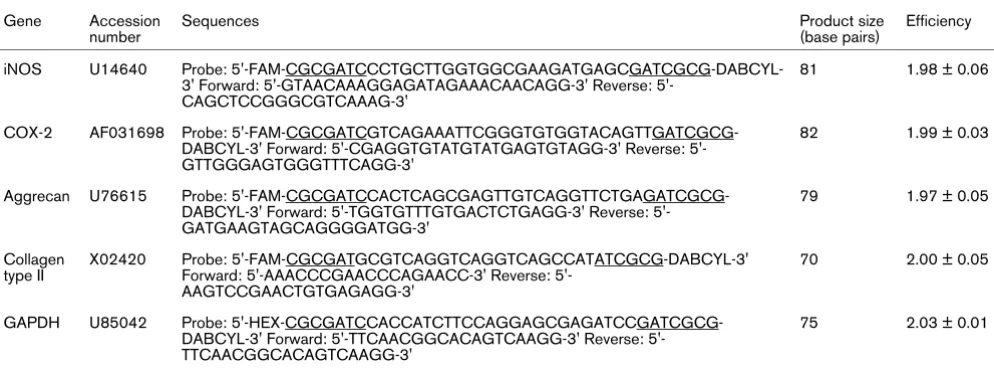
Molecular beacons were introduced for real-time detection of PCR products and were synthesized from oligonucleotides (Sigma Genosys Limited, Cambridge, UK) using the Beacon Designer software (Premier Biosoft International, California, USA). Probes have a hairpin structure and contain fluorescein (FAM) or 6-carboxyhexafluorescein (HEX) as the 5'-reporter dye and 4-(4'-dimethylaminophenylazo)benzoic acid as the 3'-quencher (Table 1). Sequences were designed to avoid regions of cross homology and analyzed using the Basic Local Alignment Search Tool to verify specificity. Additionally, tem-plates were folded and secondary structures avoided using MFold programme and beacon hairpin melting temperatures were calculated using the Zucker software. Primer accession numbers (EMBL [European Molecular Biology Laboratory]; Table 1) were obtained from previously published studies uti-lizing bovine chondrocytes [44-46]. Primers used in PCR experiments with molecular beacons produced amplicons, which were between 70 and 82 base pairs (Table 1).
PCR efficiencies for optimal primer pair and probe concentra-tions were derived from standard curves (n = 3) by preparing a 10-fold serial dilution of cDNA from a sample that repre-sented the untreated control at time zero conditions. The real-time PCR efficiencies (E) of amplification for each target was defined according to the following relationship: E = 10(-1/slope). The R2 value of the standard curve exceeded 0.9998 and revealed efficiency values presented in table 1.
Data normalization and statistical analyses
Relative quantification of iNOS, COX-2, aggrecan and colla-gen type II signals was accomplished by normalizing each tar-get gene to GAPDH and to the calibrator sample by a comparative Ct approach [47,48]. For the free-swelling exper-iments, the difference in cycle threshold (ΔCt) for the target was calculated by subtracting the mean Ct value for the time zero control (calibrator) from the Ct value of the target sample. For the mechanical loading studies, ΔCt was calculated by subtracting the mean Ct value for the unstrained, no treatment control (calibrator) from the target sample. The ΔCt of the tar-get was then normalized to the ΔCt of the reference gene, namely GAPDH. Thus, for each sample, the ratio of the relative expression level of target ΔCt and reference ΔCt was calcu-lated, as shown in the following equation.
E represents the efficiencies obtained for the target and refer-ence gene. ΔCttarget represents the difference in Ct values for the mean calibrator or sample for the target gene. ΔC
tRefer-encerepresents the difference in Ct values for the mean calibra-tor or sample for the reference gene GAPDH. Ratios were expressed on a logarithmic scale (arbitrary units).
Nitrite and prostaglandin E2 analysis
Quantification of nitrite and PGE2 release were previously described in detail [16-18,38]. Absolute concentrations of nitrite (μmol/l), a stable end-product of NO, were determined in the media using a spectrophotometric method based on the Griess reaction. PGE2 production was measured in the culture
media using an enzyme immunoassay kit (Amersham Bio-sciences, Buckinghamshire, UK).
Statistical analysis
For the time course studies under free-swelling conditions, data represent the mean and standard error of the mean val-ues of six replicates from three separate experiments. Two-way analysis of variance and the post hoc Bonferroni-cor-rected t-test was used to examine data for constructs cultured in the presence or absence of IL-1β and/or SB203580. For the mechanical loading experiments, data represent the mean and standard error of the mean values of replicates indicated in the individual figure legend. Statistical analysis was per-formed by a two-way analysis of variance and the post hoc
Bonferroni-corrected t-tests to compare differences between unstrained and strained constructs cultured under the differ-ent treatmdiffer-ent conditions. Data were also examined between unstrained constructs cultured in the absence and presence of the cytokine and/or inhibitor. In all cases, a level of 5% was considered statistically significant (P < 0.05).
Results
IL-1β activates the phosphorylation of p38 MAPK
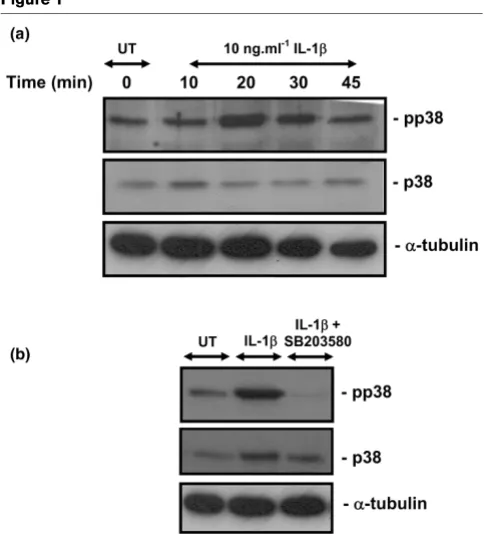
We examined the ability of IL-1β to activate the p38 pathway by Western blot analysis using antibodies directed against the Thr180/Tyr182 phosphorylated p38 (Figure 1). The activation of p38 MAPK was detected 20 minutes after the addition of IL-1β and declined thereafter, when compared with time zero. Co-stimulation with IL-1β and the p38 MAPK inhibitor (10
[image:4.612.58.555.116.301.2]μmol/l SB203580) abolished the activation of p38 at 20 min-utes in chondrocyte/agarose constructs (Figure 1b).
Table 1
Description of the Beacon designer sequences used to quantify gene expression and real-time reaction efficiencies of PCR assays
Gene Accession number
Sequences Product size
(base pairs)
Efficiency
iNOS U14640 Probe: FAM-CGCGATCCCTGCTTGGTGGCGAAGATGAGCGATCGCG-DABCYL-3' Forward: GTAACAAAGGAGATAGAAACAACAGG-FAM-CGCGATCCCTGCTTGGTGGCGAAGATGAGCGATCGCG-DABCYL-3' Reverse:
5'-CAGCTCCGGGCGTCAAAG-3'
81 1.98 ± 0.06
COX-2 AF031698 Probe: FAM-CGCGATCGTCAGAAATTCGGGTGTGGTACAGTTGATCGCG-DABCYL-3' Forward: CGAGGTGTATGTATGAGTGTAGG-3' Reverse: 5'-GTTGGGAGTGGGTTTCAGG-3'
82 1.99 ± 0.03
Aggrecan U76615 Probe: FAM-CGCGATCCACTCAGCGAGTTGTCAGGTTCTGAGATCGCG-DABCYL-3' Forward: TGGTGTTTGTGACTCTGAGG-3' Reverse: 5'-GATGAAGTAGCAGGGGATGG-3'
79 1.97 ± 0.05
Collagen type II
X02420 Probe: 5'-FAM-CGCGATGCGTCAGGTCAGGTCAGCCATATCGCG-DABCYL-3' Forward: AAACCCGAACCCAGAACC-3' Reverse:
5'-AAGTCCGAACTGTGAGAGG-3'
70 2.00 ± 0.05
GAPDH U85042 Probe: HEX-CGCGATCCACCATCTTCCAGGAGCGAGATCCGATCGCG-DABCYL-3' Forward: TTCAACGGCACAGTCAAGG-3' Reverse: 5'-TTCAACGGCACAGTCAAGG-3'
75 2.03 ± 0.01
The Beacon Designer software was used to design forward and reverse primer and probe sequences for molecular beacon applications and were synthesized by Sigma Genosys Ltd, Cambridge, UK. Secondary structures were avoided using the Mfold programme and sequences were analyzed using Basic Local Alignment Search Tool to verify specificity. Probes contain fluorescein (FAM) or 6-carboxyhexafluorescein (HEX) as a 5'-reporter dye and 4-(4'-dimethylaminophenylazo)benzoic acid (DABCYL) as 3'-quencher. Note that the arm sequences are underlined. COX, cyclo-oxygenase; GAPDH, glyceraldehyde 3-phosphate dehydrogenase; iNOS, inducible nitric oxide synthase.
Ratio of the relative ression level ET et T et Ct Mea
exp
( arg )
arg (
= 1+
Δ nn calibrator sample
E ference ference
Ct Mean calibra − +
)
( Re )
Re (
IL-1β induced iNOS and COX-2 and inhibited aggrecan expression
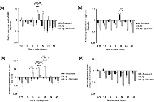
Figure 2 illustrates the effects of IL-1β on the relative expres-sion levels of iNOS, COX-2, aggrecan and collagen type II in constructs cultured under free-swelling conditions in the pres-ence and abspres-ence of the p38 MAPK inhibitor SB203580. In the absence of the cytokine, the expression levels of iNOS and COX-2 appeared to decrease over a period of 48 hours when compared with time zero (Figure 2 panels a and b, respec-tively). However, the level of downregulation was not statisti-cally significant when compared with time zero. IL-1β induced a transient increase in iNOS levels, with a peak in expression levels at 6 hours (sevenfold increase; P < 0.001) and 12 hours (fourfold increase; P < 0.05), which decreased thereafter (Fig-ure 2a). At 6 hours, SB203580 partially reduced the IL-1β induced expression of iNOS to approximately fourfold when compared with constructs treated with IL-1β only (P < 0.01; Figure 2a). The IL-1β induced iNOS expression was downreg-ulated with the p38 MAPK inhibitor at 12 hours (P < 0.01; Fig-ure 2a). The induction of COX-2 by IL-1β was detected at 3 hours (fourfold increase; P < 0.05) and 6 hours (14-fold
increase; P < 0.001), with a peak in expression at 12 hours (19-fold increase; P < 0.001) when compared with time zero (Figure 2b). The presence of SB203580 inhibited the IL-1β induced expression of COX-2 (P < 0.05 at 3 hours; P < 0.001 at 6 and 12 hours; Figure 2b).
In the absence of the cytokine, there was a downregulation of aggrecan expression up to 6 hours of culture when compared with time zero (Figure 2c). Aggrecan expression levels peaked at 12 hours (threefold increase; P < 0.01; Figure 2c) when compared with time zero and was not significantly detected at any other time point. At 12 hours, aggrecan expression was inhibited by the presence of the cytokine (P < 0.001) but was not significantly influenced by the addition of SB203580 (Fig-ure 2c). In contrast, the expression levels of collagen type II appeared to be consistently downregulated in the presence or absence of the cytokine or with the addition of the inhibitor (Figure 2d). However, the level of inhibition under the various treatment conditions did not differ significantly relative to that at time zero.
IL-1β stimulates the production of nitrite and PGE2
release
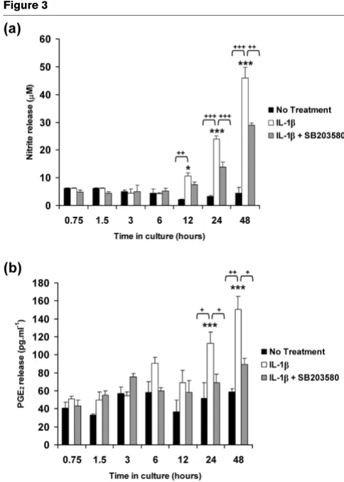
The cumulative production of nitrite and PGE2 release were assessed as presented in Figure 3. In the absence of the cytokine, the levels of nitrite and PGE2 release did not change over the 48-hour culture period. IL-1β induced nitrite and PGE2 release with significant differences measured at 24 and 48 hours (all P < 0.001; Figure 3 panels a and b, respectively). SB203580 partially reduced the IL-1β induced nitrite release at 24 hours (24 μmol/l decrease to 14 μmol/l) and at 48 hours (46 μmol/l decrease to 29 μmol/l). SB203580 abolished the IL-1β induced PGE2 release to basal levels (both P < 0.05; Figure 3b).
Dynamic compression inhibited IL-1β induced iNOS and
COX-2 expression, production of nitrite and PGE2
release
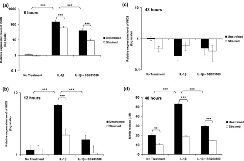
The relative expression levels of iNOS expression in unstrained constructs and constructs subjected to 15% dynamic compressive strain for 6, 12 and 48 hours are pre-sented in Figure 4. The temporal profile of iNOS expression in response to IL-1β was similar to that in free-swelling culture, with a peak in expression at 6 hours (P < 0.001; Figure 4a). However, the magnitude of the peak expression induced by the cytokine was markedly greater than under free-swelling conditions, with approximately 145-fold increase in unstrained constructs (Figure 4a) as compared with sevenfold increase in free-swelling culture (Figure 4a). Stimulation by dynamic com-pression or SB203580 in isolation significantly reduced the IL-1β induced iNOS expression at both 6 and 12 hours (both
[image:5.612.56.298.89.356.2]P < 0.001; Figure 4a,b). However, co-stimulation with dynamic compression and the inhibitor produced an effect greater than each in isolation and resulted in iNOS expression returning to basal values (Figure 4a,b,c). iNOS induction at 6 Figure 1
Activation of p38 MAPK by IL-1β
and 12 hours closely correlated with the production of nitrite, which was maximal after 48 hours of incubation with the cytokine (P < 0.001; Figure 4d). The IL-1β induced nitrite release was inhibited by dynamic compression in the absence and presence of SB203580 (both P < 0.001; Figure 4d).
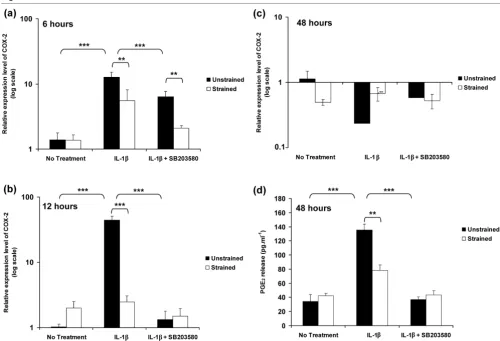
The temporal profile of COX-2 expression in unstrained and strained constructs, cultured for 6, 12 and 48 hours, is shown in Figure 5. The induction of COX-2 expression in response to IL-1β was similar to that in free-swelling culture, with peak expression at 12 hours (P < 0.001; Figure 5b). The relative magnitudes of the peak expression at 12 hours with the cytokine were 20-fold (Figure 2b) and 44-fold (Figure 5b) in free-swelling and unstrained constructs, respectively. The application of dynamic compression or presence of SB203580 reduced the IL-1β induced COX-2 expression at 6 hours (P < 0.01; Figure 5a). Stimulation by dynamic com-pression or SB203580 alone for 12 hours inhibited the
cytokine-induced induction of COX-2 (both P < 0.001; Figure 5b). Co-stimulation with both dynamic compression and SB203580 had no further effect when compared with each in isolation at 12 hours (Figure 5b), with values returning to basal levels at 48 hours (Figure 5c). COX-2 expression correlated with PGE2 production with levels unaltered in unstrained and strained constructs cultured for 48 hours under no treatment conditions (Figure 5d). Dynamic compression (P < 0.01) or the presence of SB203580 (P < 0.001) alone abolished the IL-1β induced PGE2 release (Figure 5d). However, co-stimula-tion with both dynamic compression and SB203580 had no further effect compared with each in isolation (Figure 5d).
Dynamic compression restored IL-1β induced inhibition of aggrecan expression
[image:6.612.57.554.95.423.2]The expression levels for aggrecan and collagen type II in unstrained constructs and constructs subjected to dynamic compressive strain for 6, 12 and 48 hours are shown in Figure Figure 2
IL-1β stimulates iNOS and COX-2 expression
IL-1β stimulates iNOS and COX-2 expression. Temporal profile of IL-1β on (a) iNOS, (b) COX-2, (c) aggrecan and (d) collagen type II expression by chondrocyte/agarose constructs cultured under free-swelling conditions with 0 or 10 ng/ml IL-1β and/or 10 μmol/l SB203580. Bars represent the mean and standard error of the mean of six replicates from three separate experiments. The ratio of the relative expression level for the target gene was calibrated to the mean value at time = 0 and normalized to the reference gene GAPDH. Ratios were expressed on a logarithmic scale (arbitrary units). Two-way analysis of variance with post hoc Bonferroni-corrected t-tests was used to compare data under the different treatments: *P < 0.05, **P < 0.01 and ***P < 0.001 for comparisons between time zero with IL-1β; +P < 0.05, ++P < 0.01 and +++P < 0.001 for comparisons
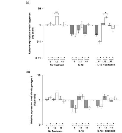
6. In the absence of the cytokine, dynamic compression increased aggrecan expression to approximately threefold at 12 hours (P < 0.001; Figure 6a). IL-1β inhibited aggrecan expression in unstrained constructs at 6, 12 and 48 hours (all
P < 0.05), and the levels were not influenced significantly by dynamic compression. However, co-stimulation with dynamic compression and SB203580 increased aggrecan expression at 12 hours (P < 0.05; Figure 6a). By contrast, the expression levels of collagen type II were not influenced by dynamic com-pression at 6, 12, or 48 hours in the presence or absence of the cytokine (Figure 6b). Moreover, the presence of the p38 MAPK inhibitor did not significantly influence collagen type II expression in unstrained or strained constructs.
Discussion
IL-1β plays a pivotal role in both the pathogenesis of OA and as a potent mediator of catabolic processes in articular chondrocytes. The inflammatory process involves the exces-sive production of NO, PGs, matrix metalloproteinases, aggre-canases, and cytokines associated with the IL-1 and tumour necrosis factor families [5-9]. The net result is a tissue milieu with increased levels of inflammatory mediators that lead to cellular stress, chondrocyte apoptosis and loss of matrix tissue [5-9]. There is clinical evidence to support the beneficial effects of controlled moderate exercise in relieving adults with OA, in combination with chondroprotective agents [49,50]. However, the success of these treatments largely relies on the validation of the drugs and their pathophysiological interac-tions within the OA affected joint. Consequently, in vitro biore-actor systems have been developed as model systems to investigate the mechano-sensitive and cytokine-sensitive intra-cellular pathways. More specifically, a number of studies have shown that the application of controlled loading regimens at physiological magnitude prevents the harmful effects of IL-1β by blocking the release of NO, PGE2 and matrix metalloprotei-nases, and restoring extracellular matrix production [10-18,38,39,45,46]. Consequently, the tissue maintains a bal-ance in matrix turnover and regulates cytokine-induced path-ways during articular cartilage loading.
In the present study, we utilized real-time quantitative PCR assays coupled with a novel fluorescent probe known as the molecular beacon to detect the catabolic (iNOS and COX-2) and anabolic (aggrecan and collagen type II) genes utilizing the chondrocyte/agarose model in conjunction with a well characterized bioreactor system [40,41]. The data provide striking evidence that dynamic compression antagonizes the IL-1β induced expression of iNOS and COX-2 levels and pro-duction of NO and PGE2 release. Additionally, we provide evi-dence to support restoration of aggrecan levels by both types of stimuli. These data support our previous studies that show that dynamic compression counteracts the IL-1β induced production of NO and PGE2 release by full-depth chondrocytes and superficial zone chondrocytes, and restores cell proliferation and proteoglycan synthesis [16,17].
[image:7.612.54.296.85.424.2]The free-swelling studies were undertaken to determine how the cytokine influenced gene expression levels with time, in the absence of the bioreactor system. IL-1β activated the phos-phorylation of p38 MAPK and was inhibited by the p38 MAPK inhibitor (Figure 1). The cytokine additionally induced a tran-sient increase in the expression levels of both iNOS and COX-2 (Figure COX-2). COX-COX-2 expression appeared to be more sensitive to the cytokine and to the presence of SB203580 (Figure 2b), suggesting that COX-2 activation may occur independently of NO and primarily involves a p38 MAPK dependent pathway. We observed a similar effect with the release of NO (Figure 3a) and PGE2 production (Figure 3b) in response to IL-1β, in which SB203580 had a more potent effect on PGE2 release Figure 3
IL-1β stimulates the production of nitrite and PGE2 release
IL-1β stimulates the production of nitrite and PGE2 release.
Tempo-ral profile of IL-1β on the production of (a) nitrite and (b) PGE2 release by chondrocyte/agarose constructs cultured under free-swelling condi-tions with 0 or 10 ng/ml IL-1β and/or 10 μmol/l SB203580. Bars repre-sent the mean and standard error of the mean of six replicates from three separate experiments. Two-way analysis of variance with post hoc Bonferroni-corrected t-tests was used to compare data under the dif-ferent treatments: *P < 0.05 and ***P < 0.001 for comparisons between time zero with IL-1β; +P < 0.05, ++P < 0.01 and +++P < 0.001
than on NO production. It could be argued that the p38 MAPK inhibitor may have nonspecific effects. Previous studies have shown inhibition of NO release by SB203580 at a concentra-tion of 1 and 10 μmol/l in IL-1β treated chondrocytes [20-22,24,51]. However, at concentrations at 10 μmol/l or greater, SB203580 was demonstrated to inhibit JNK in chondrocyte monolayers, suggesting less specificity at a higher dose [52]. Accordingly, iNOS induction and NO release may therefore involve activation of multiple MAPK pathways, in particular JNK. A number of previous studies have reported differential mechanisms through which IL-1β upregulates iNOS and COX-2 expression that involve activating transcription factor-2, NF-κB, IL-6, cAMP responsive element binding protein-1, or activator protein-2 transcription factors and activation of all members of the MAPKs [20,23,53]. Nevertheless, NF-κB appears to be the primary transcription factor that influences iNOS expression and NO production, and has been shown to strongly inhibit PGE2 synthesis in OA affected cartilage [52]. Our studies utilizing human chondrocyte/agarose constructs
cultured with selective inhibitors of iNOS (1400W) and COX-2 (NS-398), also suggest that NO could have a negative influence on PGE2 production [18]. However, whether NO induces or suppresses PGE2 release in the presence of IL-1β remains controversial [9,18,54].
[image:8.612.57.554.96.424.2]In separate experiments, constructs were subjected to dynamic compression or remained unstrained within a biore-actor system. The present data shows that unstrained culture within the bioreactor system induced a significant enhance-ment in the levels of NO release in the presence and absence of IL-1β (Figure 4d), when compared with free-swelling culture conditions (Figure 2a), whereas PGE2 release (Figures 2b and 5d) was largely unaffected, a phenomenon that concurs with our previous studies [16,17]. A similar effect was noted for iNOS induction in response to IL-1β, with a sevenfold increase in free-swelling conditions (Figure 2a), as compared with a 145-fold increase in unstrained constructs cultured within the bioreactor without the application of dynamic compression Figure 4
Dynamic compression inhibits IL-1β induced iNOS expression and production of nitrite release
(Figure 4a). In accordance with the PGE2 release data, the fold increase in expression of COX-2 was broadly similar under both culture conditions (Figures 2b and 5). There are marked differences in the diffusional constraints in unstrained con-structs in the bioreactor, involving contact of the upper surface with a fluid impermeable loading pin. Accordingly, the mass transport of oxygen into the construct may be impaired, lead-ing to increased hypoxia in the bioreactor system, compared with constructs cultured under free-swelling conditions. A number of recent studies support the hypothesis that oxygen tension can influence the production of inflammatory media-tors in cartilage. For example, hypoxia dramatically increased iNOS expression and NO production by bovine chondrocytes in response to IL-1β, whereas COX-2 expression was not strongly influenced by oxygen tension [44,55,56].
Co-stimulation with dynamic compression and inhibitor for 6 hours completely abolished iNOS induction in response to
[image:9.612.55.555.92.432.2]IL-1β (Figure 4a). Interestingly, neither dynamic compression or the inhibitor alone were able to abolish the IL-1β induced iNOS expression. The effect was more striking for COX-2 with levels returning to basal values after application of dynamic compression or the p38 MAPK inhibitor alone for 12 hours (Figure 5b). The data suggest interactions between mechano-sensitive and p38 MAPK dependent pathways in mediating the inhibitory effect of dynamic compression on iNOS and COX-2 expression. The importance of these findings is sup-ported by recent studies utilizing monolayer chondrocytes, which showed sustained levels of iNOS and COX-2 expression in response to IL-1β for up to 24 hours, with a dra-matic reduction by cyclic tensile strain for 8 hours over a 24-hour loading regimen [15]. Additionally, the mechanism was shown to involve NF-κB activation, which could act down-stream of p38 MAPK [13]. Nonetheless, static or intermittent compression of different magnitudes applied to cartilage explants increased NO and PGE2 production [28,29]. These Figure 5
Dynamic compression inhibits IL-1β induced COX-2 expression and production of PGE2 release
Dynamic compression inhibits IL-1β induced COX-2 expression and production of PGE2 release. Effects of 15% dynamic compressive strain
differences may potentially be attributed to both the nature of the mechanical stimulus and the level of oxygen tension, as indicated in recent studies [56-58].
We initially examined the involvement of the p38 MAPK because this kinase was influenced by either IL-1β or mechan-ical loading [24,52]. We demonstrated activation of the p38 MAPK following 20 minutes of stimulation with the cytokine (Figure 1). However, a major limitation of the bioreactor system
[image:10.612.55.467.96.545.2]is the time taken to retrieve constructs at the end of an exper-iment (up to 10 minutes). This causes difficulties associated with analyzing transient phosphorylation events, because alter-ations in activation state may occur after the end of the mechanical loading regimen. Accordingly, the time courses of phosphorylation events in response to both IL-1β and dynamic compression are not presented here, but work is actively ongoing. In addition, we feel that the regulation of the IL-1β pathways may be through many factors, and dynamic Figure 6
IL-1β and dynamic compression influences aggrecan expression but not collagen type II
IL-1β and dynamic compression influences aggrecan expression but not collagen type II. Effects of 15% dynamic compressive strain on (a) aggrecan and (b) collagen type II expression in unstrained (-) and strained (+) constructs cultured under no treatment conditions or with 10 ng/ml IL-1β and/or 10 μmol/l SB203580 for 6, 12 and 48 hours. The ratio of the relative expression levels of the target gene was calibrated to the mean value for the unstrained (untreated) control and normalized to GAPDH. Ratios were expressed on a logarithmic scale (arbitrary units). Bars represent the mean and standard error of the mean of 10 replicates from three separate experiments. Two-way analysis of variance with post hoc Bonferroni-corrected t-tests was used to compare data: *P < 0.05 and ***P < 0.001 between unstrained and strained values; and +P < 0.05 between
compression may actually target a global mechanism involving JNK or NF-κB, as indicated in previous studies [13,51,52].
In addition to the regulation of iNOS and COX-2 by dynamic compression, co-stimulation with the inhibitor was important in restoring the cytokine-induced inhibition of aggrecan expres-sion. This is in contrast to collagen type II, in which no response to IL-1β or dynamic compression was observed. It is highly likely that matrix gene expression is controlled by sient factors, and could involve rapid activation of multiple tran-scription factors that are influenced by a combination of mechanical loading and IL-1β [19,22,31,32]. Consequently, the multiple events activated in a time-dependent manner will influence the rate of expression and assembly of the extracel-lular matrix molecules, causing flare-ups in gene expression levels. This phenomenon may start as early as 1 hour after mechanical stimulation, as reported with aggrecan and colla-gen type II expression [37,59].
Overall, these observations indicate a hypothetical mechanism for the activation of iNOS by IL-1β that are, respectively, dependent on p38 MAPK signalling and could involve activa-tion of other MAPKs. In free-swelling condiactiva-tions the p38 inde-pendent pathway is predominantly activated, as indicated by the partial effect of the p38 inhibitor on iNOS expression at later time points. However, at early time points the p38 inhibi-tor blocked activation of the p38 MAPK. Incorporation of the construct into the bioreactor system, with its inherent diffusional constraints, dramatically enhances iNOS induction, potentially via preferential activation of the p38 MAPK depend-ent pathway and possibly through a hypoxia-driven mecha-nism. Accordingly, the inhibitor has a greater effect on iNOS expression under these conditions. Dynamic compression substantially reduces iNOS expression and co-stimulation with both dynamic compression and the p38 MAPK inhibitor abolished iNOS expression when compared with each in iso-lation, suggesting that dynamic compression targets the p38 MAPK dependent pathway. By contrast, COX-2 activation by IL-1β appears to be primarily mediated by a p38 MAPK dependent pathway that is largely unaffected by transfer to the bioreactor system but is highly susceptible to the p38 MAPK inhibitor or dynamic compression. In addition to the regulation of iNOS and COX-2 expression by dynamic compression, co-stimulation with the inhibitor was important in restoring the cytokine-induced inhibition of aggrecan expression. This is in contrast to collagen type II, in which we observed no response. It is probable that matrix gene expression is control-led by transient factors and could involve rapid activation of multiple MAPKs and transcription factors. Ultimately, elucida-tion of the intracellular pathways will enable the identificaelucida-tion of appropriate pharmacological agents and provide a clinical rationale for promoting the benefits of controlled physical activity to manage and treat OA.
Conclusion
It is important to define the mechanistic pathways induced by physiologically relevant mechanical signals in the presence of proinflammatory mediators, because this information will pro-vide key parameters for the safe application of pharmacologi-cal therapies, in conjunction with biophysipharmacologi-cal treatments for OA.
Competing interests
The authors declare that they have no competing interests.
Authors' contributions
TC supervised SA and OOA, who performed cell culture experiments and analysis by real-time PCR. DS and JB partic-ipated in Western blot analysis and TC carried out the bio-chemical assays, performed the statistical analysis and drafted the manuscript. DS, DB and DL participated in its design and coordination and helped to draft the manuscript. All authors read and approved the final manuscript.
Acknowledgements
This study was supported by The Wellcome Trust (project grant: 073972). Dr Chowdhury would like to thank Drs Kerry Elliot, Lindsay Ramage and Ying Zhou for their excellent support at the Queens Medi-cal Research Institute, Edinburgh University.
References
1. Millward-Sadler SJ, Salter DM: Integrin-dependent signal cas-cades in chondrocyte mechanotransduction. Ann Biomed Eng 2004, 32:435-446.
2. Griffin TM, Guilak F: The role of mechanical loading in the onset and progression of osteoarthritis. Exerc Sport Sci Rev 2005, 33:195-200.
3. Brandt KD: Response of joint structures to inactivity and to reloading after immobilization. Arthritis Rheumatism 2003, 49:267-271.
4. Guilak F, Fermor B, Keefe FJ, Kraus VB, Olson SA, Pisetsky DS, Setton LA, Weinberg JB: The role of biomechanics and inflam-mation in cartilage repair and injury. Clin Orthop Relat Res 2004, 423:17-26.
5. Studer RK, Jaffurs D, Stefanovic Racic M, Robbins PD, Evans CH: Nitric oxide in osteoarthritis. Osteoarthritis Cartilage 1999, 7:377-379.
6. Lotz M: The role of nitric oxide in articular cartilage damage. Rheum Dis Clin North Am 1999, 25:269-282.
7. Evans CH, Watkins SC, Stefanovic Racic M: Nitric oxide and car-tilage metabolism. Methods Enzymol 1996, 269:7-13. 8. Scher JU, Pillinger MH, Abramson SB: Nitric oxide synthases
and osteoarthritis. Curr Rheumatol Rep 2007, 9:9-15. 9. Weinberg JB, Fermor B, Guilak F: NOS and COX interactions in
cartilage and meniscus: relationships to joint physiology, arthritis and tissue repair. Subcell Biochem 2007, 42:31-62. 10. Gassner R, Buckley MJ, Georgescu H, Studer R, Stefanovic-Racic
M, Piesco NP, Evans CH, Agarwal S: Cyclic tensile stress exerts anti-inflammatory actions on chondrocytes by inhibiting induc-ible nitric oxide synthase. J Immunol 1999, 163:2187-2192. 11. Xu Z, Buckley MJ, Evans CH, Agarwal S: Cyclic tensile strain acts
as an antagonist of IL-1 beta actions in chondrocytes. J Immunol 2000, 165:453-460.
12. Deschner J, Hofman CR, Piesco NP, Agarwal S: Signal transduc-tion by mechanical strain in chondrocytes. Curr Opin Clin Nutr Metab Care 2003, 6:289-293.
14. De Croos JN, Dhaliwal SS, Grynpas MD, Pilliar RM, Kandel RA: Cyclic compressive mechanical stimulation induces sequen-tial catabolic and anabolic gene changes in chondrocytes resulting in increased extracellular matrix accumulation. Matrix Biol 2006, 25:323-331.
15. Madhavan S, Anghelina M, Rath-Deschner B, Wypasek E, John A, Deschner J, Piesco N, Agarwal S: Biomechanical signals exert sustained attenuation of proinflammatory gene induction in articular chondrocytes. Osteoarthritis cartilage 2006, 14:1023-1032.
16. Chowdhury TT, Bader DL, Lee DA: Dynamic compression inhib-its the synthesis of nitric oxide and PGE2 by IL-1β stimulated
chondrocytes cultured in agarose constructs. Biochem Bio-phys Res Commun 2001, 285:1168-1174.
17. Chowdhury TT, Bader DL, Lee DA: Dynamic compression coun-teracts IL-1β induced release of nitric oxide and PGE2 by
superficial zone chondrocytes cultured in agarose constructs. Osteoarthritis Cartilage 2003, 11:688-696.
18. Chowdhury TT, Bader DL, Lee DA: Dynamic compression coun-teracts IL-1beta induced iNOS and COX-2 activity by human chondrocytes cultured in agarose constructs. Biorheology 2006, 43:413-429.
19. Mengshol JA, Vincenti MP, Coon CI, Barchowsky A, Brinckerhoff CE: IL-1 induction of MMP-13 gene expression in chondro-cytes requires p38, c-Jun N-terminal kinase, and nuclear factor kappaB: differential regulation of collagenase 1 and colla-genase 3. Arthritis Rheum 2000, 43:801-811.
20. Nieminen R, Leinonen S, Lahti A, Vuolteenaho K, Jalonen U, Kankaanranta H, Goldring MB, Moilanen E: Inhibitors of mitogen-activated protein kinases downregulate COX-2 expression in human chondrocytes. Mediators Inflamm 2005, 5:249-255. 21. Scherle PA, Pratta MA, Feeser WS, Tancula EJ, Arner EC: The
effects of IL-1 on mitogen-activated protein kinases in rabbit articular chondrocytes. Biochem Biophys Res Commun 1997, 230:573-577.
22. Liacini A, Sylvester J, Li WQ, Zafarullah M: Inhibition of IL-1 stim-ulated MAP kinases, AP-1 and NF-kappa B transcription fac-tors down-regulates MMP gene expression in articular chondrocytes. Matrix Biol 2002, 21:251-262.
23. Malemud CJ: Protein kinases in chondrocyte signaling and osteoarthritis. Clin Orthop Relat Res 2004, 427(suppl):145-151.
24. Badger AM, Cook MN, Lark MW, Newman-Tarr TM, Swift BA, Nel-son AH, Barone FC, Kumar S: SB203580 inhibits p38 mitogen-activated protein kinase, nitric oxide production, and inducible nitric oxide synthase in bovine cartilage-derived chondrocytes. J Immunol 1998, 161:467-473.
25. Meyer M, Schreck R, Bauerle PA: H2O2 and antioxidants have
opposite effects on activation of NF-kB and AP-1 in intact cells: AP-1 as secondary antioxidant-responsive factor. EMBO J 1993, 12:2005-2015.
26. Mendes AF, Carvalho AP, Caramona MM, Lopes MC: Role of nitric oxide in the activation of NF-kappaB, AP-1 and NOS II expression in articular chondrocytes. Inflamm Res 2002, 51:369-375.
27. Marks-Konczalik J, Chu SC, Moss J: Cytokine-mediated tran-scriptional induction of the human inducible nitric oxide syn-thase gene requires both AP-1 and NF-κB binding sites. J Biol Chem 1998, 273:22201-22208.
28. Fermor B, Weinberg JB, Pisetsky DS, Misukonis MA, Banes AJ, Guilak F: The effects of static and intermittent compression on nitric oxide production in articular cartilage explants. J Orthop Res 2001, 19:729-737.
29. Fermor B, Weinburg JB, Pisetsky DS, Misukonis MA, Fink C, Gui-lak F: Induction of cyclooxygenase-2 by mechanical stress through a nitric oxide-regulated pathway. Osteoarthritis Cartilage 2002, 10:792-798.
30. Gosset M, Berenbaum F, Levy A, Pigenet A, Thirion S, Saffar JL, Jacques C: PGE2 synthesis in cartilage explants under
com-pression: mPGES1 is a mechanosensitive gene. Arthritis Res Ther 2006, 8:R135.
31. Fanning PJ, Emkey G, Smith RJ, Grodzinsky AJ, Szasz N, Trippel SB: Mechanical regulation of MAPK signalling in articular cartilage. J Biol Chem 2003, 278:50940-50948.
32. Fitzgerald JB, Jin M, Dean D, Wood DJ, Zheng MH, Grodzinsky AJ: Mechanical compression of cartilage explants induces multi-ple time-dependent gene expression patterns and involves
intracellular calcium and cyclic AMP. J Biol Chem 2004, 279:19502-19511.
33. Hung CT, Henshaw DR, Wang CC, Mauck RL, Raia F, Palmer G, Chao PH, Mow VC, Ratcliffe A, Valhmu WB: Mitogen-activated protein kinase signaling in bovine articular chondrocytes in response to fluid flow does not require calcium mobilization. J Biomech 2000, 33:73-80.
34. Murata M, Bonassar LJ, Wright M, Mankin HJ, Towle CA: A role for the interleukin-1 receptor in the pathway linking static mechanical compression to decreased proteoglycan synthe-sis in surface articular cartilage. Arch Biochem Biophys 2003, 413:229-235.
35. Vincent TL, McLean CJ, Full LE, Peston D, Saklatvala J: FGF-2 is bound to perlecan in the pericellular matrix of articular carti-lage, where it acts as a chondrocyte mechanotransducer. Osteoarthritis Cartilage 2007, 15:752-763.
36. Li KW, Wang AS, Sah RL: Microenvironment regulation of extracellular signal-regulated kinase activity in chondrocytes: effects of culture configuration, interleukin-1, and compres-sive stress. Arthritis Rheum 2003, 48:689-699.
37. Ragan PM, Badger AM, Cook M, Chin VI, Gowen M, Grodzinsky AJ, Lark MW: Down-regulation of chondrocyte aggrecan and type-II collagen gene expression correlates with increases in static compression magnitude and duration. J Orthop Res 1999, 17:836-842.
38. Chowdhury TT, Appleby RN, Salter DM, Bader DA, Lee DA: Integrin-mediated mechanotransduction in IL-1β stimulated chondrocytes. Biomech Model Mechanobiol 2006, 5:192-201. 39. Lee DA, Frean SP, Lees P, Bader DL: Dynamic mechanical
com-pression influences nitric oxide production by articular chondrocytes seeded in agarose. Biochem Biophys Res Commun 1998, 251:580-585.
40. Lee DA, Bader DL: Compressive strains at physiological fre-quencies influence the metabolism of chondrocytes seeded in agarose. J Orthop Res 1997, 15:181-188.
41. Lee DA, Knight MM: Mechanical loading of chondrocytes embedded in 3D constructs: in vitro methods for assessment of morphological and metabolic response to compressive strain. Methods Mol Med 2004, 100:307-324.
42. Cuenda A, Rose J, Doza YN, Meier R, Cohen P, Gallagher TF, Young PR, Lee JC: SB203580 is a specific inhibitor of a MAPK homologue which is stimulated by cellular stresses and IL-1. FEBS Lett 1995, 364:229-233.
43. Campbell JJ, Blain EJ, Chowdhury TT, Knight MM: Loading alters actin dynamics and upregulates coflin gene expression in chondrocytes. Biochem Biophys Res Commun 361:329-334. 44. Mathy-Hartert M, Burton S, Deby-Dupont G, Davel P, Reginster JY,
Henrotin Y: Influence of oxygen tension on nitric oxide and prostaglandin E2 synthesis by bovine chondrocytes. Osteoar-thritis Cartilage 2005, 13:74-79.
45. Blain EJ, Mason DJ, Duance VC: The effect of cyclical compres-sive loading on gene expression in articular cartilage. Biorhe-ology 2001, 40:111-117.
46. Mio K, Saito S, Tomatsu T, Toyama Y: Intermittent compressive strain may reduce aggrecanase expression in cartilage: a study of chondrocytes in agarose gel. Clin Orthop Relat Res 2005, 433:225-232.
47. Nolan T, Hands RE, Bustin SA: Quantification of mRNA using real-time RT-PCR. Nat Protocols 2006, 1:1559-1582.
48. Pfaffl MW, Horgan GW, Dempfle L: Relative expression soft-ware tool (REST) for group wise comparison and statistical analysis of relative expression results in real time PCR. Nucleic Acids Res 2002, 30:e3.
49. Roddy E, Doherty M: Changing life-styles and osteoarthritis: what is the evidence. Best Pract Res Clin Rheumatol 2006, 20:81-97.
50. Stitik TP, Blacksin MF, Stiskal DM, Kim JH, Foye PM, Schoenherr L, Choi ES, Chen B, Saunders HJ, Nadler SF: Efficacy and safety of hyaluronan treatment in combination therapy with home exercise for knee osteoarthritis pain. Arch Phys Med Rehabil 2007, 88:135-141.
51. Saklatvala J: Inflammatory signalling in cartilage. MAPK and NF-κB pathways in chondrocytes and the use of inhibitors for research into pathogenesis and therapy of OA. Curr Drug Targets 2007, 8:305-313.
pro-teoglycan synthesis and for activation of JNK1 following cycli-cal mechanicycli-cal stimulation in a human chondrocyte culture model. Osteoarthritis Cartilage 2007, 15:884-893.
53. Vaillancourt F, Morquette B, Shi Q, Fahmi H, Lavigne P, Di Battista JA, Fernandes JC, Benderdour M: Differential regulation of cyclooxygenase-2 and inducible nitric oxide synthase by 4-hydroxynonenal in human osteoarthritic chondrocytes through ATF-2/CREB-1 transactivation and concomitant inhi-bition of NF-kappaB signaling cascade. J Cell Biochem 2007, 100:1217-1231.
54. Amin AR, Attur M, Patel RN, Thakker GD, Marshall PJ, Rediske J, Stuchin SA, Patel IR, Abramson SB: Superinduction of cycloox-ygenase-2 activity in human osteoarthritis-affected cartilage. Influence of nitric oxide. J Clin Invest 1997, 99:1231-1237. 55. Henrotin Y, Kurz B, Aigner T: Oxygen and reactive oxygen
spe-cies in cartilage degradation. Osteoarthritis Cartilage 2005, 13:643-654.
56. Martin G, Andriamanalijaona R, Grassel S, Dreier R, Mathy-Hartert M, Bogdanowicz P, Boumediene K, Henrotin Y, Bruckner P, Pujol JP: Effect of hypoxia and reoxygenation on gene expression and response to interleukin-1 in cultured articular chondrocytes. Arthritis Rheum 2004, 50:3549-3560.
57. Fermor B, Weinberg JB, Pisetsky DS, Guilak F: The influence of oxygen tension on the induction of nitric oxide and prostaglan-din E2 by mechanical stress in articular cartilage. Osteoarthritis
Cartilage 2005, 13:935-941.
58. Fermor B, Christensen SE, Youn I, Cernanec JM, Davies CM, Weinberg JB: Oxygen, nitric oxide and articular cartilage. Eur Cell Mater 2007, 13:56-65.