Appendix I: The Predicted Three-Dimensional Structure of the Human
D
2Dopamine Receptor and the Binding Site and Binding Affinities for
Appendix II: Predicted 3-D Structure for Human
b
2
Appendix III: The Predicted Structure of the Human D
1The Predicted Structure of the Human D
1Dopamine Receptor
M. Yashar S. Kalani §, *
, Nagarajan Vaidehi*
& William A. Goddard III**
* Materials and Process Simulation Center, Beckman Institute, California Institute of Technology,
Pasadena, CA 91125
§ Johns Hopkins University School of Medicine, Baltimore, MD 21205
**To whom correspondence should be addressed (e-mail: [email protected])
Results:
We have predicted the structure and binding site of 11 agonists and antagonists (Shown in Figure 3-1) in D1DR using the methods detailed in the Methods and Materials
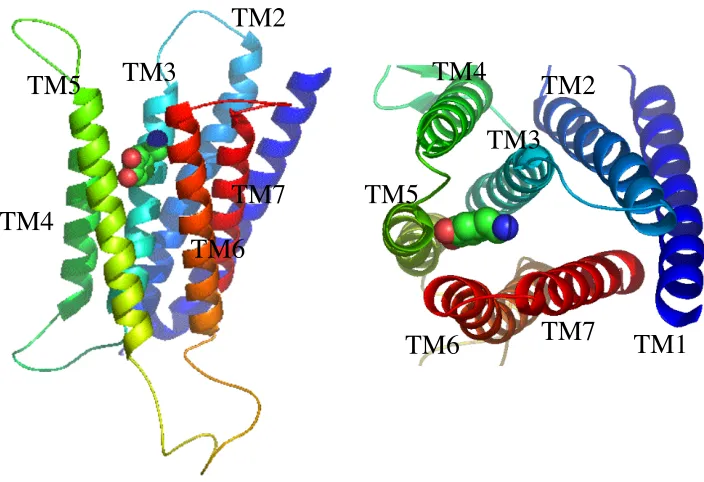
section. Figure 3-2 shows the 3D atomic resolution structure of the human D1DR
modeled based on the predicted D2 structure using the Membstruk procedure and the
predicted binding site of dopamine determined using the HierDock procedure. We refer to this predicted structure as D1DR. Using the D1DR structure, we have identified the
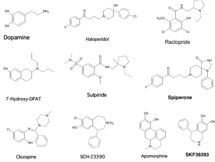
Figure 3-1. 10 agonists and antagonists studied for the human Dopamine D1 receptor. OH
OH
NH2 O
N OH Cl F OH OH N N NH O O S O O N N OH OH Cl H3CO
Cl
O HN
N O N F N NH O Cl NH N N N NCH3 Cl HO OH OH N H Dopamine Haloperidol 7-Hydroxy-DPAT Apomorphine Sulpiride Raclopride Spiperone
Figure 3-2. Predicted binding site of Dopamine (shown in sphere) in the predicted structure of human dopamine D1 receptor.
In the preliminary studies, we have validated these methods for bovine rhodopsin, human ß-2 adrenergic receptor, human s1p (sphingolipid) and lpa (lysophosphatidic acid) receptors, the human D2 dopamine receptor (Kalani 2003), and 10 mouse olfactory
receptors (Vaidehi 2002, Floriano 2000, Floriano 2003, Hall 2003). The results are in good agreement with available experimental data, indicating a good description of the binding site and relative binding energies of various ligands.
TM1
TM2
TM3
TM4
TM5
TM6
TM7
TM1
TM2
TM3
TM4
TM5
[image:18.612.148.500.92.333.2]Prediction of the Structure of Human D1DR:
The Membstruk procedure used to predict the structure is detailed in Vaidehi 2002. The homology modeling procedure used to model the structure of the D1 receptor
based on the D2 (MS) structure is outlined elsewhere. The TM2nDs procedure (Vaidehi
2002, Trabanino 2003) was utilized to identify the transmembrane (TM) spanning regions based on a hydrophobic analysis of the sequence. A seven helical motif was identified (below) ranging from 19-29 residues per helix. The highlighted residues represent TM helices, while the intervening sequences are loop regions. The same transmembrane predictions were used in the alignment to the human D2 structure to perform homology
modeling.
MRTLNTSAMDGTGLVVERDFSVRILTACFLSLLILSTLLGNTLVCAAVIRFRHLRS
KVTNFFVISLAVSDLLVAVLVMPWKAVAEIAGFWPFGSFCNIWVAFDIMCSTASI
LNLCVISVDRYWAISSPFRYERKMTPKAAFILISVAWTLSVLISFIPVQLSWHKAK
PTSPSDGNATSLAETIDNCDSSLSRTYAISSSVISFYIPVAIMIVTYTRIYRIAQKQIR
RIAALERAAVHAKNCQTTTGNGKPVECSQPESSFKMSFKRETKVLKTLSVIMGV
FVCCWLPFFILNCILPFCGSGETQPFCIDSNTFDVFVWFGWANSSLNPIIYAFNADF
RKAFSTLLGCYRLCPATNNAIETVSINNNGAAMFSSHHEPRGSISKECNLVYLIPH AVGSSEDLKKEEAAGIARPLEKLSPALSVILDYDTDVSLEKIQPITQNGQHPT
Scheme 5-1. Predicted transmembrane regions are highlighted below in the human D1
The TM prediction was utilized in building seven canonical a-helices and optimized using the procedure described in Vaidehi 2002. The helices were bundled in explicit bilayer of dilaurylphosphatidylcholine lipid molecules to mimic the biological membrane. The structural factors such as helical bend, helical tilt etc., of the predicted structure of D1DR (homology) structure compared to the crystal structure of rhodopsin
are summarized in Table 3-1.
TM
Helix
Helical
Bend
Plane
Tilt
HPM
Angle
HPM
Mag.
HPM Fit Plane CM
Dist.
Plane CM
Angle
CM Fit
1 9.3 40.7 9.3 1.1 -0.8 15.6 0.0 2.7662
2 12.2 37.9 55.8 3.5 -2.9 10.0 36.3 0.5758
3 4.6 12.3 -36.1 2.0 -0.6 4.5 120.3 2.2851
4 31.5 21.3 13.3 1.8 2.4 14.0 116.5 -4.5510
5 10.3 5.7 -68.9 1.4 -0.8 14.8 183 5.4926
6 23.3 11.1 -166.3 4.3 -5.0 11.5 238.5 2.6714
Centered Comparison Table
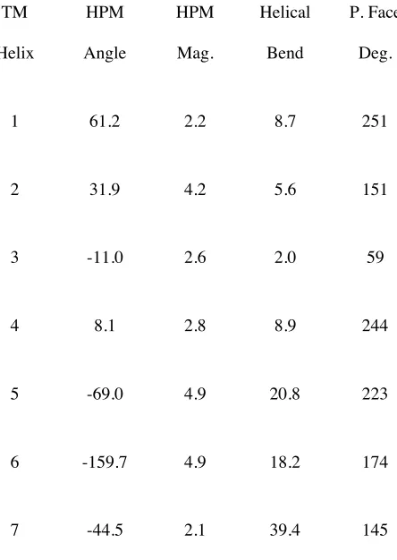
TM
Helix
HPM
Angle
HPM
Mag.
Helical
Bend
P. Face
Deg.
1 61.2 2.2 8.7 251
2 31.9 4.2 5.6 151
3 -11.0 2.6 2.0 59
4 8.1 2.8 8.9 244
5 -69.0 4.9 20.8 223
6 -159.7 4.9 18.2 174
[image:21.612.215.437.114.427.2]7 -44.5 2.1 39.4 145
Table 3-1. Comparison of the D1DR with the crystal structure of bovine rhodopsin.
RMS height of TMR: 31.2
RMS radius of TMR: 12.4
From Table 3-1 it is clear that the helical bends are very different for D1DR
compared to rhodopsin. The volume of the TM barrel for D1DR is 15111.9 Å 3
. There is one disulfide bond between Cys96 and Cys186 in EC2. Rhodopsin has a volume of 11,807.7 Å3
structure compared to the crystal structure of rhodopsin is 4.78 Å and a total residue RMS of 6.32Å (all atoms).
Prediction of the putative binding site for dopamine in D1DR: The Hierdock2.0 procedure (Vaidehi 2002) was utilized to scan all the D1DR models as described in
Materials and Methods section. We discuss the details of the predicted binding site in D1DR (homology) model for all the 11 ligands studied in this paper to capture the same
binding as those identified in the D2 (MS) for comparison reasons and explanations of
receptor specificity in future chapters.
include Trp99, and Ser107 (TM3), Phe156 (TM4), Tyr194, and Ala195 (TM5), Trp285, Phe289, and Asn292 (TM6). The first phenylalanine of the conserved WXXFF motif, Phe288 is slightly greater than 5.5 Å away from dopamine. Asn292 in TM6 is providing a very strong component of the binding energy of dopamine to the receptor. Assuming Asp103 forms a bidentate salt bridge, or a salt bridge and a hydrogen bond to the amino group of dopamine, the interaction with Asn292 fulfils the ability of the amino group to form favorable interactions with any other residues. It must be noted, however, that the
Figure 3-3. The 5.5 Å binding site of dopamine to the human D1 dopamine receptor. The
numbers in the brackets indicate the TM helices to which the residues belong to.
presence of Trp99 (3) may be the reason why dopamine exhibits reduced binding to the D1 subtype of the receptor compared to the D2 receptor. The presence of the Trp99 (3)
provides Asp103 (3) an alternate hydrogen bond donor, meaning the aspartate will share its electron density between the protonated amino group and the indole and will not
Dopamine Trp99 (3)
Asp103 (3)
Ser107 (3) Asn292 (6)
Ala195 (5)
Ser198 (5)
Ser199 (5)
Ser202 (5) Trp285 (6)
[image:23.612.197.497.277.499.2]interact as efficiently with the primary amino group. Interestingly, I predict that tertiary amino groups should have enhanced binding to this receptor since the presence of a single proton will not cause for competition for the aspartate electron density.
Figure 3-4. The 5.5 Å binding site of 7-OH DPAT to the human D1 dopamine receptor.
The numbers in the brackets indicate the TM helices to which the residues belong to.
Apomorphine: The ligand binds in the putative agonist binding site located between TM3, 4, 5, and 6. The protonated amino group of Apomorphine is salt bridged to the TM3 Asp103 (2.9 Å). The other major polar contacts are to the conserved TM5 serines198, 199 and 202. There are as previously two hydrogen bonds to Ser198 (3.8 Å) and Ser202 (3.1 Å) both to the lone hydroxyl group on Apomorphine. The contact to Ser199 is ~ 6 Å apart. Other residues form a most hydrophobic pocket around the ligand; these residues include: Trp99, Phe102, Cys106, Ser107, Ile111 (all TM3), Trp148, Phe156 (both TM4), Tyr194, Ile201 (TM5), Trp285, Phe288, Phe289, Asn292 (all TM6),
Trp99 (3) Phe102 (3) Asp103 (3) Cys106 (3) Ser107 (3)
Trp148 (4) Tyr194 (5)
Ser198 (5) Ser199 (5) Ile201 (5) Ser202 (5)
Trp285 (6) Phe288 (6) Phe289 (6) Asn292 (6)
Val317 (7) Trp321 (7)
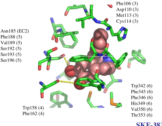
[image:25.612.178.496.94.358.2]Val317 (TM7) (Figure 3-5). Mutational experiments have shown that Phe288Ala and Phe289Ala mutants show substantial reduction in binding constants for Apomorphine (Cho et al 1995). This shows that these residues are important in ligand recognition.
Figure 3-5. The 5.5 Å binding site of Apomorphine to the human D1 dopamine receptor.
The numbers in the brackets indicate the TM helices to which the residues belong to.
Clozapine: Clozapine is a class I antagonist, meaning it binds in the putative agonist binding site located between TM3, 4, 5, and 6. There is, as always, an amino group salt bridged to the TM3 Asp103 (2.8 Å). As is the case for all antagonists studied, there is a lone hydrogen bond to the sequence of TM5 serines. Although the hydrogen bond may be to either Ser198 or Ser202, in our structure, the more reasonable distance and angle are for the interaction between Ser198 and the heteroatom (3.3 Å). There is a third
Trp99 (3) Phe102 (3) Cys106 (3) Ser107 (3) Ile111 (3) Trp148 (4)
Phe156 (4)
Tyr194 (5) Ser198 (5) Ser199 (5) Ile201 (5) Ser202 (5)
Trp285 (6) Phe288 (6) Phe289 (6) Asn292 (6)
Val317 (7)
interaction, which although cannot be categorized as a hydrogen bond, is energetically significant; this is a 3.1 Å interaction between Trp285 in TM6 and the hydrogen of the ring in Clozapine. Ser107 is 4.9 Å apart from the other nitrogen in the Clozapine ring. The remainder of the residues provide a mostly hydrophobic pocket for the ligand. There residues in close proximity of the Clozapine ligand are Trp99, Phe102, Cys106, Ser107 (TM3), Phe156 (TM4), Tyr194, Ala195 (TM5), Phe281, Phe288, Phe289, Asn292, (TM6), Val317, Trp321 (TM7) (Figure 3-6).
Figure 3-6. The 5.5 Å binding site of Clozapine to the human D1 dopamine receptor. The
numbers in the brackets indicate the TM helices to which the residues belong to.
Fenoldopam: Fenoldopam is an agonist, meaning it binds in the putative agonist binding site located between TM3, 4, 5, and 6. There is a bidentate salt bridge between Asp103 (TM3) with and the amino group with distances of 2.6 and 2.8 Å. Ser107 (TM3) is hydrogen bonded to heteroatom with a length of 3.3 Å. The catechol portion is hydrogen
Clozapine Trp99 (3) Phe102 (3) Asp103 (3) Cys106 (3) Ser107 (3) Phe156 (4)
Tyr194 (5) Ala195 (5) Ser198 (5) Ser199 (5) Ser202 (5)
Phe281 (6) Trp285 (6) Phe288 (6) Phe289 (6) Asn292 (6)
bonded to the TM5 serine networks of Ser198, Ser199, and Ser202 (TM5). There is a 3.1 Å interaction and a 2.8 Å interaction to the catechol by Ser198 and Ser202, respectively. Ser199 is too far to form a hydrogen bond to the ligand. Interestingly, it is possible for the phenol group to be pi stacked between Phe288 and Phe289 (TM6) and for its hydroxyl group to interact via a hydrogen bond with Asn202 (TM6). The remainder of the residues in the cavity provide a mostly hydrophobic pocket for the drug. These residues include: Trp99, Ile104, Ile111 (TM3), Trp148 (TM4), Ala195, Ile201 (TM5), Phe281, Trp285 (TM6), Val317 (TM7) (Figure 3-7).
Figure 3-7. The 5.5 Å binding site of Fenoldopam to the human D1 dopamine receptor.
The numbers in the brackets indicate the TM helices to which the residues belong to.
Haloperidol: The ligand binds in the putative antagonist-binding site located between TM2, TM3, TM4, TM5, TM6, and TM7. Class II antagonists such as Haloperidol are
Fenoldopam
Trp99 (3) Asp103 (3) Ile104 (3) Ser107 (3) Ile111 (3)
Trp148 (4) Ala195 (5) Ser198 (5) Ser199 (5) Ile201 (5) Ser202 (5)
Phe281 (6) Trp285 (6) Phe288 (6) Phe289 (6) Asn292 (6)
composed of two aromatic domains connected by a linker domain, which has a protonated amino group. The protonated amino group of Haloperidol is salt bridged to the TM3 Asp103 (2.8 Å). There are several heteroatom contacts that are typically too weak to be considered hydrogen bonds. There is a long hydrogen bond between the hydroxyl of Haloperidol and TM6 Trp285 (4.2 Å). Haloperidol and other class II antagonists are predicted to have low affinities to the D1 receptor; and, indeed experimental studies are in
Figure 3-8. (a) The 5.5 Å binding site of Haloperidol to the human D1 dopamine
receptor. The numbers in the brackets indicate the TM helices to which the residues belong to. (b) The binding cavity without the ligand is presented on the left, and the cavity with the ligand present on the right. The presence of the TM3 Phe102 blocks the cavity for the class II antagonist to occupy the void between TM2 and TM7.
Haloperidol
Trp80 (2) Lys81 (2) Trp99 (3)
Phe102 (3) Cys106 (3) Ser107 (3) Trp148 (4)
Val152 (4) Phe156 (4)
Tyr194 (5) Ser198 (5) Ser199 (5) Ile201 (5) Ser202 (5)
Trp285 (6) Phe288 (6) Phe289 (6) Asn292 (6)
Asp314 (7) Val317 (7) Trp318 (7) Trp321 (7)
Phe102 (3)
Figure 3-9. The 5.5 Å binding site of Raclopride to the human D1 dopamine receptor.
The numbers in the brackets indicate the TM helices to which the residues belong to.
SCH-23390: SCH-23390 is a class I antagonist. It forms a salt bridge with the TM3 Asp103 with a distance of 2.9 Å. There are two hydrogen bond contacts with the 5th
transmembrane domain. There is a 3.0-Å interaction Tyr194 (5) and a 3.4 Å interaction with Ser202 (5). The remainder of the residues create a mostly hydrophobic pocket for the ligand, with Phe288 (6) and Phe289 (6) forming excellent Van der Waals interactions with the phenyl substituent of the ligand. Other amino acids in the cavity include: Trp99 (3), Phe102 (3), Ser107 (3), Trp148 (4), Val152 (4), Phe156 (4), Ala195 (5), Ser198 (5), Ser199 (5), Trp285 (6), Asn292 (6), Cys293 (6), and Val317 (7) (Figure 3-10). There is no clash between the ligand and Phe102 (3) since the ligand does not extend into the domain between TM2 and TM7.
Raclopride Trp99 (3) Asp103 (3) Ser107 (3) Ile111 (3) Trp148 (4)
Val152 (4) Phe156 (4)
Tyr194 (5) Ser197 (5) Ser198 (5) Ser199 (5) Ile201 (5) Ser202 (5)
Trp285 (6) Phe288 (6) Phe289 (6) Asn292 (6)
Figure 3-10. The 5.5 Å binding site of SCH-23390 to the human D1 dopamine receptor.
The numbers in the brackets indicate the TM helices to which the residues belong to.
SKF-38393: SKF-38393 is an agonist. It forms a bidentate salt bridge with Asp103 with a distance of 2.8 Å. There are two hydrogen bonds to Ser198 (3.1 Å) and Ser202 (3.5 Å). The phenyl substituent is pi stacked between Phe288 (6) and Phe289 (6). The remainder of the residues form a pocket around the ligand; these residues include: Trp99 (3), Ser107 (3), Phe156 (4), Tyr194 (5), Ala195 (5), Ser199 (5), Ile201 (5), Trp285 (6), Asn292 (6), and Cys293 (6) (Figure 3-11).
SCH-23390 Trp99 (3) Phe102 (3) Asp103 (3) Ser107 (3) Trp148 (4)
Val152 (4) Phe156 (4)
Tyr194 (5) Ala195 (5) Ser198 (5) Ser199 (5) Ser202 (5)
Trp285 (6) Phe288 (6) Phe289 (6) Asn292 (6) Cys293 (6)
Figure 3-11. The 5.5 Å binding site of SKF-38393 to the human D1 dopamine receptor.
The numbers in the brackets indicate the TM helices to which the residues belong to.
Spiperone: Spiperone is a class II antagonist; meaning, it binds with its aromatic domains to the voids between TM2 & 7 and TM4 & 6, and it forms a salt bridge to TM3 and weak non-ionic interactions to TM5 and 6. The protonated amino group of the ligand is salt bridged to the TM3 Asp103 (2.9 Å). Since class II antagonists are too large to bind into the D1 class of dopamine receptors, the interaction with the TM5 serine network is
not possible. The ligand clashes with Phe102 (3), which blocks the cavity, and as a result, the heteroatom is not in a position to interact with the serine networks. The remainder of the residues in the cavity form a mostly hydrophobic pocket for the ligand. The residues in the cavity include: Leu76 (2), Val77 (2), Trp80 (2), Lys81 (2), Trp99 (3), Val100 (3), Cys106 (3), Ser107 (3), Trp148 (4), Tyr194 (5), Ser198 (5), Ser199 (5), Ile201 (5),
SKF-38393 Trp99 (3) Asp103 (3) Ser107 (3) Phe156 (4)
Tyr194 (5) Ala195 (5) Ser198 (5) Ser199 (5) Ile201 (5) Ser202 (5)
Ser202 (5), Trp285 (6), Phe288 (6), Phe289 (6), Asn292 (6), Asp314 (7), Val317 (7), Trp318 (7), Phe319 (7), Gly320 (7), and Trp321 (7) (Figure 3-12).
Figure 3-12. The 5.5 Å binding site of Spiperone to the human D1 dopamine receptor.
The numbers in the brackets indicate the TM helices to which the residues belong to.
Sulpiride: Sulpiride is a class I antagonist meaning it binds in the putative agonist binding site with minimal extension into the TM2, and 7 aromatic micro domain. Sulpiride has a salt bridge to the TM3 Asp103 (2.8 Å). Interestingly, the same aspartate is also hydrogen bonding to the amide hydrogen of Sulpiride (3.0 Å). This interaction is also found in all the Sulpiride-like ligands. Although the addition of the amide was introduced to decrease the lipophilicity of these drugs, the addition seems to fair well due to the interactions it has with the Asp103 in TM3. The sulfonamide portion of the ligand
interacts well with the sequence of Serines in TM5. There are two hydrogen bonds of 2.8 Å and 3.6 Å to Ser198 and Ser202, respectively. There is also a hydrogen bond between Tyr194 (5) and the sulfonamide group; this interaction is 3.3 Å long. The strength of these hydrogen bonds is, however, a different issue. The remainder of the residue provide a mostly hydrophobic pocket. The residues in close proximity include: Trp99 (3), Phe102 (3), Ser107 (3), Trp148 (4), Val152 (4), Phe156 (4), Ala195 (5), Ser199 (5), Ile201 (5), Trp285 (6), Phe288 (6), Phe289 (6), Asn292 (6), Asp314 (7), Val317 (7) (Figure 3-13).
Figure 3-13. The 5.5 Å binding site of Sulpride to the human D1 dopamine receptor. The
numbers in the brackets indicate the TM helices to which the residues belong to.
Comparisons of Binding Site of Agonists vs. Antagonists: Experimental studies have
outlined the binding site for agonists and antagonists. The putative agonist-binding site is located between TM3, 4, 5, & 6, and some residues in the EC2 loop (assuming this loop
Sulpride Tyr194 (5)
Ala195 (5) Ser198 (5) Ser199 (5) Ile201 (5) Ser202 (5)
Trp148 (4) Val152 (4) Phe156 (4)
Trp285 (6) Phe288 (6) Phe289 (6) Asn292 (6)
Trp99 (3) Phe102 (3) Asp103 (3) Ser107 (3)
closes during the process of activation). We have classified antagonists into two categories: Class I & II. Class I antagonists, such as Clozapine bind in the putative agonist binding pocket meaning they occupy the void between TM3, 4, 5 and 6. Class II antagonists, such as Haloperidol consists of two aromatic domains connected by a linker, which possess a protonated amino group. These antagonists bind in the cavity between TM2, 3, 4, 5, 6, and 7. There are two aromatic micro domains (pictures of aromatic micro domains goes here). The first aromatic micro-domain is located in TM4 and TM6, which is composed of the very symmetric Trp148 (4), Phe156 (4), Trp285 (6), Phe288 (6), and Phe289 (6). This aromatic micro-domain stabilizes one of the aromatic rings of the class II antagonists. TM3 provides the salt bridge to TM3 Asp103, which stabilizes the ligand in place. TM5 provides weak interactions with heteroatom functionalities such as halogens on the rings of class II antagonists. These interactions are weak but important in recognition of the correct aromatic domain for docking into the cavity. The second aromatic micro-domain composed of Trp90 (2), Trp318 (7), and Trp321 (7) stabilize the second aromatic ring group of the class II antagonists. It must be noted, however, that due to the presence of Phe102 (3) which blocks the cavity and does not allow for the longer ligands in class II to access the second aromatic micro-domain. Class II antagonists are not predicted to bind to this class of receptors.
network of TM5 serines, although this is not an absolute necessary for agonism. There is, in every case studied, favorable interaction from the first aromatic micro-domain located in TM4 and TM6. A residue that has not been appreciated in drug design is Asn292 (6). The presence of this residue contributes greatly to ligand binding, yet it appears that there few agonists utilize this residue effectively. It is important to note that the agonists studied effectively interact with both Ser198 and Ser202 in TM5. The interactions to Ser199, the third TM5 serine, are too long to be considered a hydrogen bond (on the order of ~ 5 Å). Under no circumstance, due to structural constraints, can all three serines effectively interact with the agonists studied. It appears that there could at most be two hydrogen bonds to the TM5 serines. Although in our structure Ser198 is not participating in any interactions, it is possible that in a slightly different structure, perhaps one resulting from activation, there could be interactions to Ser199 and Ser202 as opposed to Ser198 and Ser202. All agonists studied cause strong coupling of TM3 and 5. None of the agonists studied block TM3 and 6 motions. Based on structural studies of rhodopsin, it has been established that a motion between TM3 and 6 are essential for activation. The coupling of TM3 and 6 by agonists causes a decrease in distance between TM3 and 5 while allowing for motion between TM3 and 6.
Class I Antagonists: the Clozapine-like antagonists salt bridge to the TM3 Asp103 with their protonated amino group. They do not have two aromatic rings and therefore only utilize the first aromatic micro-domain between TM4 and 6. As is the case with both Class I and class II antagonists, they form only one weak interaction with the TM5 serine network. In our models the interaction may be with Ser198 or Ser202. At first glance, it appears that both the number of and strength of the interactions with the TM5 serines may be important for activation. Interestingly, however, the situation is more complicated than it appears; Strange et al. have identified agonism from a non-hydroxylated form of the DPAT series. The critical distinguishing feature of an agonist vs. antagonist appears to be its relative position to TM6. Class I antagonists burry their aliphatic domain deep into the conserved TM6 WXXFF motif. Experimental studies of rhodopsin suggest that motion of TM3 and 6 is necessary for activation. The binding of antagonists at the TM6 WXXFF would prevent any motion, specifically the hinge motion between TM3 and TM6 that is necessary for activation. It appears that the presence of one hydroxyl/one hydrogen bond donor/acceptor is not an absolute necessity for antagonism. The class I antagonists are further stabilized by Trp99 in TM3.
second ring would bind to the second aromatic micro-domain but Phe102 (3) does not allow for this binding in the second aromatic micro-domain. Since the ligand has little room to bind in the putative cavity, it fails to form adequate contacts to the TM5 serine network and is also expected to have lower efficacy as an antagonist. In some cases Asn292 (6) may also stabilize the class II antagonists. As is the case with the class I antagonists, class II antagonists prevent motion between TM3 and TM6 by burying their aliphatic portion between TM3 and TM6, thereby preventing interaction of these helices. There is very little difference between the binding sites of the class II antagonists, although some utilize the cavity better than others.
Table 3-2.Binding energies in kcal/mol for a library of 11 ligands to the human D1
dopamine receptor. Haldol and Raclopride do not bind this class of dopamine receptors due to the presence of TM3 Phe102 blocker.
D1 Receptor Experimental Ranking:
SCH23390~SKF38393~Fenoldopam>Clozapine~Haldol~Spiperone~Apomorphine~Dop amine~7OHDPAT>Sulpiride~Raclopride
Ligand B.E. (Kcal/mol)
7OHDPAT -48.7
Apomorphine -47.8
Clozapine -41.8
Dopamine -48.3
Fenoldopam -48.8
Haldol NA
Raclopride NA
SCH -60.6
SKF -45.1
Spiperone NA
D1 Receptor Ranking Theory:
Appendix IV: The Predicted Structure of the Human D
3The Predicted Structure of the Human D
3Dopamine Receptor
M. Yashar S. Kalani §, *
, Nagarajan Vaidehi*
& William A. Goddard III**
* Materials and Process Simulation Center, Beckman Institute, California Institute of Technology,
Pasadena, CA 91125
§ Johns Hopkins University School of Medicine, Baltimore, MD 21205
**To whom correspondence should be addressed (e-mail: [email protected])
Results:
We have predicted the structure and binding site of 10 agonists and antagonists (Shown in Figure 4-1) in D3DR using the methods detailed in the Methods and Materials
section. Figure 4-2 shows the 3D atomic resolution structure of the human D3DR
predicted based upon the model of the D2DR obtained using the Membstruk procedure
and the predicted binding site of dopamine determined using the HierDock procedure. We refer to this predicted structure as D3DR. Using the D3DR structure, we have
Figure 6-1. 10 agonists and antagonists studied for the human Dopamine D3 receptor. OH
OH
NH2 O
N OH Cl F OH OH N N NH O O S O O N N OH OH Cl H3CO Cl
O HN
N O N F N NH O Cl NH N N N NCH3 Cl HO OH OH N H Dopamine Haloperidol 7-Hydroxy-DPAT Apomorphine Sulpiride Raclopride Spiperone
Figure 6-2. Predicted binding site of Dopamine (shown in sphere) in the predicted structure of human dopamine D3 receptor.
In the preliminary studies, we have validated these methods for bovine rhodopsin, human ß-2 adrenergic receptor, human s1p (sphingolipid) and lpa (lysophosphatidic acid) receptors, and 10 mouse olfactory receptors (Vaidehi 2002, Floriano 2000, Floriano 2003, Hall 2003). The results are in good agreement with available experimental data, indicating a good description of the binding site and relative binding energies of various ligands.
TM1
TM2 TM3
TM4 TM5
TM6 TM7
TM1 TM2
TM3 TM4
TM5
Prediction of the Structure of Human D3DR:
The Membstruk procedure used to predict the structure is detailed in Vaidehi 2002. The TM2nDs procedure (Vaidehi 2002, Trabanino 2003) was utilized to identify the transmembrane (TM) spanning regions based on a hydrophobic analysis of the sequence. A seven helical motif was identified (below) ranging from 19-29 residues per helix. The highlighted residues represent TM helices, while the intervening sequences are loop regions.
MASLSQLSSHLNYTCGAENSTGASQARPHAYYALSYCALILAIVFGNGLVCMAV
LKERALQTTTNYLVVSLAVADLLVATLVMPWVVYLEVTGGVWNFSRICCDVFV
TLDVMMCTASILNLCAISIDRYTAVVMPVHYQHGTGQSSCRRVALMITAVWVL
AFAVSCPLLFGFNTTGDPTVCSISNPDFVIYSSVVSFYLPFGVTVLVYARIYVVLK
QRRRKRILTRQNSQCNSVRPGFPQQTLSPDPAHLELKRYYSICQDTALGGPGFQE RGGELKREEKTRNSLSPTIAPKLSLEVRKLSNGRLSTSLKLGPLQPRGVPLREKKA
TQMVAIVLGAFIVCWLPFFLTHVLNTHCQTCHVSPELYSATTWLGYVNSALNPVI
YTTFNIEFRKAFLKILSC
Scheme 4-1. Predicted transmembrane regions are highlighted below in the human D3
dopamine receptor.
explicit bilayer of dilaurylphosphatidylcholine lipid molecules to mimic the biological membrane. The structural factors such as helical bend, helical tilt etc., of the predicted structure of D3DR structure compared to the crystal structure of rhodopsin are
summarized in Table 4-1.
TM Helix Helical Bend Plane Tilt HPM Angle HPM Mag.
HPM Fit Plane CM
Dist.
Plane CM
Angle
CM Fit
1 13.9 51.2 4.0 8.2 0.0 15.9 0.0 2.2527
2 8.8 32.9 109.5 5.8 0.0 10.0 34.5 0.4490
3 5.9 11.3 63.0 2.8 0.0 4.2 119.9 2.1168
4 2.1 11.4 -9.2 9.1 0.0 14.5 125.3 -2.0740
5 11.1 7.8 -21.8 7.0 0.0 14.1 187.7 2.2381
6 22.1 12.9 -157.9 9.6 0.0 11.7 244.4 2.2205
7 8.5 20.9 72.3 5.3 0.0 10.1 301.1 -6.3052
Centered Comparison Table
1 76.5 3.2 4.4 242
2 108.1 3.4 9.3 150
3 17.7 2.5 0.0 57
4 5.7 3.6 4.7 245
Helix 5 -17.0 3.3 17.2 219
Helix 6 -126.7 5.9 17.4 201
Helix 7 11.2 4.1 2.7 157
Table 4-1. Comparison of the D3DR with the crystal structure of bovine rhodopsin
RMS height of TMR: 29.5
RMS radius of TMR: 12.1
From Table 4-1 it is clear that the helical bends are very different for D3DR
compared to rhodopsin. The volume of the TM barrel for D3DR is 13471.1 Å 3
. There is one disulfide bond between Cys103 and Cys181 in EC2. Rhodopsin has a volume of 11,807.7 Å3. The calculated RMS in the coordinates of the C
structure compared to the crystal structure of rhodopsin is 7.10 Å and a total residue RMS of 7.74 Å (all atoms).
Prediction of the putative binding site for dopamine in D3DR: The Hierdock2.0 procedure (Vaidehi 2002) was utilized to scan all the D3DR models as described in
Materials and Methods section. We discuss the details of the predicted binding site in D3DR model for all the 10 ligands studied in this paper.
(6), Phe346 (6), His349 (6), and Thr369 (7). The first phenylalanine of the conserved WXXFF motif, Phe345 is slightly greater than 5.5 Å away from dopamine. His349 in TM6 is providing a very strong component of the binding energy of dopamine to the receptor. Assuming Asp110 forms a bidentate salt bridge, or a salt bridge and a hydrogen bond to the amino group of dopamine, the interaction with His349 fulfils the ability of the amino group to form favorable interactions with any other residues.
Figure 4-3. The 5.5 Å binding site of dopamine to the human D3 dopamine receptor. The
numbers in the brackets indicate the TM helices to which the residues belong to.
7-OH DPAT: The ligand binds in putative agonist binding site located between TM3, 4, 5, and 6. The protonated amino group of 7-OH DPAT is salt bridged to the TM3 Asp110 (2.8 Å). The other major polar contacts are to the conserved TM5 serines 192, 193 and 196. There are two hydrogen bonds, one 2.9 Å to Ser192 and 3.1 Å to Ser196. The
Dopamine Phe106 (3)
Asp110 (3)
Met113 (3) Cys114 (3)
Phe162 (4) Val189 (5)
Ser192 (5)
Ser193 (5)
Ser196 (5)
Trp342 (6) Phe346 (6)
interaction to Ser193 is 5.8 Å and is too long to be considered a hydrogen bond. The remainder of the residues in the cavity are mainly hydrophobic residues that provide stabilization for the aromatic and aliphatic rings of the ligand. The residues in close hydrophobic contact of the ligands are Phe106 (3), Leu109 (3), Met113 (3), Cys114 (3), Phe162 (4), Asn185 (EC2), Val189 (5), Trp342 (6), Phe345 (6), Phe346 (6), His349 (6), Thr369 (7), and Tyr373 (7) (Figure 4-4) which provide a mainly hydrophobic pocket.
Figure 4-4. The 5.5 Å binding site of 7-OH DPAT to the human D3 dopamine receptor.
The numbers in the brackets indicate the TM helices to which the residues belong to.
Apomorphine: The ligand binds in the putative agonist binding site located between TM3, 4, 5, and 6. The protonated amino group of Apomorphine is salt bridged to the TM3 Asp110 (2.9 Å). The other major polar contacts are to the conserved TM5 serines
Phe106 (3) Leu109 (3) Asp110 (3) Met113 (3) Cys114 (3) Phe162 (4)
Asn185 (EC2) Val189 (5) Ser192 (5) Ser193 (5) Ser196 (5)
Trp342 (6) Phe345 (6) Phe346 (6) His349 (6)
Thr369 (7) Tyr373 (7)
192, 193 and 196. There are as previously two hydrogen bonds to Ser192 (3.0 Å) and Ser196 (3.2 Å) both to the hydroxyl groups on Apomorphine. The contact to Ser193 is ~ 6 Å apart. Other residues form a most hydrophobic pocket around the ligand; these residues include: Phe106 (3), Met113 (3), Cys114 (3), Trp158 (4), Phe162 (4), Phe188 (5), Val189 (5), Phe338 (6), Trp342 (6), Phe345 (6), Phe346 (6), His349 (6), and Thr369 (7) (Figure 4-5). Mutational experiments have shown that Phe345Ala and Phe346Ala mutants show substantial reduction in binding constants for Apomorphine (Cho et al 1995). This shows that these residues are important in ligand recognition.
Figure 4-5. The 5.5 Å binding site of Apomorphine to the human D3 dopamine receptor.
The numbers in the brackets indicate the TM helices to which the residues belong to. Phe106 (3)
Asp110 (3) Met113 (3) Cys114 (3) Trp158 (4)
Phe162 (4)
Phe188 (5) Val189 (5) Ser192 (5) Ser193 (5) Ser196 (5)
Phe338 (6) Trp342 (6) Phe345 (6) Phe346 (6) His349 (6)
Thr369 (7)
Figure 4-6. The 5.5 Å binding site of Clozapine to the human D3 dopamine receptor. The
numbers in the brackets indicate the TM helices to which the residues belong to.
Fenoldopam: Fenoldopam is an agonist, meaning it binds in the putative agonist binding site located between TM3, 4, 5, and 6. There is a bidentate salt bridge between Asp110 (TM3) with and the amino group with a distances of 2.6 and 2.8 Å. Cys114 (TM3) is hydrogen bonded to heteroatom with a length of 3.4 Å, although the strength of this hydrogen bond is much weaker than that present when serine in a similar position in the D1-Like receptors interacts with this ligand. The catechol portion is hydrogen bonded to
Phe188 (5) Val189 (5) Ser192 (5) Ser193 (5) Ser196 (5)
Phe338 (6) Trp342 (6) Phe345 (6) Phe346 (6) His349 (6) Phe162 (4)
Phe106 (3) Leu109 (3) Asp110 (3) Met113 (3) Cys114 (3)
Trp85 (2)
Thr369 (7) Tyr373 (7)
the TM5 serine networks of Ser192, Ser193, and Ser196 (TM5). There is a 3.2 Å interaction and a 2.9 Å interaction to the catechol by Ser192 and Ser196, respectively. Ser193 is too far to form a hydrogen bond to the ligand. Interestingly, it is possible for the phenol group to be pi stacked between Phe345 and Phe346 (TM6) and for its hydroxyl group to interact via a hydrogen bond with His349 (6), although this interaction is again weaker than that present in the D1-like receptor when there is an asparagine
Figure 4-7. The 5.5 Å binding site of Fenoldopam to the human D3 dopamine receptor.
The numbers in the brackets indicate the TM helices to which the residues belong to.
Haloperidol: The ligand binds in the putative antagonist-binding site located between TM2, TM3, TM4, TM5, TM6, and TM7. Class II antagonists such as Haloperidol are composed of two aromatic domains connected by a linker domain, which has a protonated amino group. The protonated amino group of Haloperidol is salt bridged to the TM3 Asp110 (2.8 Å). There are several heteroatom contacts that are typically too weak to be considered hydrogen bonds. There is a weak interaction between the chlorine atom and TM5 Ser196 (3.2 Å). There is also a hydrogen bond between the hydroxyl of Haloperidol and TM6 Trp342 (3.4 Å). There is a third weak interaction between the fluorine atom on the second aromatic portion of Haloperidol and TM2 Trp85 (2.9 Å). Other residues form a mostly hydrophobic pocket around the ligand; these residues include: Val82 (2), Leu89 (2), Phe106 (3), Leu109 (3), Met113 (3), Cys114 (3), Trp158
Fenoldopam
Phe338 (6) Trp342 (6) Phe345 (6) Phe346 (6) His349 (6)
Thr369 (7) Tyr373 (7) Val189 (5)
Ser192 (5) Ser193 (5) Val195 (5) Ser196 (5)
Trp158 (4) Phe162 (4)
(4), Phe162 (4), Phe188 (5), Val189 (5), Val195 (5), Trp342 (6), Phe345 (6), Phe346 (6), His349 (6), Ser366 (7), Ala367 (7), Thr369 (7), Trp370 (7), and Tyr373 (7) (Figure 4-8).
Figure 4-8. The 5.5 Å binding site of Haloperidol to the human D3 dopamine receptor.
The numbers in the brackets indicate the TM helices to which the residues belong to.
Raclopride: Raclopride binds in the class I antagonist-binding site, although it begins to extend to the TM2 and 7 aromatic micro-domain. The critical contact points are the salt bridge between TM3 Asp110 and the protonated amino group of the ligand (2.8 Å), and interactions with the TM3 Cys114 (3.6 Å) and Ser192 (3.8 Å) and Ser196 (3.9 Å), the latter of which is too long to be considered a hydrogen bond. The remainder of the residues provide a mainly hydrophobic pocket for the remainder of the ligand. The
[image:57.612.161.473.174.431.2]residues in close proximity of the ligand include: Phe106 (3), Val107 (3), Met113 (3), Cys114 (3), Trp158 (4), Phe162 (4), Asn185 (EC2), Phe188 (5), Val189 (5), Trp342 (6), Phe345 (6), Phe346 (6), His349 (6), Ser366 (7), and Thr369 (7) (Figure 4-9).
Figure 4-9. The 5.5 Å binding site of Raclopride to the human D3 dopamine receptor.
The numbers in the brackets indicate the TM helices to which the residues belong to.
SCH-23390: SCH-23390 is a class I antagonist. It forms a salt bridge with the TM3 Asp110 with a distance of 2.8 Å. There are two hydrogen bond contacts with the 5th
transmembrane domain. There is a 3.0-Å interaction with Ser192 (5) and a 3.6 Å interaction with Ser196 (5). The remainder of the residues create a mostly hydrophobic pocket for the ligand, with Phe345 (6) and Phe346 (6) forming excellent Van der Waals interactions with the phenyl substituent of the ligand. Other amino acids in the cavity
Trp342 (6) Phe345 (6) Phe346 (6) His349 (6) Asn185 (EC2)
Phe188 (5) Val189 (5) Ser192 (5) Ser196 (5)
Trp158 (4) Phe162 (4)
Phe106 (3) Val107 (3) Asp110 (3) Met113 (3) Cys114 (3)
Ser366 (7) Thr369 (7)
[image:58.612.187.467.191.437.2]include: Phe106 (3), Met113 (3), Cys114 (3), Trp158 (4), Phe162 (4), Asn185 (EC2), Phe188 (5), Val189 (5), Ser193 (5), Trp342 (6), His349 (6), Val350 (6), Thr353 (6), Thr369 (7) (Figure 4-10).
Figure 4-10. The 5.5 Å binding site of SCH-23390 to the human D3 dopamine receptor.
The numbers in the brackets indicate the TM helices to which the residues belong to.
SKF-38393: SKF-38393 is an agonist. It forms a bidentate salt bridge with Asp110 with a distance of 2.8 Å. There are two hydrogen bonds to Ser192 (3.0 Å) and Ser196 (3.8 Å). The phenyl substituent is pi stacked between Phe345 (6) and Phe346 (6). The remainder of the residues form a pocket around the ligand; these residues include: Phe106 (3), Met113 (3), Cys114 (3), Trp158 (4), Phe162 (4), Asn185 (EC2), Phe188 (5), Val189 (5), Ser193 (5), Trp342 (6), His349 (6), Val350 (6), Thr353 (6) (Figure 4-10).
SCH-23390
Asn185 (EC2) Phe188 (5) Val189 (5) Ser192 (5) Ser193 (5) Ser196 (5)
Trp342 (6) Phe345 (6) Phe346 (6) His349 (6) Val350 (6) Thr353 (6)
Thr369 (7) Phe106 (3)
Asp110 (3) Met113 (3) Cys114 (3) Trp158 (4)
Figure 4-11. The 5.5 Å binding site of SKF-38393 to the human D3 dopamine receptor.
The numbers in the brackets indicate the TM helices to which the residues belong to.
Spiperone: Spiperone is a class II antagonist; meaning, it binds with its aromatic domains to the voids between TM2 & 7 and TM4 & 6, and it forms a salt bridge to TM3 and weak non-ionic interactions to TM5 and 6. The protonated amino group of the ligand is salt bridged to the TM3 Asp110 (2.9 Å). There is a hydrogen bond between the fluorine of the ligand and Ser192 (2.8 Å). Another weak, non-ionic interaction is to the TM6 Trp342 (3.0 Å). The remainder of the residues in the cavity create a mostly hydrophobic pocket for the aliphatic portions of the ligand. Residues present in the cavity include: Leu81 (2), Trp85 (2), Val105 (3), Phe106 (3), Val107 (3), Thr108 (3), Leu109 (3), Met113 (3), Cys114 (3), Trp158 (4), Phe162 (4), Val189 (5), Trp342 (6), Phe345 (6), Phe346 (6), His349 (6), Ser366 (7), Thr369 (7), Trp370 (7), Leu371 (7), Gly372 (7), Tyr373 (7) (Figure 4-12).
SKF-38393 Asn185 (EC2)
Phe188 (5) Val189 (5) Ser192 (5) Ser193 (5) Ser196 (5)
Trp158 (4) Phe162 (4)
Figure 4-12. The 5.5 Å binding site of Spiperone to the human D3 dopamine receptor.
The numbers in the brackets indicate the TM helices to which the residues belong to.
Sulpiride: Sulpiride is a class I antagonist meaning it binds in the putative agonist binding site with minimal extension into the TM2 and 7 aromatic micro-domain. Sulpiride has a salt bridge to the TM3 Asp110 (2.8 Å). The sulfonamide portion of the ligand interacts well with the sequence of Serines in TM5. There are two hydrogen bonds of 2.9 Å and 3.5 Å to Ser192 and Ser196, respectively. The strength of these hydrogen bonds is, however, a different issue. The remainder of the residue provide a mostly hydrophobic pocket. The residues in close proximity include: Trp85 (2), Phe106 (3), Leu109 (3), Met113 (3), Cys114 (3), Trp158 (4), Phe162 (4), Phe188 (5), Val189 (5),
Ser193 (5), Trp342 (6), Phe345 (6), Phe346 (6), His349 (6), Ser366 (7), Thr369 (7), Trp370 (7), and Tyr373 (7) (Figure 4-13).
Figure 4-13. The 5.5 Å binding site of dopamine to the human D3 dopamine receptor.
The numbers in the brackets indicate the TM helices to which the residues belong to.
Comparisons of Binding Site of Agonists vs. Antagonists: Experimental studies have
outlined the binding site for agonists and antagonists. The putative agonist-binding site is located between TM3, 4, 5, & 6, and some residues in the EC2 loop (assuming this loop closes during the process of activation). We have classified antagonists into two categories: Class I & II. Class I antagonists, such as Clozapine bind in the putative agonist binding pocket meaning they occupy the void between TM3, 4, 5 and 6. Class II antagonists, such as Haloperidol consists of two aromatic domains connected by a linker,
Sulpiride Phe188 (5)
Val189 (5) Ser192 (5) Ser193 (5) Ser196 (5)
Trp158 (4) Phe162 (4)
Trp342 (6) Phe345 (6) Phe346 (6) His349 (6)
Ser366 (7) Thr369 (7) Trp370 (7) Tyr373 (7) Trp85 (2)
which possess a protonated amino group. These antagonists bind in the cavity between TM2, 3, 4, 5, 6, and 7. There are two aromatic micro domains (pictures of aromatic micro domains goes here). The first aromatic micro-domain is located in TM4 and TM6, which is composed of the very symmetric Trp158 (4), Phe162 (4), Trp342 (6), and Phe346 (6). This aromatic micro-domain stabilizes one of the aromatic rings of the class II antagonists. TM3 provides the salt bridge to TM3 Asp110, which stabilizes the ligand in place. TM5 provides weak interactions with heteroatom functionalities such as halogens on the rings of class II antagonists. These interactions are weak but important in recognition of the correct aromatic domain for docking into the cavity. The second aromatic micro-domain composed of Trp85 (2), Phe106 (3), Trp370 (7), and Tyr373 (7) stabilize the second aromatic ring group of the class II antagonists.
order of ~ 5 Å). Under no circumstance, due to structural constraints, can all three serines effectively interact with the agonists studied. It appears that there could at most be two hydrogen bonds to the TM5 serines. Although in our structure Ser193 is not participating in any interactions, it is possible that in a slightly different structure, perhaps one resulting from activation, there could be interactions to Ser193 and Ser196 as opposed to Ser192 and Ser196. All agonists studied cause strong coupling of TM3 and 5. None of the agonists studied block TM3 and 6 motions. Based on structural studies of rhodopsin, it has been established that a motion between TM3 and 6 are essential for activation. The coupling of TM3 and 6 by agonists causes a decrease in distance between TM3 and 5 while allowing for motion between TM3 and 6.
Comparison of antagonists binding site: The antagonists studied were classified into two categories: Class I antagonists (exemplified by Clozapine), which bind in the putative agonist binding pocket; and class II antagonists (exemplified by Haloperidol), which bind in the cavity between TM2, 3, 4, 5, 6, and 7.
than it appears; Strange et al. have identified agonism from a non-hydroxylated form of the DPAT series. The critical distinguishing feature of an agonist vs. an antagonist appears to be its relative position to TM6. Class I antagonists burry their aliphatic domain deep into the conserved TM6 WXXFF motif. Experimental studies of rhodopsin suggest that motion of TM3 and 6 is necessary for activation. The binding of antagonists at the TM6 WXXFF would prevent any motion, specifically the hinge motion between TM3 and TM6 that is necessary for activation. It appears that the presence of one hydroxyl/one hydrogen bond donor/acceptor is not an absolute necessity for antagonism. The class I antagonists are further stabilized by Phe106 in TM3.
Table 4-2. Binding energies in kcal/mol for a library of 11 ligands to the human D3
dopamine receptor.
D3 Receptor Experimental Ranking:
Raclopride~Spiperone~7-OHDPAT>Haldol~Sulpiride~Dopamine~Apomorphine> Clozapine~SCH23390~SKF38393
D3 Receptor Ranking Theory:
Haldol~Spiperone~Raclopride~Apomorphine~Sulpiride>SCH23390~SKF38393~7-OHDPAT~Fenoldopam~Dopamine~Clozapine
Ligand B.E. (Kcal/mol)
7OHDPAT -95
Apomorphine -107.4 Clozapine -79.8
Dopamine -91.9
Fenoldopam -92.2
Haldol -123.5
Raclopride -108.4
SCH -102.3
SKF -96.9
[image:66.612.224.384.102.264.2]Appendix V: The Predicted Structure of the Human D
4The Predicted Structure of the Human D
4Dopamine Receptor
M. Yashar S. Kalani §, *
, Nagarajan Vaidehi*
& William A. Goddard III**
* Materials and Process Simulation Center, Beckman Institute, California Institute of Technology,
Pasadena, CA 91125
§ Johns Hopkins University School of Medicine, Baltimore, MD 21205
**To whom correspondence should be addressed (e-mail: [email protected])
Results:
We have predicted the structure and binding site of 10 agonists and antagonists (Shown in Figure 5-1) in D4DR using the methods detailed in the Methods and Materials
section. Figure 5-2 shows the 3D atomic resolution structure of the human D4DR
modeled from the predicted structure of D2DR using the Membstruk procedure and the
predicted binding site of dopamine determined using the HierDock procedure. We refer to this predicted structure as D4DR. Using the D4DR structure, we have identified the
residues in the binding site of all the 10 ligands and analyze the similarities and differences in their binding sites.
Figure 5-1. 10 agonists and antagonists studied for the human Dopamine D4 receptor. OH
OH
NH2 O
N OH Cl F OH OH N N NH O O S O O N N OH OH Cl H3CO Cl
O HN
N O N F N NH O Cl NH N N N NCH3 Cl HO OH OH N H Dopamine Haloperidol 7-Hydroxy-DPAT Apomorphine Sulpiride Raclopride Spiperone
[image:69.612.113.541.192.535.2]Figure 5-2. Predicted binding site of Dopamine (shown in sphere) in the predicted structure of human dopamine D4 receptor.
In the preliminary studies, we have validated these methods for bovine rhodopsin, human ß-2 adrenergic receptor, human s1p (sphingolipid) and lpa (lysophosphatidic acid) receptors, and 10 mouse olfactory receptors (Vaidehi 2002, Floriano 2000, Floriano 2003, Hall 2003). The results are in good agreement with available experimental data, indicating a good description of the binding site and relative binding energies of various ligands.
TM1 TM2
TM3
TM4 TM5
TM6 TM7
TM1 TM2
TM3 TM4
TM5
Prediction of the Structure of Human D4DR:
The Membstruk procedure used to predict the structure is detailed in Vaidehi 2002. The TM2nDs procedure (Vaidehi 2002, Trabanino 2003) was utilized to identify the transmembrane (TM) spanning regions based on a hydrophobic analysis of the sequence. A seven helical motif was identified (below) ranging from 19-29 residues per helix. The highlighted residues represent TM helices, while the intervening sequences are loop regions.
MGNRSTADADGLLAGRGPAAGASAGASAGLAGQGAAALVGGVLLIGAVLAGN
SLVCVSVATERALQTPTNSFIVSLAAADLLLALLVLPLFVYSEVQGGAWLLSPRL
CDALMAMDVMLCTASIFNLCAISVDRFVAVAVPLRYNRQGGSRRQLLLIGATW
LLSAAVAAPVLCGLNDVRGRDPAVCRLEDRDYVVYSSVCSFFLPCPLMLLLYW
ATFRGLQRWEVARRAKLHGRAPRRPSGPGPPSPTPPAPRLPQDPCGPDCAPPAPG
LPRGPCGPDCAPAAPGLPPDPCGPDCAPPAPGLPQDPCGPDCAPPAPGLPRGPCG PDCAPPAPGLPQDPCGPDCAPPAPGLPPDPCGSNCAPPDAVRAAALPPQTPPQTR RRRRAKITGRERKAMRVLPVVVGAFLLCWTPFFVVHITQALCPACSVPPRLVSA
VTWLGYVNSALNPVIYTVFNAEFRNVFRKALRACC
Scheme 5-1. Predicted transmembrane regions are highlighted below in the human D4
The TM prediction was utilized in building seven canonical a-helices and optimized using the procedure described in Vaidehi 2002. The helices were bundled in explicit bilayer of dilaurylphosphatidylcholine lipid molecules to mimic the biological membrane. The structural factors such as helical bend, helical tilt etc., of the predicted structure of D4DR structure compared to the crystal structure of rhodopsin are
summarized in Table 5-1.
TM
Helix
Helical
Bend
Plane
Tilt
HPM
Angle
HPM
Mag.
HPM Fit Plane CM
Dist.
Plane CM
Angle
CM Fit
1 10.1 32.3 -89.0 9.1 0.0 15.9 0.0 1.3762
2 14.6 33.0 -12.1 7.3 0.0 10.2 37.0 0.6935
3 3.9 13.4 9.0 2.8 0.0 3.9 124.1 2.7529
4 4.5 4.5 -81.7 8.2 0.0 14.5 127.0 -2.9960
5 8.7 10.4 -110.0 7.3 0.0 14.1 189.6 2. 6744
6 21.8 15.5 87.4 7.9 0.0 11.7 246.8 1.9452
Centered Comparison Table
Table 5-1. Comparison of the D4DR with the crystal structure of bovine rhodopsin.
RMS height of TMR: 30.4
RMS radius of TMR: 12.0
From Table 5-1 it is clear that the helical bends are very different for D4DR
compared to rhodopsin. The volume of the TM barrel for D4DR is 13810.2 Å 3
. There is
TM
Helix
HPM
Angle
HPM
Mag.
Helical
Bend
P. Face
Deg.
1 -65.7 3.1 21.8 238
2 33.2 3.8 10.8 165
3 5.5 3.0 0.0 68
4 -69.1 4.1 3.5 242
5 24.4 3.8 15.3 221
6 -153.4 5.5 15.2 202
one disulfide bond between Cys108 and Cys185 in EC2. Rhodopsin has a volume of 11,807.7 Å3
. The calculated RMS in the coordinates of the Ca atoms of the D4DR
structure compared to the crystal structure of rhodopsin is 6.81 Å and a total residue RMS of 7.39 Å (all atoms).
Prediction of the putative binding site for dopamine in D4DR: The Hierdock2.0 procedure (Vaidehi 2002) was utilized to scan all the D4DR models as described in
Materials and Methods section. We discuss the details of the predicted binding site in D4DR model for all the 10 ligands studied in this paper.
residues in the binding pocket provide mostly hydrophobic packing for the ligand. These residues include Leu113 (3), Leu118 (3), Cys119 (3), Val193 (5), Trp407 (6), Phe411 (6), His414 (6), Ile415 (6), and Thr434 (7) (Figure 5-3). The first phenylalanine of the conserved WXXFF motif, Phe410 is slightly greater than 5.5 Å away from dopamine. His414 in TM6 is providing a very strong component of the binding energy of dopamine to the receptor. Assuming Asp115 forms a bidentate salt bridge, or a salt bridge and a hydrogen bond to the amino group of dopamine, the interaction with His414 fulfils the ability of the amino group to form favorable interactions with any other residues. It is essential to know that a Phe-Ala164 mutation in TM4 has removed the stabilization of the aliphatic/aromatic portions of the ring. Additionally, Trp160 (4), usually present in the binding site of dopamine and other ligands is absent in this bound conformation.
Figure 5-3. The 5.5 Å binding site of dopamine to the human D4 dopamine receptor. The
numbers in the brackets indicate the TM helices to which the residues belong to.
Leu111 (3)
Asp115 (3)
Cys119 (3)
Dopamine
His414 (6)
Ile415 (6) Val193 (5)
Ser196 (5)
Ser197 (5)
Ser200 (5)
[image:75.612.167.480.397.632.2]7-OH DPAT: The ligand binds in putative agonist binding site located between TM3, 4, 5, and 6. The protonated amino group of 7-OH DPAT is salt bridged to the TM3 Asp115 (2.8 Å). The other major polar contacts are to the conserved TM5 serines 196, 197 and 200. There are two hydrogen bonds, both 3.0 Å to Ser196 and 200. The interaction to Ser197 is 5.2 Å and is too long to be considered a hydrogen bond. The remainder of the residues in the cavity are mainly hydrophobic residues that provide stabilization for the aromatic and aliphatic rings of the ligand. The residues in close hydrophobic contact of the ligands are Leu111 (3), Met114 (3), Leu118 (3), Cys119 (3), Val193 (5), Trp407 (6), Phe410 (6), Phe411 (6), His414 (6), Ile415 (6), Thr434 (7), and Tyr438 (7), (Figure 5-4) which provide a mainly hydrophobic pocket.
Figure 5-4. The 5.5 Å binding site of 7-OH DPAT to the human D4 dopamine receptor.
The numbers in the brackets indicate the TM helices to which the residues belong to.
7OH-DPAT
Val193 (5) Ser196 (5) Ser197 (5) Ser200 (5)
Trp407 (6) Phe410 (6) Phe411 (6) His414 (6) Ile415 (6)
Thr434 (7) Tyr438 (7) Leu111 (3)
[image:76.612.186.467.393.618.2]Apomorphine: The ligand binds in the putative agonist binding site located between TM3, 4, 5, and 6. The protonated amino group of Apomorphine is salt bridged to the TM3 Asp115 (2.9 Å). The other major polar contacts are to the conserved TM5 serines 196, 197 and 200. There are as previously two hydrogen bonds to Ser196 (3.1 Å) and Ser200 (3.2 Å) both to the hydroxyl groups on Apomorphine. The contact to Ser197 is ~ 6 Å apart. Other residues form a most hydrophobic pocket around the ligand; these residues include: Leu111 (3), Leu118 (3), Cys119 (3), Trp160 (4), Val193 (5), Phe403 (6), Trp407 (6), Phe410 (6), Phe411 (6), His414 (5), Ile415 (6), Trp434 (7) (Figure 5-5). Mutational experiments have shown that Phe410Ala and Phe411Ala mutants show substantial reduction in binding constants for Apomorphine (Cho et al 1995). This shows that these residues are important in ligand recognition.
Figure 5-5. The 5.5 Å binding site of Apomorphine to the human D4 dopamine receptor.
The numbers in the brackets indicate the TM helices to which the residues belong to.
Apomorphine
Val193 (5)Ser196 (5) Ser197 (5) Ser200 (5)
Phe403 (6) Trp407 (6) Phe410 (6) Phe411 (6) His414 (6) Ile415 (6)
Leu111 (3) Asp115 (3) Leu118 (3) Cys119 (3)
Trp160 (4)
[image:77.612.168.452.418.659.2]Clozapine: Clozapine is a class I antagonist, meaning it binds in the putative agonist binding site located between TM3, 4, 5, and 6. There is, as always, an amino group salt bridged to the TM3 Asp115 (2.8 Å). As is the case for all antagonists studied, there is a lone hydrogen bond to the sequence of TM5 serines. Although the hydrogen bond may be to either Ser196 or Ser200, in our structure, the more reasonable distance and angle are for the interaction between Ser196 and the heteroatom (3.2 Å). Cys119 is 4.2 Å apart from the ring nitrogen in the Clozapine ring. The remainder of the residues provide a mostly hydrophobic pocket for the ligand. The residues in close proximity of the Clozapine ligand are Leu90 (2), Leu111 (3), Met114 (3), Leu118 (3), Cys119 (3), Ser122 (3), Tyr192 (5), Val193 (5), Phe403 (7), Trp407 (7), Phe410 (7), Phe411 (7), His414 (6), Thr434 (7), and Tyr438 (7) (Figure 5-6).
Figure 5-6. The 5.5 Å binding site of Clozapine to the human D4 dopamine receptor. The
numbers in the brackets indicate the TM helices to which the residues belong to. Clozapine Phe403 (6)
Trp407 (6) Phe410 (6) Phe411 (6) His414 (6) Tyr192 (5)
Val193 (5) Ser196 (5) Ser197 (5) Ser200 (5)
Thr434 (7) Tyr438 (7)
Leu90 (2) Leu111 (3)
[image:78.612.159.445.412.631.2]Figure 5-7. The 5.5 Å binding site of Fenoldopam to the human D4 dopamine receptor.
The numbers in the brackets indicate the TM helices to which the residues belong to.
Haloperidol: The ligand binds in the putative antagonist-binding site located between TM2, TM3, TM4, TM5, TM6, and TM7. Class II antagonists such as Haloperidol are composed of two aromatic domains connected by a linker domain, which has a protonated amino group. The protonated amino group of Haloperidol is salt bridged to the TM3 Asp115 (2.8 Å). There are several heteroatom contacts that are typically too weak to be considered hydrogen bonds. There is a weak interaction between the chlorine atom and TM5 Ser200 (3.2 Å). There is also a weak interaction between the hydroxyl of Haloperidol and TM6 Trp407 (4.1 Å). There is a third weak interaction between the fluorine atom on the second aromatic portion of Haloperidol and TM2 Ser431 (2.9 Å). In
Fenoldopam
Phe403 (6)Trp407 (6) Phe410 (6) Phe411 (6) His414 (6)
Thr434 (7) Val193 (5)
Ser196 (5) Ser197 (5) Cys199 (5) Ser200 (5)
Trp160 (4)
[image:80.612.180.461.93.343.2]cases where there is a TM2 trypthopan, the interaction with the fluorine is provided by the tryptophan. Other residues form a mostly hydrophobic pocket around the ligand; these residues include: Leu90 (2), Phe91 (2), Ser94 (2), Leu111 (3), Met114 (3), Leu118 (3), Cys119 (3), Trp160 (4), Val193 (5), Cys199 (5), Trp407 (6), Phe410 (6), Phe411 (6), His414 (6), Ile415 (6), Val430 (7), Ser431 (7), Ala432 (7), Thr434 (7), Trp435 (7), Tyr438 (7) (Figure 5-8).
Figure 5-8. The 5.5 Å binding site of Haloperidol to the human D4 dopamine receptor.
The numbers in the brackets indicate the TM helices to which the residues belong to.
Haloperidol
Val193 (5) Ser196 (5) Ser197 (5) Cys199 (5)
Ser200 (5) Trp407 (6)
Phe410 (6) Phe411 (6) His414 (6) Ile415 (6) Trp160 (4)
Leu111 (3) Met114 (3) Asp115 (3) Leu118 (3) Cys119 (3)
Leu90 (2) Phe91 (2) Ser94 (2)
[image:81.612.173.464.262.539.2]Raclopride: Raclopride binds in the class I antagonist-binding site, although it begins to extend to the TM2 and 7 aromatic micro-domain. The critical contact points are the salt bridge between TM3 Asp115 and the protonated amino group of the ligand (2.9 Å), and interactions with the TM3 Cys119 (4.3 Å) and Ser200 (3.8 Å) both of which is too long to be considered a hydrogen bond. The remainder of the residues provide a mainly hydrophobic pocket for the remainder of the ligand. The residues in close proximity of the ligand include: Leu111 (3), Met112 (3), Leu118 (3), Trp160 (4), Ala164 (4), Phe192 (5), Val193 (5), Trp407 (6), Phe410 (6), Phe411 (6), His414 (6), Ile415 (6), and Thr434 (7) (Figure 5-9).
Figure 5-9. The 5.5 Å binding site of Raclopride to the human D4 dopamine receptor.
The numbers in the brackets indicate the TM helices to which the residues belong to. Raclopride
Tyr192 (5) Val193 (5) Ser196 (5) Ser200 (5)
Trp160 (4) Ala164 (4)
Trp407 (6) Phe410 (6) Phe411 (6) His414 (6) Ile415 (6)
[image:82.612.196.445.337.583.2]SCH-23390: SCH-23390 is a class I antagonist. It forms a salt bridge with the TM3 Asp115 with a distance of 2.9 Å. There are two hydrogen bond contacts with the 5th
[image:83.612.174.410.370.584.2]transmembrane domain. There is a 3.1-Å interaction with Ser196 (5); there is a second interaction, which is too weak to be considered a hydrogen bond with Ser200 (5) with a distance of 3.8 Å. The remainder of the residues create a mostly hydrophobic pocket for the ligand, with Phe410 (6) and Phe411 (6) forming excellent Van der Waals interactions with the phenyl substituent of the ligand. Other amino acids in the cavity include: Leu111 (3), Met112 (3), Leu118 (3), Cys119 (3), Phe192 (5), Val193 (5), Ser197 (5), Trp407 (6), His414 (6), Ile415 (6), and Thr434 (7) (Figure 5-10).
Figure 7-10. The 5.5 Å binding site of SCH-23390 to the human D4 dopamine receptor.
The numbers in the brackets indicate the TM helices to which the residues belong to. SCH-23390
Tyr192 (5) Val193 (5) Ser196 (5) Ser197 (5) Ser200 (5)
Trp407 (6) Phe410 (6) Phe411 (6) His414 (6) Ile415 (6)
Leu111 (3) Met112 (3) Asp115 (3) Leu118 (3) Cys119 (3)
SKF-38393: SKF-38393 is an agonist. It forms a bidentate salt bridge with Asp115 with a distance of 2.8 Å. There are two hydrogen bonds to Ser196 (3.0 Å) and Ser200 (3.3 Å). The phenyl substituent is pi stacked between Phe410 (6) and Phe411 (6). The remainder of the residues form a pocket around the ligand; these residues include: Leu111 (3), Leu118 (3), Cys119 (3), Phe192 (5), Val193 (5), Ser197 (5), Trp407 (5), His414 (6), and Ile415 (6) (Figure 5-11).
Figure 5-11. The 5.5 Å binding site of SKF-38393 to the human D4 dopamine receptor.
The numbers in the brackets indicate the TM helices to which the residues belong to.
SKF-38393
Tyr192 (5) Val193 (5) Ser196 (5) Ser197 (5) Ser200 (5)
Trp407 (6) Phe410 (6) Phe411 (6) His414 (6) Ile415 (6)
Spiperone: Spiperone is a class II antagonist; meaning, it binds with its aromatic domains to the voids between TM2 & 7 and TM4 & 6, and it forms a salt bridge to TM3 and weak non-ionic interactions to TM5 and 6. The protonated amino group of the ligand is salt bridged to the TM3 Asp115 (2.9 Å). There is a hydrogen bond between the fluorine of the ligand and Ser196 (2.9 Å). Another weak, non-ionic interaction is to the TM6 Trp407 (3.0 Å). The remainder of the residues in the cavity create a mostly hydrophobic pocket for the aliphatic portions of the ligand. Residues present in the cavity include: Leu86 (2), Val87 (2), Leu90 (2), Phe91 (2), Ser94 (2), Leu111 (3), Met112 (3), Met114 (3), Leu118 (3), Cys119 (3), Trp160 (4), Val193 (5), Ser197 (5), Ser200 (5), Trp407 (6), Phe410 (6), Phe411 (6), His414 (6), Ser431 (7), Thr434 (7), Trp435 (7), and Tyr438 (7) (Figure 5-12).
Figure 5-12. The 5.5 Å binding site of Spiperone to the human D4 dopamine receptor.
The numbers in the brackets indicate the TM helices to which the residues belong to.
Figure 5-13. The 5.5 Å binding site of Sulpiride to the human D4 dopamine receptor. The
numbers in the brackets indicate the TM helices to which the residues belong to.
Comparisons of Binding Site of Agonists vs. Antagonists: Experimental studies have
outlined the binding site for agonists and antagonists. The putative agonist-binding site is located between TM3, 4, 5, & 6, and some residues in the EC2 loop (assuming this loop closes during the process of activation). We have classified antagonists into two categories: Class I & II. Class I antagonists, such as Clozapine bind in the putative agonist binding pocket meaning they occupy the void between TM3, 4, 5 and 6. Class II antagonists, such as Haloperidol consists of two aromatic domains connected by a linker, which possess a protonated amino group. These antagonists bind in the cavity between
Sulpiride
Tyr192 (5) Val193 (5) Ser196 (5) Ser197 (5) Ser200 (5)
Trp407 (6) Phe410 (6) Phe411 (6) His414 (6) Ile415 (6)
Thr434 (7)
Trp160 (4)
TM2, 3, 4, 5, 6, and 7. There are two aromatic micro domains (pictures of aromatic micro domains goes here). The first aromatic micro-domain is located in TM4 and TM6, which is composed of the very symmetric Trp160 (4), Ala164 (mutated in this receptor) (4), Trp407 (6), and Phe411 (6). This aromatic micro-domain stabilizes one of the aromatic rings of the class II antagonists. TM3 provides the salt bridge to TM3 Asp115, which stabilizes the ligand in place. TM5 provides weak interactions with heteroatom functionalities such as halogens on the rings of class II antagonists. These interactions are weak but important in recognition of the correct aromatic domain for docking into the cavity. The second aromatic micro-domain composed of Phe91 (2), Leu111 (mutated in this receptor) (3), Trp435 (7), and Tyr438 (7) stabilize the second aromatic ring group of the class II antagonists.
order of ~ 5 Å). Under no circumstance, due to structural constraints, can all three serines effectively interact with the agonists studied. It appears that there could at most be two hydrogen bonds to the TM5 serines. Although in our structure Ser197 is not participating in any interactions, it is possible that in a slightly different structure, perhaps one resulting from activation, there could be interactions to Ser197 and Ser200 as opposed to Ser196 and Ser200. All agonists studied cause strong coupling of TM3 and 5. None of the agonists studied block TM3 and 6 motions. Based on structural studies of rhodopsin, it has been established that a motion between TM3 and 6 are essential for activation. The coupling of TM3 and 6 by agonists causes a decrease in distance between TM3 and 5 while allowing for motion between TM3 and 6.
Comparison of antagonists binding site: The antagonists studied were classified into two categories: Class I antagonists (exemplified by Clozapine), which bind in the putative agonist binding pocket; and class II antagonists (exemplified by Haloperidol), which bind in the cavity between TM2, 3, 4, 5, 6, and 7.