Cyclophilin D controls mitochondrial pore–
dependent Ca
2+
exchange, metabolic flexibility,
and propensity for heart failure in mice
John W. Elrod, … , Elizabeth Murphy, Jeffery D. Molkentin
J Clin Invest.
2010;
120(10)
:3680-3687.
https://doi.org/10.1172/JCI43171
.
Cyclophilin D (which is encoded by the
Ppif
gene) is a mitochondrial matrix peptidyl-prolyl
isomerase known to modulate opening of the mitochondrial permeability transition pore
(MPTP). Apart from regulating necrotic cell death, the physiologic function of the MPTP is
largely unknown. Here we have shown that
Ppif
–/–mice exhibit substantially greater cardiac
hypertrophy, fibrosis, and reduction in myocardial function in response to pressure overload
stimulation than control mice. In addition,
Ppif
–/–mice showed greater hypertrophy and lung
edema as well as reduced survival in response to sustained exercise stimulation.
Cardiomyocyte-specific transgene expression of cyclophilin D in
Ppif
–/–mice rescued the
enhanced hypertrophy, reduction in cardiac function, and rapid onset of heart failure
following pressure overload stimulation. Mechanistically, the maladaptive phenotype in the
hearts of
Ppif
–/–mice was associated with an alteration in MPTP-mediated Ca
2+efflux
resulting in elevated levels of mitochondrial matrix Ca
2+and enhanced activation of Ca
2+-dependent dehydrogenases. Elevated matrix Ca
2+led to increased glucose oxidation
relative to fatty acids, thereby limiting the metabolic flexibility of the heart that is critically
involved in compensation during stress. These findings suggest that the MPTP maintains
homeostatic mitochondrial Ca
2+levels to match metabolism with alterations in myocardial
workload, thereby suggesting a physiologic function for the MPTP.
Research Article
Cardiology
Find the latest version:
Cyclophilin D controls mitochondrial
pore–dependent Ca
2+
exchange,
metabolic flexibility, and propensity
for heart failure in mice
John W. Elrod,1 Renee Wong,2 Shikha Mishra,3 Ronald J. Vagnozzi,4 Bhuvana Sakthievel,5
Sanjeewa A. Goonasekera,1 Jason Karch,1 Scott Gabel,2 John Farber,5 Thomas Force,4
Joan Heller Brown,3 Elizabeth Murphy,2 and Jeffery D. Molkentin1
1Department of Pediatrics, University of Cincinnati, Cincinnati Children’s Hospital Medical Center, Howard Hughes Medical Institute, Cincinnati, Ohio, USA. 2Translational Medicine Branch, National Heart, Lung, and Blood Institute, NIH, Bethesda, Maryland, USA. 3Department of Pharmacology,
University of California, San Diego, La Jolla, California, USA. 4Center for Translational Medicine and Cardiology Division and 5Department of Pathology,
Thomas Jefferson University, Philadelphia, Pennsylvania, USA.
Cyclophilin D (which is encoded by the
Ppif
gene) is a mitochondrial matrix peptidyl-prolyl isomerase known
to modulate opening of the mitochondrial permeability transition pore (MPTP). Apart from regulating
necrot-ic cell death, the physiolognecrot-ic function of the MPTP is largely unknown. Here we have shown that
Ppif
–/–mice
exhibit substantially greater cardiac hypertrophy, fibrosis, and reduction in myocardial function in response
to pressure overload stimulation than control mice. In addition,
Ppif
–/–mice showed greater hypertrophy and
lung edema as well as reduced survival in response to sustained exercise stimulation. Cardiomyocyte-specific
transgene expression of cyclophilin D in
Ppif
–/–mice rescued the enhanced hypertrophy, reduction in cardiac
function, and rapid onset of heart failure following pressure overload stimulation. Mechanistically, the
mal-adaptive phenotype in the hearts of
Ppif
–/–mice was associated with an alteration in MPTP-mediated Ca
2+efflux resulting in elevated levels of mitochondrial matrix Ca
2+and enhanced activation of Ca
2+-dependent
dehydrogenases. Elevated matrix Ca
2+led to increased glucose oxidation relative to fatty acids, thereby limiting
the metabolic flexibility of the heart that is critically involved in compensation during stress. These findings
suggest that the MPTP maintains homeostatic mitochondrial Ca
2+levels to match metabolism with alterations
in myocardial workload, thereby suggesting a physiologic function for the MPTP.
Introduction
Opening of the mitochondrial permeability transition pore (MPTP) is critically involved in regulating cell death by inducing a sustained and irreversible loss of inner mitochondrial membrane potential, coinciding with mitochondrial swelling and rupture (1). The pro-teins that constitute the MPTP, localized between the inner and outer mitochondrial membranes, remain unknown. Cyclophilin D (CypD), a mitochondrial matrix peptidyl-prolyl cis-trans isomerase, stands as the only genetically verified component of the MPTP that functions in gating the pore (2–5). Cyclosporine A (CsA) and its analogs inhibit CypD, leading to desensitization of MPTP opening and reduced necrotic cell death following Ca2+ overload or reactive
oxygen species stimulation (6–8). Genetic deletion of Ppif (the gene encoding CypD) in the mouse demonstrates that MPTP opening is critically involved in long-term degenerative diseases such as mus-cular dystrophy, Alzheimer disease, Parkinson disease, and mul-tiple sclerosis, as well as acute cellular loss following myocardial infarction and stroke (3, 4, 9–11). While such translational studies clearly demonstrate that the MPTP functions as a final mechanism for cellular necrosis in various pathologic states, the baseline or homeostatic function of the MPTP in healthy cells remains largely
unknown, although regulation of mitochondrial matrix Ca2+ levels
has been suggested (12–14). The heart is an ideal organ system for evaluating the potential physiologic function of the MPTP, given that mitochondria constitute approximately one-third the volume of an adult cardiomyocyte and the intimate dependence of the heart on Ca2+ and high-energy phosphate production at all times.
Given that loss of CypD benefits the heart acutely following myocardial ischemia-reperfusion injury by preventing necrotic cell death, we initially hypothesized that Ppif–/– mice would fare better
in heart failure, especially since the development of heart failure is associated with a cumulative loss of myocytes over time that is thought to contribute to dysfunction (15, 16). However, in direct contrast to our hypothesis, we observed that Ppif–/– mice were more
susceptible to heart failure initiated by several stimuli, including physiologic exercise-induced hypertrophy, and that this progres-sive myocardial dysfunction was not associated with changes in myocardial cell death rates as analyzed by histology and TUNEL staining. These surprising results laid the groundwork for the cur-rent study, wherein we propose a model for the physiologic func-tion of the mitochondrial permeability pore independent of its well-regarded role in cell death.
Results
Baseline characterization and aging phenotype of Ppif–/– mice. Mice null
for the Ppif gene (CypD protein) displayed normal LV dimension
Authorship note: John W. Elrod and Renee Wong contributed equally to this work.
Conflict of interest: The authors have declared that no conflict of interest exists.
research article
The Journal of Clinical Investigation http://www.jci.org Volume 120 Number 10 October 2010 3681
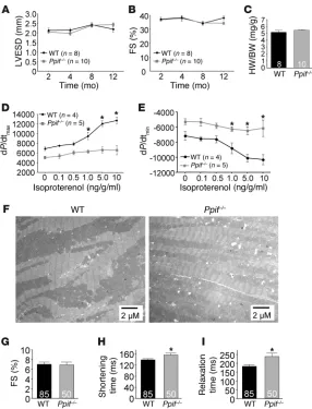
and performance measured by echocardiography at 2, 4, 8, and 12 months of age (Figure 1, A and B). There was also no difference in heart weights of mice at 12 months of age, nor any defects in myofilament or mitochondrial architecture, as assessed by trans-mission electron microscopy (Figure 1, C and F). However, while baseline contractility did not differ between WT and Ppif–/– mice
at 8 months of age, maximum contractile reserve, revealed by infusion of isoproterenol (a β-adrenergic receptor agonist), was compromised in Ppif–/– mice (dP/dtmax, Figure 1D), as was
relax-ation (dP/dtmin, Figure 1E). To determine whether the observed
myocardial dysfunction was myocyte specific, we examined sarco-meric shortening and cytosolic Ca2+ transients in isolated adult
cardiomyocytes. While the baseline percent fractional shortening was comparable to that in WT myocytes, the shortening time and relaxation time were significantly increased in Ppif–/– myocytes
(Figure 1, G–I), consistent with our in vivo findings. In addition, cytosolic Ca2+ transients from adult Ppif–/– cardiomyocytes showed
a longer time of decay, substantiating the observed increase in relaxation time (Supplemental Figure 1; supplemental material available online with this article; doi:10.1172/JCI43171DS1).
Ppif–/– mice are more susceptible to myocardial hypertrophy and heart
failure following pressure overload. To explore this maladaptive phe-notype further, we subjected Ppif–/– mice to stress stimulation as
a means of unmasking underlying problems associated with a
loss in functional reserve. Indeed, Ppif–/– mice were
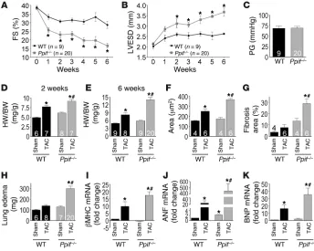
unable to compensate for the increase in afterload caused by transaortic constriction (TAC), display-ing a 42% reduction in fractional shortendisplay-ing at the 6-week time point as compared with WT mice (Fig-ure 2A). Ppif–/– mice decompensated within 1 week of
TAC stimulation and showed significant ventricular dilation by 2 weeks, with equivalent pressure gradi-ents across the aortic constrictions compared with WT mice (Figure 2, B and C). Consistent with these results, hearts from Ppif–/– mice hypertrophied to a
greater extent than WT mice after 2 and 6 weeks of TAC stimulation (Figure 2, D and E). Indeed, at the 6-week time point, Ppif–/– mice displayed a
signifi-cantly greater increase (41%) in heart weight normal-ized to body weight and LV wall thickness compared with WT TAC controls (Figure 2E and Supplemental Table 1), which correlated with approximately the same increase in hypertrophy of individual myocytes in the heart (Figure 2F). Fur-ther histological analysis of hearts showed a substantial increase in fibrosis following TAC in Ppif–/– mice compared with WT TAC
con-trols (Figure 2G), and indices of congestive heart failure, such as pulmonary edema, were also prominent in Ppif–/– animals (Figure
2H). However, we observed no difference in TUNEL-positive nuclei of cardiomyocytes or interstitial cells at 2 and 6 weeks of TAC stimulation in Ppif–/– compared with WT hearts, suggesting that
the rate of cellular demise was unchanged (Supplemental Figure 2, A–D). The more severe cardiac pathology observed in Ppif–/– mice
also correlated with stronger activation of the fetal gene program after TAC, including the β–myosin heavy chain gene, atrial natri-uretic factor (ANF), and B-type natrinatri-uretic peptide (BNP) genes (Fig-ure 2, I–K). Taken together, these results suggest that loss of CypD increases sensitivity to cardiac stress stimulation and disease.
Ppif–/– mice display increased mortality and heart failure in pathologic
and physiologic models of hypertrophy. To further characterize this mal-adaptive phenotype due to hypertrophic stimulation, we crossed
Ppif–/– mice with Ca2+/calmodulin-dependent protein kinase IIδc–
[image:3.585.43.329.80.455.2]transgenic mice (CaMKII-Tg). CaMKIIδc overexpression results in heart failure characterized by a loss of LV function and signifi-cant mortality, but more importantly these mice show alterations in intracellular Ca2+ handling and a predisposition to increased
Figure 1
Baseline characterization and aging of Ppif–/– mice. (A)
WT and Ppif–/– mice were evaluated from 2 months to
1 year of age for changes in LV end-systolic dimen-sion (LVESD). (B) LV percent fractional shortening (FS) from 2 to 12 months of age. (C) Heart weight to body weight (HW/BW) ratio in mice 12 months of age. (D) LV contractility (dP/dtmax) in mice 8 months of age
following isoproterenol dose escalation. (E) LV relax-ation (dP/dtmin) in mice 8 months of age following
mitochondrial Ca2+ (17–19). As will be presented in figures below,
this predisposition to elevated mitochondrial Ca2+ with CaMKIIδc
overexpression is similar to alterations observed in Ppif–/– hearts,
making the cross mechanistically germane. Indeed, hypertro-phy associated with the CaMKII-Tg was synergistically increased when mice were crossed into the Ppif–/– background, and the cross
also resulted in earlier lethality (Figure 3, A and B). Importantly, isolation of mitochondria from the hearts of these crossed mice showed that MPTP formation after Ca2+ stimulation was inhibited
by genetic loss of Ppif, even with the CaMKII-Tg (Figure 3C). The rapid onset of cardiac decompensation in Ppif–/– mice after
TAC stimulation coupled with their inability to even modestly increase cardiac contractility following acute β-adrenergic stimula-tion suggested a loss in funcstimula-tional reserve. To address this possibil-ity, and minimize the confounding effect of a pathologic insult, we subjected Ppif–/– mice to swimming exercise to induce physiologic
hypertrophy. Remarkably, swimming resulted in significant mor-tality, with more than 40% of Ppif–/– mice having died by the
con-clusion of the training period due to extreme fatigue and drown-ing (Figure 3D). Histological analysis showed no overt pathology, including no induction of cell death (Supplemental Figure 2, E
and F, and data not shown). Swim-ming also induced significantly greater cardiac hypertrophy and pul-monary edema in Ppif–/– mice as
com-pared with WT controls, suggesting an inability to compensate for the higher workload (Figure 3, E and F). In total, these results suggest that loss of CypD compromises the functional reserve of the heart and renders it more susceptible to failure, even with physiologic stimulation that is nor-mally cardioprotective.
Cardiac-restricted transgenic rescue of Ppif–/– pathology following TAC. We
hypothesized that the extreme predis-position to decompensation in Ppif–/–
mice with stress stimulation was due to a defect in mitochondrial function within myocytes themselves. However,
Ppif–/– mice lack the gene in all cells;
hence, it was formally possible that the cardiac defect observed was not myocyte autonomous. To address this issue, we crossed transgenic mice expressing CypD under control of the α–myosin heavy chain promoter (CypD-Tg) into the Ppif–/– background.
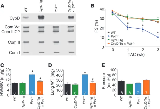
Western blotting demonstrated that
Ppif–/– mice lacked CypD protein in
the heart, while CypD-Tg × Ppif–/–
crossed mice had restored myocyte CypD protein, albeit to levels slightly higher than in the WT control (Figure 4A). After 3 weeks of TAC stimulation,
Ppif–/– mice from the same backcross
again showed rapid loss of ventricu-lar performance, greater increases in cardiac hypertrophy, and pulmonary edema, all of which were rescued back to WT levels by the CypD-Tg cross (Figure 4, B, C, and D). Importantly, pressure gradients across the aortic constriction did not differ among the groups (Figure 4E). These results suggest that the maladaptive cardiac phenotype observed in Ppif–/– mice is myocyte dependent.
Ppif–/– mice display substantial metabolic reprogramming. To further
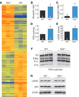
examine the mechanism whereby CypD loss from cardiac mito-chondria sensitizes to failure, we performed a genome-wide array using mRNA isolated from LV tissue of 8-week-old WT and Ppif–/–
mice. Affymetrix gene array analysis revealed that 362 probe sets were significantly different (ANOVA, P < 0.01), with a minimum difference in gene expression of 25% (Figure 5A). Unbiased func-tional annotation revealed numerous metabolism-associated gene groups that were significantly enriched, suggesting an alteration in mitochondrial function (Table 1). To examine this possible mechanism further, we evaluated substrate utilization in a work-ing heart preparation with 13C NMR spectrometry. The analysis
showed that Ppif–/– hearts had a significant increase in the glucose
[image:4.585.42.386.83.358.2]to palmitate ratio, suggesting a metabolic shift away from fatty acid oxidation toward glycolysis (Figure 5B). We also observed an increase in the succinate to glutamate ratio in Ppif–/– hearts,
Figure 2
Ppif–/– mice rapidly develop hypertrophy and heart failure following pressure overload. (A) LV
per-cent FS following TAC in WT and Ppif–/– mice. (B) LVESD following TAC. (C) Pressure gradients
(PG) across the TAC 1 week after TAC. (D) HW/BW ratio 2 weeks following TAC or sham operation. (E) HW/BW ratio 6 weeks following TAC. (F) Cardiomyocyte cross-sectional area (μm2) after TAC
research article
The Journal of Clinical Investigation http://www.jci.org Volume 120 Number 10 October 2010 3683
and while the significance of this change is uncertain, it does sug-gest increased TCA cycle flux (Figure 5C). Associated with these changes in substrate utilization, the activities of pyruvate dehy-drogenase (PDH) and α–ketoglutarate dehydrogenase (αKGD), two Ca2+-regulated mitochondrial enzymes with rate-limiting
metabolic properties, were significantly enhanced in Ppif–/– hearts
(Figure 5, D and E). Importantly, protein expression levels for the PDH subunits did not differ between WT and Ppif–/– hearts, nor
were expression levels for αKGD and two other Ca2+-regulated
mito-chondrial dehydrogenases changed (Figure 5, F and G), suggesting that the increase in activity was due to increased mitochondrial Ca2+.
Ppif–/– mice display altered
mito-chondrial Ca2+ exchange. Given the
observed metabolic changes and increased αKGD and PDH activity, we suspected an increase in mito-chondrial matrix Ca2+. Indeed, we
previously showed that mitochon-dria from Ppif–/– mice take up more
Ca2+ before undergoing MPTP
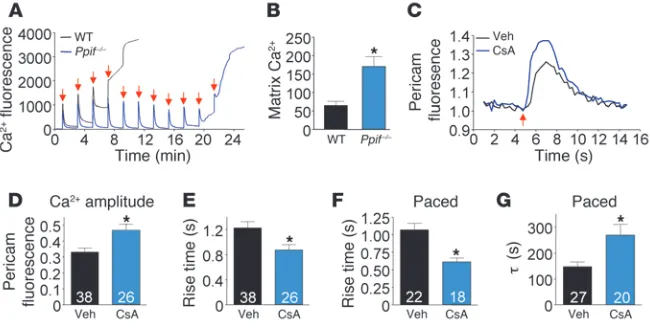
for-mation (3). Reexamination of this phenomenon with purified mito-chondria from Ppif–/– mice showed
nearly 3-fold-greater Ca2+
accumu-lation prior to MPTP opening com-pared with that in WT mitochon-dria (Figure 6A). More importantly, direct measurement of total mito-chondrial Ca2+ content of Ppif–/–
hearts showed a 2.6-fold elevation compared with WT mitochondria (Figure 6B). Taken together, these results suggest that in the absence of CypD mitochondrial matrix Ca2+
levels are reset and constitutively elevated, consistent with the observed metabolic alterations.
We hypothesized that the elevated level of matrix Ca2+ observed
in mitochondria from hearts of Ppif–/– mice was due to reduced
physiological opening of the MPTP, as its low conductance state has been implicated in affecting Ca2+ homeostasis (20, 21). To
address this hypothesis, we used cultured neonatal rat ventricular cardiomyocytes infected with an adenovirus encoding a mitochon-drial-targeted ratiometric-pericam (mt-pericam) Ca2+ sensor
pro-Figure 3
Ppif–/– mice display increased mortality and heart failure in pathologic and physiologic models of
hyper-trophy. (A) HW/BW ratios in the indicated groups of mice at 4 weeks of age. (B) Kaplan-Meier survival plot in the groups of mice shown. (C) Mitochondrial swelling following Ca2+ addition (indicated by red
[image:5.585.225.540.525.740.2]arrow), measured as change in absorbance (520 nm) over time (representative tracing of 3 indepen-dent experiments). (D) Kaplan-Meier survival plot during swimming exercise. (E) Ventricular weight/tibia length (VW/TL) ratios in mice at completion of the swimming protocol. (F) Lung edema (wet weight in mg – dry weight in mg) at completion of the swimming protocol. All values are reported as mean ± SEM. Sample size for each group is indicated inside the respective bar. *P < 0.05 versus WT control (A, B, and D) or rested group (E); #P < 0.05 versus CaMKII and Ppif–/– (A–C) or versus WT swim (E and F).
Figure 4
Cardiac-restricted transgenic rescue of Ppif–/–
tein (22). This system permits direct measurement of mitochon-drial matrix Ca2+ levels in real time and, in conjunction with CsA,
allowed examination of MPTP-dependent Ca2+ handling effects.
In agreement with previous reports, we found mt-pericam to be exclusively expressed in mitochondria (data not shown) (22). Fol-lowing a single 80-V pulse, CsA-treated myocytes showed a sig-nificantly greater mitochondrial Ca2+ transient compared with
untreated myocytes (Figure 6C). Quantification of this effect in multiple independent myocytes showed a quantitative increase in the amplitude of the Ca2+ transient, decreased rise time, and
increased rise slope (Figure 6, D and E, and data not shown). We also performed continuous pacing (80 v, 0.2 Hz, 100 seconds) to allow greater mitochondrial matrix Ca2+ accumulation for
examin-ing extrusion dynamics. Under paced conditions, MPTP desensiti-zation with CsA also decreased the rise time in Ca2+ accumulation,
but more importantly, it prolonged the recovery time after pac-ing (Figure 6, F and G). Both of these effects are consistent with a proposed homeostatic function of the MPTP as a Ca2+ “leak” or
release channel to prevent Ca2+ overload in cardiac mitochondria.
Discussion
CypD inhibition is known to allow greater accumulation of mito-chondrial Ca2+ in vitro, yet the mechanism proposed for this
phe-nomenon has always been tied to cell death. However, our data taken together with past studies suggest a critical role for CypD and the MPTP in controlling mitochondrial Ca2+ homeostasis for
proper metabolic function. For example, past in vitro studies
sug-gested that transient opening of the MPTP might allow the exchange of matrix Ca2+ across the inner membrane
without causing cellular demise. Evidence for this hypothesis emerged in 1992 in a study by Altschuld and colleagues, who showed that CsA increased mito-chondrial Ca2+ loading and inhibited efflux in
iso-lated rabbit cardiomyocytes (23). This hypothesis was extended by Ichas et al., who presented a biophysical model of Ca2+-induced mitochondrial Ca2+ release that
involved low-conductance MPTP opening for mito-chondrial Ca2+ efflux (20). Similarly, Siemen and
col-leagues used mitoplasts and direct patching to exam-ine MPTP opening in the mitochondrial membrane. They showed a large conductance current, with multi-ple subconductance states, that was Ca2+ activated and
reversibly inhibited with CsA (24). Our results extend this Ca2+ homeostasis model by showing that genetic loss of CypD
results in mitochondria with higher resting Ca2+ levels in the heart
and that CsA treatment acutely reduces mitochondrial Ca2+ efflux
and leak in cultured cardiomyocytes.
In the current study, we provide in vivo evidence linking MPTP-mediated Ca2+ efflux to global changes in metabolic substrate
uti-lization, a mechanism that appears crucial to the heart’s ability to maintain contractility in response to increased workload (25). For example, increases in cardiac contractility are tied to greater intracellular Ca2+ cycling rates in myocytes, which has the net
effect of enhancing mitochondrial Ca2+ load through the Ca2+
uni-porter (MCU). Mitochondrial extrusion rates through the mito-chondrial Na+/Ca2+ exchanger or other transporters/channels lag
behind the cytoplasmic Ca2+ transient, resulting in accumulation
of Ca2+ at higher beating rates until physiologic opening of the
MPTP occurs. The net increase in mitochondrial Ca2+ leads to
acti-vation of 3 mitochondrial dehydrogenases important in substrate utilization and oxidative metabolism, thereby increasing energy supply during increased workloads (26). Indeed, simultaneous measurements of mitochondrial Ca2+ and NADH have correlated
increased mitochondrial Ca2+ load with increased oxidative
phos-phorylation and ATP production (27). Moreover, pharmacologic inhibition of the MCU reduced the activity of the aforementioned intra-mitochondrial dehydrogenases by reducing Ca2+ influx (28,
29). Analogous to this model, we found enhanced enzymatic activ-ity for PDH and αKGD at baseline in Ppif–/– hearts, consistent with
[image:6.585.44.320.79.417.2]the finding of increased basal Ca2+ in the mitochondrial matrix.
Figure 5
Ppif–/– mice display significant metabolic alterations. (A)
Heat map displaying gradient differences in gene expres-sion between WT and Ppif–/– ventricular tissue at 8 weeks
of age (ANOVA, P < 0.01). (B) Ratio of glucose/palmitate (Glu/palm) consumption in working hearts using 13C NMR
spectroscopy. (C) Ratio of succinate/cytosolic glutamate (Succ/glut) in working hearts using 13C NMR
spectros-copy. (D) PDH enzymatic activity in mitochondria isolated from mouse hearts (y axis indicates change in mOD/min/ mg protein where r2 = 1). (E) αKGD enzymatic activity in
research article
The Journal of Clinical Investigation http://www.jci.org Volume 120 Number 10 October 2010 3685
PDH in particular is central in linking glycolysis to the citric acid cycle by converting pyruvate to acetyl-CoA. We hypothesize that the mitochondrial calcium-mediated increase in PDH activity may account for the observed elevation in glucose oxidation. For example, inhibition of PDH kinase, which increases PDH activity, enhances glucose oxidation in rat heart (30).
Increased basal Ca2+ in the mitochondria mimics higher contractile
workloads, which on the surface would not appear to be detrimen-tal, likely explaining why Ppif–/– mice are relatively unaffected when
not stressed. However, our working hypothesis is that such a state of constitutively higher intramitochondrial Ca2+ reduces the dynamic
range in cardiac metabolism, ultimately decreasing the heart’s meta-bolic reserve capacity and rendering it more
sensitive to decompensation with even subtle stress provocation (such as sustained exer-cise). At first glance, these results seem con-trary to our previous report that Ppif deletion rescued heart disease in β2a-Tg mice, a trans-genic model with severe heart failure due to massive Ca2+ overload and myocyte necrosis
(31). However, the loss of Ppif in β2a-Tg mice was likely protective, blocking the pathologic function of the MPTP in its large-conduc-tance state and secondary myocyte necrosis, hence predominating over the physiologic function of the MPTP in regulating mito-chondrial matrix Ca2+.
A hallmark of pathologic hypertrophy is metabolic reprogramming back toward a fetal-like gene program, favoring glycolysis over fatty acid oxidation (32). While this metabolic reprogramming was originally postulated to be compensatory, studies in genetically modified mouse models predict that any switching of major metabolic sub-strate utilization in the heart is detrimental (16, 33). For example, Ppara–/– mice
dis-play an increase in glucose oxidation and a decrease in fatty acid oxidation resulting in myocardial fibrosis and an inability to
tol-erate increased workloads (34–36). In parallel,
Ppard–/– (PPARγ protein) mice display increased
glucose uptake, with a decrease in fatty acid
β-oxidation and likewise exhibit systolic dys-function and the rapid onset of heart failure (37). Even more striking, a recent attempt to genetically overcome a shift in fatty acid utili-zation by overexpression of Glut1 resulted in vast metabolic reprogramming and myocardial pathology (38). The Ppif–/– phenotype strongly
fits this paradigm, in that a shift in substrate utilization precedes any overt cardiac pathol-ogy, and upon stress-stimulation the observed deficit in metabolic reserve leads to disease.
In conclusion, we have established that CypD and the MPTP represents an important regula-tor of mitochondrial Ca2+ exchange in myocytes
that is pivotal in maintaining metabolic plas-ticity, thereby facilitating adaptation to disease states or increased workload. Thus, our results suggest that the modulation of mitochondrial Ca2+ exchange may
be an exciting new avenue for the treatment of heart failure.
Methods
Targeted and transgenic mice. The generation of Ppif-null mice and CypD-transgenic mice has previously been reported (3). For our experiments,
Ppif–/– mice were crossed with αMHC-CypD-Tg mice and heterozygous
(Ppif+/–) offspring carrying the CypD transgene crossed with Ppif+/– void of
the transgene. The offspring of this cross were again mated to generate the 4 genotypes of mice used in the current study: WT, Ppif–/–, α
MHC-CypD-Tg, and Ppif–/–×αMHC-CypD-Tg. The generation of CaMKII-Tg mice has
[image:7.585.51.340.103.260.2]previously been reported (17). For our experiments Ppif–/– mice were crossed Table 1
Ppif–/– hearts display significant metabolic reprogramming
Functional group P Fold enrichment
Pigment biosynthetic process 6.94 × 10⁻⁴ 12.18
Pigment metabolic process 1.40 × 10⁻³ 10.15
Secondary metabolic process 5.81 × 10⁻³ 6.89
Porphyrin biosynthesis 6.73 × 10⁻³ 23.44
Heme biosynthesis 8.34 × 10⁻³ 21.09
Cellular metabolic process 7.14 × 10⁻³ 1.19
Metabolic process 1.64 × 10⁻² 1.15
Primary metabolic process 2.13 × 10⁻² 1.16
Amino acid catabolic process 3.22 × 10⁻² 5.73
Di-, tri-valent inorganic cation transport 5.36 × 10⁻³ 3.77
Cation transmembrane transporter activity 2.85 × 10⁻² 1.95
Substrate-specific transmembrane transporter activity 4.97 × 10⁻² 1.63
DAVID functional annotation microarray analysis of gene class changes in Ppif–/– hearts, which
[image:7.585.218.543.456.620.2]showed many alterations in mitochondrial genes and genes involved in metabolism.
Figure 6
Ppif–/– mice display increased mitochondrial Ca2+. (A) Representative tracing of Ca2+ uptake
in mitochondria isolated from WT and Ppif–/– mice (calcium green-5n fluorescence intensity,
485excitation/528emission; red arrow indicates Ca2+ injection). (B) Matrix-free Ca2+ in mitochondria
isolated from ventricular samples (nmol/mg protein). (C) Representative tracing from cultured neonatal cardiomyocytes infected with mt-pericam treated with vehicle (Veh) or 1 μM CsA. Pericam fluorescence is the ratio of 490/415excitation, 530emission; red arrow indicates field
stimula-tion. (D) Amplitude of mitochondrial Ca2+ transients in neonatal myocytes following single-pulse
stimulation. (E) Rise time of mitochondrial Ca2+ transients following single-pulse stimulation.
(F) Average rise time of mitochondrial Ca2+ transients during continuous pacing (0.2 Hz for
with CaMKII-Tg mice in the same manner as they were with CypD-Tg. All experiments in the current study utilized littermate controls of matched age and sex. The IACUC of Cincinnati Children’s Hospital approved all experiments involving animals.
Western blot analysis. Sample preparation and gel electrophoresis were pre-viously described in detail (39). The following antibodies were used in the current study: CypD (Mitosciences), respiratory chain complexes (OXPHOS blot, Mitosciences), PDH subunits (Mitosciences), αKGD, COX II (Santa Cruz Biotechnologies Inc.), and isocitrate dehydrogenase (Abcam).
Isolation of adult cardiomyocytes, cytosolic Ca2+ current, and myocyte shortening.
Ventricular myocytes were isolated as described previously (31). Myocytes were maintained at room temperature in 5% CO2 and 95% O2 and used within 3
hours of isolation. Myocytes were placed in a chamber mounted on an inverted Nikon microscope and perfused with normal physiological solution (Ringer) containing 2 mM Ca2+. Contractions were measured with video edge detection,
and intracellular Ca2+ was measured with Fura-2 as described previously (31).
Mitochondria isolation and analysis of mitochondrial Ca2+ content. Briefly, the
heart was quickly excised and mitochondria isolated in buffers free of EGTA/EDTA, pH 7.4, at 4°C. For measurement of Ca2+ content,
mito-chondria were isolated and washed repeatedly in EGTA/EDTA-free buffers and resuspended in 0.6 N HCl as previously reported (40). Concentration was then determined spectrophotometrically using the O-Cresolphthalein Complexone assay (TECO Diagnostics).
Isolated mouse heart preparation and 13C metabolite NMR. Mice were
anesthe-tized with an intraperitoneal injection of 0.10 ml pentobarbital sodium diluted 1:5 in perfusate. The abdominal cavity was exposed with a transverse incision, and 0.05 ml heparin was administered to the inferior vena cava. The heart was quickly isolated and placed in ice-cold Krebs-Henseleit (KH) buf-fer (25 mM NaHCO3, 120 mM NaCl, 11 mM glucose, 4.7 mM KCl, 1.2 mM
KH2PO4, 1.2 mM MgSO4, and 1.75 mM CaCl2) to arrest the heart. The heart
was perfused via the aorta. A water-filled latex balloon was inserted into the LV to measure hemodynamic parameters using a PowerLab 2/25 and Chart v5.5 software (AD Instruments). All hearts were perfused with KH buffer gassed with 95% O2 and 5% CO2 and maintained at 37°C. 13C metabolite
labeling was performed as previously described (41). Following equilibration, hearts were perfused in phosphate-free KH buffer with 8 mM glucose, 2 mM 1-13C-glucose, 0.5 mM uniformly labeled 13C-palmitate, and 3% (w/v) BSA for
20 minutes. The hearts were freeze-clamped, extracted with cold 3.6% perchlo-ric acid, and centrifuged at 14,000 g. The resulting supernatant was neutral-ized on ice with 5 M KOH to a pH of approximately 8, centrifuged at 14,000 g, and lyophilized. Samples were dissolved in D2O for 13C NMR measurement,
and proton-decoupled 13C spectra were acquired on a Varian Inova 500-MHz
NMR spectrometer. By analyzing the scalar coupling pattern of the C-4 car-bon resonance of glutamate, we were able to measure the relative contribu-tion of 13C-enriched glucose versus palmitate to the acetyl-CoA pool.
Dehydrogenase activity assays. PDH activity was assayed by using an immu-nocapture technique and then calculating the mean maximal rate of NADH production (change in mean OD/min where r2 = 1) as described in
the manufacturer’s instructions (Mitosciences). αKGD activity was moni-tored using NADH-coupled reactions. Briefly, mitochondria were lysed in 120 mM KCl, 10 mM Tris, 5 mM MOPS, 5 mM KH2PO4, and 1% Triton
X-100. The homogenate was immediately added to 5 mM MgCl2, 5 mM
αKG, 0.2 mM coenzyme A, 0.4 mM thiamine pyrophosphate, and 1 mM NAD+. NADH produced was measured at an absorbance of 340 nm.
Neonatal myocyte isolation and pericam imaging and analysis. Neonatal rat cardiomyocytes were isolated from Sprague-Dawley rat pups as previously described (42). Cardiomyocytes were allowed to mature for 72 hours in media containing 15% fetal bovine serum. Myocytes were then infected with mt-pericam adenovirus for 6 hours (22). Twenty-four hours follow-ing infection, myocytes were utilized in Ca2+ experiments. Prior to
experi-ments, myocytes were switched to standard Ringer solution containing 4 mM Ca2+, as previously reported (43). The pericam fluorescence ratio
was determined at room temperature using a Delta Scan dual-beam spec-trofluorophotometer (Photon Technology International), operated at an emission wavelength of 530 nM, with excitation wavelengths of 490 and 415 nM. The stimulating frequency for Ca2+ transient measurements was
0.2 Hz. All data were recorded and analyzed using Chart v5.6 (AdInstru-ments) and IonWizard (IonOptix) software.
Isolation of mRNA, Taqman qPCR, and gene array. mRNA was isolated using the RNeasy Protect Midi Kit according to manufacturer’s instructions (QIAGEN). Reverse transcription was performed in a standard fashion with QuantiTect Reverse Transcription Kit (QIAGEN) supplemented with DNase treatment. Taqman qPCR was carried out according to the manu-facturer’s instructions using probe sets for αMHC, βMHC, ANP, BNP, and GAPDH. Analysis was carried out using the ΔΔ-CT method with αMHC and GAPDH correction and reported as fold change versus WT sham. Gene expression analysis was performed using Affymetrix Mouse Gene Array 1.0ST. Results were filtered for a raw value greater than 5, followed by ANOVA (P < 0.01) statistical analysis of WT versus Ppif–/–. A raw filter
for values greater than 6 was then applied, followed by a volcano plot for WT versus Ppif–/– (fold difference >1.25, P < 0.05). Results were then
ana-lyzed for functional group classification using DAVID functional annota-tion microarray analysis (http://david.abcc.ncifcrf.gov/home.jsp) (44, 45). The data have been deposited into the Gene Expression Omnibus database (http://www.ncbi.nlm.nih.gov/geo/; accession number GSE23028).
Histology, image analysis, and electron microscopy. For histological analysis, hearts were collected at the indicated times, fixed in 10% buffered for-malin, and embedded in paraffin. Serial 5-μm heart sections from each group were stained with H&E and Masson’s trichrome (to detect fibrosis). Myocyte cross-sectional areas were analyzed in slides stained with wheat germ agglutinin–FITC conjugate at 50 μg/ml to accurately identify sar-colemmal membrane as previously reported (39). ImageJ (http://rsbweb. nih.gov/ij/) analysis was then performed. All analysis was performed in a blinded fashion. Transmission electron microscopy and TUNEL staining were performed as previously described (11, 39).
Echocardiography, invasive hemodynamics, swimming protocol, and pressure overload. Hypertrophy studies using the TAC model, swimming exercise, and invasive hemodynamics have been previously described (39, 46).
Statistics. All results are presented as mean ± SEM. Statistical analysis was per-formed using Prism 4.0 (GraphPad Software) for unpaired 2-tailed t test (for 2 groups) and 1-way ANOVA with Bonferroni correction (for groups of 3 or more). For experiments performed over time, 2-way ANOVA with Bonferroni post-hoc analysis was used. P values less than 0.05 were considered significant.
Acknowledgments
The authors greatly appreciate the technical expertise of Allen York and Michelle Sargent. This work was supported by grants from the NIH to J.W. Elrod (5F32HL092737), J.D. Molkentin (HL62927, HL81104), and to J.H. Brown (HL80101), as well as by an interna-tional grant in heart failure research from the Fondation Leducq and the Howard Hughes Medical Institute.
Received for publication March 31, 2010, and accepted in revised form August 4, 2010.
research article
The Journal of Clinical Investigation http://www.jci.org Volume 120 Number 10 October 2010 3687
1. Kroemer G, Galluzzi L, Brenner C. Mitochondrial membrane permeabilization in cell death. Physiol Rev. 2007;87(1):99–163.
2. Nakagawa T, et al. Cyclophilin D-dependent mito-chondrial permeability transition regulates some necrotic but not apoptotic cell death. Nature. 2005; 434(7033):652–658.
3. Baines CP, et al. Loss of cyclophilin D reveals a criti-cal role for mitochondrial permeability transition in cell death. Nature. 2005;434(7033):658–662. 4. Schinzel AC, et al. Cyclophilin D is a component of
mitochondrial permeability transition and mediates neuronal cell death after focal cerebral ischemia. Proc Natl Acad Sci U S A. 2005;102(34):12005–12010. 5. Basso E, Fante L, Fowlkes J, Petronilli V, Forte MA,
Bernardi P. Properties of the permeability transi-tion pore in mitochondria devoid of Cyclophilin D.
J Biol Chem. 2005;280(19):18558–18561. 6. Le Quoc K, Le Quoc D. Involvement of the ADP/ATP
carrier in calcium-induced perturbations of the mito-chondrial inner membrane permeability: importance of the orientation of the nucleotide binding site. Arch Biochem Biophys. 1988;265(2):249–257.
7. Halestrap AP, Davidson AM. Inhibition of Ca2(+)-induced large-amplitude swelling of liver and heart mitochondria by cyclosporin is probably caused by the inhibitor binding to mitochondrial-matrix peptidyl-prolyl cis-trans isomerase and preventing it interacting with the adenine nucleotide translo-case. Biochem J. 1990;268(1):153–160.
8. Bernardi P. The permeability transition pore. Con-trol points of a cyclosporin A-sensitive mitochon-drial channel involved in cell death. Biochim Biophys Acta. 1996;1275(1–2):5–9.
9. Forte M, et al. Cyclophilin D inactivation protects axons in experimental autoimmune encephalomy-elitis, an animal model of multiple sclerosis. Proc Natl Acad Sci U S A. 2007;104(18):7558–7563. 10. Du H, et al. Cyclophilin D deficiency attenuates
mitochondrial and neuronal perturbation and ameliorates learning and memory in Alzheimer’s disease. Nat Med. 2008;14(10):1097–1105. 11. Millay DP, et al. Genetic and pharmacologic
inhibi-tion of mitochondrial-dependent necrosis attenuates muscular dystrophy. Nat Med. 2008;14(4):442–447. 12. Ichas F, Mazat JP. From calcium signaling to cell
death: two conformations for the mitochondrial permeability transition pore. Switching from low- to high-conductance state. Biochim Biophys Acta. 1998;1366(1–2):33–50.
13. Griffiths EJ. Mitochondrial calcium transport in the heart: physiological and pathological roles.
J Mol Cell Cardiol. 2009;46(6):789–803.
14. Spat A, Szanda G, Csordas G, Hajnoczky G. High- and low-calcium-dependent mechanisms of mito-chondrial calcium signalling. Cell Calcium. 2008; 44(1):51–63.
15. Foo RS, Mani K, Kitsis RN. Death begets failure in the heart. J Clin Invest. 2005;115(3):565–571. 16. Mudd JO, Kass DA. Tackling heart failure in the
twen-ty-first century. Nature. 2008;451(7181):919–928. 17. Zhang T, et al. The deltaC isoform of CaMKII is
activated in cardiac hypertrophy and induces dilated cardiomyopathy and heart failure. Circ Res. 2003;92(8):912–919.
18. Zhang T, et al. Phospholamban ablation rescues sarcoplasmic reticulum Ca(2+) handling but exac-erbates cardiac dysfunction in CaMKIIdelta(C) transgenic mice. Circ Res. 2010;106(2):354–362. 19. Sag CM, et al. Calcium/calmodulin-dependent
pro-tein kinase II contributes to cardiac arrhythmogenesis in heart failure. Circ Heart Fail. 2009;2(6):664–675. 20. Ichas F, Jouaville LS, Mazat JP. Mitochondria are
excitable organelles capable of generating and conveying electrical and calcium signals. Cell. 1997;89(7):1145–1153.
21. Bernardi P, Petronilli V. The permeability transi-tion pore as a mitochondrial calcium release chan-nel: a critical appraisal. J Bioenerg Biomembr. 1996; 28(2):131–138.
22. Nagai T, Sawano A, Park ES, Miyawaki A. Circu-larly permuted green fluorescent proteins engi-neered to sense Ca2+. Proc Natl Acad Sci U S A. 2001; 98(6):3197–3202.
23. Altschuld RA, Hohl CM, Castillo LC, Garleb AA, Starling RC, Brierley GP. Cyclosporin inhibits mitochondrial calcium efflux in isolated adult rat ventricular cardiomyocytes. Am J Physiol. 1992; 262(6 pt 2):H1699–H1704.
24. Loupatatzis C, Seitz G, Schonfeld P, Lang F, Sie-men D. Single-channel currents of the permeabil-ity transition pore from the inner mitochondrial membrane of rat liver and of a human hepatoma cell line. Cell Physiol Biochem. 2002;12(5–6):269–278. 25. Liu T, O’Rourke B. Regulation of mitochondrial Ca2+ and its effects on energetics and redox balance in normal and failing heart. J Bioenerg Biomembr. 2009;41(2):127–132.
26. Denton RM. Regulation of mitochondrial dehydro-genases by calcium ions. Biochim Biophys Acta. 2009; 1787(11):1309–1316.
27. Brandes R, Bers DM. Simultaneous measure-ments of mitochondrial NADH and Ca(2+) during increased work in intact rat heart trabeculae. Bio-phys J. 2002;83(2):587–604.
28. McCormack JG, England PJ. Ruthenium red inhibits the activation of pyruvate dehydrogenase caused by positive inotropic agents in the perfused rat heart. Biochem J. 1983;214(2):581–585. 29. Unitt JF, McCormack JG, Reid D, MacLachlan LK,
England PJ. Direct evidence for a role of intramito-chondrial Ca2+ in the regulation of oxidative phos-phorylation in the stimulated rat heart. Studies using 31P n.m.r. and ruthenium red. Biochem J. 1989; 262(1):293–301.
30. Piao L, et al. The inhibition of pyruvate dehydro-genase kinase improves impaired cardiac function and electrical remodeling in two models of right ventricular hypertrophy: resuscitating the hibernat-ing right ventricle. J Mol Med. 2010;88(1):47–60. 31. Nakayama H, et al. Ca2+- and
mitochondrial-depen-dent cardiomyocyte necrosis as a primary mediator of heart failure. J Clin Invest. 2007;117(9):2431–2444. 32. Lehman JJ, Kelly DP. Gene regulatory mechanisms
governing energy metabolism during cardiac hyper-trophic growth. Heart Fail Rev. 2002;7(2):175–185. 33. Taegtmeyer H, Wilson CR, Razeghi P, Sharma S.
Metabolic energetics and genetics in the heart. Ann N Y Acad Sci. 2005;1047:208–218.
34. Barger PM, Brandt JM, Leone TC, Weinheimer CJ, Kelly DP. Deactivation of peroxisome proliferator-activated receptor-alpha during cardiac hypertro-phic growth. J Clin Invest. 2000;105(12):1723–1730. 35. Watanabe K, et al. Constitutive regulation of car-diac fatty acid metabolism through peroxisome proliferator-activated receptor alpha associated with age-dependent cardiac toxicity. J Biol Chem. 2000;275(29):22293–22299.
36. Leone TC, Weinheimer CJ, Kelly DP. A critical role for the peroxisome proliferator-activated receptor alpha (PPARalpha) in the cellular fasting response: the PPARalpha-null mouse as a model of fatty acid oxidation disorders. Proc Natl Acad Sci U S A. 1999; 96(13):7473–7478.
37. Cheng L, et al. Cardiomyocyte-restricted peroxisome proliferator-activated receptor-delta deletion per-turbs myocardial fatty acid oxidation and leads to cardiomyopathy. Nat Med. 2004;10(11):1245–1250. 38. Yan J, Young ME, Cui L, Lopaschuk GD, Liao R,
Tian R. Increased glucose uptake and oxidation in mouse hearts prevent high fatty acid oxidation but cause cardiac dysfunction in diet-induced obesity.
Circulation. 2009;119(21):2818–2828.
39. Maillet M, Lynch JM, Sanna B, York AJ, Zheng Y, Molkentin JD. Cdc42 is an antihypertrophic molec-ular switch in the mouse heart. J Clin Invest. 2009; 119(10):3079–3088.
40. Picard M, et al. Resistance to Ca2+-induced opening of the permeability transition pore differs in mitochon-dria from glycolytic and oxidative muscles. Am J Physiol Regul Integr Comp Physiol. 2008;295(2):R659–R668. 41. Gabel SA, Walker VR, London RE, Steenbergen
C, Korach KS, Murphy E. Estrogen receptor beta mediates gender differences in ischemia/reperfu-sion injury. J Mol Cell Cardiol. 2005;38(2):289–297. 42. De Windt LJ, Lim HW, Haq S, Force T, Molkentin
JD. Calcineurin promotes protein kinase C and c-Jun NH2-terminal kinase activation in the heart. Cross-talk between cardiac hypertrophic signaling pathways. J Biol Chem. 2000;275(18):13571–13579. 43. Kettlewell S, Cabrero P, Nicklin SA, Dow JA, Davies
S, Smith GL. Changes of intra-mitochondrial Ca2+ in adult ventricular cardiomyocytes examined using a novel fluorescent Ca2+ indicator targeted to mito-chondria. J Mol Cell Cardiol. 2009;46(6):891–901. 44. Huang da W, et al. The DAVID Gene Functional
Classification Tool: a novel biological module-cen-tric algorithm to functionally analyze large gene lists. Genome Biol. 2007;8(9):R183.
45. Huang da W, Sherman BT, Lempicki RA. System-atic and integrative analysis of large gene lists using DAVID bioinformatics resources. Nat Protoc. 2009; 4(1):44–57.