S T U D Y P R O T O C O L
Open Access
3D vs. 4K Display System - Influence of
“
State-of-the-art
”
-Display Technique On
Surgical Performance (IDOSP-Study) in
minimally invasive surgery: protocol for a
randomized cross-over trial
Roger Wahba
1*†, Rabi Raj Datta
1†, Andrea Hedergott
2, Jana Bußhoff
1, Thomas Bruns
1, Robert Kleinert
1, Georg Dieplinger
1,
Hans Fuchs
1, Caroline Giezelt
2, Desdemona Möller
3, Martin Hellmich
4, Christiane J. Bruns
1and Dirk L. Stippel
1Abstract
Background:Three-dimensional (3D) stereoscopic vision is crucial to perform any kind of manual task. The reduction from real life 3D to virtual two-dimensional (2D) sight is a major challenge in minimally invasive surgery (MIS). A 3D display technique has been shown to reduce operation time and mistakes and to improve the learning curve. Therefore, the use of a3D display technique seems to optimize surgical performance for novice and experienced surgeons. Inspired by consumer electronics, a 4K display technique was recently introduced to MIS. Due to its high resolution and zoom effect, surgeons should benefit from it. The aim of this study is to evaluate if
“state-of-the-art”3D- vs. 4K-display techniques could influence surgical performance.
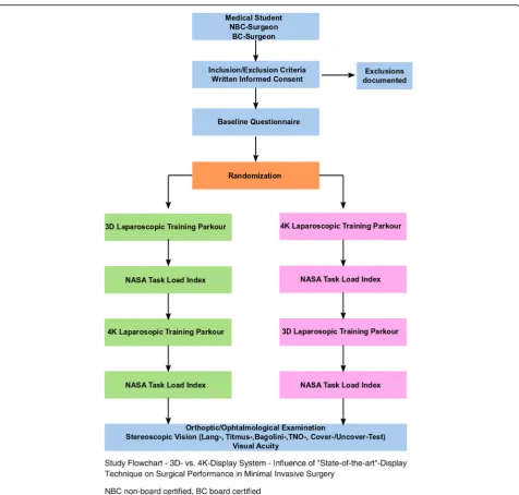
Methods:A randomized, cross-over, single-institution, single-blinded trial is designed. It compares the primary outcome parameter “surgical performance”, represented by “performance time”and “number of mistakes”, using a passive polarizing 3D and a 4K display system (two arms) to perform different tasks in a minimally invasive/laparoscopic training parkour. Secondary outcome parameters are the mental stress load (National Aeronautics and Space Administration (NASA) Task Load Index) and the learning curve. Unexperienced novices (medical students), non-board-certified, and board-certified abdominal surgeons participate in the trial (i.e., level of experience, 3 strata). The parkour consists of seven tasks (for novices, five tasks), which will be repeated three times. The 1st run of the parkour will be performed with the randomized display system, the 2nd run with the other one. After each run, the mental stress load is measured. After completion of the parkour, all participants are evaluated by an ophthalmologist for visual acuity and stereoscopic vision with five tests. Assuming a correlation of 0.5 between measurements per subject, a sample size of 36 per stratum is required to detect a standardized effect of 0.5 (including an additional 5% for a non-parametric approach) with a power of 80% at a two-sided type I error of 5%. Thus, altogether 108 subjects need to be enrolled.
(Continued on next page)
© The Author(s). 2019Open AccessThis article is distributed under the terms of the Creative Commons Attribution 4.0 International License (http://creativecommons.org/licenses/by/4.0/), which permits unrestricted use, distribution, and reproduction in any medium, provided you give appropriate credit to the original author(s) and the source, provide a link to the Creative Commons license, and indicate if changes were made. The Creative Commons Public Domain Dedication waiver (http://creativecommons.org/publicdomain/zero/1.0/) applies to the data made available in this article, unless otherwise stated. * Correspondence:[email protected]
Roger Wahba and Rabi Datta contributed equally to the study.
1Department of General, Visceral and Cancer Surgery, University Hospital of
Cologne, University of Cologne, Kerpener Straße 62, 50937 Cologne, Germany
(Continued from previous page)
Discussion: Complex surgical procedures are performed in a minimally invasive/laparoscopic technique. This study should provide some evidence to decide which display technique a surgeon could choose to optimize his performance.
Trial registration: ClinicalTrials.gov, NCT03445429. Registered on 7 February 2018.
Keywords: Minimally invasive surgery, Laparoscopic, 3D, 4K, Surgical performance, Learning curve, Surgical training
Background
Laparoscopic and minimally invasive operation tech-niques/surgery (MIS) have become the standard in basic (e.g. cholecystectomy [1]) as well as in complex surgical procedures (e.g. living donor nephrectomy [2]). In gen-eral, the learning curve for MIS is prolonged compared to open surgery [3] and even longer for complex opera-tions [4]. One challenge is the reduction from real life three-dimensional (3D) stereoscopic vision to virtual two-dimensional (2D) sight. 3D vision is very important to perform any kind of manual task [5]. Therefore, opti-mizing the visualization of the operative field is required, especially in MIS. A 2D full-high-definition technique was one step used to improve vision. The passive polar-izing 3D display technique reintroduces natural stereo-scopic view and orientation to MIS. It leads to shorter operation times and seems to optimize surgical perform-ance compared to standard 2D imaging in basic proce-dures [6, 7]. Novices as well as experienced surgeons seem to benefit from the 3D passive polarizing tech-nique [8]. The learning curve and performance, espe-cially in complex surgical procedures, e.g. vascular preparation during retroperitoneoscopic donor nephrec-tomy, could be optimized and simplified [9]. The recent European Association for Endoscopic Surgery (EAES) consensus statement recommended the use of 3D vision to reduce operative time [7]. As a disadvantage of the technique, the surgeon must wear glasses and the equip-ment is expensive. Furthermore, a relevant percentage of the population has deficits in binocular and stereoscopic vision, which could induce dizziness and nausea when using the passive polarizing 3D video technique [5]. This could result in a deterioratingsurgical performance. In-spired by consumer electronics, the 4K-display tech-nique has reached medicine. It creates a high resolution image with 4098 × 2160 pixels on a large-scale 55″ monitor (140 cm), resulting in an up to 30 times zoom. Due to these features, it should also optimize surgical performance in MIS and could be an alternative to the passive polarizing 3D display technique. Data comparing these techniques are scarce. Therefore, both techniques are compared in this randomized cross-over setting. The aim of this study is to evaluate if “state-of-the-art”
display techniques could influence surgical performance, represented by the outcome parameters “performance time ”and “number of mistakes” in different tasks of a minimally invasive/laparoscopic training parkour.
Methods/design
the study flowchart. According to the Standard Proto-col Items: Recommendations for Interventional Trials (SPIRIT) 2013 guidelines, a trial schedule (Table 1) and a trial checklist (Additional file 1) are part of the protocol [10, 11].
Outcome measures and data collection
The primary outcome measure is the surgical perform-ance measured by the items “time in seconds” and
“number of mistakes”. Both items are measured for each task separately and for all tasks together. The mistakes are defined for every task as any deviation from perfect performance (general and special mistakes). Secondary
outcome parameters are the scores of the NASA-TLX and the learning curves. Learning curves will be de-scribed as performance (time, errors) over repetitions with an added standard CUSUM analysis [12]. Moreover, performance indicators are investigated for possible interaction of replication, technique, and sequence (3D after 4K or vice versa).
Baseline characteristics are acquired by a questionnaire. The MIS tasks are recorded as standard 2D videos. NASA-TLX is performed as a pen-and-paper version. An ophthalmological examination is performed and docu-mented on a separate case report form (CRF). When the data for one subject are complete, they will be transferred
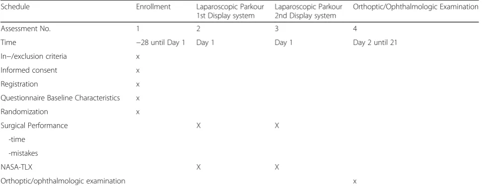
[image:3.595.60.537.84.538.2]to the data trustee, who pseudonymized the data and vid-eos. The videos will be sent back to the investigators for evaluation of the primary outcome measure. Each video will be assessed by two evaluation-trained investigators. Inter-rater reliability will be evaluated by contingency table analysis (kappa statistics or intraclass correlation). Large differences, i.e., larger than 1.96*standard devia-tions, will be reevaluated by two additional raters and de-scribed qualitatively. This will be documented on a CRF and retransferred to the data trustee, who will reunite it all. Then the pseudonymized data are sent to the investi-gators and statistician for final evaluation. Table1 shows the type and time of data collection. At the time of pub-lishing the study protocol, the trial is still recruiting.
Inclusion criteria
Subjects fulfilling the following inclusion criteria may be enrolled in the study:
1. Medical student, surgeons in training, board-certified surgeons
2. Those who have given written informed consent 3. Those aged > 18 years
Exclusion criteria
The following criteria will exclude subjects from the study:
1. Medical students with any experience in laparoscopic surgery
2. Experience in the laparoscopic training parkour (all subjects)
3. Non-correctable vision disorders 4. Known impaired stereoscopic vision 5. Manual skill disorders
Randomization and blinding
Randomization of subjects to sequences is based on per-muted blocks and stratified by level of experience. This will be performed by the data trustee. After that subjects will perform the laparoscopic parkour and the NASA-TLX and be examined by the ophthalmologist. The performance of the laparoscopic parkour will be video documented as standard 2D videos. After completion of the study examin-ation, all data including the videos will be collected by the data trustee and pseudonymized and stored on a secured data base with routine backup. To guarantee blinding, only pseudonymized data are sent back to the investigators for evaluation of the final study data. Therefore, the evaluating investigators are not able to find out whether the 3D or 4K display system was used during the laparoscopic training parkour.
Data management
Data evaluation and entry to the study data base will be double checked and performed by two investigators. Final access to the data base is given to the sponsor, re-sponsible party, and authors of the protocol. It will not be provided to any third party.
Interim analysis and stopping guidelines
There is no interim analysis planned. There are no stopping guidelines due to the fact that the trial does not evaluate an US–Food and Drug Administration (FDA)-regulated drug product or a US FDA-regulated device product.
Sample size
[image:4.595.56.542.99.287.2]Assuming a correlation of 0.5 between measurements per subject, a sample size of 36 per stratum is required to de-tect a standardized effect of 0.5 (including an additional 5% for a non-parametric approach) with a power of 80% Table 1Trial Schedule–3D vs. 4K Display System - Influence of“State-of-the-art”-Display Technique on Surgical Performance
Schedule Enrollment Laparoscopic Parkour
1st Display system
Laparoscopic Parkour 2nd Display system
Orthoptic/Ophthalmologic Examination
Assessment No. 1 2 3 4
Time −28 until Day 1 Day 1 Day 1 Day 2 until 21
In−/exclusion criteria x
Informed consent x
Registration x
Questionnaire Baseline Characteristics x
Randomization x
Surgical Performance X X
-time
-mistakes
NASA-TLX X X
Orthoptic/ophthalmologic examination x
at a two-sided type I error of 5%. This is a cautious esti-mate since a considerably larger effect size of 1.0 was re-ported by Smith et al. [8] for the improvement in the median time and for completion of the entire protocol, al-beit in a parallel-group setting. Similarly, for the median number of errors, an effect size of 1.95 was observed. Also, preliminary cases supported the sample size calcula-tion. Thus, altogether 108 subjects need to be enrolled [8]. Subjects who drop out of the study may be replaced.
Statistical analyses
Quantitative variables are summarized by mean ± standard deviation and percentiles (0, 25, 50, 75, and 100), qualita-tive variables by count and percentage. Outcome mea-sures are evaluated by modeling; specifically (generalized) linear mixed models for repeated measures (MMRM) with main effects modality, stratum, and period (type III SS, REML, unstructured covariance matrix). Estimated mar-ginal means and contrasts are derived. Interaction effects, particularly stratum*modality, are explored. Two-sided p values < 0.05 are interpreted to indicate statistical signifi-cance. Missing data will substituted by multiple imputa-tions. Subgroup analysis will be performed according to the above mentioned strategies.
Trial organization
The IDOSP trial is an investigator-initiated trial without external funding. The trial is sponsored by the University Hospital of Cologne. The Department of General, Visceral and Cancer Surgery is responsible for the coordination of the trial.
Ethics
Ethics Committee approval was obtained before the study (Ethikkommission der Medizinischen Fakultät der Universität zu Köln, Number 17-388, date 26 October 2017). Written informed consent will be given by all subjects before study inclusion and randomization. The pseudonymized data management is guaranteed by the data trustee. The study is performed in accordance with German national laws and guidelines, Good Clinical Practice, and the Dec-laration of Helsinki. The study is registered at ClinicalTrials.gov (trial number NCT03445429).
Dissemination policy
Trial results will be published by the authors of this protocol in scientific journals and on ClinicalTrials.gov.
3D- and 4K-display system
A commercially available passive polarizing 3D lap-aroscopic system consisting of the Einstein Vision® 2.0, 30° camera, 10 mm, 3D full high-definition 32“monitor, Aesculap AG, Tuttlingen, Germany is
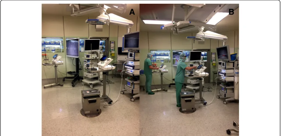
used. Also, a commercially available 4K system, the Visera 4K UltraHighDefinition, 30° camera, 10 mm, 4K big screen 55” monitor, Olympus Medical system, Olympus Europa SE & Co. KG, Hamburg, Germany is used. The position of the complete laparoscopic training parkour, the camera position in the laparo-scopic training system, and the distance from study subject to the screens are standardized. All positions are marked with signs on the training system or on the floor in the operating theater.
Laparoscopic training parkour
The laparoscopic training parkour consists of the train-ing simulator (eoSim, eoSurgical Ltd., Edinburgh, UK) wherein the tasks are performed. eoSIM is compatible with the Fundamentals of Laparoscopic Surgery (FLS) trainer. Construct validity for the system was shown pre-viously [13]. Integrated in the training simulator is a video camera system connected to a standard tablet computer. It documents the tasks for evaluation in the 2D video standard. The training simulator is connected to the 3D or 4K display system via the main camera and monitor. The complete setup of the parkour is shown in Figs.2and3. To minimize the potential bias of two sim-ultaneous participants at a training parkour described by Kowalewski et al. [14], participants start in a “time de-layed” manner. The display systems are placed 1.5 m away from each other. The working direction is turned by 45°, so that the participants are looking in different directions. Additionally, there are always two investiga-tors in the operation theater, who observe the partici-pants and prevent “copying”. Seven different tasks (novices 5) with increasing difficulty are performed by the subjects. Each task is performed three times in a row. Then the next task follows. The tasks are called
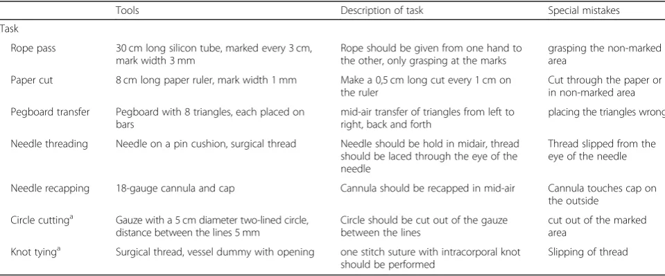
“rope pass”, “paper cut”, “pegboard transfer”, “needle threading”,“needle recapping”,“circle cutting”and“knot tying”(Fig.4). Table2briefly describes the tasks.
grasping the non-marked area, for“paper cut”a too long cut through the paper and for “knot tying” a slipping of the prepared loop. In addition the task will be rated ac-cording to the Global Operative Assessment of Laparo-scopic Skills (GOALS) [15].
Discussion
More and more complex surgical procedures are per-formed in minimally invasive/laparoscopic technique. Optimal visualization of the surgical field is one of the key aspects in this context: the better a surgeon can see,
the more subtle preparation of damageable tissue (e.g. small vessel, liver parenchyma) becomes possible. State-of-the-art display technique supports this progress. This study compares in a randomized controlled setting, the use of 3D vs. 4K display technique and its influence on surgical performance. The hypothesis is that one of both techniques could facilitate minimally invasive sur-gery. This should result in a shorter operating/perform-ance time and a minimized mistake rate. Finally, this would lead to a better outcome for the patient. Depend-ing on the different factors (e.g. structured teachDepend-ing
Fig. 2Set-Up - Laparoscopic training parkour. Laparoscopic training simulator in combination with the 4K (a) and the 3D Display system (b)
[image:6.595.58.539.87.285.2] [image:6.595.56.540.468.702.2]programs, talent of the surgeon, kind of operation, equipment), 30–100 procedures could be necessary to adopt a complex minimally invasive operation [3, 4, 9]. It seems possible, that an optimal display system could also optimize this teaching and learning process. It could help novice surgeons to improve faster during their training, especially in times of highly specialized surgical centers, external quality control and bench marking with demanding low complication rates. Experienced sur-geons, who have learned over the years to deal with re-duced standard 2D vision in MIS, could also benefit from optimal display technique. Reducing the task load by optimal intraoperative vison could help to perform long lasting minimally invasive procedures. In terms of working conditions (e.g. retirement at the age of 67 as a
surgeon in Germany), optimized intraoperative vision in MIS seems to become an important aspect in the future. Using an in-vitro setting in the study many aspects could be evaluated easier and less biased compared to a clinical trial. In times of offensive marketing and economical in-fluenced decision making in medicine, the authors hope with this investigator initiated trial to improve evidence in this field of minimally invasive surgery and help to choose optimal equipment for the future operation theater.
Trial status
This protocol represents the trial protocol version 1.0, first posted on the 7th of February 2018. The recruit-ment began at the 28th February 2018 and will be com-pleted approximately at the 01th May 2019.
[image:7.595.58.540.88.270.2]Fig. 4Tasks of the laparoscopic training parkour. Rope pass (a), paper cut (b), pegboard transfer (c), needle threading (d), needle recapping (e), circle cutting (f), and knot tying (g)
Table 2Description of the task performed during the laparoscopic training parkour - 3D- vs. 4K-displaysystem
Tools Description of task Special mistakes
Task
Rope pass 30 cm long silicon tube, marked every 3 cm, mark width 3 mm
Rope should be given from one hand to the other, only grasping at the marks
grasping the non-marked area
Paper cut 8 cm long paper ruler, mark width 1 mm Make a 0,5 cm long cut every 1 cm on the ruler
Cut through the paper or in non-marked area
Pegboard transfer Pegboard with 8 triangles, each placed on bars
mid-air transfer of triangles from left to right, back and forth
placing the triangles wrong
Needle threading Needle on a pin cushion, surgical thread Needle should be hold in midair, thread should be laced through the eye of the needle
Thread slipped from the eye of the needle
Needle recapping 18-gauge cannula and cap Cannula should be recapped in mid-air Cannula touches cap on the outside
Circle cuttinga Gauze with a 5 cm diameter two-lined circle, distance between the lines 5 mm
Circle should be cut out of the gauze between the lines
cut out of the marked area
Knot tyinga Surgical thread, vessel dummy with opening one stitch suture with intracorporal knot should be performed
Slipping of thread
a
[image:7.595.57.537.525.724.2]Additional file
Additional file 1:SPIRIT 2013 checklist: recommended items to address in a clinical trial protocol and related documents. (DOC 122 kb)
Abbreviations
2D:Two-dimensional; 3D: Three-dimensional; 4K: 4K resolution; CRF: Case report form; MIS: Minimally invasive surgery; NASA-TLX: NASA Task Load Index
Acknowledgements
The authors would like to thank Christoph Denz, MD and David Jones for their support of the study during daily operation theater routine.
Funding
This trial was conducted with no external funding. For this investigator initiated trial personal and technical resources were provided by the Department of General, Visceral and Cancer Surgery, University Hospital of Cologne. The participating researchers were released from clinical routine to perform the study. Technical resources were available at the department and could be used for the study after daily clinical routine use in the operating theater. There was no internal financial for the study.
Availability of data and materials
The datasets generated and/or analyzed during the current study are not publicly available due the data security concept of the study and the General Data Protection Regulation of the European Union but are available from the corresponding author on reasonable request.
Authors’contributions
RW and RD designed study, performed study, collected data, analyzed data, wrote the paper and contributed equally to the study; RK, HF, GD, JB and TB collected data, analyzed data, and performed study, AH and CG are the ophthalmologist and performed study, collected data, wrote the paper; DM the data trustee, performed randomization, designed study, collected data; MH is the medical statistician and designed study, analyzed data, wrote the paper, CJB designed study, analyzed data, wrote the paper, DLS designed study, analyzed data, wrote the paper. All authors read and approved the final manuscript.
Ethics approval and consent to participate
The study has been approved by the Ethics Committee of the University of Cologne (Ethikkommission der Medizinischen Fakultät der Universität zu Köln, Nummer 17–388). Written informed consent will be given by all subjects before study inclusion and randomization. Written informed consents of the subjects during this study were only obtained by the authors of the protocol.
Consent for publication
People photographed in Fig.3consented to their photo being included in this study/publication.
Competing interests
The authors declare that they have no competing interests.
Publisher’s Note
Springer Nature remains neutral with regard to jurisdictional claims in published maps and institutional affiliations.
Author details
1Department of General, Visceral and Cancer Surgery, University Hospital of
Cologne, University of Cologne, Kerpener Straße 62, 50937 Cologne, Germany.2Department of Ophthalmology, University Hospital of Cologne, University of Cologne, Cologne, Germany.3Faculty of Management, Economics and Social Sciences, Department of Business Administration and Health Care Management, University of Cologne, Cologne, Germany. 4Institute of Medical Statistics and Computational Biology, Faculty of
Medicine and University Hospital of Cologne, University of Cologne, Cologne, Germany.
Received: 10 December 2018 Accepted: 25 March 2019
References
1. Keus F, de Jong JA, Gooszen HG, van Laarhoven CJ. Laparoscopic versus small-incision cholecystectomy for patients with symptomatic cholecystolithiasis. Cochrane Database Syst Rev. 2006;4:CD006229. 2. Wilson Colin H, Sanni A, Rix David A, Soomro Naeem A. Laparoscopic versus
open nephrectomy for live kidney donors. Cochrane Database Syst Rev. 2011;(11). Accessed 1 Dec 2018.
3. Suguita FY, Essu FF, Oliveira LT, et al. Learning curve takes 65 repetitions of totally extraperitoneal laparoscopy on inguinal hernias for reduction of operating time and complications. Surg Endosc. 2017;31(10):3939–45. 4. Serrano OK, Bangdiwala AS, Vock DM, et al. Defining the tipping point in
surgical performance for laparoscopic donor nephrectomy among transplant surgery fellows: a risk-adjusted cumulative summation learning curve analysis. Am J Transplant. 2017;17(7):1868–78.
5. Tidbury LP, Black RH, O’Connor AR. Clinical assessment of stereoacuity and 3-D stereoscopic entertainment. Strabismus. 2015;23(4):164–9.
6. Gurusamy KS, Sahay S, Davidson BR. Three dimensional versus two dimensional imaging for laparoscopic cholecystectomy. Cochrane Database Syst Rev. 2011;1:CD006882.
7. Arezzo A, Vettoretto N, Francis NK, et al. The use of 3D laparoscopic imaging systems in surgery: EAES consensus development conference 2018. Surg Endosc. 2018.https://doi.org/10.1007/s00464-018-06612-x.
8. Smith R, Schwab K, Day A, et al. Effect of passive polarizing three-dimensional displays on surgical performance for experienced laparoscopic surgeons. Br J Surg. 2014;101(11):1453–9.
9. Wahba R, Kleinert R, Hellmich M, et al. Optimizing a living kidney donation program: transition to hand-assisted retroperitoneoscopic living donor nephrectomy and introduction of a passive polarizing three-dimensional display system. Surg Endosc. 2017;31(6):2577–85.
10. Chan AW, Tetzlaff JM, Altman DG, Dickersin K, Moher D. SPIRIT 2013: new guidance for content of clinical trial protocols. Lancet. 2013;381(9861):91–2. 11. Chan AW, Tetzlaff JM, Gotzsche PC, et al. SPIRIT 2013 explanation and
elaboration: guidance for protocols of clinical trials. BMJ. 2013;346:e7586. 12. Noyez L. Control charts, Cusum techniques and funnel plots. A review of methods for monitoring performance in healthcare. Interact Cardiovasc Thorac Surg. 2009;9(3):494–9.
13. Hennessey IA, Hewett P. Construct, concurrent, and content validity of the eoSim laparoscopic simulator. J Laparoendosc Adv Surg Tech A. 2013;23(10): 855–60.
14. Kowalewski KF, Minassian A, Hendrie JD, et al. One or two trainees per workplace for laparoscopic surgery training courses: results from a randomized controlled trial. Surg Endosc. 2018.https://doi.org/10.1007/ s00464-018-6440-5.