TOWARDS DEVELOPING AN EFFECTIVE TECHNIQUE TO
COMPRESS MEDICAL IMAGES
1K.GOPI,2 Dr.T.RAMASHRI
1
Asst.professor, Department of ECE, Sreenivasa Institute of
Technology and Management Studies ,chittoor 2
Professor, Department of ECE, S.V University College of Engineering, Tirupati
E-mail:[email protected]
ABSTRACT
The images play an important role in many of the applications. There are several techniques to compress these images in order to reduce the size of the image without any loss of data. This paper proposes an effective technique to compress the medical images using an improved ridgelet transform. This improved technique replaces the discrete wavelet transform in the ridgelet transform with the slantlet transform. This slantlet transform is an orthogonal DWT and has two zero moments with improved time localization. In this proposed technique the medical image is compressed using the improved ridgelet transform and then we apply thresholding and quantization methods to the ridgelet coefficients of the compressed medical image. Also the proposed technique requires only minimum number of coefficients for reconstructing the medical image without any loss of data. Comparison is made between the ridgelet transform and the proposed improved ridgelet transform in terms of SDME and this technique assures to provide higher signal to noise ratio.
Keywords: Ridgelet Transform, Discrete Wavelet Transform (DWT), Slantlet Transform (SLT), Thresholding, Quantization.
1. INTRODUCTION
Images play an important role in the world of multimedia and its transmission with storage has become really a big burden as it occupy more space in memory [1]. A thousand words stored on a digital computer require very little capacity, but a single picture/image can require much more. Such images require huge volume of data to describe them and this greatly slows down the transmission and makes storage prohibitively costly [3]. For example, to store a color image of a moderate size, e.g.512×512 pixels, one needs 0.75 MB of disk space. A 35mm digital image with a resolution of 12μm requires 18 MB. To store these images, and make them available over network (e.g. the internet), compression techniques are needed [5].
When a digital image is transmitted through a communication channel, the cost of transmission depends on the size of the data. To reduce the transmission cost, the data need to be compressed [4]. To meet the demand for high speed transmission of image; efficient image storage, remote treatment and efficient image compression techniques is essential [13]. Image compression has
been an important research topic for many years. There are many kinds of compression techniques. Lots of research literature is available which explains the importance and techniques of Image compression. Recently this field has gained an immense attention of scientists and researchers [2].
Lossless compression is preferred for archival purposes and often for medical imaging, technical drawings, clip art, or comics [14]. The lossy compression methods are especially suitable for natural images such as photographs in applications where minor (sometimes imperceptible) loss of fidelity is acceptable to achieve a substantial reduction in bit rate. The lossy compression that produces imperceptible differences may be called visually lossless [15]. The development of efficient compression techniques will continue to be a design challenge for future communication systems and advanced multimedia applications [11]. Hence, it is essential to analyze and suggest a best technique for lossless image compression [1]. Multispectral images are widely used in geoscience and remote sensing. Their use in other applications field like medical imagery, quality control in industry, meteorology, and exact color measurements are increasing [10]. Medical images like MRI and CT are Special images, which require lossless compression as a minor loss that can cause adverse effects [16].
2. RELATED WORKS
Chakrapani et al. [17] have applied the technique of Genetic Algorithm (GA) for Fractal Image Compression (FIC). With the help of this evolutionary algorithm, effort has been made to reduce the search complexity of matching between range block and domain block. One of the image compression techniques in the spatial domain is Fractal Image Compression but the main drawback of FIC is that it involved more computational time due to global search. In order to improve the computational time and also the acceptable quality of the decoded image, Genetic algorithm has been proposed. Experimental results have been showed that the Genetic Algorithm was a better method than the traditional exhaustive search method.
Kilari Veera Swamy et al. [18] have proposed a compression technique and image watermarking algorithm based on Contourlet Transform (CT). For image compression, an energy based quantization is used. Scalar quantization is explored for image watermarking. Double filter bank structure is used in CT. The Laplacian Pyramid (LP) is used to capture the point discontinuities, and then followed by a Directional Filter Bank (DFB) to link point discontinuities. The coefficients of down sampled low pass version of LP decomposed image are re-ordered in a pre-determined manner and prediction algorithm is used to reduce entropy (bits/pixel). In addition, the coefficients of CT are quantized based
on the energy in the particular band. The superiority of proposed algorithm to JPEG is observed in terms of reduced blocking artifacts. The results are also compared with wavelet transform (WT). Superiority of CT to WT is observed when the image contains more contours. The watermark image is embedded in the low pass image of contourlet decomposition. The watermark can be extracted with minimum error. In terms of PSNR, the visual quality of the watermarked image is exceptional. The proposed algorithm is robust to many image attacks and suitable for copyright protection applications
Anna Saro Vijendran et al. [19] have presented an interpolation method, which is proposed for compression technique. Their proposed method is the localizing of spatial and frequency correlation from wavelets. Modified Forward Only Counter Propagation Neural Network (MFOCPN) is used for the classification and functional task. The wavelet based technique decomposes the lower sub band consisting of non significant coefficients and are eliminated. The significant smooth and sharp coefficients are found using interpolation methods. Here a technique is proposed called the cosine interpolation, which is an alternative to the nearest neighborhood interpolation method. The proposed methodology of interpolation is proved to be an efficient approach for mapping all significant coefficients and thus resulting in improved quality. Hence the comparison is made between nearest neighborhood interpolation and cosine interpolation. The experimental results are tested on various standard images, where these results yield a better PSNR value compared with the existing nearest neighbor interpolation method.
Parveen Banu et al. [20] have discussed the Imaging applications that generate large volumes of data leading to challenges for transmission and storage. They have proposed a hybrid image compression technique for efficient storage and delivery of data. It is based on decomposing the data using daubechies-4 wavelet in combination with the lifting scheme and entropy encoding. The proposed scheme has been concerned with the compression ratio, bits per pixel and peak signal to noise ratio. Experimental results illustrated that the proposed scheme is efficient and feasible in terms of compression ratio, bits per pixel and peak signal to noise ratio.
artificial neural network uses the different Back-Propagation artificial neural networks in processing of the image. The original images taken, for instance 256*256 pixels of bitmap image, each block of image into one network selection, according to each block the value of pixels in image complexity value is calculated. For estimation each value of the images in a block can be evaluated and trained. Best PSNR in selecting images to be compressed with a modification Levenberg-Marquart for MLP neural network is taken. The algorithm taken gives a good research result to each block of image. The time taken for the learning procedure for running each block of images is reduced. Finally, a neural network is taken for the Back Propagation artificial neural network.
3. THE RIDGELET TRANSFORM
For many of the image processing techniques that take advantage of the sparse representation of the image, the wavelet transform was mostly used. But these wavelets fail in effectively representing the line singularities along two-dimension. So, to overcome the issues in the wavelet transform, Candes and Donoho established a new scheme named ridgelet transform which effectively represents the line singularities in two-dimension. The main idea behind this ridgelet transform is to map the line singularity into point singularity and then make use of the wavelet transform for representing this point singularity.
In the ridgelet transform fig 1, initially Two-dimensional Discrete Fourier Transform (2D DFT) is applied to the 2D medical image. Then to map the line singularities into point singularities the radon transform is applied. For this the One-dimensional Inverse Discrete Fourier Transform (1-D I(1-DFT) is applied to each column of the 2(1-D image. Finally, wavelet transform is performed on each column of the two-dimensional medical image.
Fig 1: Discrete Ridgelet Transform
The continuous ridgelet transform (CRT) of a bivariate function f(x) in R2 is defined by,
(ab θ) ( ) ( )x f xdx
R
ψ
CRT
f =∫
2 abθ, , ,
, (1)
where the ridgelets
ψ
( )xθ b a, ,
are defined from
the 1D wavelet Ψ
( )
x as,( )
x a(
(
x θ x θ b)
a)
θ b a
/ sin cos
2 1
2 / 1 ,
, = Ψ + −
−
Ψ
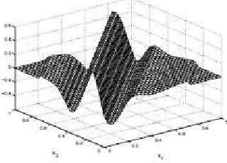
(2) [image:3.595.322.482.308.423.2]The fig 2. Shows a typical ridgelet function and it is oriented along the angle θ and is constant along the line x1 cos θ + x2 sin θ = const.
Fig 2: Typical Ridgelet Function.
On the other hand the continuous wavelet transform (CWT) of f(x) in R2 is given by,
(
)
( ) ( )
∫ Ψ
=
R a a b b x f x dx
b b a a CWT f
2
2 1 2 1
2 1 2 1
, , ,
, , ,
(3)
where the wavelets in 2D are tensor products
( ) , ( )1 , ( )2 ,
, ,
2 2 2
1 2
1 2 1
x x
x
b a b
a b
b a
a
Ψ
Ψ
Ψ
=(4)
of the 1D wavelets, Ψa,b
( )
t =a−1/2Ψ(
(
t−b)
/a)
In comparison, the CRT is similar to the 2D CWT except that the point parameters are replaced with the line coordinates. The wavelets are efficient at representing objects with point singularities where as the ridgelets are effective at representing the objects along the line.
The radon transform is denoted as,
(
)
( )
(
x θ x θ t)
dxδ x f t
θ R
R
f
∫
− +
= 2
sin cos
,
2 1
(5)
(abθ)
ψ
( )tR
( )θtdtCRT
f , , R ab f ,,
∫
= (6)
4. IMAGE REPRESENTATION USING IMPROVED RIDGELET TRANSFORM
As mentioned above, the ridgelet transform is the application of 1D wavelet transform to the radon transform and it consists of four parts for processing the image.
(a) Two dimensional Discrete Fourier Transform (2-D DFT)
(b) Radon Transform
(c) One dimensional Inverse Discrete Fourier Transform (1-D IDFT)
(d) One dimensional Discrete Wavelet Transform (1-D DWT) In this improved ridgelet transform, the one dimensional discrete wavelet transform is replaced by the slantlet transform (SLT). This slantlet transform is an orthogonal DWT and provides improved time localization with two zero moments. Hence, this improved ridgelet transform is expected to provide higher performance than the existing ridgelet transform.
4.1 2-D Discrete Fourier Transform
The Discrete Fourier Transform (DFT) is easy to handle the 2 dimensional images using the tensor product form of the orthogonal basis. Assume the data set as N x M array,
[
n,m]
,0 ≤ n ≤ N −1,0 ≤ m ≤ M −1.F (7)
Then the fourier transform of f is defined as,
[
]
− − =∑ ∑
− = − = M lm π i N kn π i m n f l k F N n M m 2 exp 2 exp , ] , [ 1 0 10 (8)
Whereas the vector uk defined as in (9) forms the orthonormal basis of the space CN of complex valued N vectors.
[ ]
= N kn π i nu
k exp 2 (9)and the tensor product vector ukl is given by,
]
[
1 0 , 1 0 , 2 exp 2 exp , − ≤ ≤ − ≤ ≤ = M m N n M lm π i N kn π i m nu
kl (10)This set forms an orthogonal basis of the space CN x CM. = CNM. This form of DFT is employed in MATLAB which is defined as
F=fft2(f).
Let the medical image be I size n x m. Then the 2D DFT coefficients of the medical image is given by,
[
]
− − =∑ ∑
− = − = ∧ M lmI j N nI j I I f m n I m n N n M m m n π π 2 exp 2 exp , ] , [ 1 0 1 0 (11)4.2 Radon Transform
The Radon transform was developed by J.Radon in 1917. The Radon transform is defined as the summation of image pixels over a certain set of lines. The order of coefficients of the 2D DFT coefficients of the medical image is controlled by direction of set of normal vectors (ak,bk), where k=0,1,…p. The line integral along a particular direction is called as projection. The radon transform maps the Cartesian rectangular coordinates (x,y) to a distance and an angle (ρ,θ). The optimum number of projections for the radon transform is p+1, and the best ordering of the 2D DFT coefficients can be achieved if the vectors are determined from,
(
)
(
( )
( )
)
)
(
a b{
nu n p}
b C a C b a k k k k p k p k k ≤ ≤ ∈ = 1 : , , , min arg ,
,Cp( )bk ≥0 (12)
Here, Cp(x) is the centralized function and it is given by,
Cp(x) = x – p.round(x/p) (13) and
(
Cp( )
ak ,Cp( )
bk)
denotes the distance ofthe vectors from the origin on the fourier plane. The optimal vectors have angles (0,π) and after reordering, the Fourier matrix I^ is named as Iopt^ and the size of this matrix will be p x (p + 1). Then to obtain the Radon coefficients of the matrix, apply the 1D IDFT to each column of the
I
opt^ matrix. The Radon coefficients are givenby,
(
)
(
)
+ − + =∑
− = 1 * 2 exp 1 * 1 ] , [ 1 0 ^ ^ p p kn j I p p m n I N k opt R π (14)4.3 Slantlet Transform
is an orthogonal DWT with two zero moments and improved time localization. Unlike DWT, it is not based on filterbank iteration and instead different filters are used for different scales of this transform. An orthogonal discrete time basis having an octave-band characteristic can be easily designed by the usual filter bank iteration. But it does not yield discrete time basis optimal with respect to time localization for a fixed number of zero moments. The filters in slantlet transform are piecewise linear and discontinuous.
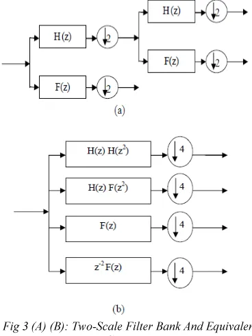
Fig 3 (A) (B): Two-Scale Filter Bank And Equivalent Structure.
The fig 3(a), shows a two-scale iterated filter bank and fig 3(b), an equivalent filter bank structure. The slantlet filter bank is similar to the second filter bank in which the length of the filter is restricted. But in slantlet filter bank, the structure will be occupied by different filters and they won’t be the products. With this possibility to have different filters, the slantlet filter bank is designed with filters of shorter length and also satisfies orthogonality and zero moment conditions.
[image:5.595.124.510.78.544.2]For a two-channel case, the filter developed by Daubechies has the orthogonal filter bank and K zero moments. For K = 2 zero moments, the iterated filters of Fig 3(b), are of length 10 and 4. But the slantlet filter bank with K = 2 zero moments as in fig 4, has filter length 8 and 4. This is 2 filters less than that of the Daubechies filter.
Fig 4: Two-Scale Slantlet Filter Bank.
[image:5.595.94.278.259.504.2]( )
(
)
− = − + − = + = + 1 2 .... 2 , 2 1 2 .... 0 , 1 1 , 1 0 , 1 1 , 0 0 , 0 i i i i n for n a a n for n a a ng (15)
( )
(
)
− = − + − = + = + 1 2 .... 2 , 2 1 2 .... 0 , 1 1 , 1 0 , 1 1 , 0 0 , 0 i i i i n for n b b n for n b b nh (16)
( )
(
)
− = − + − = + = + 1 2 .... 2 , 2 1 2 .... 0 , 1 1 , 1 0 , 1 1 , 0 0 , 0 i i i i n for n c c n for n c c nf (17)
Then, the slantlet coefficients of the image I is given by,
[
n m]
g(
I[
n m]
)
IS^ , = R^ , (18)
And the same approach stands for both the f(n) and h(n) filters.
5. THE PROPOSED EFFECTIVE TECHNIQUE FOR MEDICAL IMAGE COMPRESSION
The medical image compression is needed not only to take care of the file size of the medical image, but also to take care the time taken to transfer the medical image. There are several techniques for the compression of medical images. This paper proposes an effective technique for the medical image compression. This technique makes use of an improved ridgelet transform in which the discrete wavelet transform (DWT) in ridgelet transform is replaced with the slantlet transform (SLT). This proposed technique for medical image compression has two phases namely the medical image compression (encoding) and medical image decompression (decoding).
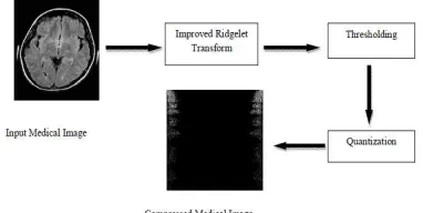
5.1 Medical image compression
The steps for the compression of the medical images are as follows. Initially the input medical image is taken and resized. Then the medical image is processed with the improved ridgelet transform, and it is converted to ridgelet coefficients. Many values in the ridgelet coefficient may be equal to zero. Using a technique called thresholding, these coefficients are modified so that the slantlet coefficient values contain large number of zeros. Thresholding may be an operation that performs test against a function T which is of the form,
)
(
))
(
(
[
x y p x y f x y]
T
T = , , , , , (19)
where f(x,y) is the grey level at (x,y) and p(x,y) describes some of the local properties of the point. The thresholded image is defined by,
(
)
(
)
(
)
≥ > = T y x f if T y x f if y x g , 0 , 1, (20)
Here, pixels labeled 1 corresponds to image and pixels labeled 0 correspond to the background.
[image:6.595.310.501.260.356.2]In the final step of the medical image compression, the set of floating point values are converted to the nearby integer values. This method is called as quantization. The simplest method of quantization is to round-off the floating values to the nearby integer values.
Fig 5: Medical Image Encoder
5.2 Medical image decompression
[image:6.595.313.503.478.534.2]The decoding of the compressed medical image is the inverse process of medical image compression (encoding). The compressed medical image is decoded by applying the inverse improved ridgelet transform. The fig shows the process of medical image decoding.
Fig 6: Medical Image Decoder.
6. RESULT AND DISCUSSION
The Second Derivative-like Measure of Enhancement (SDME) is calculated for the medical images compressed using both the transforms. The SDME is an enhancement measure based on a contrast measure based on the concept of second derivative. The SDME is given by,
l k l k center l
k
l k l k center l
k k
l k
k I I I
I I
I k k SDME
, min; , ; ,
max;
, min; , ; ,
max;
1 1
1 1
2 2 ln
20 1
1 2
+ −
+ −
− =
∑ ∑
= =
(21)
The SDME comparison between the ridgelet transform and the improved ridgelet transform is given in the table below.
Image Transform SDME PSNR MSE Encoding
Time
Decoding Time
Compression rate
1.
Ridgelet Transform -9.3599 81.2067 4.9637 0.0234 0.6057 98%
Improved Ridgelet Transform -8.2356 24.5411 230.33 0.0121 0.6934 100%
2.
Ridgelet Transform -10.0706 77.8831 0.0011 0.0151 0.6401 94%
Improved Ridgelet Transform -9.8685 20.2495 618.76 0.0120 0.7540 100%
3.
Ridgelet Transform -11.5102 73.9670 0.0026 0.0156 0.2425 92%
Improved Ridgelet Transform -8.7840 18.2005 991.81 0.0121 0.5974 100%
From the results in the table above, it is clear that the compression of medical images using the proposed, Improved Ridgelet transform yields better SDME values when compared to the SDME values of the same images compressed using Ridgelet transform. Also in the Improved Ridgelet transform the compression rate of the images are high.
7. CONCLUSION
For effective compression of the medical images, the images must be processed using effective techniques. In this paper, the improved ridgelet transform has been used for compressing the medical images and the compressed images that are reconstructed with less data loss. This improved ridgelet transform replaces the wavelet transform in the ridgelet transform with the slantlet transform which is piecewise linear and also has filters of smaller length. This improved ridgelet transform proves to be the effective technique for compressing the medical images. This is shown by the comparison of SDME values of the reconstructed medical images of the ridgelet transform and the improved ridgelet transform. Thus by implementing the improved ridgelet transform for compression of medical images it is concluded that the compression based on the slantlet transform yields better performance than the discrete wavelet transform.
REFERENCES
[1] Senthilkumaran, "Neural Network Technique for Lossless Image Compression Using X-Ray Images", International Journal of Computer and Electrical Engineering, Vol.3, No.1, pp.17-23 February, 2011.
[2] Sajjad Mohsin and Sadaf Sajjad, "Codebook Generation for Image Compression with Simple and Ordain GA", International Journal of Computers and Communications, Vol.1, No.2, pp.35-40, July, 2007.
[3] Sudhakar Radhakrishnan and Jayaraman Subramaniam, "Novel Image Compression Using Multiwavelets with SPECK Algorithm",
The International Arab Journal of Information Technology, Vol.5, No.1, January 2008. [4] Preeti Aggarwal& Babita Rani, "Performance
Comparison Of Image Compression Using Wavelets", International Journal Of Computer Science & Communication, Vol. 1, No. 2, pp. 97-100, December 2010.
[5] Jagadish H. Pujar, Lohit M. Kadlaskar, "A New Lossless Method of Image Compression and Decompression Using Huffman Coding Techniques”, Journal of Theoretical and Applied Information Technology, Vol.15, No.1, pp.18-23, 2010.
[6] Vijaya Prakash and Gurumurthy, "A Novel VLSI Architecture for Digital Image Compression Using Discrete Cosine Transform and Quantization", International Journal of Computer Science and Network Security, Vol.10, No.9, pp.175-182, September 2010. [7] Nikkoo Khalsa, Sarate and Ingole, "Factors
Influencing The Image Compression Of Artificial And Natural Image Using Wavelet Transform", International Journal of Engineering Science And Technology, Vol. 2, No. 11, pp. 6225-6233, 2010.
International Journal (SPIJ), Vol. 2, No. 5, pp. 17-26, October 2008.
[10] Satish K.Singh and Shishir Kumar, "Mathematical Transforms and Image Compression: A Review", Maejo International Journal of Science and Technology, Vol.4, No.2, pp.235-249, 2010.
[11] Jonathan Delcourt, Alamin Mansouri, Tadeusz Sliwa and Yvon Voisin, "An Adaptive Multiresolution-Based Multispectral Image Compression Method", International Journal Of Future Generation Communication And Networking Vol. 3, No. 4,Pp.1-10,December, 2010.
[12] Saudagar Abdul Khader Jilani, "JPEG Image Compression Using FPGA with Artificial Neural Networks", International Journal of Engineering and Technology, Vol.2, No.3, Pp.252-257, June 2010.
[13] Lalitha and Latte, "Lossless and Lossy Compression of DICOM images with scalable ROI", IJCSNS, Vol.10, No.1, pp.276-281, July 2010.
[14] Bhawna Gautam, "Image Compression Using Discrete Cosine Transform & Discrete Wavelet Transform", Technical Report, National Institute of Technology, Rurekela, May 2010. [15] Nishat Kanvel and Elwin Chandra Monie,
"Performance Measure of Different Wavelets for a Shuffled Image Compression Scheme",
IJCSNS, Vol.9, No.3, pp.217-222, March 2009. [16] Ramesh and Shanmugam,"Medical image
compression using wavelet decomposition for prediction method", IJCSIS, Vol.7, No.1, pp.262-265, 2010.
[17] Chakrapani and Soundara Rajan, "Genetic Algorithm Applied to Fractal Image Compression", ARPN Journal of Engineering and Applied Sciences, Vol.4, No.1, pp.53-58, FEBRUARY 2009.
[18] Kilari Veera Swamy, Chandra Mohan, Bhaskar Reddy, Srinivas Kumar, "image compression and watermarking scheme using scalar quantization", The International Journal of next generation network, Vol.2, No.1, pp,37-47, March 2010.
[19] Anna Saro Vijendran and Vidhya, "A Hybrid Image Compression Technique Using Wavelet Transformation - MFOCPN and Interpolation",
Global Journal of Computer Science and Technology, Vol.11, No.3, pp.57-62, March 2011.
[20] Parveen Banu and Venkataraman, "An Efficient Hybrid Image Compression Scheme based on Correlation of Pixels for Storage and Transmission of Images", International Journal of Computer Applications, Vol.18, No.3, pp.6-9, March 2011.
[21] Prema Karthikeyan and Narayanan Sreekumar, "A Study on Image Compression with Neural Networks Using Modified Levenberg Maruardt Method", Global Journal of Computer Science and Technology, Vol.11, No.3, pp.1-6, March 2011.
[22] Yicong Zhou, Karen Panetta and Sos S. Agaian “Nonlinear filtering for enhancing prostate MR images via alpha-trimmed Mean Separation”, In proceeding of the IEEE International Conference on Systems, Man and Cybernetics, Istanbul, Turkey, October 10-13, 2010.