The combined role of P- and E-selectins in
atherosclerosis.
Z M Dong, … , R O Hynes, D D Wagner
J Clin Invest.
1998;
102(1)
:145-152.
https://doi.org/10.1172/JCI3001
.
P- and E-selectins are adhesion molecules mediating the first step in leukocyte
extravasation. Because their function in leukocyte adhesion is overlapping, we
hypothesized that there might be a combined effect of these selectins on the development of
atherosclerotic lesions. We bred P- and E-selectin-double-deficient mice onto the
low-density lipoprotein receptor (LDLR)-deficient background (LDLR-/- P/E-/-) and compared
lesion development in these mice to that in mice wild type for both selectins
(LDLR-/-P/E+/+). After 8 wk on atherogenic diet, the LDLR-/- P/E-/- mice developed fatty streaks in
the aortic sinus that were five times smaller than those in LDLR-/- P/E+/+ mice. The density
of macrophages in the fatty streaks was comparable between P/E+/+ and
LDLR-/-P/E-/- mice. After 22 wk on the diet, the lesions spread throughout the aorta but this process
was delayed in LDLR-/- P/E-/- mice. At 37 wk on diet, the lesions progressed to the fibrous
plaque stage in both genotypes. However, the lesions in the aortic sinus in LDLR-/-
P/E-/-mice were 40% smaller and less calcified than those of LDLR-/- P/E +/+ P/E-/-mice. Our results
suggest that P- and E-selectins together play an important role in both early and advanced
stages of atherosclerotic lesion development.
Research Article
Find the latest version:
J. Clin. Invest.
© The American Society for Clinical Investigation, Inc. 0021-9738/98/07/0145/08 $2.00
Volume 102, Number 1, July 1998, 145–152 http://www.jci.org
The Combined Role of P- and E-Selectins in Atherosclerosis
Zhao Ming Dong,*‡ Susan M. Chapman,* Allison A. Brown,* Paul S. Frenette,*§ Richard O. Hynes,i and Denisa D. Wagner*‡
*Center for Blood Research, ‡Department of Pathology, and §Department of Medicine, Harvard Medical School, Boston, Massachusetts
02115; iHoward Hughes Medical Institute, Center for Cancer Research, Department of Biology, Massachusetts Institute of Technology, Cambridge, Massachusetts 02139
Abstract
P- and E-selectins are adhesion molecules mediating the first step in leukocyte extravasation. Because their function in leukocyte adhesion is overlapping, we hypothesized that there might be a combined effect of these selectins on the development of atherosclerotic lesions. We bred P- and E-selectin–double-deficient mice onto the low-density lipo-protein receptor (LDLR)–deficient background (LDLR2/2 P/E2/2) and compared lesion development in these mice to that in mice wild type for both selectins (LDLR2/2 P/E1/1). After 8 wk on atherogenic diet, the LDLR2/2 P/E2/2 mice developed fatty streaks in the aortic sinus that were five times smaller than those in LDLR2/2 P/E1/1 mice. The density of macrophages in the fatty streaks was compa-rable between LDLR2/2 P/E1/1 and LDLR2/2 P/E2/2 mice. After 22 wk on the diet, the lesions spread throughout the aorta but this process was delayed in LDLR2/2 P/E2/2 mice. At 37 wk on diet, the lesions progressed to the fibrous plaque stage in both genotypes. However, the lesions in the aortic sinus in LDLR2/2 P/E2/2 mice were 40% smaller and less calcified than those of LDLR2/2 P/E 1/1 mice. Our results suggest that P- and E-selectins together play an important role in both early and advanced stages of athero-sclerotic lesion development. (J. Clin. Invest. 1998. 102:145– 152.) Key words: leukocyte adhesion • animal model for
atherosclerosis • macrophage • smooth muscle cells • lipo-fuscin
Introduction
Adhesion of circulating leukocytes, especially monocytes, to the endothelial lining is an early event in atherosclerosis. The active stages of atherosclerosis are marked by the extensive in-filtration of these monocytes through the endothelium into the arterial intima (1). It is now well documented that leukocyte and endothelial adhesion molecules play an important role in the emigration of leukocytes from the blood into foci of in-flammation (2–3), and similar processes could be at work in atherosclerosis. However, it is unclear which cell adhesion molecules are important in atherogenesis.
Leukocyte rolling along the endothelial surface is the first step in leukocyte adhesion to endothelial cells. Multiple stud-ies indicate that leukocyte rolling is mediated by the selectin family of adhesion receptors and their carbohydrate ligands (4). L-selectin is constitutively expressed on the surfaces of most leukocytes. E-selectin is found only on endothelial cells stimulated by inflammatory cytokines. P-selectin is rapidly re-leased from storage granules in activated platelets and endo-thelial cells. The generation of selectin-deficient mice has con-firmed the role of selectins in the rolling of blood cells and provided evidence indicating that there is an overlapping func-tion of these selectins in the regulafunc-tion of inflammatory pro-cesses (5–9). In our previous study, P-selectin–deficient (P2/2) mice were bred onto a low density lipoprotein receptor (LDLR)1–deficient background that represents an animal
model of familial hypercholesterolemia (10). We found that absence of P-selectin partially reduced fatty streak formation but did not significantly affect lesion progression to fibrous plaque stage in LDLR-deficient mice (11). These results sug-gested that other selectins such as E-selectin may be able to promote monocyte recruitment in the LDLR2/2 P2/2 mice in late stages of lesion development. To investigate this possi-bility, we have generated a line of LDLR2/2 P/E2/2 mice, and compared lesion development in these mice to LDLR2/2 mice lacking just P-selectin or wild type for both selectins.
Methods
Mice, genotype analysis, and diets.P- and E-selectin double–deficient (P/E2/2) mice were generated in our laboratory and were on a mixed C57BL/6J 3 129Sv background (7). LDLR-deficient mice (LDLR2/2) were provided by Joachim Herz at University of Texas Southwestern Medical Center (Dallas, TX) and were also on a mixed C57BL/6J 3 129Sv background (10). Because the selectin family of genes is clustered on chromosome 1 within 300 kb, we were able to produce mice deficient in LDLR (the gene is located on chromosome 9) and both selectins (LDLR2/2 P/E2/2) by mating LDLR2/2 mice to P/E2/2 mice. Littermates from the F2 generation of this in-tercross were genotyped by PCR analysis and used to establish LDLR2/2 P/E2/2 matings and LDLR2/2 P/E1/1 matings. The progeny of these matings were used in our study. PCR analysis was performed for genotyping for selectins (7) and LDLR (12).
Before starting on an atherogenic diet, mice of all genotypes were maintained on a standard low fat mouse chow containing 5% fat (wt/wt) (Prolab 3000; PMI Feeds, St. Louis, MO). When they were 5–7-wk-old, mice were placed on an atherogenic diet for indicated lengths of time. The atherogenic diet (ICN Biomedicals; Aurora, OH) contained 1.23% (wt/wt) cholesterol, 17.84% (wt/wt) butter, 0.98% (wt/wt) corn oil, and 0.48% (wt/wt) sodium cholate.
Cholesterol determination. Mice were fasted overnight before transport to the laboratory and blood was collected through the ret-roorbital venous plexus. Cholesterol concentrations in total plasma Address correspondence to Denisa D. Wagner, Center for Blood
Re-search, Harvard Medical School, 800 Huntington Avenue, Boston, MA 02115. Phone: 617-278-3344; FAX: 617-278-3368.
Received for publication 5 February 1997 and accepted in revised form 30 April 1998.
were determined using an enzymatic assay according to the manufac-turer’s instructions (Cholesterol kit 352-50, Sigma Chemical Co.).
Quantitation of aortic sinus lesions. The sections containing aor-tic sinus lesions were prepared as described (11). Four consecutive 10-mm sections were collected for each slide. 10 slides were made from each animal. Odd-numbered slides were stained with oil red-O and hematoxylin, counterstained with light green, and examined for lesion size and calcium deposits. Even-numbered slides were kept for histological and immunohistochemical analysis. Five sections, each 80 mm apart, were scored for each animal without knowledge of the genotype. The area of the lesion was measured with a Leica Q500MC image analysis program (Leica Inc., Deerfield, IL). Values reported represent the mean lesion area from five sections for each animal.
Quantitation of lesions in entire aorta. Mice were perfused with 4% paraformaldehyde for 15–20 min, and aortae were collected be-tween the subclavian and ilial branches, placed in 4% paraformalde-hyde for 4–6 h, followed by placement in 30% sucrose (wt/vol) for 2–3 d, and then stored in 10% formalin for later analysis. The entire aortae were stained with Sudan IV, as described (11). After staining, aortae were opened longitudinally and mounted on slides using glyc-erol gelatin. For quantitation of Sudan IV-stained surface area, mounted aortae were visualized through a JVL TK-1280U color video camera into a Leica Q500MC image analysis program. Percent-age of area covered by lesion was determined by Sudan IV–stained area divided by total aorta area.
Histological analysis. Calcium deposits were identified by the he-matoxylin stain, as described (13). Odd-numbered slides stained with oil red-O, hematoxylin, and light green were examined by light mi-croscopy for presence of calcification. A total of 20 sections (five slides) from the aortic sinus were examined for each animal. Mice were considered positive if calcium deposits were observed in one or more sections.
To assess lipofuscin accumulation on the surface of the aortic valve leaflets, unstained even-numbered slides were examined by light microscopy for the presence of yellow-brown lipofuscin pigment deposits (14). The presence of lipofuscin in any 1 of 20 sections (five slides) defined the mouse as lipofuscin-positive.
To quantitate cell density in the 8-wk lesions, the four sections on slide No. 6 for each mouse were stained with hematoxylin and num-bers of the blue cell nuclei in the lesion area were counted through a light microscopy. Cell density was expressed as the number of cell nu-clei per square millimeter of atherosclerotic lesion area.
Immunohistochemical analysis. To identify macrophages and T cells in aortic sinus lesions from mice on atherogenic diet for 8 wk, frozen sections (10-mm-thick) were fixed in cold acetone for 5 min, and then incubated with avidin-blocking solution followed by biotin-blocking solution for 30 min each. Endogenous peroxidase activity was blocked by incubating slides in a solution of 3% hydrogen perox-ide for 5 min. Slperox-ides were then incubated with a biotin-conjugated anti–Mac-1 antibody (No. 01712D, dilution 1:10; PharMingen, San Diego, CA) for identification of macrophages, or a biotin-conjugated anti-CD3 antibody (No. 01082A, dilution 1:25; PharMingen) for iden-tification of T cells. Antibodies were visualized by an avidin/biotin peroxidase–linked detection system from Vector Laboratories (Bur-lingame, CA). Antibody was omitted from the control sections. De-tection of intracellular TNF-a on frozen sections of 8-wk lesions was performed as described (15). A rat anti–mouse TNF-a monoclonal antibody (No. 18131D, dilution 1:4; PharMingen) was used. To quan-titate smooth muscle cells in 22- and 37-wk lesions, two sections on slide No. 8 for each mouse were stained with a mouse monoclonal an-tibody against human a-actin directly coupled to horseradish peroxi-dase (No. U7033, dilution 1:2; DAKO Corp., Carpinteria, CA) as de-scribed (11), and the other two sections on the same slide were incubated either with diluted normal mouse serum or without the an-tibody as controls. The a-actin positive area of the lesion was mea-sured with a Leica Q500MC image analysis program. Percentage of lesion area positive for a-actin was determined by a-actin positive area of the lesion divided by total atherosclerotic lesion area.
Statistical analysis. Data are presented as mean6SEM. Student’s t test and chi-square analyses were performed using software for the MacIntosh.
Results
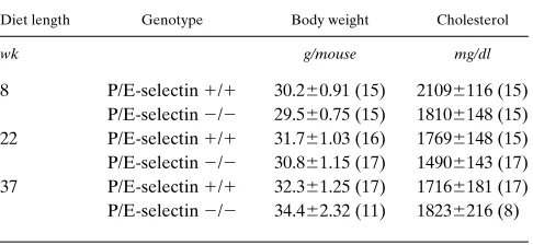
Generation of mice with combined P- and E-selectin and LDLR deficiencies. To study the role of P- and E-selectins in atherosclerotic lesion development, we bred P/E2/2 mice with LDLR2/2 mice to generate LDLR2/2 P/E1/1 and LDLR2/2 P/E2/2 mice. Like the P/E2/2 mice that present some degree of immunodeficiency (7), LDLR2/2 P/E2/2 mice suffered from a cutaneous infection starting at z 16 wk of age; this occurred at a higher percentage in the female mice. To rule out infection as a factor affecting atherosclerotic lesion development in LDLR2/2 P/E2/2 mice, in the long-term ex-periments (diet for 22 or 37 wk) we only collected data from male mice without skin infections. LDLR2/2 P/E1/1 and LDLR2/2 P/E2/2 male mice responded similarly to the atherogenic diet, as demonstrated by similar body weight and comparable levels of cholesterol in the blood at all three obser-vation times (Table I). Because most older female LDLR2/2 P/E2/2 mice developed skin infections, we were only able to collect data from females on the diet for 8 wk; again, there were no statistically significant differences between the two genotypes in body weight and total plasma cholesterol (data not shown).
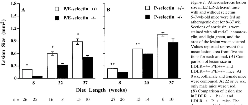
[image:3.612.315.558.561.673.2]P- and E-selectin deficiency inhibits formation of atheroscle-rotic lesions in the proximal aorta. The proximal aorta is par-ticularly prone to the development of intimal lesions in mouse models of atherosclerosis, and the extent of lesions in the prox-imal aorta has been shown to correlate with that of the entire aorta in LDLR2/2 mice (16). To examine the effect of P- and E-selectin deficiency on the development of atherosclerotic le-sions in the proximal aorta, mean lesion areas were measured in oil red-O–stained tissue sections from LDLR2/2 P/E1/1 and LDLR2/2 P/E2/2 mice on atherogenic diet for 8–37 wk. Previously, we reported that, after 8 wk on the diet, the lesion areas in LDLR2/2 P2/2 male mice are twofold smaller than those in LDLR2/2 P1/1 male mice, but the lesion areas in females are similar for both genotypes (11). Surprisingly, after
Table I. Body Weight and Total Plasma Cholesterol in LDLR-deficient Male Mice on Atherogenic Diet
Diet length Genotype Body weight Cholesterol
wk g/mouse mg/dl
8 P/E-selectin 1/1 30.260.91 (15) 21096116 (15) P/E-selectin 2/2 29.560.75 (15) 18106148 (15) 22 P/E-selectin 1/1 31.761.03 (16) 17696148 (15) P/E-selectin 2/2 30.861.15 (17) 14906143 (17) 37 P/E-selectin 1/1 32.361.25 (17) 17166181 (17) P/E-selectin 2/2 34.462.32 (11) 18236216 (8)
8 wk on the diet, LDLR2/2 P/E2/2 male mice had lesions 5.43 smaller than the LDLR2/2 P/E1/1 males (P , 0.0001), and LDLR2/2 P/E2/2 female mice had lesions 43 smaller than the LDLR2/2 P/E1/1 females (P , 0.0001). The com-bined data from males and females are shown in Fig. 1 A. In-terestingly, lesions in LDLR2/2 P/E2/2 mice were 3.33 smaller than those in the LDLR2/2 P2/2 mice. In addition, after 8 wk on the diet, 78% of the aortic sinus circumference in LDLR2/2 P/E1/1 mice was covered by oil red-O–stained positive lesions, as compared to 57% found in LDLR2/2 P/E2/2 mice (P , 0.0001, n 5 25–26, data not shown). There-fore, LDLR2/2 P/E2/2 mice had more lesion-free aortic si-nus surface as well as smaller lesions than did LDLR2/2 P/E1/1 mice.
With time on the atherogenic diet, lesions grew in both LDLR2/2 P/E1/1 and LDLR2/2 P/E2/2 mice (Fig. 1 A). Previously, we have shown that, after 20 wk on the diet, a slightly smaller lesion was observed in LDLR2/2 P2/2 mice as compared to LDLR2/2 P1/1 mice, and at 37 wk the lesion size difference was no longer significant (Fig. 1 B). However, after either 22 or 37 wk on the diet, lesion size in LDLR2/2 P/E2/2 mice was significantly smaller than that in LDLR2/2 P/E1/1 mice (Fig. 1 A). Actually, LDLR2/2 P/E2/2 mice on diet for 37 wk had a comparable lesion size to LDLR2/2 P/E1/1 mice on diet for 22 wk (Fig. 1 A), indicating that P/E-selectin deficiency could delay lesion growth in LDLR2/2 mice by z 15 wk, even at late stages of lesion development. Unlike at 8 wk, after 22 wk, the percentage of lesion coverage on the aortic sinus circumference was similar in both LDLR2/2 P/E1/1 and LDLR2/2 P/E2/2 mice (z 90–95%, data not shown).
P- and E-selectin deficiency delays atherosclerotic lesion de-velopment throughout the entire aorta. The extent of athero-sclerosis was also determined in a defined segment of the aorta, from the subclavian branch to the iliac bifurcation. Al-though, after 8 wk on the atherogenic diet, we found no
obvi-ous lesions in this segment in either mobvi-ouse genotype, lesions were numerous after 22 wk on the diet. Comparison of the per-centage of surface area occupied by the lesions in mice on the diet for 22 or 37 wk showed that the lesions were signifi-cantly more extensive in LDLR2/2 P/E1/1 mice relative to LDLR2/2 P/E2/2 mice (Fig. 2). Consistent with the findings in the proximal aorta, after 22 wk on the diet, the percentage of coverage in LDLR2/2 P/E1/1 mice was two times more than that of LDLR2/2 P/E2/2 mice. Similarly, LDLR2/2 P/E2/2 mice on diet for 37 wk had a comparable percentage lesion coverage of the entire aorta to LDLR2/2 P/E1/1 mice on diet for only 22 wk (Fig. 2). Our results show that absence of P- and E-selectins in LDLR2/2 mice reduced diet-induced atherosclerotic lesion progression in both the aortic sinus and the entire aorta.
[image:4.612.57.556.59.272.2]rophages (Fig. 3, E and F) and only 1–2% were CD3-positive T cells (data not shown). The distributions of these cells were approximately uniform throughout 8-wk fatty streak lesions. By counting the cell nuclei per unit of lesion area, we found that there was no difference in cell density between the two genotypes of mice (2,6106125/mm2 in LDLR2/2 P/E1/1
mice versus 2,6406198/mm2 in LDLR2/2 P/E2/2 mice, P .
0.05, n 5 25–26). However, because lesions were smaller in the LDLR2/2 P/E2/2 mice, total cell numbers per section in these mice were significantly lower than those in the LDLR2/2 P/E1/1 mice (125623 in LDLR2/2 P/E2/2 mice versus 577655 in LDLR2/2 P/E1/1 mice, P , 0.001, n 5 25–26). Furthermore, the percentage of cells that express TNF-a on 8-wk lesions was similar in both genotypes of mice (z 90%, Fig. 3, G and H). We also found that there were no differences in percent lesion area positive for a-actin staining in both geno-types of mice after 22 or 37 wk on diet (Table II). Our results indicate that smooth muscle cell growth in the fibrous plaque stage of lesions of LDLR2/2 P/E2/2 mice still reached a nor-mal proportion despite their snor-maller lesion size as compared to LDLR2/2 P/E1/1 mice. However, after 37 wk on the diet, fi-brous plaque lesions in most of the LDLR2/2 P/E1/1 mice had calcified; whereas calcification was rare in the lesions from LDLR2/2 P/E2/2 mice (Table II; Fig. 4, C and D). There-fore, the absence of P- and E-selectin in LDLR2/2 mice did delay the formation of some components of atherosclerotic le-sions, such as calcification. In addition, a lower incidence of cardiac lipofuscin was observed in LDLR2/2 P/E2/2 mice lesions throughout the experimental period (Table II; Fig. 3, C and D).
Discussion
Pathogenesis of atherosclerosis is a complex process. A variety of cells, growth factors, cytokines, and vasoregulatory mole-cules participate in this disease (1). Here we present data showing that the endothelial selectins (P- and E-selectins), to-Figure 2. Atherosclerotic lesions in entire aorta of LDLR-deficient
[image:5.612.59.292.58.363.2]mice with and without P/E-selectins. 5–7-wk-old male mice were fed an atherogenic diet for 22 or 37 wk. Aortae were collected between the subclavian and ilial branches and stained with Sudan IV. Sudan IV–stained surface area was measured by an image analysis program. Percentage of area covered by lesion (% coverage) was calculated (Sudan IV–stained area divided by total aorta area 1003). Represen-tative specimens collected from both genotypes of mice on the diet for 22 (A) and 37 wk (B) are shown.
[image:5.612.57.394.483.736.2]gether, play a critical role in both early and late stages of ath-erosclerotic lesion development. The finding is based on the following observations. First, the size of the fatty streaks formed in the aortic sinus of LDLR-deficient mice on a high fat/cholesterol diet (atherogenic diet) for 8 wk was two times smaller in absence of P-selectin, but was five times smaller in the absence of both P- and E-selectins. Second, the size of the fibrous plaque formed in the aortic sinus of LDLR-deficient mice on an atherogenic diet for 37 wk was identical in the
[image:6.612.59.402.58.597.2]pres-ence and abspres-ence of P-selectin, but was significantly reduced by 40% when both selectins were absent (Fig. 1). Similarly, the area occupied by atherosclerotic lesions in the entire aorta was comparable for LDLR2/2 P1/1 and LDLR2/2 P2/2 mice on the diet for 37 wk (11) but was decreased in LDLR2/2 P/E2/2 mice (Fig. 2). In addition, in a preliminary study, we have found that after 8 wk on an atherogenic diet, the size of the fatty streaks formed in the aortic sinus of LDLR-deficient mice without E-selectin (LDLR2/2 E2/2) was not smaller
than that in LDLR2/2 E1/1 mice (data not shown). This suggests that a deficiency of E-selectin alone does not have a major protective effect on atherosclerotic lesion development. Multiple studies have indicated that the major function of selectins is to mediate neutrophil, monocyte, lymphocyte, and platelet rolling along the venular wall and their effects in regu-lating inflammatory processes seem to be overlapping (4, 17). For example, mice lacking E-selectin display no obvious changes in the trafficking of leukocytes. However, treatment of the E-selectin–deficient animals with an anti–P-selectin an-tibody significantly inhibits leukocyte emigration, whereas the same treatment has no effect in wild-type mice (6). Mice lack-ing P-selectin present a complete absence of leukocyte rolllack-ing in exteriorized venules, whereas both P- and E-selectins have to be absent to abolish rolling under inflammatory condi-tions (7). Recently, it has been found that the absence of either P-selectin or E-selectin alone had no notable effect on skin wound healing, but mice deficient in both P- and E-selectins had markedly reduced recruitment of inflammatory cells and impaired closure of the wound (18). Our present results extend these studies and further confirm the importance of the inflam-matory component in atherosclerosis.
Decreased atherosclerotic lesion size in LDLR2/2 P/E2/2 mice is mainly due to a reduction in monocyte infiltration into the intima. The evidence is that, after 8 wk on the atherogenic diet, the density of macrophages in the fatty streaks was com-parable between LDLR2/2 P/E1/1 and LDLR2/2 P/E2/2 mice, but total cell numbers accumulated in the aortic sinus were five times less in LDLR2/2 P/E2/2 mice. It is well known that monocyte adhesion to the endothelium and migra-tion into the intima is a crucial early event in development of atherosclerosis, and that migration continues in late stages of lesions (1). Macrophages in the subendothelium may protect against atherosclerosis by scavenging noxious materials, such as oxidized lipoproteins, but it is also hypothesized that they contribute to oxidized LDL formation and to the fibroprolifer-ative process through secretion of numerous growth factors (1, 19). P- and E-selectin deficiency leads to a reduction in both
early and late stages of atherosclerotic lesion development, in-dicating that under conditions that are optimal for lesion de-velopment, the net effect of the macrophage recruitment is proatherogenic. The lipofuscin data suggest that decreased macrophage infiltration into lesions in the absence of both se-lectins may lead to a reduction in oxidized LDL production; the lipofuscin pigment consists of terminally oxidized polyun-saturated lipid and protein complexes, and there are common pathways contributing to oxidized LDL, lipofuscin, and fatty streak development (14). Decreased oxidized LDL in lesions may result in reduction in lesion progression because oxidized LDL is known to stimulate expression of adhesion molecules on endothelium leading to additional cellular infiltration and is the main source of lipid in foam cells (1). Our results do not clarify why monocytes but not neutrophils are recruited specif-ically into atherosclerotic lesions. It does not appear that P/E-selectins provide the necessary clues. The absence of endothe-lial selectins reduces recruitment of neutrophils to inflamed peritoneum to similar extent (7), as it diminishes accumulation of monocytes in the fatty streaks. The adhesion receptors, or more likely, the chemokines and their receptors that direct the specific extravasation of monocytes into the atherosclerotic le-sions are still elusive.
In vitro studies suggest that the selectins also affect mono-cyte function, especially in the production of proinflammatory cytokines such as TNF-a (20). So it is possible that absence of both selectins prevents lesion progression through modifica-tion of macrophage funcmodifica-tion in producmodifica-tion of cytokines. In this study, we have found that most macrophages in atherosclerotic lesions of LDLR2/2 P/E2/2 mice are still able to produce TNF-a (Fig. 3, G and H). However, considering that the im-munostaining method for detection of TNF-a used in this study is not quantitative, we still cannot exclude the possibility that individual macrophage in lesions of LDLR2/2 P/E2/2 mice may produce lower amount of TNF-a, as compared to the macrophages from LDLR2/2 P/E1/1 lesions. Furthermore, the fatty streaks in LDLR2/2 P/E2/2 mice also progress to fibrous plaque stage and their fibrous plaque contains a similar ratio of smooth muscle cells to other lesion components, as seen in LDLR2/2 P/E1/1 mice (Fig. 4, A and B; Table II). This suggests that macrophages in the lesions of LDLR2/2 P/E2/2 mice likely retain the ability to induce smooth muscle cell proliferation by producing growth factors such as PDGF. Therefore, we suspect that the absence of the selectins did not greatly modify macrophage function in the atherosclerotic le-sions.
[image:7.612.57.298.94.221.2]In terms of a mouse model for atherosclerosis, it is interest-ing to see that calcifications can occur in the fibrous plaque le-sion in most LDLR2/2 mice, even though complicated lesions characterized by thrombosis have not yet been found. Calcifi-cation of atherosclerotic lesions has been shown to contribute to atherosclerotic plaque instability and myocardial infarction in humans (19). Therefore, the finding that calcification of ath-erosclerotic lesions in LDLR2/2 mice could be delayed in the absence of both P- and E-selectins may have important impli-cations for designing new methods to prevent plaque rupture. The mechanism(s) responsible for decreased calcification in LDLR2/2 P/E2/2 mice is not clear. We suspect that it is due to a smaller lesion size in the LDLR2/2 P/E2/2 mice, as presence of calcifications seems to correlate with lesion size (Table II).
Atherosclerotic lesions still form in LDLR2/2 P/E2/2 Table II. Lipofuscin, Smooth Muscle Cells, and Calcification
in Aortic Sinus Lesions of LDLR-deficient Mice on Atherogenic Diet
Diet length
(wk) Genotype
Incidence of lipofuscin*
Lesion area positive for
a-actin
Incidence of calcification*
%
8 P/E-selectin 1/1 15/24 (63%) 0 0/24 (0%) P/E-selectin 2/2 0/25 (0%)‡ 0 0/25 (0%) 22 P/E-selectin 1/1 14/16 (88%) 2061.4 3/16 (20%)
P/E-selectin 2/2 5/16 (31%)‡ 2361.5 1/17 (6%)‡ 37 P/E-selectin 1/1 11/15 (73%) 3168.8 14/15 (93%) P/E-selectin 2/2 3/10 (30%)‡ 3066.1 2/10 (20%)‡
mice, suggesting that other adhesion molecules such as vascu-lar cell adhesion molecule–1 (VCAM-1) and L-selectin may be mediating monocyte rolling, leading to the development of atherosclerosis in these animals. VCAM-1 has been found to be expressed on endothelium before macrophage accumula-tion in dietary hypercholesterolemic and Watanabe heritable hyperlipidemic rabbit models of atherosclerosis (21–22). This protein on the surface of endothelial cells interacts with the b1 integrin very late antigen–4, which is expressed mainly by mononuclear leukocytes (23). VCAM-1/very late antigen–4 pathway has been shown to mediate both rolling and firm adhesion in leukocyte transendothelial migration (24–25). L-selectin is constitutively expressed on the surface of most leukocytes. It has been shown that L-selectin mediates mono-cyte attachment to activated aortic endothelium (26). Because mice deficient in all three selectins (P-, E-, and L-selectins) have been recently generated (27), their combined role in ath-erosclerosis could be investigated.
Accumulated evidence, including our present study, sup-ports the notion that monocyte-derived macrophages play a key role in atherogenesis (1, 28–29). Finding means of control-ling macrophage participation in atherogenesis could be criti-cal in modifying lesion progression. There are many potential sites that could be targeted. These include monocyte transen-dothelial migration, where both P- and E-selectins are possible target molecules as suggested by this study, as well as the pro-cesses of monocyte differentiation into macrophage, and mac-rophage uptake of modified lipoprotein particles. At present, it is unclear which site(s) should be targeted to get better re-sults for inhibiting atherosclerotic lesion progression with fewer adverse effects. The effect of blocking monocyte differ-entiation into macrophage on the development of atheroscle-rosis was analyzed using mice with the op mutation (28). The mutation affects the gene-encoding macrophage colony–stim-ulating factor and leads to monocytopenia and decreased tis-sue macrophages. Atherosclerotic lesions in ApoE-deficient mice with op mutation were one-seventh the size of lesions in ApoE-deficient mice and did not progress to the fibroprolifer-ative stage. But these double-mutant mice have two to three times more cholesterol in their blood than do the ApoE-defi-cient mice (28). In contrast, mice lacking the endothelial selec-tins have increased levels of blood monocytes (7) and their cholesterol levels are not affected by the mutation (Table I). The handicap of a long-term treatment inhibiting the selectins is that it might render the patients more susceptible to infec-tions (7). However, it is encouraging that spontaneous skin in-fection occuring in P/E2/2 mice can be prevented with oral antibiotics (7). In addition to selectins, inhibition of the inter-cellular adhesion molecule–1/b2 integrin pathway of adhesion may also protect against atherosclerosis. C57BL/6 mice mutant in the intercellular adhesion molecule–1 or b2 integrin gene
de-velop 50–75% smaller fatty streaks, when fed atherogenic diet (30). However, a long-term treatment inhibiting these adhe-sion receptors may cause obesity in the patients (31).
Both P- and E-selectins have been found on endothelial surfaces overlying atherosclerotic lesions in humans (32–33). Recently, E-selectin polymorphism was reported to be associ-ated with human atherosclerosis (34). The resulting alteration enhances binding capacity of E-selectin to its ligands (35), and individuals possessing this polymorphism appear to have a higher risk for early severe atherosclerosis (34). These results, combined with our present study, lead us to suspect that the
endothelial selectins may also play an important role in human atherosclerosis.
Acknowledgments
We are grateful to Lesley Cowan for help with preparation of the manuscript.
The work was supported by National Institutes of Health grants HL-53756 (D.D. Wagner) and P01-HL-41484 (R.O. Hynes). Z.M. Dong was supported by a NIH fellowship grant HL09869. S.M. Chap-man and A.A. Brown were supported by a grant from Genetics Insti-tute. R.O. Hynes is an investigator of the Howard Hughes Medical Institute. P.S. Frenette is a fellow of the Medical Research Council of Canada. The care of the experimental mice was in accordance with the guidelines of the Center for Blood Research.
References
1. Ross, R. 1993. The pathogenesis of atherosclerosis: a perspective for the 1990s. Nature. 362:801–809.
2. Bevilacqua, M.P., R.M. Nelson, G. Mannori, and O. Cecconi. 1994. En-dothelial-leukocyte adhesion molecules in human disease. Annu. Rev. Med. 45: 361–378.
3. Carlos, T.M., and J.M. Harlan. 1994. Leukocyte-endothelial adhesion molecules. Blood. 84:2068–2101.
4. Frenette, P.S., and D.D. Wagner. 1997. Insights into selectin function from knockout mice. Thromb. Haemostasis. 78:60–64.
5. Mayadas, T.N., R.C. Johnson, H. Rayburn, R.O. Hynes, and D.D. Wag-ner. 1993. Leukocyte rolling and extravasation are severely compromised in P-selectin-deficient mice. Cell. 74:541–554.
6. Labow, M.A., C.R. Norton, J.M. Rumberger, K.M. Lombard-Gillooly, D.J. Shuster, J. Hubbard, R. Bertko, P.A. Knaack, R.W. Terry, M.L. Harbison et al. 1994. Characterization of E-selectin-deficient mice: demonstration of overlapping function of the endothelial selectins. Immunity. 1:709–720.
7. Frenette, P.S., T.N. Mayadas, H. Rayburn, R.O. Hynes, and D.D. Wag-ner. 1996. Susceptibility to infection and altered hematopoiesis in mice deficient in both P- and E-selectins. Cell. 84:563–574.
8. Bullard, D.C., E.J. Kunkel, H. Kubo, M.J. Hicks, I. Lorenzo, N.A. Doyle, C.M. Doerschuk, K. Ley, and A.L. Beaudet. 1996. Infectious susceptibility and severe deficiency of leukocyte rolling and recruitment in E-selectin and P-selec-tin double mutant mice. J. Exp. Med. 183:2329–2336.
9. Staite, N.D., J.M. Justen, L.M. Sly, A.L. Beaudet, and D.C. Bullard. 1996. Inhibition of delayed-type contact hypersensitivity in mice deficient in both E-selectin and P-selectin. Blood. 88:2973–2979.
10. Ishibashi, S., M.S. Brown, J.L. Goldstein, R.D. Gerard, R.E. Hammer, and J. Herz. 1993. Hypercholesterolemia in low density lipoprotein receptor knockout mice and its reversal by adenovirus-mediated gene delivery. J. Clin.
Invest. 92:883–893.
11. Johnson, R.C., S.M. Chapman, Z.M. Dong, J.M. Ordovas, T.N. Maya-das, J. Herz, R.O. Hynes, E.J. Schaefer, and D.D. Wagner. 1997. Absence of P-selectin delays fatty streak formation in mice. J. Clin. Invest. 99:1037–1043.
12. Shimada, M., S. Ishibashi, T. Inaba, H. Yagyu, K. Harada, J.I. Osuga, K. Ohashi, Y. Yazaki, and N. Yamada. 1996. Suppression of diet-induced athero-sclerosis in low density lipoprotein receptor knockout mice overexpressing lipo-protein lipase. Proc. Natl. Acad. Sci. USA. 93:7242–7246.
13. Qiao, J.H., P.Z. Xie, M.C. Fishbein, J. Kreuzer, T.A. Drake, L.L. De-mer, and A.J. Lusis. 1994. Pathology of atheromatous lesions in inbred and ge-netically engineered mice: genetic determination of arterial calcification.
Arte-rioscler. Thromb. 14:1480–1497.
14. Qiao, J.H., C.L. Welch, P.Z. Xie, M.C. Fishbein, and A.J. Lusis. 1993. Involvement of the tyrosinase gene in the deposition of cardiac lipofuscin in mice: association with aortic fatty streak development. J. Clin. Invest. 92:2386– 2393.
15. Litton, M.J., B. Sander, E. Murphy, A. O’Garra, and J.S. Abrams. 1994. Early expression of cytokines in lymph nodes after treatment in vivo with
Staph-ylococcus enterotoxin B. J. Immunol. Methods. 175:47–58.
16. Tangirala, R.K., E.R. Rubin, and W. Palinski. 1995. Quantitation of ath-erosclerosis in murine models: correlation between lesions in the aortic origin and in the entire aorta, and differences in the extent of lesions between sexes in LDL receptor-deficient and apolipoprotein E-deficient mice. J. Lipid. Res. 36: 2320–2328.
17. Frenette, P.S., R.C. Johnson, R.O. Hynes, and D.D. Wagner. 1995. Platelets roll on stimulated endothelium in vivo: an interaction mediated by en-dothelial P-selectin. Proc. Natl. Acad. Sci. USA. 92:7450–7454.
19. Berliner, J.A., M. Navab, A.M. Fogelman, J.S. Frank, L.L. Demer, P.A. Edwards, A.D. Watson, and A.J. Lusis. 1995. Atherosclerosis: basic mecha-nisms. Circulation. 91:2488–2496.
20. Weyrich, A.S., T.M. McIntyre, R.P. McEver, S.M. Prescott, and G.A. Zimmerman. 1995. Monocyte tethering by P-selectin regulates monocyte chemotactic protein-1 and tumor necrosis factor-a secretion, signal integration and NF-kB translocation. J. Clin. Invest. 95:2297–2303.
21. Cybulsky, M.I., and M.A. Gimbrone, Jr. 1991. Endothelial expression of a mononuclear leukocyte adhesion molecule during atherosclerosis. Science. 251:788–791.
22. Li, H., M.I. Cybulsky, M.A. Gimbrone, Jr., and P. Libby. 1993. An atherogenic diet rapidly induces VCAM-1, a cytokine-regulatable mononuclear leukocyte adhesion molecules, in rabbit aortic endothelium. Arterioscler.
Thromb. 13:197–204.
23. Elices, M.J., L. Osborn, Y. Takada, C. Crouse, S. Luhowskyj, M.E. Hemler, and R.R. Lobb. 1990. VCAM-1 on activated endothelium interacts with the leukocyte integrin VLA-4 at a site distinct from the VLA-4/fibronectin binding site. Cell. 60:577–584.
24. Berlin, C., R.F. Bargatze, J.J. Campbell, U.H. von Andrian, M.C. Szabo, S.R. Hasslen, R.D. Nelson, E.L. Berg, S.L. Erlandsen, and E.C. Butcher. 1995. a4 integrins mediate lymphocyte attachment and rolling under physiologic flow. Cell. 80:413–422.
25. Alon, R., P.D. Kassner, M.W. Carr, E.B. Finger, M.E. Hemler, and T.A. Springer. 1995. The integrin VLA-4 supports tethering and rolling in flow on VCAM-1. J. Cell Biol. 128:1243–1253.
26. Giuffre, L., A.S. Cordey, N. Monai, Y. Tardy, M. Schapira, and O. Sper-tini. 1997. Monocyte adhesion to activated aortic endothelium: role of L-selectin and heparan sulfate proteoglycans. J. Cell Biol. 136:945–956.
27. Robinson, S.D., P.S. Frenette, H. Rayburn, M. Cummiskey, M. Ullman-Cullere, D.D. Wagner, and R.O. Hynes. 1997. The generation of double and tri-ple-deficient selectin mice. Microcirculation. 4:64a. (Abstr.).
28. Smith, J.D., E. Trogan, M. Ginsberg, C. Grigaux, J. Tian, and M. Miyata. 1995. Decreased atherosclerosis in mice deficient in both macrophage colony-stimulating factor (op) and apolipoprotein E. Proc. Natl. Acad. Sci.
USA. 92:8264–8268.
29. Breslow, J.J. 1996. Mouse models of atherosclerosis. Science. 272:685–688. 30. Nageh, M.F., E.T. Sandberg, K.R. Marotti, A.H. Lin, E.P. Melchior, D.C. Bullard, and A.L. Beaudet. 1997. Deficiency of inflammatory cell adhe-sion molecules protects against atherosclerosis in mice. Arterioscler. Thromb.
Vasc. Biol. 17:1517–1520.
31. Dong, Z.M., J.C. Gutierrez-Ramos, A. Coxon, T.N. Mayadas, and D.D. Wagner. 1997. A new class of obesity genes encodes leukocyte adhesion recep-tors. Proc. Natl. Acad. Sci. USA. 94:7526–7530.
32. Van der Wal, A.C., P.K. Das, and A.J. Tigges. 1992. Adhesion mocules on the endothelium and mononuclear cells in human atherosclerotic le-sions. Am. J. Pathol. 141:1427–1433.
33. Johnson-Tidey, R.R., J.L. McGregor, P.R. Taylor, and R.N. Poston. 1994. Increase in the adhesion molecule P-selectin in endothelium overlying atherosclerotic plaques. Am. J. Pathol. 144:952–961.
34. Wenzel, K., S. Felix, F.X. Kleber, R. Brachold, T. Menke, S. Schattke, K.L. Schulte, C. Glaser, K. Rohde, G. Baumann, and A. Speer. 1994. E-selectin polymorphism and atherosclerosis: an association study. Hum. Mol. Genet. 3: 1935–1937.