link in four Fanconi anemia complementation
groups.
Y Li, H Youssoufian
J Clin Invest.
1997;
100(11)
:2873-2880.
https://doi.org/10.1172/JCI119836
.
Fanconi anemia (FA) consists of a group of at least five autosomal recessive disorders that
share both clinical (e.g., birth defects and hematopoietic failure) and cellular (e.g., sensitivity
to cross-linking agents and predisposition to apoptosis) features with each other. However,
a common pathogenetic link among these groups has not been established. To identify
genetic pathways that are altered in FA and characterize shared molecular defects, we used
mRNA differential display to isolate genes that have altered expression patterns in FA cells.
Here, we report that the expression of an interferon-inducible gene, MxA, is highly
upregulated in cells of FA complementation groups A, B, C, and D, but it is suppressed in
FA group C cells complemented with wild-type FAC cDNA as well as in non-FA cells. A
posttranscriptional mechanism rather than transcriptional induction appears to account for
MxA overexpression. Forced expression of MxA in Hep3B cells enhances their sensitivity to
mitomycin C and induces apoptosis, similar to the FA phenotype. Thus, MxA is a
downstream target of FAC and is the first genetic marker to be identified among multiple FA
complementation groups. These data suggest that FA subtypes converge onto a final
common pathway, which is intimately related to the interferon signaling mechanism.
Constitutive activity of this pathway may explain a number of the phenotypic features of FA,
particularly the […]
Research Article
Find the latest version:
J. Clin. Invest.
© The American Society for Clinical Investigation, Inc. 0021-9738/97/12/2873/08 $2.00
Volume 100, Number 11, December 1997, 2873–2880 http://www.jci.org
MxA Overexpression Reveals a Common Genetic Link in Four Fanconi Anemia
Complementation Groups
Youlin Li and Hagop Youssoufian
Department of Medicine, Hematology-Oncology Division, Brigham and Women’s Hospital, Harvard Medical School, Boston, Massachusetts 02115
Abstract
Fanconi anemia (FA) consists of a group of at least five au-tosomal recessive disorders that share both clinical (e.g., birth defects and hematopoietic failure) and cellular (e.g., sensitivity to cross-linking agents and predisposition to apop-tosis) features with each other. However, a common patho-genetic link among these groups has not been established. To identify genetic pathways that are altered in FA and characterize shared molecular defects, we used mRNA dif-ferential display to isolate genes that have altered expres-sion patterns in FA cells. Here, we report that the expresexpres-sion
of an interferon-inducible gene, MxA, is highly upregulated
in cells of FA complementation groups A, B, C, and D, but it is suppressed in FA group C cells complemented with
wild-type FAC cDNA as well as in non-FA cells. A
post-transcriptional mechanism rather than post-transcriptional in-duction appears to account for MxA overexpression. Forced expression of MxA in Hep3B cells enhances their sensitivity to mitomycin C and induces apoptosis, similar to the FA phenotype. Thus, MxA is a downstream target of FAC and is the first genetic marker to be identified among multiple FA complementation groups. These data suggest that FA subtypes converge onto a final common pathway, which is intimately related to the interferon signaling mechanism. Constitutive activity of this pathway may explain a number of the phenotypic features of FA, particularly the pathogenesis
of bone marrow failure. (J. Clin. Invest. 1997. 100:2873–
2880.) Key words: apoptosis • cross-linker • differential
dis-play • gene regulation • interferon
Introduction
Phenotypic manifestations of Fanconi anemia (FA)1 include
skeletal and visceral malformations, predisposition to cancer, and progression to bone marrow failure and acute leukemia (1–3). A pathognomonic finding in FA cells is the presence of
spontaneous chromosomal gaps and breaks and their accentu-ation by bifunctional alkylating agents (cross-linkers), such as mitomycin C (MMC). Somatic cell hybrid studies have identi-fied at least five distinct complementation groups of FA (4–6), and the genes mutant in groups A, C, and D have been mapped to chromosomes 16q, 9q, and 3p, respectively (5, 7, 8). Furthermore, the genes for complementation groups A (FAA; reference 9) and C (FAC; references 10 and 11) have been cloned, and despite their ability to suppress MMC cytotoxicity in cells of appropriate FA complementation groups, these genes and their protein products share little sequence homol-ogy with each other or with any other known genes or pro-teins.
We and others have been investigating the subcellular loca-tion and biochemical interacloca-tions of FA gene products in or-der to unor-derstand their function. FAC is a 63-kD cytoplasmic protein (12, 13) that is thought to function as a biological sen-sor and prevent or attenuate DNA damage (14). FAC has been shown to interact with at least three cytoplasmic proteins in vitro (15), suggesting that it is a component of a multimeric complex. One of the interacting proteins is NADPH cyto-chrome P-450 reductase, a microsomal enzyme involved in xe-nobiotic metabolism (Youssoufian, H., T. Hoshino, J.M. Liu, P. Joseph, and A.K. Jaiswal, manuscript submitted for publica-tion); FAC suppresses the catalytic activity of this reductase. By contrast, much less is known about the localization and function of FAA. It is considerably larger than FAC with a predicted molecular mass of 163 kD and contains both a puta-tive nuclear localization signal and an imperfect leucine zipper. Thus, FAA may function within the nucleus in a manner dis-tinct from FAC.
To understand the molecular pathogenesis of FA in the context of its genetic heterogeneity, we used mRNA differen-tial display analysis (16) to characterize genetic alterations in FA cells. Here we report that the expression of an IFN-induc-ible gene, MxA (17–19), is highly upregulated in cells of multi-ple FA commulti-plementation groups. In addition, wild-type FAC can suppress the expression of MxA in lymphoblastoid cells from FA complementation group C (FA-C) and restore resis-tance to MMC. MxA is a GTPase (20) that belongs to the Mx family of proteins, which are ubiquitous in eukaryotes (21–25) and mediate a number of fundamental cellular processes, in-cluding resistance to certain viral infections in mice and hu-mans (26–30) and vacuolar sorting in Saccharomyces cerevisiae
(25). Our results suggest that the activities of at least four dis-tinct FA genes converge onto a common genetic pathway, which is intimately related to IFN signaling and susceptibility to apoptosis.
H. Youssoufian’s present address is Department of Molecular and Human Genetics, Baylor College of Medicine, Houston, Texas 77030. Address correspondence to Hagop Youssoufian, M.D., Depart-ment of Molecular and Human Genetics, Baylor College of Medi-cine, One Baylor Plaza, Houston, TX 77030. Phone: 713-798-8311; FAX: 713-798-5386.
Received for publication 24 June 1997 and accepted in revised form 26 September 1997.
Methods
Cell culture. Epstein-Barr virus-transformed human lymphoblastoid cell lines were maintained in RPMI 1640 (GIBCO BRL, Gaithers-burg, MD) and 15% FBS. In addition, stably transfected FA-C lym-phoblastoid cells HSC536-Vect (transfected with the empty DRA-CD vector) and HSC536-FAC (transfected with DRA-FAC) described previously (15) were supplemented with 200 mg/ml hygromycin B (Boehringer Mannheim Biochemicals, Indianapolis, IN). Other FA cell lines used were HSC99 (FA-A; reference 5), HSC230 (FA-B; ref-erence 5), RA568 (FA-C; refref-erence 15), and HSC230 and PD-20 (FA-D; references 5 and 8). Non-FA hematopoietic cells were grown in RPMI 1640–10% FBS. COS-1 and Hep3B cells were grown in Dul-becco’s modified essential medium (DMEM) and 10% FBS. Primary human fibroblasts (GM02149; American Tissue Type Collection, Rockville, MD) were grown in DMEM–20% FBS and supplemented with vitamins as well as essential and nonessential amino acids. All cells were cultured at 378C in a humidified atmosphere containing 5% CO2.
Expression plasmids. Full-length human FAC cDNA cloned into the mammalian episomal expression vector DRA-CD has been de-scribed previously (15). p78/2-8B containing the full-length coding se-quence of MxA (19) was obtained from the American Tissue Type Collection. Wild-type FAC, a mutant FAC allele (L554P), and full-length MxA were also cloned into pcDNA3 (Invitrogen Corp., San Diego, CA) for expression in COS-1 and Hep3B cells. pXP2-MxA809 was constructed by inserting a PCR-amplified 809-bp genomic frag-ment of the MxA promoter (19) into the KpnI and BglII sites of the promoterless luciferase reporter plasmid pXP2 (gift of Dr. D. Tenen, Beth Israel Hospital, Boston, MA).
Antibodies. The generation of anti-FAC antiserum has been de-scribed previously (13–15). An aliquot of goat polyclonal anti-human MxA antibodies was kindly provided by Dr. M.A. Horisberger (Ciba-Geigy Ltd., Basel, Switzerland) and MxA rabbit polyclonal anti-bodies were subsequently generated by us against a 30-kD histidine-tagged fusion protein (MxA amino acids 241–499). Other antisera used in this study were obtained from commercial sources and in-cluded anti-human STAT1 p84/p91, STAT2 p113, and STAT3 p92 (Santa Cruz Biotechnology Inc., Santa Cruz, CA), anti-human IP10 (PeproTech, Inc., Rocky Hill, NJ), anti-human–b2-microglobulin and anti-human HLA class I antigen (Sigma Chemical Co., St. Louis, MO), and anti–b-tubulin (Boehringer Mannheim Biochemicals).
mRNA differential display. 0.5 mg of total RNA treated with RNase-free DNase I was reverse transcribed and the cDNAs were amplified by PCR using one base–anchored oligo-dT primers (Gen-Hunter Corporation, Nashville, TN; reference 16) in the presence of [a35S]dATP. The PCR products were resolved on 6%
urea-polyacryl-amide sequencing gels. Differentially expressed bands were cut from gels, reamplified for 30 cycles, cloned into the vector pCR2.1 (Invitro-gen Corp.), and sequenced. Reamplified cDNAs were also labeled with [a32P]dCTP by random-prime labeling (Boehringer Mannheim
Biochemicals) and used as hybridization probes in Northern analyses.
Northern analysis. Total RNA or poly A–selected mRNA iso-lated by previously described methods (31) was fractionated on 1% agarose gels containing formaldehyde, transferred to Biotrans nylon membranes (ICN Biomedicals, Inc., Irvine, CA), and fixed by baking at 808C for 30 min. Prehybridization and hybridization were per-formed according to the manufacturers’ suggestions in a buffer con-taining 50% formamide. Final washing was in 0.1 3SSC and 0.1% SDS at 558C for 30 min.
Western analysis. For Western (or immunoblotting) experiments, total cell lysates or immune complexes obtained by immunoprecipita-tion of cytosolic lysates were resolved by SDS-PAGE and transferred to polyvinyldifluoride membranes (DuPont-Merck Pharmaceutical Co., Wilmington, DE). After blocking, membranes were reacted se-quentially with a primary antibody followed by horseradish peroxi-dase–conjugated secondary antibodies (GIBCO BRL) and visualized by chemiluminescence (DuPont-Merck Pharmaceutical Co.).
Transfections. COS-1 cells were grown to z 50% confluence in
10-cm dishes and transfected transiently with 1 mg/ml plasmid DNA by the DEAE-dextran as described previously (13), while Hep3B cells were stably transfected using a calcium phosphate coprecipita-tion method. Stable Hep3B transfectants were selected in the pres-ence of G418 (400 mg/ml).
Nuclear run-on assay. Nuclei were isolated by sucrose gradient centrifugation, and in vitro transcription was performed as described (31). [a32P]UTP–labeled transcripts were isolated as described (32),
and 6.3 3 105 cpm/ml was used to probe identical strips of
Hybond-N1 (Amersham Life Sciences-USB, Arlington Heights, IL) filters containing 10 mg of linearized plasmid DNA. Hybridization and washing conditions were similar to the Northern blots.
Promoter analysis. Lysates from COS-1 cells transfected with promoter and reporter vectors were prepared using the Luciferase Assay System (Promega Corp., Madison, WI), and luciferase activity was measured with a luminometer. To control for the efficiency of transfection, 100-ml aliquots of lysates were also incubated with 3.5 mM O-nitrophenyl-b-D-galactopyranoside (Sigma Chemical Co.) at 378C for 30 min and the activity of b-galactosidase was assessed by light absorbance measured at 420 nm.
Determination of cell survival in response to MMC. Cell survival in response to MMC (Sigma Chemical Co.) was performed by incuba-tion of 1 3 104 viable cells with different concentrations of MMC for
6 d and counting of live cells by trypan blue exclusion. Data obtained from at least three independent experiments are shown.
Apoptosis assays. Two different assays were used for the detec-tion of apoptotic cells in Hep3B cells overexpressing MxA. First, cov-erslips containing transfected Hep3B cells were fixed with 3.5% paraformaldehyde for 30 min, and the terminal transferase-mediated UTP nick end-labeling (TUNEL) assay was performed using termi-nal transferase and fluorescein-conjugated dUTP (Boehringer Mann-heim Biochemicals). Apoptotic cells were visualized by fluorescence microscopy, and fluorescent cells in five different fields on each cov-erslips were counted. Second, genomic DNA enriched for small frag-ments was isolated from parental and MxA-expressing cells using a modification of the Hirt procedure. Briefly, Hep3B cells were washed with PBS containing Ca21/Mg21, detached by scraping, and resus-pended in a buffer containing 10 mM Tris-HCl (pH 8.0), 10 mM EDTA, 20 mg/ml RNase A, and 0.6% SDS. After 15 min at 378C, NaCl was added to a final concentration of 1.2 M and the mixture was precipitated overnight at 48C. After spinning in a microcentrifuge for 5 min, the supernatant was extracted with phenol and chloroform, precipitated with 2 vol of ethanol, resuspended in TE buffer, and re-solved on 2% agarose gel in Tris-acetate buffer.
Results
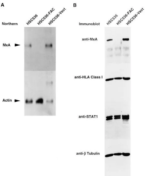
MxA gene is overexpressed in FA-C cells.Approximately 20% of the total cellular RNA from HSC536 and HSC536-FAC cells was analyzed by mRNA differential display. As demon-strated previously (15), electroporation with the episomal vec-tor DRA-FAC and stable expression of FAC in HSC536 cells restores their resistance to MMC. Thus, any differentially ex-pressed product may correlate with the cellular phenotype of MMC sensitivity. Using primers HT11 C and HAP12 (Gen-Hunter kit 2), we detected a 340-bp fragment that was uniquely expressed in HSC536 but not FAC-complemented cells (Fig. 1). Cloning of this fragment and partial sequencing showed a high degree of identity with nucleotides 2529–2784 of the human MxA cDNA (also called p78; reference 19). The differential expression of MxA mRNA from HSC536 was fur-ther investigated by Norfur-thern hybridization using a 32P-labeled
con-taining RNA from parental and vector-transfected HSC536 cells but not from FAC-transfected cells (Fig. 2 A). Six other differentially-displayed bands showed no consistent differ-ences in their expression patterns by Northern analysis and were not analyzed further (data not shown).
Altered expression of MxA but not other IFN-induced pro-teins. Consistent with the differential expression of mRNA, MxA protein expression was also detected in parental and vec-tor-transfected HSC536 cells, but not in FAC-transfected cells (Fig. 2 B). To assess the possibility that the expression of other IFN-inducible genes and STATs that transduce IFN signals are also altered in FA, we performed immunoblotting experi-ments with antibodies directed against HLA class I (Fig. 2 B) as well as b2-microglobulin and the g-IFN–inducible gene
IP-10 (data not shown). Both HLA class I antigen and b2
-micro-globulin were expressed in noncomplemented and comple-mented FA-C cells in approximately equal levels, while IP-10 expression was absent from both cell types. The expression of STAT1 (Fig. 2 B), STAT2, and STAT3 (data not shown) were similar in the three cell types. In addition, we also noted no
variations in the expression of a- and g-IFN or their receptors in HSC536 parental and FAC-complemented cells, as assessed by reverse-transcription and PCR (data not shown). There-fore, while we cannot exclude completely an altered expres-sion pattern for other genes involved in IFN-mediated signal-ing, our data demonstrate that the IFN-inducible pathway is not globally dysregulated in FA-C cells.
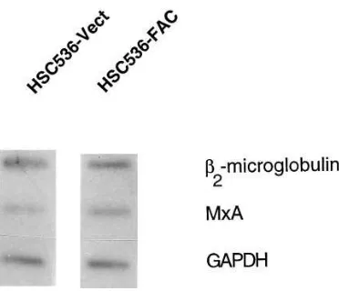
MxA is also overexpressed in FA groups A, B, and D. We also assessed the expression of MxA in a larger panel of FA and non-FA cells by Northern (Fig. 3 A) and Western (Fig. 3
B) analyses. MxA expression was upregulated in additional FA-C cells that contain different FAC gene mutations as well as in other well-defined FA cells that belong to complementa-tion groups A, B, and D. By contrast, we did not detect MxA overexpression in a number of non-FA cells, either hemato-poietic or nonhematohemato-poietic. Likewise, MxA was suppressed in the non-FA lymphoblastoid cell line LCL171 (15) and in Nalm6 cells (data not shown). These results confirm previous studies (33) that the expression of MxA is generally silent in most established tissue culture lines and primary cell types. Importantly, we demonstrate that MxA overexpression is a cellular phenotype that is shared by at least four different FA complementation groups.
Accumulation of MxA mRNA in FA-C cells is mainly post-transcriptional. Both transcriptional and posttranscriptional mechanisms may be responsible for the observed differences in the steady-state levels of MxA mRNA. To estimate the rela-tive transcription rates of MxA and assess whether the differ-Figure 1. mRNA
[image:4.612.63.246.59.483.2]differen-tial display analysis in FA-C cells. Total cellular RNA from HSC536 and HSC536-FAC cells was analyzed by mRNA differential dis-play. To minimize artifacts, two independent reverse-transcribed reaction prod-ucts from HSC536 (2) and HSC536-FAC (1) HSC536 cells were used for subsequent PCR ampli-fications. Results obtained with three different primer pairs (A, B, and C) are shown. Primers HT11 C and HAP12 (GenHunter RNA image kit 2) were used to obtain the display pattern in panel B. A 340-bp corresponding to a par-tial cDNA clone of MxA from the 39 end is shown (arrow).
[image:4.612.315.555.389.680.2]ential accumulation of MxA mRNA is regulated by transcrip-tional or posttranscriptranscrip-tional mechanisms, we performed in vitro transcription on isolated nuclei from HSC536-Vect and HSC536-FAC cells by radiolabeling of RNA and using it as a probe on filters that contain denatured, immobilized plasmid DNA. No differences were observed in the transcriptional rate of MxA in the two different cellular milieu (Fig. 4). There were also no differences in the transcriptional rates of b2
-microglobulin and glyceraldehyde-3-phosphate dehydroge-nase (GAPDH). These results suggest that the differential reg-ulation of MxA in FA-C cells is primarily posttranscriptional.
MxA promoter is not a direct target of FAC. While the above results implicate a posttranscriptional mechanism for the differential regulation of MxA, a dual regulatory mecha-nism consisting of both transcriptional and posttranscriptional regulation cannot be excluded. Given that FAC is primarily cytoplasmic, several potential scenarios can be envisioned that may contribute to the transcriptional regulation of target genes. It is possible that a small amount of FAC protein may enter the nucleus and affect gene expression directly by inter-action with the MxA promoter. Alternately, FAC may affect gene expression indirectly by either protein interactions with spatially-regulated transcription factors or proteins that affect mRNA stability. To evaluate these possibilities, the MxA pro-moter (Fig. 5 A; references 34 and 35) was cloned into a pro-moterless expression plasmid encoding luciferase and trans-fected into COS-1 cells with or without expression plasmids encoding FAC. Coexpression of either wild-type FAC or an al-lele containing a deleterious missense mutation (L554P) failed to suppress the baseline luciferase activity of this promoter (Fig. 5 B). Transfection of the MxA promoter construct into
HSC536 and HSC536-FAC lymphoblasts also failed to reveal any differences in promoter activity (data not shown). These results demonstrate that the MxA promoter is not a direct tar-get of FAC.
[image:5.612.65.403.58.367.2]MxA activation is independent of STAT status. Because STAT proteins play critical roles in gene regulation initiated by IFN signaling, we also assessed whether the expression, ac-tivation or DNA-binding of STAT isoforms are different in
Figure 3. Expression of MxA in FA and non-FA cells. (A) Northern analysis of total RNA from the following FA cells: HSC99 (FA-A, lane 1), HSC230 (FA-B, lane 2), RA568 (FA-C, lane 3), GM4510 (FA-C, lane 4), PD-20 (FA-D, lane 5), and HSC62 (FA-D, lane 6). The positions of MxA, 28S rRNA and 18S rRNA are shown. (B) Immunoblot of lysates from the following cells probed with the indicated antisera: HSC99 (FA-A, lane 1), HSC230 (FA-B, lane 2), PD-20 (FA-D, lane 3), GM02149 (primary human fibroblast, lane
4), K563 (myeloid/monocytic cell line, lane
5), Hep3B (human hepatoma cell line, lane 6) Hep3B cells stably transfected with pcDNA3-MxA (lane 7), COS-1 (monkey kidney cell line, lane 8), and COS-1 cells transfected with pcDNA3-MxA (lane 9).
[image:5.612.320.506.512.672.2]parental and complemented FA-C cells by a number of differ-ent assays. First, steady-state levels of STAT1 (Figs. 2 B and 3
B) as well as STAT2 and STAT3 (data not shown) analyzed by immunoblotting showed no differences in the overall expres-sion levels of these proteins. Second, using nuclear extracts from HSC536 parental and FAC-complemented cells, we found no differences in the activation and DNA-binding pro-file of STATs and STAT-associated proteins by mobility-shift assays (data not shown). Finally, using sequential immunopre-cipitation with anti-FAC antibodies and immunoblotting with anti-STAT antibodies from lysates of transfected COS-1 cells and HeLa cells, we failed to detect interactions between FAC and STATs 1, 2, or 3 under relatively mild binding conditions (e.g., 50 mM NaCl, 0.1% Triton X-100; data not shown), which have been used by us previously to detect interactions with other FAC-binding proteins (15; Youssoufian, H., T. Hoshino, J.M. Liu, P. Joseph, and A.K. Jaiswal, manuscript submitted for publication). Thus, MxA induction in FA group C cells ap-pears to be independent of the expression or activation of STATs 1, 2, and 3. A direct interaction between FAC and STATs is also not supported by these studies.
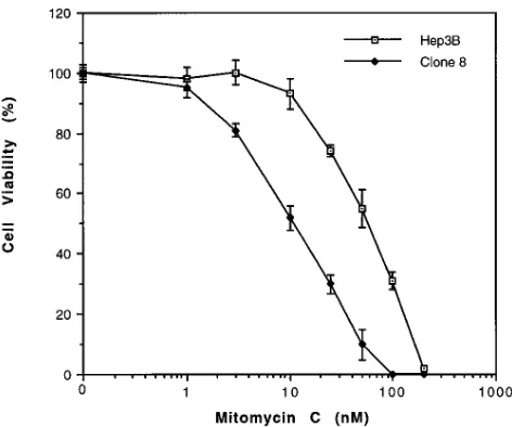
Phenotypic effects of MxA overexpression. These studies demonstrate that MxA is downstream of FAC. Because MMC sensitivity and apoptosis are also consequences of FAC defi-ciency, it is possible that MxA overexpression can lead directly to these phenotypic effects and thus constitute a linear path-way of pathogenesis. Alternately, the phenotypic features of FA may result from the activities of pathways that are
inde-pendent of MxA overexpression. To distinguish between these possibilities, we transfected MxA into a non-FA cell line, Hep3B, and obtained low-level expression of MxA in a popu-lation of cells that were resistant to G418 (Fig. 3 B). We then isolated individual clones and evaluated their response to MMC. In general, it was difficult to isolate clones that showed high levels of MxA by this strategy, suggesting that ectopic overexpression of MxA may be toxic to these cells. However, two clones (Hep3B clone No. 8 and No. 11) showed relatively high MxA levels and were chosen for subsequent studies. Hep3B clone No. 8 was significantly more sensitive to the cyto-toxic effects of MMC than the parental counterpart (Fig. 6). The average dose of MMC reducing the survival of these cells to 50% of control levels (EC50) was 12 nM, while the average EC50 of parental Hep3B cells to MMC was 60 nM. This result supports the linear-pathway model of FA pathogenesis.
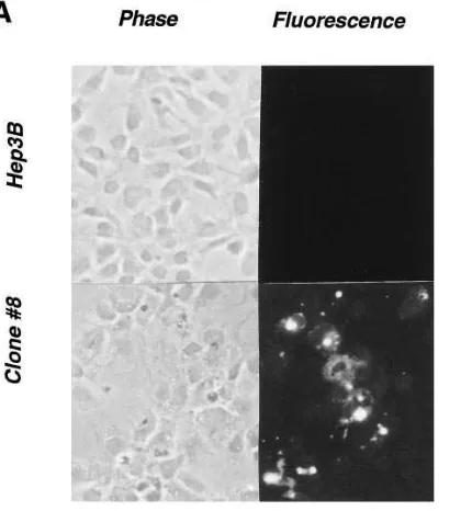
These cells were also examined for the presence of apop-totic markers by two different assays. First, the TUNEL assay was performed using a commercial kit and fluorescence mi-croscopy. A significantly greater proportion of the Hep3B clone No. 8 cells were apoptotic compared with the parental cells (Fig. 7 A). Quantitation of five different fields per cover-slip that had cell densities of z 50% confluence revealed 1463
[image:6.612.57.296.57.246.2]fluorescent cells per 100 cells for clone No. 8 and 060 for pa-rental Hep3B cells. Pretreatment with MMC for 24 h did not enhance the number of apoptotic cells (data not shown). Sec-ond, genomic DNA prepared from parental Hep3B, clone No. 8 and clone No. 11 was also examined for the presence of DNA laddering characteristic of apoptosis. Both clones over-expressing MxA showed a greater degree of ladder formation than the parental cells (Fig. 7 B). As with the TUNEL assay, the presence of MMC did not appear to enhance the extent of ladder formation. These results demonstrate that MxA over-expression is sufficient to induce apoptosis in Hep3B cells. Figure 5. The MxA promoter is not a direct target of FAC. (A)
Sche-matic representation of the MxA promoter showing consensus bind-ing sites for STATs. Numbers below the diagram show the base loca-tions in the MxA promoter. To facilitate subcloning, KpnI and BglII sites were added during PCR amplification. (B) Activity of the MxA promoter in the presence of wild-type (black columns) and mutant (white columns) FAC isoforms. COS-1 cells were cotransfected with the MxA promoter fused to the reporter gene luciferase in the vector pXP2 as well as expression constructs encoding FAC isoforms and
b-galactosidase. The luciferase activity in cell lysates was normalized for b-galactosidase activity. For dose–response studies, different quantities of plasmid DNA encoding FAC isoforms were used, as in-dicated. The mean and SEM are shown.
[image:6.612.317.553.474.671.2]Discussion
In this study, we demonstrate that MxA, an IFN-inducible gene, is overexpressed in FA cells of complementation groups A, B, C, and D in the absence of exogenous stimulation by IFN. The overexpression of MxA is reversible by wild-type FAC, suggesting that FAC is a natural inhibitor of MxA in vivo. By contrast, several other IFN-inducible genes—includ-ing HLA human class I antigen, b2-microglobulin, and IP10—
do not appear to be subject to regulation by FAC. Thus, the pathogenesis of FA may involve a constitutive, albeit selective, signaling by IFN. Our combined data on the altered expression of MxA demonstrates that the mechanism is primarily on a posttranscriptional level, although the precise mode of post-transcriptional control remains to be established.
Among the many cellular activities of IFNs is their ability to inhibit cellular proliferation in vivo and in culture (36). In fi-broblasts stimulated by growth factors, a- or b-IFN delays the onset of DNA synthesis. In addition, IFNs antagonize the ac-tion of some positive growth factors, which may be particularly relevant to the growth of blood cell production where these polypeptides act as negative regulators (37, 38). Thus, high lev-els of IFNs may be expected to suppress hematopoiesis and have indeed been used clinically in myeloproliferative disor-ders where such activities would be desirable. IFNs have also been suspected to play a role in the hematopoietic failure of FA. The most compelling evidence comes from animal model-ing attempts of FA. Targeted disruption of the murine fac gene recapitulates the chromosomal instability and other features of the human disorder (39, 40). It has been noted that the colony-forming capacity of murine progenitor cells declines progres-sively, and that low doses of g-IFN profoundly inhibit the growth of erythroid and myeloid progenitors (40, 41). In addi-tion, both human and murine primary hematopoietic progeni-tor cells are susceptible to apoptosis at lower doses of g-IFN than their normal counterparts (41). It was, therefore, gratify-ing that our unbiased approach of mRNA differential display identified a gene that is associated with IFN signaling.
An increasing body of evidence also demonstrates that FA genes may have anti-apoptotic functions (41–44). Increased susceptibility to cross-link specific apoptosis in FA-C cells by a p53-independent mechanism has been described previously (43). In addition, FAC overexpression in either human or mu-rine myeloid IL-3–dependent progenitor cells was also noted to delay the onset of apoptosis after withdrawal of the critical growth factor (44). Taken together, these results show that FAC is coupled to fundamental pathways that regulate cell death. In this study, our observations that MxA overexpres-sion is coupled to apoptosis leads us to propose the following model (Fig. 8). Constitutive overexpression of MxA by IFNs or viral infection (i.e., exogenous stimuli) could result in grad-ual loss of stem cells from the bone marrow. Similarly, an in-trinsic cellular defect (i.e., FAC and perhaps other FA gene mutations) could also lead to apoptosis and progressive cell loss. The inability to replace apoptotic cells efficiently may eventually lead to marrow aplasia. In principle, this mecha-nism could also operate in other cell types and may account for additional phenotypic features of FA. Although transgenic mice expressing high levels of the human MxA protein have been described and shown to be resistant to viral infections (45), the effect of MxA overexpression on blood cell formation was not analyzed, possibly because of inadequate expression
transfected clone overexpressing MxA (clone No. 8) were grown on coverslips under identical condi-tions, fixed, and stained with fluo-rescein-dUTP for the detection of apoptotic cells. Phase contrast and fluorescent photomicrographs were obtained at a magnification of 100. (B) Increased DNA ladder forma-tion in Hep3B cells overexpressing MxA with or without pretreatment with 100 nM MMC for 24 h, as indi-cated. Lanes 1 and 4, parental Hep3B cells; lanes 2 and 5, MxA-expressing Hep3B clone No. 8; lanes
[image:7.612.63.278.61.294.2]3 and 6, MxA-expressing Hep3B clone No. 11. Molecular size mark-ers are shown on the left lane.
[image:7.612.317.554.612.672.2]of the transgene in these cells or to embryonic lethality. Hence, an evaluation of the in vivo effects of MxA on hematopoiesis may have to await the generation of more appropriate animal models.
A number of critical questions remain about the relevance of this novel pathway to the pathogenesis of FA. The observa-tion that MxA is a GTPase raises the issue of in vivo targets for this enzyme, particularly those that are associated with known pathways of apoptosis. In addition, it is reasonable to suspect that an as yet unknown GTPase-associated protein could dampen or reverse the GTPase activity of MxA. The expres-sion of the latter putative molecule may be particularly impor-tant for the modulation of disease severity in FA.
Finally, although controversial, resistance to infection by microorganisms has been invoked as a selective force in the maintenance of otherwise deleterious, mutant alleles in het-erozygotes (46). For example, certain forms of thalassemia, heterozygosity for the sickle cell mutation, and G6PD defi-ciency are all thought to confer protection against malaria (46, 47). It has also been postulated that heterozygotes for cystic fi-brosis may have a selective advantage for resistance to cholera (48) as well as to noninfectious or to other potentially-infec-tious disorders, such as bronchial asthma (49). Similarly, it may be worth speculating that in asymptomatic carriers (or possibly in affected homozygotes or compound heterozygotes) FA mu-tations could confer protection against respiratory infections caused by influenza or related viruses.
Acknowledgments
We wish to thank M.A. Horisberger for contributing a sample of the MxA antibody, M. Grompe for the PD20 cells, A.D. Auerbach for the LCL171 cells, and D. Tenen for the luciferase expression vectors. This study was supported by grants from the National Institutes of Health (HL52138), the Leukemia Society of America, and the Lawrence Family Fund.
References
1. Fanconi, G. 1967. Familial panmyelocytopathy, Fanconi’s anemia (FA). I. Clinical aspects. Semin. Hematol. 4:233–240.
2. Alter, B.P., and N.S. Young. 1993. The bone marrow failure syndromes.
In Hematology of Infancy and Childhood. D.G. Nathan and F.A. Oski, editors. W.B. Saunders Company, Philadelphia. 216–316.
3. Liu, J.M., M. Buchwald, C.E. Walsh, and N.S. Young. 1994. Fanconi ane-mia and novel strategies for therapy. Blood. 84:3995–4007.
4. Duckworth-Rysiecki, G., K. Cornish, C.A. Clarke, and M. Buchwald. 1985. Identification of two complementation groups in Fanconi anemia. So-matic. Cell Mol. Genet. 11:35–41.
5. Strathdee, C.A., A.M.V. Duncan, and M. Buchwald. 1992. Evidence for at least four Fanconi anaemia genes including FACC on chromosome 9. Nat. Genet. 1:196–198.
6. Joenje, H., J.R. Lo Ten Foe, A.B. Oostra, C.G.M. van Berkel, M.A. Roo-imans, T. Schroeder-Kurth, R.-D. Wegner, J.J.P. Gille, M. Buchwald, and F. Arwert. 1995. Classification of Fanconi anemia patients by complementation analysis: evidence for a fifth genetic subtype. Blood. 86:2156–2160.
7. Pronk, J.C., R.A. Gibson, A. Savoia, M. Wijker, N.V. Morgan, S. Mel-chionda, D. Ford, S. Temtamy, J.J. Ortega, S. Jansen, et al. 1995. Localisation of the Fanconi anaemia complementation group A gene to chromosome 16q24.3.
Nat. Genet. 11:338–340.
8. Whitney, M., M. Thayer, C. Reifsteck, S. Olson, L. Smith, P.M. Jakobs, R. Leach, S. Naylor, H. Joenje, and M. Grompe. 1995. Microcell mediated chro-mosome transfer maps the Fanconi anaemia group D gene on chrochro-mosome 3p.
Nat. Genet. 11:341–343.
9. Strathdee, C.A., H. Gavish, W.R. Shannon, and M. Buchwald. 1992. Cloning of cDNAs for Fanconi’s anaemia by functional complementation. Na-ture. 356:763–767.
10. Lo Ten Foe, J.R., M.A. Rooimans, L. Bosnoyan-Collins, N. Alon, M. Wijker, L. Parker, J. Lightfoot, M. Carreau, D.F. Callen, A. Savoia, et al. 1996.
Expression cloning of a cDNA for the major Fanconi anemia gene, FAA. Nat. Genet. 14:320–323.
11. The Fanconi Anaemia/Breast Cancer Consortium. 1996. Positional cloning of the Fanconi anaemia group A gene. Nat. Genet. 14:324–328.
12. Yamashita, T., D.L. Barber, Y. Zhu, N. Wu, and A.D. D’Andrea. 1994. The Fanconi anemia polypeptide, FACC, is localized to the cytoplasm. Proc. Natl. Acad. Sci. USA. 91:6712–6716.
13. Youssoufian, H. 1994. Localization of Fanconi anemia C protein to the cytoplasm of mammalian cells. Proc. Natl. Acad. Sci. USA. 91:7975–7979.
14. Youssoufian, H. 1996. Cytoplasmic localization of FAC is essential for the correction of a prerepair defect in Fanconi anemia group C cells. J. Clin. In-vest. 97:2003–2010.
15. Youssoufian, H., A.D. Auerbach, P.C. Verlander, V. Steimle, and B. Mach. 1995. Identification of cytosolic proteins that bind to the Fanconi anemia complementation group C polypeptide in vitro. J. Biol. Chem. 270:9876–9882.
16. Liang, P., and A.B. Pardee. 1992. Differential display of eukaryotic mes-senger RNA by means of the polymerase chain reaction. Science. 257:967–971.
17. Horisberger, M.A., P. Staeheli, and O. Haller. 1983. Interferon induces a unique protein in mouse cells bearing a gene for resistance to influenza virus.
Proc. Natl. Acad. Sci. USA. 80:1910–1914.
18. Aebi, M., J. Fah, N. Hurt, C.E. Samuel, D. Thomas, L. Bazzigher, J. Pavlovic, O. Haller, and P. Staeheli. 1989. cDNA structures and regulation of two interferon-induced human Mx proteins. Mol. Cell. Biol. 9:5062–5072.
19. Horisberger, M.A., G.K. McMaster, H. Zeller, M.G. Wathelet, J. Dellis, and J. Content. 1990. Cloning and sequence analyses of cDNAs for interferon-and virus-induced human Mx proteins reveal that they contain putative guanine nucleotide-binding sites: functional study of the corresponding gene promoter.
J. Virol. 64:1171–1181.
20. Horisberger, M.A. 1992. Interferon-induced human protein MxA is a GTPase which binds transiently to cellular proteins. J. Virol. 66:4705–4709.
21. Dreiding, P., P. Staeheli, and O. Haller. 1985. Interferon-induced pro-tein Mx accumulates in nuclei of mouse cells expressing resistance to influenza virus. Virology. 140:192–196.
22. Meier, E., J. Fah, M.S. Grob, R. End, P. Staeheli, and O. Haller. 1988. A family of interferon-induced Mx-related mRNAs encodes cytoplasmic and nu-clear proteins in rat cells. J. Virol. 62:2386–2393.
23. Staehli, P., and J.G. Sutcliffe. 1988. Identification of a second interferon-regulated murine Mx gene. Mol. Cell. Biol. 8:4524–4528.
24. Staehli, P., Y.-X. Yu, R. Grob, and O. Haller. 1989. Double-stranded RNA inducible fish gene homologous to the murine influenza virus resistance gene Mx. Mol. Cell. Biol. 9:3117–3121.
25. Rothman, J.H., C.K. Raymond, T. Gilbert, P.J. O’Hara, and T.H. Stevens. 1990. A putative GTP binding protein homologous to interferon-inducible Mx proteins performs an essential function in yeast protein sorting.
Cell. 61:1063–1074.
26. Krug, R.M., M. Shaw, B. Broni, G. Shapiro, and O. Haller. 1985. Inhibi-tion of influenza viral mRNA synthesis in cells expressing the interferon-induced
Mx gene product. J. Virol. 56:201–206.
27. Staehli, P., O. Haller, W. Boll, J. Lindenmann, and C. Weissmann. 1986. Mx protein: constitutive expression in 3T3 cells transformed with cloned Mx cDNA confers selective resistance to influenza virus. Cell. 44:147–158.
28. Staehli, P., and O. Haller. 1987. Interferon-induced Mx protein: a medi-ator of cellular resistance to influenza virus. Interferon. 8:1–23.
29. Arnheiter, H., and O. Haller. 1988. Antiviral state against influenza vi-rus neutralized by microinjection of antibodies to interferon-induced Mx pro-teins. EMBO (Eur. Mol. Biol. Organ.) J. 7:1315–1320.
30. Pavlovic, J., T. Zurcher, O. Haller, and P. Staeheli. 1990. Resistance to influenza virus and vesicular stomatitis virus conferred by expression of human MxA protein. J. Virol. 64:3370–3375.
31. Ausubel, F.M., R. Brent, R.E. Kingston, D.D. Moore, J.G. Seidman, J.A. Smith, and K. Struhl, editors. 1994. Current Protocols in Molecular Biol-ogy. Greene Publishing Associates and John Wiley & Sons, New York.
32. Sambrook, J., E.F. Fritsch, and T. Maniatis. 1989. Molecular Cloning: A Laboratory Manual, Second Ed. Cold Spring Harbor Press, Cold Spring Har-bor, NY.
33. Ronni, T., K. Melen, A. Malygin, and I. Julkunen. 1993. Control of IFN-inducible MxA gene expression in human cells. J. Immunol. 150:1715–1726.
34. Chang, K.C., E. Hansen, L. Foroni, L. Lida, and G. Goldpink. 1991. Mo-lecular and functional analysis of the virus- and interferon-inducible human MxA promoter. Arch. Virol. 117:1–15.
35. Fu, X.-Y., C. Schindler, T. Improta, R. Aebersold, and J.E. Darnell. 1992. The proteins of ISGF-3, the interferon a-induced transcriptional activa-tor, define a gene family involved in signal transduction. Proc. Natl. Acad. Sci. USA. 89:7840–7843.
36. Petska, S., J.A. Langer, K.C. Zoon, and C.E. Samuel. 1987. Interferons and their actions. Annu. Rev. Biochem. 56:727–777.
37. Raefsky, E.L., L.C. Platanias, N.C. Zoumbos, and N.S. Young. 1985. Studies of interferon as a regulator of hematopoietic cell proliferation. J. Immu-nol. 135:2507–2512.
Reifsteck, S. Olson, R.E. Braun, M.C. Heinrich, R.K. Rathbun, G.C. Bagby, and M. Grompe. 1996. Germ cell defects and hematopoietic hypersensitivity to g-interferon in mice with a targeted disruption of the Fanconi anemia C gene.
Blood. 88:49–58
41. Rathbun, R.K., G.R. Faulkner, M.H. Ostroski, T.A. Christianson, G. Hughes, G. Jones, R. Cahn, R. Maziarz, G. Royle, W. Keeble, et al. 1997. Inac-tivation of the Fanconi anemia group C gene augments interferon-gamma–induced apoptotic responses in hematopoietic cells. Blood. 90:974–985.
42. Kruyt, F.A.E., L.M. Dijkmans, T.K. van den Berg, and H. Joenje. 1996. Fanconi anemia genes act to suppress a cross-linker-inducible p53-independent apoptosis pathway in lymphoblastoid cell lines. Blood. 87:938–948.
43. Marathi, U.K., S.R. Howell, R.A. Ashmun, and T.P. Brent. 1996. The Fanconi anemia complementation group C protein corrects DNA interstrand
Kolb, P. Staeheli, and O. Haller. 1995. Enhanced virus resistance of transgenic mice expressing the human MxA protein. J. Virol. 69:4506–4510.
46. Weatherall, D.J. 1996. Host genetics and infectious disease. Parasitol-ogy. 112(Suppl.):S23–S29.
47. Miller, L.H. 1996. Malaria: protective selective pressure. Nature. 383: 480–481.
48. Gabriel, S.E., K.N. Brigman, B.H. Koller, R.C. Boucher, and M.J. Stutts. 1994. Cystic fibrosis heterozygote resistance to cholera toxin in the cystic fibrosis mouse model. Science. 266:107–109.