Toll-like receptor-dependent production of
IL-12p40 causes chronic enterocolitis in myeloid
cell-specific Stat3-deficient mice
Masaya Kobayashi, … , Kiyoshi Takeda, Shizuo Akira
J Clin Invest.
2003;
111(9)
:1297-1308.
https://doi.org/10.1172/JCI17085
.
Stat3 plays an essential role in IL-10 signaling pathways. A myeloid cell-specific deletion of
Stat3 resulted in inflammatory cytokine production and development of chronic enterocolitis
with enhanced Th1 responses in mice. In this study, we analyzed the mechanism by which
a Stat3 deficiency in myeloid cells led to the induction of chronic enterocolitis in vivo. Even
in the absence of Stat1, which is essential for IFN-
g
signaling pathways, Stat3 mutant mice
developed chronic enterocolitis. TNF-
a
/Stat3 double-mutant mice developed severe chronic
enterocolitis with enhanced Th1 cell development. IL-12p40/Stat3 double-mutant mice,
however, showed normal Th1 responses and no inflammatory change in the colon.
RAG2/Stat3 double-mutant mice did not develop enterocolitis, either. These findings
indicate that overproduction of IL-12p40, which induces potent Th1 responses, is essential
for the development of chronic enterocolitis in Stat3 mutant mice. Furthermore, enterocolitis
was significantly improved and IFN-
g
production by T cells was reduced in TLR4/Stat3
double-mutant mice, indicating that TLR4-mediated recognition of microbial components
triggers aberrant IL-12p40 production by myeloid cells, leading to the development of
enterocolitis. Thus, this study clearly established a sequential innate and acquired immune
mechanism for the development of Th1-dependent enterocolitis.
Article
Immunology
Find the latest version:
Introduction
Human inflammatory bowel diseases (IBD) including Crohn diseases and ulcerative colitis are chronic immune-mediated disorders whose pathogenesis and etiology are largely unknown (1). A number of gene-manipulated murine models of colitis have been devel-oped. These include IL-10–deficient, T cell
receptor-α–deficient, Gαi2–deficient, Stat4-transgenic, and T-bet-transgenic mice (2–5). Experimentally induced models of colitis have also been established by recon-stituting immunocompromised mice with CD4+
CD45RBhigh T cells. All of these models are used in
attempts to elucidate the pathogenesis of IBD. As a result, it has been proposed that an exaggerated bias toward Th1 or Th2 polarization in the mucosal immune system is a key factor in the pathogenesis of colitis (6, 7). In IL-10–deficient mice, CD4+T cells residing in the
lamina propria region of intestinal tract preferentially produced IFN-γ(8). Immunocompromised mice trans-ferred with T cells from IL-10–deficient mice developed colitis with skewed Th1 responses (9). Furthermore, treatment with anti–IFN-γAb significantly delayed the onset of the disease and reduced the severity of colitis in young IL-10–deficient mice (8). Treatment of anti-IL-12 Ab prevented the development of the disease in young IL-10–deficient, but not in aged IL-10–deficient, mice (7, 10). Thus, the involvement of Th1 cells has become clear in the pathogenesis of early phase of colitis in IL-10–defi-cient mice. IL-10 is a well-known regulatory cytokine, which can provide positive and negative signals on a vari-ety of immune cells including B cells, T cells, NK cells, mast cells, and myeloid cells (11). Additionally, IL-10 has recently been shown to play an important role in the dif-ferentiation and function of regulatory T cells, which possess a capacity to inhibit harmful immune disorders including chronic colitis (12, 13). Although it is now
Toll-like receptor-dependent
production of IL-12p40 causes
chronic enterocolitis in myeloid
cell-specific Stat3-deficient mice
Masaya Kobayashi,
1Mi-Na Kweon,
2Hirotaka Kuwata,
1Robert D. Schreiber,
3Hiroshi Kiyono,
2,4Kiyoshi Takeda,
1,5and Shizuo Akira
1,51Department of Host Defense, Research Institute for Microbial Diseases, Osaka University, Osaka, Japan 2Department of Mucosal Immunology, Research Institute for Microbial Diseases, Osaka University, Osaka, Japan 3Department of Pathology and Immunology, Center for Immunology, School of Medicine, Washington University,
St. Louis, Missouri, USA
4Division of Mucosal Immunology, Institute of Medical Science, The University of Tokyo, Tokyo, Japan 5Solution Oriented Research for Science and Technology, Japan Science and Technology Corp., Tokyo, Japan
Stat3 plays an essential role in IL-10 signaling pathways. A myeloid cell-specific deletion of Stat3 resulted in inflammatory cytokine production and development of chronic enterocolitis with enhanced Th1 responses in mice. In this study, we analyzed the mechanism by which a Stat3 defi-ciency in myeloid cells led to the induction of chronic enterocolitis in vivo. Even in the absence of Stat1, which is essential for IFN-γsignaling pathways, Stat3 mutant mice developed chronic entero-colitis. TNF-α/Stat3 double-mutant mice developed severe chronic enterocolitis with enhanced Th1 cell development. IL-12p40/Stat3 double-mutant mice, however, showed normal Th1 responses and no inflammatory change in the colon. RAG2/Stat3 double-mutant mice did not develop enterocoli-tis, either. These findings indicate that overproduction of IL-12p40, which induces potent Th1 responses, is essential for the development of chronic enterocolitis in Stat3 mutant mice. Further-more, enterocolitis was significantly improved and IFN-γproduction by T cells was reduced in TLR4/Stat3 double-mutant mice, indicating that TLR4-mediated recognition of microbial compo-nents triggers aberrant IL-12p40 production by myeloid cells, leading to the development of entero-colitis. Thus, this study clearly established a sequential innate and acquired immune mechanism for the development of Th1-dependent enterocolitis.
J. Clin. Invest.111:1297–1308 (2003). doi:10.1172/JCI200317085.
Received for publication October 8, 2002, and accepted in revised form January 28, 2003.
Address correspondence to:Shizuo Akira, Department of Host Defense, Research Institute for Microbial Diseases, Osaka University, 3-1 Yamada-oka, Suita Osaka 565-0871, Japan. Phone: 81-6-6879-8302; Fax: 81-6-6879-8305;
E-mail: [email protected].
Conflict of interest:The authors have declared that no conflict of interest exists.
Nonstandard abbreviations used:inflammatory bowel diseases (IBD); phycoerythrin (PE); Toll-like receptor 4 (TLR4).
clear that IL-10 plays an essential role in the negative reg-ulation of inflammatory responses in the intestine, the mechanism by which IL-10 exerts the anti-inflammato-ry responses remains unclear.
The STAT family of transcription factors, consisting of seven members, is involved in cytokine signaling. The knockout of each member of the STAT family in mice resulted in impaired responses to corresponding cytokines, demonstrating that STAT proteins have essential roles in cytokine-mediated biological respons-es (14, 15). Unlike the knockout of all other STAT family members, the knockout of Stat3 resulted in early death in embryogenesis (16). Therefore, Stat3 has been deleted in a cell- or tissue-specific manner by the Cre-loxP recombination system in order to exam-ine biological and functional roles of the transcription factor in vivo (17–19). For example, mice lacking Stat3 in myeloid cells such as macrophages, neutrophils, and dendritic cells developed chronic enterocolitis and showed exaggerated Th1 responses (20). The immunological and histological analysis revealed very similar disease development in the intestinal tract of IL-10–deficient mice. Macrophages from the Stat3 mutant mice did not show any anti-inflammatory responses mediated by IL-10 and produced increased levels of inflammatory cytokines in response to bacte-rial LPS, demonstrating that Stat3 is essential for the IL-10–mediated signaling pathways in myeloid cells (20). This study also indicated that myeloid cells, including macrophages and dendritic cells, are major targets for IL-10 exhibiting anti-inflammatory responses in vivo, and the abnormal activation of myeloid cells is involved in the pathogenesis of chron-ic colitis in mchron-ice. It remains elusive, however, how the abnormal activation of myeloid cells leads to the development of colitis in vivo.
In this study, we examined the in vivo mechanism by which activated myeloid cells in the absence of IL-10 sig-naling induce chronic colitis using Stat3 mutant mice with additional deleted alleles, such as Stat1, TNF-α, IL-12p40, RAG2, and Toll-like receptor 4 (TLR4).
Methods
Mice. Stat3 mutant (LysMcre; Stat3flox/flox) mice on a mixed 129 × C57BL/6 genetic background (backcrossed to C57BL/6 for three generations) were used in this study (17, 20). Stat1-deficient mice on a C57BL/6 background were mated with Stat3 mutant mice (21). TNF-α –defi-cient and IL-12p40–defi–defi-cient mice on a C57BL/6 back-ground were kindly provided by K. Sekikawa and J. Magram, respectively (22, 23). RAG2-deficient mice on a C57BL/6 background were kindly provided by Fred Alt (Harvard Medical School and Center for Blood Research, Boston, Massachusetts, USA). TLR4-deficient mice were generated as described previously (24). Littermate wild-type or heterozygous mutant mice were used for experi-ments as control mice. All mice were housed in a specif-ic pathogen-free facility at the Research Institute for Microbial Diseases, Osaka University (Osaka, Japan).
Histopathological analysis. Paraffin-embedded colon samples were sectioned and stained with H&E. Trans-verse sections are shown in the figures. Severity of coli-tis was evaluated by the standard scoring system as described previously (25). Each region of the colon (cecum; ascending, transverse, and descending colon; and rectum) was graded semiquantitatively from 0 (no change) to 5 (most severe change). The grading repre-sents an increasing incidence and degree of inflamma-tion, goblet cell loss, crypt abscesses and ulcerainflamma-tion, and fibrosis in the lamina propria. The summation of the score for each segment of the colon provides a total disease score per mouse (0–25) where: 0 indicates no change; 1–5, mild disease; 6–10, mild-moderate; and 11–20, severe. No mice in these studies had a score above 20 because such severe disease results in death. The scoring was performed blinded manner by two independent investigators. The colitis scores were eval-uated using the program Statview II (SAS Inc., Cary, North Carolina, USA). Pvalues less than 0.05 were assumed to be statistically significant.
Cytokine production by macrophages. Mice were intraperi-toneally injected with 2 ml of 4% thioglycollate medium (Sigma-Aldrich, St. Louis, Missouri, USA). Three days later, peritoneal exudate cells were isolated from the peritoneal cavity by washing with ice-cold HBSS (Sigma-Aldrich). Cells were cultured for 2 h and washed with HBSS to remove nonadherent cells. Adherent monolayer cells were used as peritoneal macrophages. Peritoneal macrophages were cultured with 10 ng/ml of LPS (Escherichia coliserotype O55:B5), 1 µM of CpG DNA, or 50 U/ml of IFN-γ(Genzyme, Cambridge, Mass-achusetts, USA) plus LPS for 24 h. Concentrations of TNF-α, IL-6, and IL-12p40 in the culture supernatants were measured by ELISA according to the manufactur-er’s instructions (Genzyme; Endogen Inc., Woburn, Massachusetts, USA). Production of nitric oxide was measured by the Greiss method using a NO2/NO3Assay
Kit (DOJINDO, Kumamto, Japan).
Cytokine production by dendritic cells isolated from the large intestine. The large intestine was removed aseptically. After removal of colonic patches, the large intestine was washed with RPMI-1640 on ice and stirred with RPMI-1640 containing 20 mM HEPES, 50 µg/ml of gen-tamycin, 100 U/ml penicillin, 100 µg/ml streptomycin, 10% FCS, 5 mM EDTA for 60 min at 37°C. The tissues were then digested with collagenase D (400 U/ml; Boehringer Mannheim GmbH, Mannheim, Germany) in RPMI-1640 containing 10% FCS for 30 min at 37°C and further incubated in the presence of 5 mM EDTA for 5 min. The isolated cells were purified by centrifugation through a 14.5% Accudenz gradient (Accurate Chemical & Scientific Corp., Westbury, New York, USA). Cells were further incubated with mouse CD11c microbeads (Mil-tenyi Biotech, Bergisch Gladbach, Germany) and select-ed on autoMACS separation columns (Miltenyi Biotech). The purified dendritic cell fractions were more than 95% CD11c+. These cells (105) were seeded on
100 ng/ml LPS for 24 h. Concentrations of IL-6 and IL-12 in the culture supernatants were analyzed by ELISA.
Cytokine production by splenic and colonic CD4+T cells. Spleens were removed aseptically and single-cell sus-pensions were prepared. After the lysis of red blood cells, cells were incubated with CD4 microbeads and passed through a magnetized column (Miltenyi Biotech). The purified T cell fractions were more than 95% CD4+and more than 99% viable. Lymphocytes of
the lamina propria of the large intestine were isolated as previously described (26). In brief, after excision of colonic patches, the intestinal tissues were treated with RPMI-1640 medium containing 1 mM EDTA for 20 min to remove the epithelial cells. The tissue was then digested with 0.5 mg/ml of collagenase (400 U/ml; Boehringer Mannheim GmbH) for 20 min at 37°C, and this step was repeated two times. The collected cells were washed and filtered through a nylon mesh. CD4+T cells were purified by the magnetic activated
cell-sorter system (Miltenyi Biotech) as described above. CD4+T cells were seeded on anti-CD3 Ab-coated (10
µg/ml in PBS) 96-well plates and cultured for 24 h. Concentrations of IFN-γin the culture supernatants were analyzed by ELISA, according to the manufactur-er’s directions (Genzyme).
Immunohistochemical analysis. Freshly obtained large intestine was washed with PBS and frozen in Tissue-Tec OCT compound (Sakura, Tokyo, Japan). Cryostat sec-tions (6 µm) were fixed with cold acetone for 2 min and dried and rehydrated with PBS. This step was followed by blocking with PBS containing 20 mM HEPES, 10% FBS, and 1 µg of Fc-blocking mAb (2.4G2; BD PharMin-gen, San Diego, California, USA). Sections were stained with FITC-conjugated anti-CD11c (HL3; BD PharMin-gen) and phycoerythrin-conjugated (PE-conjugated) anti-CD3ε(145-2C11; BD PharMingen) or PE-conju-gated anti-CD11b (M1/70; BD PharMingen) mAbs for 2 h at room temperature. After washing with PBS, the sections were analyzed by confocal microscopy (Bio-Rad Laboratories Inc., Hercules, California, USA).
For IL-12 staining, tyramide signal amplification method was adopted (TSA Biotin System; NEN Life Science Products Inc., Boston, Massachusetts, USA). Briefly, fixed cryostat sections were incubated with biotin-conjugated IL-12 (p40/p70) mAb (C17.8; BD PharMingen) for 1 h at room temperature, and washed with washing buffer. Then, sections were stained with streptavidin-HRP and biotinyl tyramide amplification reagent. The sections were then incubated with strep-tavidin-PE (BD PharMingen) and FITC-conjugated anti-CD11c (HL3; BD PharMingen) or FITC-conjugat-ed anti-CD11b (M1/70; BD PharMingen).
Northern blot analysis. Peritoneal macrophages were treated with LPS for 2 h. Total RNA (10 µg) was extract-ed using the TRIzol reagent (Invitrogen Corp., San Diego, California, USA), electrophoresed, transferred to a nylon membrane, and hybridized with cDNA probe specific for SOCS3, IL-12p40, and β-actin.
Results
Stat1/Stat3 double-mutant mice developed chronic colitis. In macrophages from myeloid-specific Stat3 mutant mice, Stat1 is constitutively phosphorylated (20). Stat1 has been shown to be essential for signaling mediated by IFN-γ, which is known to be an important cytokine for the priming of macrophage activity (21). Therefore, we expected the constitutive activation of Stat1 to confer abnormal activity of Stat3 mutant macrophages. To address this issue, we generated mice lacking both Stat1 and Stat3.
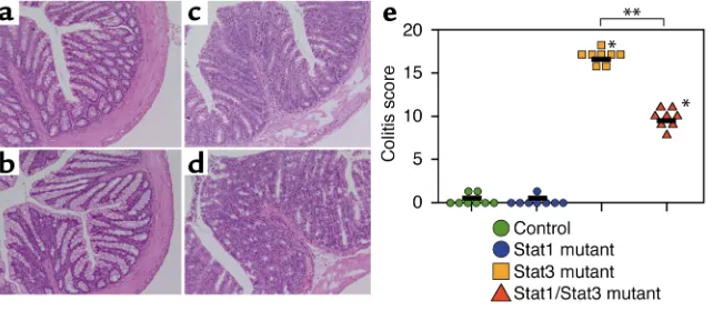
[image:4.576.149.470.538.678.2]We performed a histopathological analysis on colons of 8-week-old Stat1/Stat3 double-mutant mice (Figure 1, a–d). Control and Stat1 mutant mice did not show any sign of pathological changes. On the contrary, the colonic wall of Stat1/Stat3 double-mutant mice was thickened, and the gland number was severely reduced, as is the case in Stat3 mutant mice. These histopatho-logical changes were observed as early as 5–6 weeks of age in both Stat3 mutant and Stat1/Stat3 double-mutant mice. Almost all regions of the colon were
Figure 1
affected in Stat3 mutant mice, but not in Stat1/Stat3 double-mutant mice at 24 weeks of age. In addition, occurrence of ulcer and abscess was reduced in Stat1/Stat3 double-mutant mice compared with Stat3 single-mutant mice. Thus, Stat1/Stat3 double-mutant mice developed chronic enterocolitis, although the severity of the colitis was diminished compared with Stat3 mutant mice (Figure 1e).
We next analyzed production of IFN-γby CD4+T
cells isolated from spleens of Stat1/Stat3 double-mutant and Stat3 double-mutant mice. Splenic T cells were stimulated with PMA and ionomycin, and production of IFN-γand IL-4 by CD4+cells was analyzed by
intra-cellular staining (Figure 2a). IFN-γ–producing CD4+T
cells were significantly elevated in Stat3 single-mutant mice. IFN-γ–producing cells were also increased in Stat1/Stat3 double-mutant mice compared with that of control mice, albeit the reduced numbers were in comparison to Stat3 mutant T cells. We further puri-fied CD4+T cells from the intestinal lamina propria of
these mice and analyzed for IFN-γproduction by ELISA (Figure 2b). Intestinal CD4+T cells from Stat1/Stat3
double-mutant mice produced increased level of IFN-γ compared with control T cells. Thus, these findings demonstrated that double mutation of Stat1 and Stat3 resulted in the increase of IFN-γproduction by intes-tinal CD4+T cells in addition to splenic CD4+T cells,
[image:5.576.59.327.58.229.2]indicating an enhanced Th1 response. Figure 2
Enhanced IFN-γproduction in Stat1/Stat3 double-defi-cient mice. (a) Spleen cells were stimulated with PMA and ionomycin for 6 h, then stained for CD4, fixed, and finally stained for IFN-γand IL-4. Cells were analyzed on FACS by gating on CD4+population. The frequency of
cytokine-producing CD4+ cells is indicated as a
per-centage. (b) CD4+T cells were purified from lamina
[image:5.576.130.455.434.654.2]propria and stimulated with plate-bound anti-CD3 Ab. The concentration of IFN-γwas measured by ELISA. (c) Mice were intraperitoneally injected with thioglycollate, and 3 days later peritoneal macrophages were isolated. Macrophages were stimulated with 10 ng/ml LPS for 24 h. Concentrations of TNF-α, IL-6, and IL-12p40 in the culture supernatants were measured.
Figure 3
Based on the result obtained by the analysis of CD4+
T cells, our next experiment was to characterize macrophages. Although the analysis of the peritoneal macrophages does not directly reflect involvement of macrophages in intestinal inflammation, the inflam-matory response can be estimated in cells of other ori-gins, because the enhanced IFN-γ production was observed not only in the intestinal T cells but also in splenic T cells (Figure 2, a and b). Peritoneal macrophages isolated from Stat1/Stat3 double-mutant mice produced increased levels of TNF-αand IL-6 com-pared with control or Stat1 mutant macrophages, albeit lower compared with Stat3 mutant macrophages (Figure 2c). IL-12p40 production from Stat1/Stat3
double-mutant macrophages was comparable to that of Stat3 single-mutant macrophages. From these find-ings we speculated that inflammatory cytokine syn-thesis contributes to the development of chronic ente-rocolitis in Stat1/Stat3 double-mutant mice.
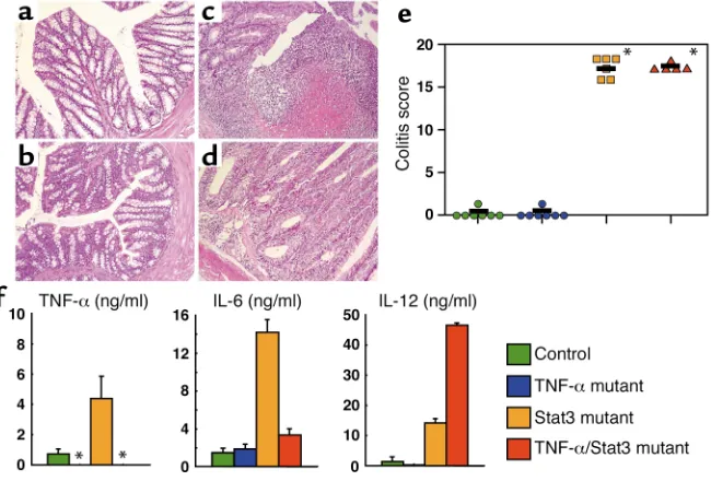
TNF-α/Stat3 double-deficient mice suffered from severe chron-ic enterocolitis. TNF-αis involved in a variety of inflamma-tory processes, including the development of IBD (27). Indeed, our previous and present studies demonstrated that the level of TNF-αproduction was increased in Stat3 mutant mice (Figure 2c). Therefore, we next generated mice lacking both Stat3 and TNF-α. Surprisingly, inflam-matory changes including loss of gland, infiltration by inflammatory cells, crypt abscess, and ulcers were observed in the colon of TNF-α/Stat3 double-mutant mice. The severity of disease was similar to that of Stat3 mutant mice (Figure 3, a–e). All of the TNF-α/Stat3 double-mutant mice analyzed (six mice) developed severe chronic enterocolitis, as did Stat3 mutant mice (Figure 3e). We next analyzed the LPS-induced inflammatory cytokine production by peritoneal macrophages. TNF-α/Stat3 double-mutant macrophages produced increased levels of IL-6 and IL-12 compared with macrophages isolated from control mice (Figure 3f). In particular, an extremely high level of IL-12p40 was pro-duced by TNF-α/Stat3 double-mutant macrophages.
In the next set of experiments, production of IFN-γby CD4+T cells isolated from spleen or intestinal lamina
propria of TNF-α/Stat3 double-mutant mice with coli-tis was analyzed (Figure 4). CD4+T cells isolated from
[image:6.576.76.277.55.196.2]both spleen and intestinal lamina propria of TNF-α/ Stat3 double-mutant mice as well as Stat3 single-mutant mice produced increased levels of IFN-γ. Thus, TNF-α/Stat3 double-mutant mice also showed skewed Th1 responses and developed chronic enterocolitis. Figure 4
IFN-γproduction by CD4+T cells in spleen and lamina propria of
TNF-α/Stat3 double-mutant mice. CD4+T cells were purified from
spleen and lamina propria (LPL), and stimulated with plate-bound anti-CD3 Ab. The concentration of IFN-γwas measured by ELISA. CD4+T cells from TNF-α/Stat3 double-mutant mice produced
sig-nificantly increased levels of IFN-γ, similar to Stat3 mutant mice.
Figure 5
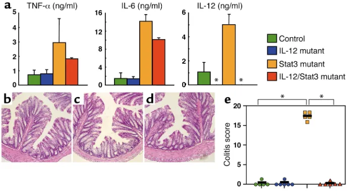
[image:6.576.126.471.475.669.2]IL-12p40/Stat3 double-mutant mice did not develop chron-ic enterocolitis. Based on the data obtained by the analy-sis of Stat3 mutant mice inserted with additional gene manipulation (Figures 1–3), it was obvious that elevat-ed IL-12p40 synthesis by macrophages was consistent-ly associated with the colitis development. Thus, it was logical to generate IL-12p40/Stat3 double-mutant mice. The analysis of the LPS-induced production of inflammatory cytokines by peritoneal macrophages revealed that IL-12p40/Stat3 double-mutant macro-phages produced increased levels of TNF-αand IL-6, which was comparable to those of Stat3 mutant macrophages (Figure 5a). The double-mutant mice,
however, did not show any pathological sign of inflam-mation in the colon (Figure 5, b–e). They also did not develop colitis even at over 24 weeks of age, when Stat3 mutant mice suffered from severe chronic enterocoli-tis (data not shown). These findings suggested a criti-cal role of IL-12p40 for the development of colitis asso-ciated with Stat3 deficiency.
To further support this view, we next analyzed the production of IFN-γby CD4+ T cells isolated from
spleen and colonic lamina propria of the double-mutant mice (Figure 6). Colonic lamina propria CD4+
T cells of Stat3 mutant mice showed increased pro-duction of IFN-γ; however, IFN-γproduction by colonic lamina propria CD4+T cells of IL-12p40/Stat3
double-mutant mice was reduced to the level of control mice. Similar results were obtained when splenic CD4+T cells
were examined (Figure 6). Taken together, these results demonstrated that IL-12p40/Stat3 double-mutant mice did not develop chronic enterocolitis with no pathological IFN-γproducing Th1-type responses. We thus suggest that IL-12–induced development of Th1 cells leads to the induction of severe chronic entero-colitis in Stat3 mutant mice.
[image:7.576.73.272.57.195.2]RAG2/Stat3 double-mutant mice did not develop chronic enterocolitis. To further confirm the critical role of Th1 cells in the development of chronic enterocolitis in Stat3 mutant mice, we next generated RAG2/Stat3 double-mutant mice, which lack T cells. Similar to macrophages obtained from Stat3 mutant mice, macrophages isolated from these double-mutant mice produced increased levels of inflammatory cytokines such as TNF-α, IL-6, and IL-12p40 (Figure 7a). RAG2/ Stat3 double-mutant mice, however, did not show any inflammatory change in the colon (Figure 7, b–e). Figure 6
Normal production of IFN-γby CD4+T cells in spleen and lamina
propria of IL-12/Stat3 double-mutant mice. CD4+T cells were
puri-fied from spleen and lamina propria (LPL) and stimulated with plate-bound anti-CD3 Ab. The concentration of IFN-γwas measured by ELISA. CD4+T cells from IL-12/Stat3 double-mutant mice produced
normal levels of IFN-γ, unlike those from Stat3 mutant mice.
Figure 7
[image:7.576.127.469.470.669.2]None of the double-mutant mice analyzed developed chronic enterocolitis even after 24 weeks of age (data not shown). These findings further support our hypothesis that myeloid cell-originated IL-12p40 medi-ates the induction of Th1 cells contributing to the devel-opment of chronic enterocolitis in Stat3 mutant mice.
TLR4 is involved in the pathogenesis of colitis in Stat3 mutant mice. In the absence of Stat3, macrophages were abnormally activated and produced increased levels of inflammatory cytokines such as IL-12p40 (Figures 2, 3, 5, 7). It was thus important to elucidate the triggering mechanism for the increased production of IL-12p40 by Stat3 mutant macrophages. The biosynthesis of inflammatory cytokines including IL-12p40 is known to be induced by stimulation with bacterial compo-nents such as LPS (28). Bacterial compocompo-nents have recently been shown to be specifically recognized by the pattern recognition family of TLRs (29). Among the family members, TLR4 is an essential receptor for the recognition of LPS (24, 30). Therefore, we finally gen-erated TLR4/Stat3 double-mutant mice and analyzed
whether TLR4 is involved in the abnormal activation of Stat3 mutant macrophages leading to the pathological Th1 cell–mediated large intestinal inflammation. As one might expect, LPS-induced production of inflam-matory cytokines was not observed at all in macro-phages isolated from TLR4/Stat3 double-mutant mice (Figure 8a). We further analyzed the production of IFN-γby CD4+T cells of TLR4/Stat3 double-mutant
mice (Figure 8b). The level of IFN-γ production by CD4+T cells isolated from TLR4/Stat3 double-mutant
[image:8.576.130.470.51.242.2]mice was significantly decreased compared with Stat3 mutant mice. Thus, aberrant production of an inflam-matory initiator cytokine, IL-12, by myeloid cells and Figure 8
Improved colitis in TLR4/Stat3 double-mutant mice. (a) Peritoneal macrophages were stimulated with 10 ng/ml LPS for 24 h. Concentra-tions of TNF-α, IL-6, and IL-12p40 in the culture supernatants were measured. LPS-induced production of inflammatory cytokines was abol-ished in TLR4/Stat3 double-mutant macrophages. *Not detected. (b) CD4+T cells were purified from spleen and stimulated with
plate-bound anti-CD3 Ab. The concentration of IFN-γwas measured by ELISA. CD4+T cells from TLR4/Stat3 double-mutant mice produced
reduced levels of IFN-γcompared with Stat3 mutant mice. (c–e) Histopathology of the colon of (c) control, (d) TLR4-deficient, and (e) TLR4/Stat3 double-mutant mice at 16 weeks. H&E staining is shown. ×20. (f) The colitis score of TLR4/Stat3 double-mutant mice at 16 weeks of age. TLR4/Stat3 double-mutant mice showed no or moderate inflammatory changes in the colon. *P< 0.001 between Stat3 mutant mice and control or TLR4/Stat3 double-mutant mice.
Figure 9
[image:8.576.350.513.540.733.2]the Th1 cell development were improved in the absence of TLR4. This notion was further supported by the analysis of the histopathological change in the colon of TLR4/Stat3 double-mutant mice (Figure 8, c–e). At 16 weeks of age, the inflammatory change was scarcely detected in the colon of TLR4/Stat3 double-mutant mice (Figure 8f). These findings clearly demonstrated that removal of the initiation signal by LPS by TLR4 inhibited the abnormal activation of Stat3 mutant macrophages, causing the development of colitis. It is important to note, however, that some TLR4/Stat3 double-mutant mice (three of six mice analyzed) showed some signs of inflammatory change in the colon at around 24 weeks of age, although the severity of affected region was reduced compared with Stat3 mutant mice (data not shown). Thus, in the absence of TLR4, the onset of chronic enterocolitis was delayed, and the severity and incidence of colitis was improved. Taken together, these findings indicate that TLR4 is involved in an initiation step of abnormal activation of myeloid cells leading to the development of chronic enterocolitis in Stat3 mutant mice.
Reduced expression of SOCS3 in Stat3 mutant macrophages. Findings obtained by analysis of TLR4/Stat3 double-mutant mice suggest that IL-10 exerts an
anti-inflam-matory activity by suppressing LPS responsiveness in myeloid cells. Recently, genes induced by IL-10 stimula-tion in macrophages have been identified and charac-terized (31, 32). Among these gene products, SOCS3 has been shown to inhibit the LPS-induced expression of TNF-α, IL-6, and iNOS in macrophages (32). We there-fore analyzed IL-10–induced mRNA expression of SOCS3 in peritoneal macrophages of Stat3 mutant mice (Figure 9a). In control mice, IL-10 stimulation resulted in robust expression of SOCS3 mRNA. IL-10–induced SOCS3 expression, however, was not observed in Stat3 mutant mice. Accordingly, IL-10–mediated suppression of LPS-induced IL-12p40 expression, which was ob-served in control macrophages, was abrogated in Stat3 mutant macrophages (Figure 9b). These findings indi-cate that IL-10–induced activation of Stat3 is critical to suppression of LPS-induced IL-12p40 production in macrophages, possibly through expression of anti-inflammatory molecules such as SOCS3.
[image:9.576.58.322.53.428.2]Dendritic cells and macrophages in the large intestine were increased in number and showed enhanced IL-12 production in Stat3 mutant mice. The inflammatory change observed in Stat3 mutant mice was restricted to the large intestine. Furthermore, our present findings described above pro-vided strong evidence that aberrant production of
Figure 10
IL-12p40 by Stat3 mutant myeloid cells directly con-tribute to the development of colonic inflammation by the generation of pathological Th1 cell environment. Therefore, we speculate that myeloid cells residing in the large intestine play important roles in the develop-ment of colitis in the mutant mice. To directly examine the involvement of mucosal dendritic cells and macrophages in the pathogenesis, immunohistochem-ical study was carried out in the large intestine of Stat3 mutant mice with colitis. The number of accumulated CD11c+dendritic cells and CD11b+macrophages was
significantly increased in the large intestine of Stat3 mutant mice compared with those of control mice (Fig-ure 10, a–d). Dendritic cells were mainly observed in the epithelium region and some in the lamina propria and the submucosal regions (Figure 10, a and b). CD11b+
macrophages were also observed in the epithelium and lamina propria region of the large intestine. Colocaliza-tion of CD3+T cells with CD11c+dendritic cells was rare
in the lamina propria of control mice (Figure 10e). Large numbers of CD3+T cells, however, were located adjacent
to CD11c+dendritic cells in Stat3 mutant mice (Figure
10f). These findings suggest a possibility that localized direct interaction between dendritic cells and T cells at the mucosal surface may initiate the creation of IL-12– mediated pathological Th1 environment leading to the development of colitis.
To examine this possibility, we next purified CD11c+
dendritic cells from the large intestine of Stat3 mutant mice for the analysis of cytokine production (Figure 10g). Dendritic cells isolated from control mice did not show any production of IL-12p40 upon stimulation with LPS. In contrast, dendritic cells obtained from Stat3 mutant mice showed marked production of IL-12p40 without stimulation (Figure 10g). The level of IL-12p40 production was further enhanced when cultured with LPS. These findings clearly demonstrat-ed that Stat3 mutant dendritic cells are committdemonstrat-ed for the pathological production of IL-12p40. In addition to the elevated IL-12p40 production, Stat3 mutant dendritic cells produced increased level of IL-6 com-pared with the cells isolated from control mice, although mucosal dendritic cells did not produce any detectable level of TNF-αin both control and Stat3 mutant mice (Figure 10g, data not shown).
Finally, we examined expression of IL-12 in intes-tinal macrophages and dendritic cells by immunohis-tochemistry. In control mice, CD11b+macrophages
and CD11c+dendritic cells scarcely expressed IL-12
(data not shown). In contrast, both CD11b+
macro-phages and CD11c+dendritic cells expressed IL-12 in
the large intestine of Stat3 mutant mice (Figure 10h). Thus, macrophages and dendritic cells residing in the large intestine were increased in number, showed enhanced production of IL-12, and many of the den-dritic cells colocalized with CD3+T cells. These
find-ings suggest that macrophages and dendritic cells residing in the large intestine are involved in the development of chronic enterocolitis by instruction
of Th1 cell development through IL-12p40 produc-tion in Stat3 mutant mice.
Discussion
In the present study, we analyzed the molecular mech-anism by which abnormally activated Stat3 mutant myeloid cells induce development of chronic entero-colitis in vivo using several gene-manipulated mice where additional gene deletion were introduced into Stat3 mutant mice. Previous study indicated that IL-10 is an important cytokine that prevents the develop-ment of chronic enterocolitis (2). CD4+ T cells of
IL-10–deficient mice showed the Th1 phenotype by producing an increased level of IFN-γ(8), and the adop-tive transfer of these CD4+T cells into
immunocom-promised mice resulted in the induction of colitis (9). Treatment with neutralizing anti–IL-12 Ab prevented the colitis development in young IL-10–deficient mice (10). These lines of evidence indicate that the entero-colitis observed in IL-10–deficient mice is associated with the increased Th1-type responses. It remains unclear, however, how the IL-10 deficiency led to the development of chronic enterocolitis in vivo. This study demonstrated that defective IL-10 signaling caused by the absence of Stat3 in myeloid cells resulted in the Th1-mediated enterocolitis in mice. Further-more, aberrant production of IL-12p40 by Stat3 mutant myeloid cells is a key pathological element for the development of Th1-dependent enterocolitis. In the absence of IL-10 or IL-10 signaling represented by IL-10 knockout or Stat3 deficiency, respectively, myeloid cells including macrophages and dendritic cells were activated and produced increased levels of inflammatory cytokines. Among these inflammatory cytokines, IL-12 instructs the development of Th1 cells, which confer the pathogenesis of enterocolitis. IL-12p40 associates with p35 to form bioactive IL-12 and addi-tionally engages p19 to form IL-23, which has several overlapping activities with IL-12 (33). Since IL-12p40– deficient mice, which lack both IL-12 and IL-23, were used in this study, we cannot exclude a possible patho-logical role of IL-23 in addition to IL-12 for the devel-opment of Th1-dependent diseases.
IL-10 has been shown to instruct the development of regulatory T cells, which have a capacity to prevent the development of chronic enterocolitis induced by Th1-type responses (12, 13). In the Stat3 mutant mice, LPS-induced IL-10 production was significantly increased (20). Furthermore, a population of CD4+CD25+
regula-tory T cells in the spleen of the Stat3 mutant mice was increased compared with control mice (14–17% versus 5–6%; our unpublished data). These regulatory T cells had a low capacity to proliferate and furthermore had an ability to inhibit proliferation of CD4+T cells,
IL-10 has been shown to induce the development of regulatory T cells (34). Therefore, overproduction of IL-10 may account for the increased number of regula-tory T cells in the Stat3 mutant mice. Although regu-latory T cells have been well established to inhibit the development of colitis (12, 13), our present and ongo-ing studies suggest that colitis is developed in the Stat3 mutant mice, despite regulatory T cells increasingly present. These findings imply that even in the presence of IL-10 and regulatory T cells, exaggerated Th1 responses due to the aberrant activation of myeloid cells lead to the development of chronic inflammation in the Stat3 mutant mice.
In the Stat3 mutant mice, there is a possibility that not only Th1 cells but also inflammatory cytokines contribute to the pathogenesis of enterocolitis. In par-ticular, TNF-αis known to be a potent inducer of sev-eral inflammatory diseases (27). Indeed, anti–TNF-α Ab has been used successfully in a clinical setting for the treatment of IBD such as Crohn disease (35). Mice that overproduced TNF-αdue to a deletion of adeno-sine-uracil–rich elements from the 3′-untranslated region of the TNF-αgene developed Crohn-like colitis (36). In this model, colitis was not induced in the RAG1–/–background, indicating that lymphocytes are
critical mediators for the development of colitis. Addi-tionally, Crohn disease patients who responded to anti–TNF-αAb showed downregulation of TNF-α, and subsequently IFN-γ, indicating that increased Th1 responses evoked by TNF-αin the mucosa are critical for pathogenesis (37, 38). TNF-α/Stat3 double-mutant mice developed severe chronic enterocolitis (Figure 3). Similarly, treatment with anti–TNF-αAb in young IL-10–deficient mice did not prevent colitis (39). Although a report showed that colitis was improved in TNF-α/IL-10 double-deficient mice, some of these mice developed severe colitis (40). Thus, TNF-αdoes not seem to be directly involved in the pathogenesis of colitis in Stat3 mutant and IL-10–deficient mice. Fur-thermore, reconstitution of RAG2-deficient mice with CD4+ T cells from Stat3 mutant mice induced the
development of chronic enterocolitis (our unpublished data). Therefore, we conclude that Th1 cells are the most important inducers of chronic enterocolitis in Stat3 mutant mice. This model clearly demonstrated that abnormal activity by myeloid cells is a first trigger to induce Th1-dependent inflammatory diseases. In TNF-α/Stat3 double-mutant mice, IL-12p40 produc-tion was significantly increased even when compared with Stat3 single-mutant mice (Figure 3f). TNF-αhas been shown to be one of important cytokines that are involved in prevention of microbial infection (41, 42). In the apical surface of the large intestine of TNF-α/ Stat3 double-mutant mice, many of rod-type bacteria can be easily detected in comparison with Stat3 single-mutant and control mice (our unpublished data). Therefore, although precise mechanism by which IL-12p40 production was increased is unclear, we could provide a speculation that the deficiency of TNF-αmay
lead to the impairment of mucosal innate immunity for the control of intestinal microflola, including the invasion of pathogen via the epithelium. Thus, several intestinal pathogens or commensal bacteria may easi-ly invade into the intestinal tissues, which may result in an increased chronic stimulation of myeloid cell lead-ing to the overproduction of IL-12p40.
Although Stat1 was constitutively activated in Stat3 mutant macrophages by unknown mechanisms (20), the insertion of Stat1 deficiency only partially improved the severity of colitis in Stat3 mutant mice (Figure 1). Stat1 plays an essential role in the signaling pathway of IFN-γ, which is an important mediator of Th1 responses (21). Therefore, Th1-mediated responses should be impaired in Stat1/Stat3 double-deficient mice, and thus lack of the disease development was expected. Our find-ings, however, demonstrated that the insertion of Stat1 deficiency into Stat3 mutant mice did not prevent the development of chronic enterocolitis. For the explana-tion of this unexpected result, we propose two possibil-ities. One is as follows: several recent reports demon-strated the presence of a Stat1-independent pathway in IFN-γsignaling (43–46). In Stat1/Stat3 double-deficient mice, IFN-γproduction from CD4+T cells was
signifi-cantly increased, indicating that these mice exhibited skewed Th1 responses even in the absence of Stat1 (Fig-ure 2, b and c). Therefore, IFN-γ–mediated Stat1-inde-pendent signaling may confer the induction of chronic colitis in these double-mutant mice. Alternatively, the adoptive transfer of CD4+T cells isolated from IFN-γ–
deficient mice has been shown to induce Th1-mediated colitis in immunocompromised mice (47). Treatment with anti–IFN-γAb was less effective than anti–IL-12 Ab in preventing the colitis development in IL-10–deficient mice (8, 10). These results indicate that destructive cell-mediated immune responses can be provided by Th1 cells without the involvement of IFN-γproduction for the development of colitis.
triggering mucosal inflammation could be “intestinal microflora,” commensal microorganisms resident on the mucosal surface (7). Therefore, we propose the fol-lowing hypothesis: TLRs expressed on myeloid cells rec-ognize microbial components of the mucosal microflo-ra and activate myeloid cells to produce inflammatory cytokines including IL-10, IL-12, and TNF-α. In normal conditions, IL-10, which is produced by myeloid cells and regulatory T cells, suppresses the activity of myeloid cells. In the absence of IL-10 signaling, however, myeloid cells are abnormally activated through TLRs and pro-duce increased levels of inflammatory cytokines. Among these, IL-12 induces the development of colitis through the generation of aberrant Th1 cells.
Some of TLR4/Stat3 double-mutant mice developed colitis at old age, indicating that other TLRs may be involved in the initiation of pathogenesis. TLR9, which induces potent Th1 responses through recognition of CpG DNA in vivo, is one such candidate (50). CpG DNA induced increased production of inflammatory cytokines, including IL-12p40, in TLR4/Stat3 double-mutant macrophages (our unpublished data). There-fore, we speculate that constitutive exposure of micro-bial components such as CpG DNA may eventually prime myeloid cells to produce increased level of inflammatory cytokines in the TLR4/Stat3 double-mutant mice. Even in the absence of LPS signaling, the abnormal activity of myeloid cells may eventually lead to the induction of pathological Th1 responses in the double-mutant mice. It should be noted, however, that the level of the CpG DNA/TLR9–mediated IL-12p40 production by the double-mutant macrophages was lower when compared with the Stat3 mutant macrophages, which may explain one of the causes for the improvement of colitis in the double-mutant mice (our unpublished data). In addition to TLR9, TLR5, which has been shown to be expressed on the basolat-eral side of intestinal epithelia and recognize bacterial flagellin, may also be involved in the initiation of inflammation cascade in the large intestine (51, 52).
One of the other interesting findings of the present study is the demonstration of the accumulation of den-dritic cells and macrophages in the disease site of Stat3 mutant mice (Figure 10). These myeloid lineages of mucosal APCs produced increased level of inflammato-ry cytokines such as IL-12p40 and IL-6. It should also be noted that colocalization of mucosal dendritic cells and CD3+T cells was directly demonstrated in the large
intestine of Stat3 mutant mice with colitis. Further-more, intestinal dendritic cells have been shown to extend their dendron to the lumen side of the intestine (53). Thus, the direct communication between the den-dritic cell/T cell clusters and outside environments, including the commensal microflora, may occur through TLRs and microbial components, respectively, at the intestinal mucosa, which instructs the initiation of large-intestinal inflammation in Stat3 mutant mice. Finally, we demonstrated that IL-10–induced expres-sion of SOCS3 is dependent on Stat3 in macrophages
(Figure 9, a–f). SOCS3 has been shown to inhibit the LPS-induced expression of TNF-α, IL-6, and iNOS in macrophages (32). Our present results show that the severe reduction in the expression of SOCS3 well corre-lates with the impaired IL-10–mediated suppression of the LPS-induced IL-12p40 expression in Stat3 mutant macrophages (Figure 9). Interestingly, expression of IL-12p40 mRNA induced by 2-h LPS stimulation was comparable between control and Stat3 mutant macro-phages (Figure 9b). IL-12p40 protein production during 24-h stimulation, however, was significantly increased in Stat3 mutant mice (Figures 2, 3, 5, 7). These findings may indicate that the initial LPS response was equivalent between control and Stat3 mutant macrophages, but after several hours of LPS stimulation, the difference may arise for the induction of IL-10–inducible genes associated with suppressor activity. Our findings suggest that one candidate would be IL-10–mediated expression of SOCS3 that suppresses macrophage activity. One must emphasize, however, that several other genes are also induced in response to IL-10 in macrophages (ref. 31 and our unpublished data). Therefore, there is a pos-sibility that an additional gene product other than SOCS3 may also be responsible for the IL-10–mediated suppression of macrophages. Precise analysis of the mechanism by which IL-10 suppresses activity of myeloid cells, especially in response to LPS stimulation, would be an interesting issue, which needs to be eluci-dated in the future.
Acknowledgments
We thank Irmgard Förster, Kenji Sekikawa, Jeanne Magram, and Fred Alt for providing LysMcre mice,
TNF-α-, IL-12-, and RAG2-deficient mice, respectively. We also thank T. Tsujimura for histopathological analysis, N. Okita for technical assistance, and E. Horita for secre-tarial assistance. This work was supported by grants from Special Coordination Funds, the Ministry of Edu-cation, Culture, Sports, Science and Technology, and the Japan Research Foundation for Clinical Pharmacology.
1. Podolsky, D.K. 1991. Inflammatory bowel disease. N. Engl. J. Med. 325:928–937.
2. Kuhn, R., Lohler, J., Rennick, D., Rajewsky, K., and Muller, W. 1993. Interleukin-10-deficient mice develop chronic enterocolitis. Cell.
75:263–274.
3. Mombaerts, P., et al. 1993. Spontaneous development of inflammatory bowel disease in T cell receptor mutant mice. Cell.75:274–282. 4. Wirtz, S., et al. 1999. Cutting edge: Chronic intestinal inflammation in
STAT-4 transgenic mice: characterization of disease and adoptive trans-fer by TNF- plus IFN-γ-producing CD4+ T cells that respond to bacter-ial antigens. J. Immunol. 162:1884–1888.
5. Neurath, M.F., et al. 2002. The transcription factor T-bet regulates mucosal T cell activation in experimental colitis and Crohn’s disease.
J. Exp. Med. 195:1129–1143.
6. Neurath, M.F., Finotto, S., and Glimcher, L.H. 2002. The role of Th1/Th2 polarization in mucosal immunity. Nat. Med. 8:567–573. 7. Strober, W., Fuss, I.J., and Blumberg, R.S. 2002. The immunology of
mucosal models of inflammation. Annu. Rev. Immunol. 20:495–549. 8. Berg, D.M., et al. 1996. Enterocolitis and colon cancer in
interleukin-10-deficient mice are associated with aberrant cytokine production and CD4(+) TH1-like responses. J. Clin. Invest. 98:1010–1020.
9. Davidson, N.J., et al. 1996. T helper cell 1-type CD4+ T cells, but not B cells, mediate colitis in interleukin 10-deficient mice. J. Exp. Med. 184:241–251.
sus-taining the chronic phase of colitis in IL-10-deficient mice. J. Immunol. 161:3143–3149.
11. Moore, K.W., de Waal Malefyt, R., Coffman, R.L., and O’Garra, A. 2001. Interleukin-10 and the interleukin-10 receptor. Annu. Rev. Immunol. 19:683–765.
12. Asseman, C., Mauze, S., Leach, M.W., Coffman, R.L., and Powrie, F. 1999. An essential role for interleukin 10 in the function of regulatory T cells that inhibit intestinal inflammation. J. Exp. Med. 190:995–1004. 13. Maloy, K.J., and Powrie, F. 2001. Regulatory T cells in the control of
immune pathology. Nat. Immunol. 2:816–822.
14. Takeda, K., and Akira, S. 2000. STAT family of transcription factors in cytokine-mediated biological responses. Cytokine Growth Factor Rev.
11:199–207.
15. O’Shea, J.J., Gadina, M., and Schreiber, R.D. 2002. Cytokine signaling in 2002: new surprises in the Jak/Stat pathway. Cell.109:S121–131. 16. Takeda, K., et al. 1997. Targeted disruption of the mouse Stat3gene leads
to early embryonic lethality. Proc. Natl. Acad. Sci. U. S. A.94:3801–3804. 17. Takeda, K., et al. 1998. Stat3 activation is responsible for IL-6-depend-ent T cell proliferation through prevIL-6-depend-enting apoptosis: Generation and characterization of T cell-specific Stat3-deficient mice. J. Immunol. 161:4652–4660.
18. Akira, S. 2000. Roles of STAT3 defined by tissue-specific gene targeting.
Oncogene.19:2607–2611.
19. Levy, D.E., and Lee, C. 2002. What does Stat3 do? J. Clin. Invest. 109:1143–1148. doi:10.1172/JCI200215650.
20. Takeda, K., et al. 1999. Enhanced Th1 activity and development of chronic enterocolitis in mice devoid of Stat3 in macrophages and neu-trophils. Immunity.10:39–49.
21. Meraz, M.A., et al. 1996. Targeted disruption of the Stat1 gene in mice reveals unexpected physiologic specificity in the JAK-STAT signaling pathway. Cell.84:431–442.
22. Magram, J., et al. 1996. IL-12-deficient mice are defective in IFNγ pro-duction and type 1 cytokine responses. Immunity.4:471–481. 23. Tagawa, Y., Sekikawa, K., and Iwakura, Y. 1997. Suppression of
con-canavalin A-induced hepatitis in IFN-γ (–/–) mice, but not in TNF-α(–/–) mice: role for IFN-γin activating apoptosis of hepatocytes. J. Immunol. 159:1418–1428.
24. Hoshino, K., et al. 1999. Cutting edge: Toll-like receptor 4 (TLR4)-defi-cient mice are hyporesponsive to lipopolysaccharide: evidence for TLR4 as the Lpsgene product. J. Immunol. 162:3749–3752.
25. Fort, M.M., Leach, M.W., and Rennick, D.M. 1998. A role for NK cells as regulators of CD4+ T cells in a transfer model of colitis. J. Immunol. 161:3256–3261.
26. Kweon, M.-N., et al. 2000. Development of antigen induced colitis in SCID mice reconstituted with spleen derived memory type CD4+
CD45RB+T cells. Gut.50:299–306.
27. Vassalli, P. 1992. The pathophysiology of tumor necrosis factors. Annu. Rev. Immunol. 10:411–452.
28. Janeway, C.A., Jr., and Medzhitov, R. 2002. Innate immune recognition.
Annu. Rev. Immunol.20:197–216
29. Akira, S., Takeda, K., and Kaisho, T. 2001. Toll-like receptors: critical pro-teins linking innate and acquired immunity. Nat. Immunol.2:675–680. 30. Poltorak, A., et al. 1998. Defective LPS signaling in C3H/HeJ and
C57BL/10ScCr mice: mutation in Tlr4gene. Science.282:2085–2088. 31. Lang, R., Patel, D., Morris, J.J., Rutschman, R.L., and Murray, P.J. 2002.
Shaping gene expression in activated and resting primary macrophages by IL-10. J. Immunol. 169:2253–2263.
32. Berlato, C., et al. 2002. Involvement of suppressor of cytokine signaling-3 as a mediator of the inhibitory effects of IL-10 on
lipopolysaccharide-induced macrophage activation. J. Immunol. 168:6404–6411. 33. Oppmann, B., et al. 2000. Novel p19 protein engages IL-12p40 to form
a cytokine, IL-23, with biological activities similar as well as distinct from IL-12. Immunity.13:715–725.
34. Groux, H., et al. 1997. A CD4+T-cell subset inhibits antigen-specific
T-cell responses and prevents colitis. Nature.389:737–742.
35. Targan, S.R, et al. 1997. A short-term study of chimeric monoclonal anti-body cA2 to tumor necrosis factor alpha for Crohn’s disease. N. Engl. J. Med. 337:1029–1035.
36. Kontoyiannis, D., Pasparakis, M., Pizarro, T.T., Cominelli, F., and Kol-lias, G. 1999. Impaired on/off regulation of TNF biosynthesis in mice lacking TNF AU-rich elements: implications for joint and gut-associat-ed immunopathologies. Immunity.10:387–398.
37. Plevy, S.E., et al. 1997. A role for TNF-αand mucosal T helper-1 cytokines in the pathogenesis of Crohn’s disease. J. Immunol. 159:6276–6282. 38. Papadakis, K.A., and Targan, S.R. 2000. Role of cytokines in the
patho-genesis of inflammatory bowel diseases. Annu. Rev. Med. 51:289–298. 39. Davidson, N.J., Fort, M.M., Müller, W., Leach, M.W., and Rennick, D.M. 2000. Chronic colitis in IL-10–/– mice: insufficient counter regulation of a Th1 response. Int. Rev. Immunol. 19:91–121.
40. Kontoyiannis, D., et al. 2001. Interleukin-10 targets p38 MAPK to mod-ulate ARE-dependent TNF mRNA translation and limit intestinal pathology. EMBO J. 20:3760–3770.
41. Rothe, J., et al. 1993. Mice lacking the tumour necrosis factor receptor 1 are resistant to TNF-mediated toxicity but highly susceptible to infec-tion by Listeria monocytogenes. Nature.364:798–802
42. Pfeffer, K., et al. 1993. Mice deficient for the 55 kd tumor necrosis factor receptor are resistant to endotoxic shock, yet succumb to L. monocyto-genesinfection. Cell.73:457–467.
43. Gil, M.P., et al. 2001. Biologic consequences of Stat1-independent IFN signaling. Proc. Natl. Acad. Sci. U. S. A.98:6680–6685.
44. Ramana, C.V., et al. 2000. Regulation of c-myc expression by IFN-γthrough Stat1-dependent and -independent pathways. EMBO J.19:263–272. 45. Ramana, C.V., et al. 2001. Stat1-independent regulation of gene
expres-sion in response to IFN-γ. Proc. Natl. Acad. Sci. U. S. A.98:6674–6679. 46. Ramana, C.V., Gil, M.P., Schreiber, R.D., and Stark, G.R. 2002.
Stat1-dependent and -inStat1-dependent pathways in IFN-γ-dependent signaling.
Trends Immunol. 23:96–101.
47. Simpson, S.J., et al. 1998. T cell-mediated pathology in two models of experimental colitis depends predominantly on the interleukin 12/sig-nal transducer and activator of transcription (Stat)-4 pathway, but is not conditional on interferon gamma expression by T cells. J. Exp. Med. 187:1225–1234.
48. Sellon, R.K., et al. 1998. Resident enteric bacteria are necessary for devel-opment of spontaneous colitis and immune system activation in inter-leukin-10-deficient mice. Infect. Immun. 66:5224–5231.
49. Madsen, K.L., et al. 2000. Antibiotic therapy attenuates colitis in inter-leukin 10 gene-deficient mice. Gastroenterology.118:1094–1105. 50. Hemmi, H., et al. 2000. A Toll-like receptor recognizes bacterial DNA.
Nature. 408:740–745.
51. Hayashi, F., et al. 2001. The innate immune response to bacterial fla-gellin is mediated by Toll-like receptor 5. Nature.410:1099–1103. 52. Gewirtz, A.T., Navas, T.A., Lyons, S., Godowski, P.J., and Madara, J.L.
2001. Cutting edge: Bacterial flagellin activates basolaterally expressed TLR5 to induce epithelial proinflammatory gene expression. J. Immunol. 167:1882–1885.