Original Article
HISTOLOGICAL EVALUATION OF THE EFFECTIVENESS OF THREE
ROTARY SYSTEMS FOR CLEANING THE APICAL THIRD OF ROOT
CANALS
Vikram Shetty, * Laxmikant Kamatagi, ** Raghu M, *** Naveen Chabbra
†* Senior Lecturer, Department of Conservative Dentistry and Endodontics, Mansarovara Dental College and Hospital, Bhopal, Madhya Pradesh, India
**Reader, Department of Conservative Dentistry and Endodontics, PM Nadagouda Memorial Dental College and Hospital, Bagalkot, India
***Senior lecturer, Department of Conservative Dentistry and Endodontics, Malabar Dental College and Hospital, Edappal, Kerala, India
†Reader, Department of Conservative Dentistry and Endodontic, KM Shah Dental College and Hospital, Vadodara, Gujarat, India
________________________________________________________________________
ABSTRACT
Introduction and Objective: The aim of this in vitro study was to compare the efficacy of protaper Next, lightspeed and protaper rotary instruments in removing pulp tissue and debris in the apical third of curved canals.
Material and Methods: Sixty extracted human permanent mandibular molars were accessed and divided in three groups. Only mesial roots were used. Group I was instrumented using Protaper Next rotary instruments. Group II was prepared with lightspeed instruments and Group III with Protaper universal instruments. After preparation, the mesial roots were sectioned at coronal portions and remaining apical portion was decalcified and then histological cuts were made using microtome. Histological sections were stained with haematoxylin and eosin and were observed under light microscope for remaining debris and pulp tissue remnants. Results: The result of this in vitro study showed significant difference in removing Pulp tissue and debris in the apical third of curved canals for LightSpeed Vs ProTaper (<0.0001) and ProTaper Vs Protaper Next (P<0.0026). There was no significant difference between LightSpeed and Protaper Next. Considering the parameters in this study the LightSpeed system proved to perform better than the other two groups. Conclusion: Under the conditions of this in vitro study, Light Speed rotary instruments showed less debris score than the other two rotary systems.
KEYWORDS: Rotary instruments; pulp tissue;
debris
INTRODUCTION
Cleaning and shaping of root canal systems is a critical component of endodontic therapy. Cleaning involves the removal of bacteria, their products and degenerated tissues and can be carried out by means of the mechanical action of both the endodontic instruments and the flow and backflow of irrigant solution.[1] Thorough instrumentation of the apical region has long been considered to be an essential component in the cleaning and shaping process. It was discussed as a critical step as early as 1931 by Groove.[2] Simon later recognized the apical area as the critical zone for instrumentation.[3] It has been demonstrated that cleaning of the root canal is not always easily accomplished, especially during the preparation of narrow and curved canals.[4] To deal with the complex problem of preparing curved root canals, several instrumentation techniques and modified instrument designs have been proposed. Moreover, recent advances in technology allowed the introduction of endodontic files manufactured from a nickel-titanium (Ni-Ti) alloy, with more elastic flexibility, as well as improved resistance to torsional fracture.[5] It has been reported that Ni-Ti instruments caused significantly less canal transportation than conventional files, providing preparations more centered and tapered.[6] Larger apical canal shapes also improve debridement and disinfection of canals (Abou-Rass & Piccinino 1982). However, thorough cleaning of the most apical part of any preparation remains difficult (Wu & Wesselink 1995). The aim of root canal
Histological evaluation of three rotary systems Shetty V, Kamatagi L, Raghu M, Chabbra N
Table 1: Summary statistics of percentage of debris among three instruments
Summary Protaper Lightspeed Protaper Next
Minimum 6.91 0.89 8.62
Maximum 49.58 28.38 24.22
Mean 24.90 9.37 14.87
Median 25.28 7.10 15.65
Std.Dev. 12.55 8.29 4.20
Std error 2.81 1.85 0.94
Coefficient of variation 50.41 88.55 28.22
Table 2: Comparison of three instruments with respect to percentage of debris by one way ANOVA test
Source of Variation Degrees of freedom Sum of squares Mean sum of squares F-value P-value
Between groups 2 2480.51 1240.2526 15.2539 0.0000*
Within groups 57 4634.50 81.3071
Total 59 7115.01
Table 3: Pair wise comparison of three instruments (i.e. group I, group II and group III) with respect to percentage of debris by Tukey multiple post hoc procedure
Groups ProTaper Light Speed Protaper Next
Mean 24.8970 9.3651 14.8720
Protaper -
Light speed 0.0001* -
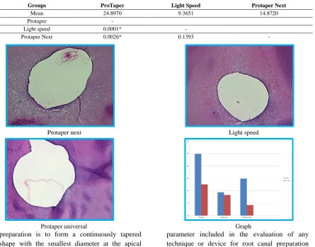
Protaper Next 0.0026* 0.1393 -
preparation is to form a continuously tapered shape with the smallest diameter at the apical foramen and the largest at the orifice to allow effective irrigation and filling. Many instruments have been recommended but only a few seem to be capable of achieving these primary objectives of root canal preparation consistently.[7] The main
parameter included in the evaluation of any technique or device for root canal preparation should be the ability to clean the root canal walls to shape the root canal without straightening. Many studies have also reported that large amount of debris and smear layer often remain after manual or automated preparation with
Protaper next Light speed
endodontic hand pieces.[8] Anatomic irregularities like isthmus between mesial canals or elliptical canals are commonly observed and are preferable sites for pulp tissues and debris to remain within the root canal system. Indeed, endodontic instruments must touch canal walls to clean and in those cases of anatomic complexities, cleaning is done solely by the flow and back flow of irrigating solutions. Nowadays nickel titanium instruments of conic shapes and variable tapers represent a hope of better instrumentation of curved canals for their ability to follow curvatures better. Recent reports relate about those instruments physical properties, cutting efficiency and apical transportation. However, very few literatures has described the efficiency of such instruments in cleaning the apical third of curved root canals therefore becoming the purpose of this study.[9] A new generation rotary endodontic instruments which have been developed from Nickel- titanium alloys has brought a path breaking change in endodontic. They potentially allow shaping of canals, with less procedural error[10] and are more effective in removing debris and smear layer in apical third of the canal as compared to hand instrumentation.[11,12] Light speed is not” just another root canal instrument”. Its design is different from all other rotary instruments , The design gives it the ability to negotiate canal curvature,’ feel’ canal diameter and instrument to an apical size large enough to clean all the walls of a canal.[13] Pro-taper represents revolutionary properties in root canal preparation procedure that was developed to facilitate instrumentation of difficult constricted, curved canals. It is designed to cover the whole range of treatment with only few files. The new file geometry allows for high efficiency and safety.[14] Thompson & Dummer (1997)[15] have shown that that automated device using rotary Nickel- Titanium instruments with various tapers leads to good instrumentation results even in severely curved canal. However little is known about their cleaning effectiveness. Considering the above-mentioned factors, the aim of present study was to evaluate the efficacy of three different rotary instruments: ProTaper Next, Lightspeed and Protaper in the removal of debris and pulp tissue from the apical third of curved root canals.
Materials and Methods
Histological evaluation of three rotary systems Shetty V, Kamatagi L, Raghu M, Chabbra N
was done till size no. 55 by progressively decreasing 1 mm from the working length. Group 3: In this group, the root canals were prepared with ProTaper Universal instruments (Dentsply-Maillefer, Ballaigues, Switzerland) used at 300 rpm with 2 Ncm torque (X‑Smart, Dentsply Maillefer). An SX file was used at 1/2 of the WL; S1 and S2 files were at 2/3 of the working length and F1, F2 and F3 files were at full WL. SX, S1 and S2 files were used in the canals with a brushing motion and the others were used with a gently in- and out-motion until the instrument had reached into the full WL. The root canals were irrigated with 1 mL distilled water after each instrument using a 31-gauge side-vent needle. Preparation for Light microscopic study After completion of the instrumentation, teeth were placed in 10% formalin solution. Coronal 2/3rd of root were removed with a 3/4-inch corborundum separating disk. The remaining apical third sectioned root was stored in 10% formalin solution again. Decalcification was done with 5% solution of Nitric acid and 5%formic acid, washed in running water overnight. The specimens were dehydrated in automated tissue processor (leica Tp10-20 Germany), wax embedded in tissue emedding station (leica EG 1160, Germany).The apical thirds were taken to a microtome (leica Rm 2165, Germany) where histological cuts done and stained with hematoxylin and eosin. All samples were observed under Lightmicroscope at X 10 magnification [ Trinocular Research microscope(Olympus BX51, Japan) with 3-chip CCD camera(proview),analyzed with Image proPlus computer program for windows( media cybernetics,USA)and computer with 17” flatron 77ST (LG) monitor to evaluate pulp tissue and debris at apical 1/3rd.
Evaluation Criteria
Cleansing of the root canals were evaluated in each cross sectional, this was calculated as a percentage figure in terms of percentage of the walls had been planed by the files. A total percentage of the walls planed for each section were estimated; all section percentages were totaled to give an average determination for each canal. Mean percentages then were complied for each of 3 groups.
RESULT
The Mean and Standard deviation values of remaining debris (percentage) according to groups are given in Table 1. The Comparison of three instruments with respect to remaining debris by ANOVA test showed a significant difference among them. Pair wise comparison of three instruments (i.e. group I, group II and group III) with respect to percentage of debris by Tukey multiple post hoc procedure & Tukey post hoc procedure showed that light speed instruments removed more debris compared to Protaper, which is statistically significant. Between protaper Next and protaper, protaper Next group showed significantly more debris removal than protaper, but there was no significant difference in debris removal between lightspeed and protaper Next instruments. Group I (Protaper) presented an average 24.90% of remaining pulp tissue and debris, while Group 2 (lightspeed) presented approximately 9.37% and Group 3 (Protaper Next) showed approximately 14.7. (Fig. 1 and Fig. 2). Statistics Analysis of one way ANOVA – p<=0,05 – showed significant difference between the three groups. LSD, Tukey and Student tests also showed a p<= 0,05 significance level demonstrating no significant statistical difference among the two instrumentation groups.
DISCUSSION
study 3% sodium hypochlorite was used as the irrigant. A thoroughly instrumented and cleaned root canal should not be expected to be free from smear layer, debris and pulpal tissue remnants. These factors should not be considered as dominant reasons for preferring one preparation technique over another. The present study recorded debris in all the specimens which is in agreement with reports in which residual debris was observed in canals after instrumentation and irrigation with Sodium hypochlorite. Mean debris score with LightSpeed 9.37% as compared with 14.87 % and 24.90 % with Protaper Next and ProTaper respectively. The better results in the LightSpeed group can be explained by the fact that spade design of instrument would allow movement of debris coronally in an irrigant flooded canal and the instrument was used in an advanced and withdrawal motion. Cutting occurs with advancement and withdrawal removes the debris. In present study, results indicated statistically significant difference for ProTaper vs LightSpeed ProTaper vs ProFile with regard to pulp tissue and debris removal. There was no significant difference between ProFile and LightSpeed. This observation was in accordance with the previous studies.[1,2,4,5,8,21,32,34,38,41,44] Protaper Next has an offset design that generates a traveling mechanical wave of motion along the active portion of a file. This swaggering effect serves to minimize the engagement between the file and dentin compared to the action of a fixed tapered file with a centered mass of rotation. A file with an offset design affords more cross-sectional space for enhanced cutting, loading and augering debris out of a canal compared to a file with a centered mass and axis of rotation. Many instruments break as a result of excessive intrablade debris packed between the cutting flutes over the active portion of a file. Importantly, an offset file design decreases the probability for laterally compacting debris and blocking root canal system anatomy. Clinically the finding of the study is more important than statistically significance difference between three rotary systems with regard to the amount of Pulp tissue and debris removal in coronal and middle portion of canals, because microorganisms remain in the apical portion of the canal and are considered to be the main cause of failure.[21] The apical preparation size is very important, because
a smaller diameter leaves more canal surface untouched, which might affect the ability to disinfect root canals.[22] Although instrumenting canals to larger sizes might not be prudent in every case, minimal apical preparations based on clinical opinions are far more detrimental to the success of root canal therapy.[23] In this study apical preparation size has been kept to #30 and taper of the preparation varied depending on the rotary system used. The clinical relevance of the current study indicated that none of the rotary instrumentation produced completely clean canal. But LightSpeed demonstrated better results as compared to other two systems. Despite these larger preparations by LightSpeed & ProTaper files, more statistically significant difference was observed among the groups with regard to debris removal. This finding is contrary to the to the study by J-Y Blum et al., which concluded that a low rubbing action or cutting action associated with less active apical portion of the instrument. This may be the result of ProTaper files applying low vertical forces and a series of withdrawing forces at the end of the working sequence.[36,49] In the present study lightspeed and Protaper Next showed significantly less debris than Protaper Universal system, but between lightspeed and Protaper Next there is no statistical significance for debris. However lightspeed files showed better results in debris score followed by Protaper Next and ProTaper. None of the rotary systems used in this study has shown debris free surface at the apical third of the canal. Further in vitro and in vivo investigation is required to evaluate the efficacy of these instruments in removal of debris and smear layer. All histological sections used for analysis were free of artifacts had an intact, undistorted root canal outline. Cross sections at 3mm from the apex of each root were examined in a microscope attached to imaging software (Image ProPlus) under 10X magnification. The use of Light microscope at magnification up to 200X allows the detection of fragments of pulp tissue and debris and a large surface area of the canal wall.[17,27]
CONCLUSION
Histological evaluation of three rotary systems Shetty V, Kamatagi L, Raghu M, Chabbra N
were found to be totally free of microorganisms, which corroborates the existing literature and reaffirms the necessity of the combination of surgical preparation and chemical disinfectants. CONFLICT OF INTEREST & SOURCE OF FUNDING
The author declares that there is no source of funding and there is no conflict of interest among all authors.
BIBLIOGRAPHY
1. Siqueira JF. Histological Evaluation of the Effectiveness of Five Instrumentation Techniques for Cleaning the Apical Third of Root Canals. J Endod. 1997;23(8):499-502. 2. Grove CJ. The value of the
dentino-cemental junction in pulp canal surgery. J Dent Res. 1931;11:466-8.
3. Simon J. The apex: how critical is it? Gen Dent. 1994;42:330-4.
4. Walton RE. Histologic evaluation of different methods of enlarging the pulp canal space. J Endodon. 1976;2:304-11. 5. Walia H, Brantley WA, Gerstein H. An
initial investigation of the bending and torsional properties of Nitinol root canal files. J Endodon. 1988;14:346-51.
6. Esposito PT, Cunningham CJ. A comparison of canal preparation with nickel-titanium and stainless steel instruments. J Endodon 1995;21:173-6.
7. Zmener O, Balbachan L. Effectiveness of nickel-titanium files for preparing curved root canals. Endod Dent Traumatol. 1995;11:121-3.
8. Yang GB, Zhou XD, Zheng L. Shaping ability of progressive versus constant taper instruments in curved root canals of extracted teeth. Int Endod J. 2007;40:707-714.
9. Versumer J, Hulsmann M, Schafers F. A comparative study of root canal preparation using ProFile .04 and Light speed rotary Ni-Ti instruments. Int Endod J. 2002;35:37-46. 10. Ferreia P, Filho G. Cleaning ability of rotary
instruments in the apical third of curved molars. Rev Fac odontol Bauru. 2002;10(4):253-256.
11. Thompson SA, Dummer PMH. Shaping Ability of Light speed Rotary Nickel-Titanium instruments in simulated Rot canals. Part2. J Endod. 1997;23(12)742-747.
12. Peters OA, Barbakow F. Effects of irrigation on Debris and Smear Layer on Canal Walls Prepared by Two Rotary Techniques: J Endod. 2000;26(1)6-10.
13. Bechelli C, Orlandini SZ, Colafranceschi M. Scanning electron microscope study on the efficacy of root canal wall debridement of hand versus Light speed instrumentation. Intl Endod. 1999;32:484-493.
14. LightSpeed Technology Inc. LightSpeed reference Guide.2001.11707 Rain drop drive, San Antonio, Tx, USA. LightSpeed Technology.
15. Clauder T, Baumann AM. ProTaper NT system. Dent Clin North Am. 2004;48(1):87-111.
16. Thompson SA, Dummer PMH. Shaping Ability of Light speed Rotary Nickel-Titanium instruments in simulated Root canals. Part 1. J Endod. 1997;23(11)698-702.
17. Simon J. The apex: how critical is it? Gen Dent. 1994;42:330-4.
18. Schilder H. Cleaning and shaping the root canal. Dent Clin North Am. 1974;18:269. 19. Lim TS, Wee TY, Koh MYC, Lim SV.
Light and scanning electron microscopic evaluation of glyde file prep in smear layer removal. Int Endod J. 2003;36:336-343. 20. Peters OA, Laib A, Gohring TA, Barbakow
F. Changes in root canal geometry after preparation assessed by high resolution computed tomography. J Endod. 2001;27:1-6.
21. Babay N. Non diseased dentinal root surface following citric acid or tetracycline hydrochloride conditioning: A scanning electron microscopic study on the effects of ultrasonic irrigation before and after root conditioning. Quintessence Int. 1997;28(2):93-7.
22. Nair PNR, Sjogren ULF. Intraradicular Bacteria and Fungi in Root-filled, Asymptomatic Human Teeth with Therapy- resistant Periapical Lesions: A Long-term Light and Electron Microscopic Follow-up Study. J endod. 1990;16(12)580-588. 23. Mickel AK, Chogle S, Liddle J, Huffaker K,
2007;33:21-3.
24. Paque F, Ganahl D, Peters AO. Effects of root canal preparation on apical geometry assessed by micro-computed tomography. J Endod. 2009;35:1056-9.