sentinels in the skin
Frank O. Nestle, Brian J. Nickoloff
J Clin Invest.
2007;
117(9)
:2382-2385.
https://doi.org/10.1172/JCI33349
.
Advances in our understanding of the skin immune system have a major impact on studies
of skin autoimmunity, graft-versus-host disease, inflammation, and cancer as well as on the
development of novel vaccines and immunotherapy approaches. In this issue of the
JCI
,
Zaba et al. carefully dissected the complex network of DCs and macrophages residing in
normal human skin and defined novel phenotypic markers for these immunocytes (see the
related article beginning on page 2517). These studies provide the basis for better insight
into the role of important immune sentinels contributing to the maintenance of skin tissue
homeostasis and lay the foundation for future studies of the skin immune system.
Commentary
Find the latest version:
Increasing existing b cell mass is a prom-ising avenue for the treatment of type 2 diabetes, and initial positive results have been obtained with one such agent, exen-din 4, an analog of glucagon-like peptide 1 (8–10). Likewise, the new model will allow researchers to uncover negative effects of commonly used drugs in b cell regen-eration. Finally, these mice will also be instrumental in elucidating the molecular mechanisms that govern the proliferative response of the mature b cell.
Address correspondence to: Klaus H. Kaest-ner, Department of Genetics and Institute for Diabetes, Obesity, and Metabolism,
560A CRB/6140, University of Pennsylva-nia, Philadelphia, Pennsylvania 19104, USA. Phone: (215) 898-8759; Fax: (215) 573-5892; E-mail: [email protected].
1. Shapiro, A.M., et al. 2000. Islet transplantation in seven patients with type 1 diabetes mellitus using a glucocorticoid-free immunosuppressive regimen.
N. Engl. J. Med. 343:230–238.
2. Bonner-Weir, S., and Weir, G.C. 2005. New sources of pancreatic beta-cells. Nat. Biotechnol. 23:857–861. 3. Dor, Y., Brown, J., Martinez, O.I., and Melton, D.A. 2004. Adult pancreatic beta-cells are formed by self-duplication rather than stem-cell differentia-tion. Nature. 429:41–46.
4. Teta, M., Rankin, M.M., Long, S.Y., Stein, G.M., and Kushner, J.A. 2007. Growth and regeneration of adult beta cells does not involve specialized pro-genitors. Dev. Cell. 12:817–826.
5. Nir, T., Melton, D.A., and Dor, Y. 2007. Recovery
from diabetes in mice by b cell regeneration. J. Clin. Invest. 117:2553–2561. doi:10.1175/JCI32959. 6. Brownlee, M. 2003. A radical explanation for
glucose-induced b cell dysfunction. J. Clin. Invest. 112:1788–1790. doi:10.1172/JCI200320501. 7. Wajchenberg, B.L. 2007. beta-cell failure in diabetes
and preservation by clinical treatment. Endocr. Rev. 28:187–218.
8. Brubaker, P.L., and Drucker, D.J. 2004. Minireview: Glucagon-like peptides regulate cell proliferation and apoptosis in the pancreas, gut, and central ner-vous system. Endocrinology. 145:2653–2659. 9. DeFronzo, R.A., et al. 2005. Effects of exenatide
(exendin-4) on glycemic control and weight over 30 weeks in metformin-treated patients with type 2 diabetes. Diabetes Care. 28:1092–1100.
10. Kendall, D.M., et al. 2005. Effects of exenatide (exendin-4) on glycemic control over 30 weeks in patients with type 2 diabetes treated with metformin and a sulfonylurea. Diabetes Care. 28:1083–1091.
Deepening our understanding
of immune sentinels in the skin
Frank O. Nestle1 and Brian J. Nickoloff2
1St. John’s Institute of Dermatology, Division of Genetics and Molecular Medicine, King’s College London School of Medicine, London, United Kingdom. 2Department of Pathology and Department of Microbiology and Immunology, Oncology Institute, Cardinal Bernardin Cancer Center,
Loyola University, Chicago, Illinois, USA.
Advances in our understanding of the skin immune system have a major
impact on studies of skin autoimmunity, graft-versus-host disease,
inflam-mation, and cancer as well as on the development of novel vaccines and
immunotherapy approaches. In this issue of the
JCI
, Zaba et al. carefully
dissected the complex network of DCs and macrophages residing in normal
human skin and defined novel phenotypic markers for these immunocytes
(see the related article beginning on page 2517). These studies provide the
basis for better insight into the role of important immune sentinels
contrib-uting to the maintenance of skin tissue homeostasis and lay the foundation
for future studies of the skin immune system.
The skin is one of the largest organs of the body and has a variety of different func-tions including providing the first line of defense against invading pathogens. Upon casual inspection, normal human skin does not appear to be a major reservoir of immune sentinels and effector cells. However, the absence of inflammation belies the significant number and pheno-typic complexity of tissue-resident T cells, macrophages, and DCs that are becoming
more widely appreciated by immunolo-gists interested in immune surveillance, autoimmunity, chronic inflammatory diseases in the skin, and beyond (1). Once the primary barrier function of skin (e.g., disruption of the outermost dead layer of cells forming the stratum corneum) is breached, immune sentinel and effector cells are poised to provide rapid and effi-cient immunological backup to restore tissue homeostasis. For example, it is not widely appreciated that there are approxi-mately 2 × 10¹⁰ skin-resident T cells, i.e.,
twice the total number of T cells in the circulation (2). A frequent misconception regarding cutaneous immunity is that “skin” equals “epidermis” (3). Once one probes beneath the basement membrane zone, there is a multitude of cells, includ-ing T lymphocytes, DCs, and macro-phages, that are regarded as components
of the dermal immune system and com- plement the overlying epidermal compo-nents to form the skin immune system. The dermal immune system provides a highly reactive immunologically based compartment that is crucially involved in the majority of chronic inflammatory skin disorders including psoriasis and atopic dermatitis (4). Here, we primarily focus on the dermal-based immune sys-tem under normal or noninflammatory skin conditions.
Skin-resident sentinels of the immune system
Skin-resident sentinels of the immune system include a spectrum of cells, rang-ing from motile DCs highly specialized in sensing danger and presenting antigens to tissue-resident macrophages specializing in phagocytosis, thereby clearing poten-tially dangerous substances and mediating tissue remodeling. Langerhans cells (LCs) are dendritic, antigen-presenting sentinels of the epidermis. LCs renew locally in epi- dermis and only recruit blood-borne pre-cursors following injury (5). Even though LCs were described 140 years ago, their in vivo function still remains elusive. Recent scientific evidence obtained using LC-depleted genetically modified mice adds
Nonstandard abbreviations used: BDCA, blood DC antigen; DC-SIGN, DC-specific ICAM-grabbing nonintegrin; DDC, dermal DC; FXIIIa, factor XIIIa; LC, Langerhans cell; PDC, plasmacytoid pre-DC. Conflict of interest: The authors have declared that no conflict of interest exists.
to potential confusion by demonstrating both immunoactivating and immunosup- pressive roles of LCs (3). While LCs are well-studied components of the skin immune system, dermal DCs (DDCs) were only identified and functionally characterized 14 years ago (6, 7). Despite the presence of significant numbers of DDCs in normal and pathological skin, the complexity of this cell type is just beginning to be under- stood. DDCs are defined by their character-istic phenotypic and functional properties including typical dendritic morphology, nonadherence to plastic, expression of high levels of adhesion and costimulatory molecules, and potent stimulatory capacity of naive and memory T cells in the context of alloantigens and nominal antigens (8). They have potential to divide locally and
thus maintain their tissue density in the steady state (9). A key unifying feature of DDCs is expression of the nonpolymorphic MHC-like lipid antigen-presenting mol-ecule blood DC antigen–1 (BDCA-1; also known as CD1c) as well as C-type lectins such as the pathogen sensor DC-specific ICAM-grabbing nonintegrin (DC-SIGN; also known as CD209) and DEC205 (also known as CD205).
Three major DDC subpopulations have been characterized in the past: BDCA-1+CD1a+CD14– and BDCA-1+CD1a–CD14–
DDCs have potent antigen-presenting capacity (6, 10), and BDCA-1+CD1a–CD14+
DDCs have low antigen-presenting capac-ity but the potential to develop into CD1a+
and Langerin-positive LCs under control of TGF-b (11). There is also an important
[image:3.585.128.458.81.356.2]subset of proinflammatory DDCs (termed “TNF- and iNOS-producing DCs” in refer-ence to their proinflammatory mouse DC counterparts; ref. 12). A recent addition to the family of skin immune sentinels is type I IFN–producing plasmacytoid pre-DCs (PDCs). These cells are rare in normal skin, but accumulate under conditions of chron-ic skin inflammation such as psoriasis and lupus erythematosus and have impor-tant functions in skin immunopathology (13, 14). A further component of the der- mal immune system is the dermal mac-rophage. Although detailed functional studies are lacking, several surface markers including CD14, CD68, HLA-DR, RM3/1, and CD11c have been used in the past to describe the phenotypic diversity of the dermal macrophage population (15, 16).
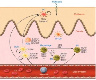
Figure 1
Normal human skin is characterized by an impressive diversity of immune sentinels. Skin-based DCs and macrophages sense invading pathogens and serve as sentinels, thereby alerting effectors of the innate and adaptive immune systems to potential danger to the host. Subsets of immune sentinels include CD1a+Langerin+ LCs located in the epidermis and various subtypes of DCs and macrophages in the dermis. In this issue of the
Diversity of DCs and macrophage subsets in the dermis
In a study reported in this issue of the
JCI, Zaba et al. (17) shed new light on the phenotype and function of DDCs and macrophages. Using a panel of mono-clonal antibodies to carefully dissect the phenotypic expression profile of DDCs and macrophages, they found 2 major subpopulations of dermal immune senti-nels. An overview of the mononuclear cell subsets located in the epidermis and der-mis of normal human skin incorporating the findings of Zaba et al. is portrayed in Figure 1. The BDCA-1+CD11c+HLA-DR+
DDCs possessed strong stimulatory capacity for allogeneic T cells, and a second macrophage-like population expressed CD163 and factor XIIIa (FXIIIa). CD163 is a hemoglobin/haptoglobin complex–binding macrophage scavenger
receptor expressed on the majority of tis-sue macrophages (18) and can be readily added to the phenotypic criteria for skin macrophages. FXIIIa, best known for its role in coagulation, is also a tissue trans-glutaminase with a potential functional role in wound healing and other cutane-ous disorders (4). Expression of FXIIIa is related to cell activation and inducible via IL-4 in alternatively activated macrophages (19). FXIIIa is also expressed in migratory DDCs (6, 11), monocyte-derived DCs (20), hematopoietic progenitor cell–derived DDC equivalents (21), and a population of MHC class II+ dendritic-appearing
cells of the dermis termed “dermal den-drocytes” (22). Despite these new find-ings, an immediate question arises: What is the biological significance of FXIIIa expression by dermal macrophages and DCs? Rather than being a lineage-specific marker, accumulating data suggest that FXIIIa expression indicates an alterna-tive cellular activation state induced by exposure of DCs and macrophages to IL-4 (19, 20). Furthermore, FXIIIa expression might indicate a potential functional role in tissue remodeling. Thus, the original description of FXIIIa dermal dendrocytes in various pathological states (22) might have included both DCs and macrophages with a dendritic morphology residing in a distinctive cytokine-containing (e.g., IL-4– rich) skin microenvironment.
Accumulating data seem to indicate that there exists a continuum of pheno- types and functions and remarkable plas-ticity between tissue-resident DCs and macrophages (23). Based on the findings by Zaba et al. (17), it is clear that certain phenotypic markers may be useful to define mononuclear cell subsets at each end of a spectrum. For example, BDCA-1+
cells identify DDCs with potent allos-timulating properties, while the CD163+
cells lacked significant allostimulatory capacity but possessed greater phagocytic activity. As portrayed in Figure 2, between these ends of the spectrum, there are der-mal mononuclear cell subsets that express various markers that may identify cell types with more flexible functionality as regards antigen presentation and phago-cytosis. Thus, it is now well established that immature DCs, including DDCs, can be phagocytic, a cellular function usu-ally attributed to macrophages (24, 25). On the other hand, macrophages might be potent antigen-presenting cells for CD8+
T cells (26). Expression of CD1a on DDCs
indicates potent antigen-presenting func-tion, while expression of CD14 indicates a precursor role for LCs (11). Thus, tissue- resident mononuclear sentinels of the der-mis are likely to exist in a pluripotent state and, depending on microenvironmental factors, may acquire an antigen-present- ing mode, migratory mode, or tissue-resi-dent phagocytic mode.
Future directions and conclusions
The present study by Zaba et al. (17) contrib- utes to a deeper understanding of the com- plex world of DCs and macrophages resid-ing in normal human skin. The study also raises important questions for future study. What is the role of human skin DCs versus macrophages in the induction of peripheral T cell unresponsiveness under steady-state conditions (27)? What is the in vivo func-tional role of human skin DCs in priming and cross-priming of a pathogen-specific immune response, and can these properties be exploited for vaccination purposes? And finally, what role are these mononuclear cells playing in conditions of chronic inflamma-tion and cancer in the skin? Despite these uncertainties, we now have better surface markers to delineate the confederacy of sen-tinel cells in skin, which will hopefully allow us to probe more deeply into proinflamma-tory, antiinflammatory, immunogenic, and tolerogenic DC and macrophage subsets and their potential roles in skin inflamma-tion and skin cancer.
Acknowledgments
The authors thank Lori Kmet and Lynn Walter for manuscript preparation and acknowledge support from the NIH (grants AR40065 and CA39542), the Wellcome Trust, and the United Kingdom Depart-ment of Health through the NIH Research Biomedical Research Centre award to Guy’s & St. Thomas’ National Health Ser-vice Foundation Trust in partnership with King’s College London.
[image:4.585.43.198.79.258.2]Address correspondence to: Brian J. Nick-oloff, Loyola University Medical Center, 2160 S. First Avenue, Building 112 Room 301, Maywood, Illinois 60513, USA. Phone: (708) 327-3241; Fax: (708) 327-3239; E-mail: [email protected]. Or to: Frank O. Nestle, Division of Genetics and Molecular Medicine, King’s College London School of Medicine, 8th Floor Guy’s Tower, Guy’s Hospital, London SE1 9RT, United King- dom. Phone: 44-20-7188-9038; Fax: 44-20-7188-2585; E-mail: [email protected].
Figure 2
1. Boyman, O., Conrad, C., Tonel, G., Gilliet, M., and Nestle, F.O. 2007. The pathogenic role of tissue-resident immune cells in psoriasis. Trends Immunol. 28:51–57.
2. Clark, R.A., et al. 2006. The vast majority of CLA+ T cells are resident in normal skin. J. Immunol. 176:4431–4439.
3. Romani, N., et al. 2006. Epidermal Langerhans cells--changing views on their function in vivo.
Immunol. Lett. 106:119–125.
4. Nickoloff, B.J. 1993. Dermal immune system. CRC Press. Boca Raton, Florida, USA. 340 pp. 5. Merad, M., et al. 2002. Langerhans cells renew in the
skin throughout life under steady-state conditions.
Nat. Immunol. 3:1135–1141.
6. Nestle, F.O., Zheng, X.G., Thompson, C.B., Turka, L.A., and Nickoloff, B.J. 1993. Characterization of dermal dendritic cells obtained from normal human skin reveals phenotypic and functionally distinctive subsets. J. Immunol. 151:6535–6545. 7. Lenz, A., Heine, M., Schuler, G., and Romani, N.
1993. Human and murine dermis contain den-dritic cells. Isolation by means of a novel method and phenotypical and functional characterization.
J. Clin. Invest. 92:2587–2596.
8. Nestle, F.O., Filgueira, L., Nickoloff, B.J., and Burg, G. 1998. Human dermal dendritic cells process and present soluble protein antigens. J. Invest. Dermatol. 110:762–766.
9. Bogunovic, M., et al. 2006. Identification of a radio-resistant and cycling dermal dendritic cell population in mice and men. J. Exp. Med. 203:2627–2638.
10. Angel, C.E., et al. 2006. Cutting edge: CD1a+ anti-
gen-presenting cells in human dermis respond rap-idly to CCR7 ligands. J. Immunol. 176:5730–5734. 11. Larregina, A.T., et al. 2001. Dermal-resident CD14+
cells differentiate into Langerhans cells. Nat. Immunol. 2:1151–1158.
12. Lowes, M.A., et al. 2005. Increase in TNF-alpha and inducible nitric oxide synthase-expressing den- dritic cells in psoriasis and reduction with efali-zumab (anti-CD11a). Proc. Natl. Acad. Sci. U. S. A. 102:19057–19062.
13. Nestle, F.O., et al. 2005. Plasmacytoid predendritic cells initiate psoriasis through interferon-alpha production. J. Exp. Med. 202:135–143.
14. Wollenberg, A., et al. 2002. Plasmacytoid dendritic cells: a new cutaneous dendritic cell subset with distinct role in inflammatory skin diseases. J. Invest. Dermatol. 119:1096–1102.
15. Djemadji-Oudjiel, N., Goerdt, S., Kodelja, V., Sch- muth, M., and Orfanos, C.E. 1996. Immunohis-tochemical identification of type II alternatively activated dendritic macrophages (RM 3/1+3, MS-1+/-, 25F9-) in psoriatic dermis. Arch. Dermatol. Res. 288:757–764.
16. Weber-Matthiesen, K., and Sterry, W. 1990. Organi- zation of the monocyte/macrophage system of nor-mal human skin. J. Invest. Dermatol. 95:83–89. 17. Zaba, L.C., Fuentes-Duculan, J., Steinman, R.M.,
Krueger, J.G., and Lowes, M.A. 2007. Normal human dermis contains distinct populations of CD11c+BDCA-1+ dendritic cells and CD163+FXIIIA+
macrophages. J. Clin. Invest. 117:2517–2525. doi:10.1172/JCI32282.
18. Fabriek, B.O., Dijkstra, C.D., and van den Berg, T.K. 2005. The macrophage scavenger receptor CD163.
Immunobiology. 210:153–160.
19. Torocsik, D., Bardos, H., Nagy, L., and Adany, R.
2005. Identification of factor XIII-A as a marker of alternative macrophage activation. Cell. Mol. Life Sci. 62:2132–2139.
20. Grassi, F., et al. 1998. Monocyte-derived dendritic cells have a phenotype comparable to that of der-mal dendritic cells and display ultrastructural granules distinct from Birbeck granules. J. Leukoc. Biol. 64:484–493.
21. Caux, C., et al. 1996. CD34+ hematopoietic pro-genitors from human cord blood differentiate along two independent dendritic cell pathways in response to GM-CSF+TNF alpha. J. Exp. Med. 184:695–706.
22. Cerio, R., Griffiths, C.E., Cooper, K.D., Nickoloff, B.J., and Headington, J.T. 1989. Characterization of factor XIIIa positive dermal dendritic cells in normal and inflamed skin. Br. J. Dermatol. 121:421–431. 23. Gordon, S., and Taylor, P.R. 2005. Monocyte and
macrophage heterogeneity. Nat. Rev. Immunol. 5:953–964.
24. Blander, J.M., and Medzhitov, R. 2006. On regula- tion of phagosome maturation and antigen presen-tation. Nat. Immunol. 7:1029–1035.
25. Filgueira, L., Nestle, F.O., Rittig, M., Joller, H.I., and Groscurth, P. 1996. Human dendritic cells phago-cytose and process Borrelia burgdorferi. J. Immunol. 157:2998–3005.
26. Pozzi, L.A., Maciaszek, J.W., and Rock, K.L. 2005. Both dendritic cells and macrophages can stimu-late naive CD8 T cells in vivo to proliferate, develop effector function, and differentiate into memory cells. J. Immunol. 175:2071–2081.
27. Steinman, R.M., Hawiger, D., and Nussenzweig, M.C. 2003. Tolerogenic dendritic cells. Annu. Rev. Immunol. 21:685–711.
Taking aim at translation for tumor therapy
Bryan C. Barnhart1 and M. Celeste Simon1,2,3,4
1Abramson Family Cancer Research Institute, University of Pennsylvania Cancer Center, 2Howard Hughes Medical Institute, and 3Department of Cancer Biology and 4Department of Cell and Developmental Biology, School of Medicine, University of Pennsylvania, Philadelphia, Pennsylvania, USA.
Increased cap-dependent mRNA translation rates are frequently observed
in human cancers. Mechanistically, many human tumors often overexpress
the cap binding protein eukaryotic translation initiation factor 4E (eIF4E),
leading to enhanced translation of numerous tumor-promoting genes. In
this issue of the
JCI
, Graff and colleagues describe potent antitumor effects
using second-generation antisense oligonucleotides for eIF4E (see the
relat-ed article beginning on page 2638). If their results are recapitulatrelat-ed in a
clinical setting, this strategy will provide a promising antitumor therapy
with broad-reaching applications.
Protein synthesis is required for many critical cellular processes, and cells regu-late mRNA translation rates accord-ing to their needs. Interestingly, dys-regulated translation has now been
linked to multiple human cancers (1, 2). Increased translation rates lead to an overproduction of proteins involved in proliferation, survival, metastasis, and other malignant characteristics (3–5). Protein synthesis regulation is complex, and its alteration in tumor cells occurs at numerous points. Many tumor-pro-moting mechanisms ultimately cause the activation of a critical regulator of cap-dependent translation, the eukaryotic translation initiation factor 4F (eIF4F) complex. Numerous human tumors
exhibit inappropriate eIF4F activation, including lymphomas and breast, pros-tate, colorectal, head and neck, cervical, bladder, and lung cancers (3). Therefore, therapeutically targeting eIF4F activity is exceedingly attractive, as it would poten-tially be applicable to a broad range of human cancers. In this issue of the JCI, Graff and colleagues (6) report such a strategy, attacking one of the important components of eIF4F, eIF4E, with striking efficacy in tumor models. If this treatment is successful in the clinic, it holds great promise for use against many human tumors and may be especially effective if used in combination with more tradition-al chemotherapeutic treatments.
Cap-dependent protein synthesis and its regulation
To provide better insight into the underly-ing mechanism for this therapy and why it might ultimately be so effective against
Nonstandard abbreviations used: ASO, antisense oligonucleotide; 4E-BP, eIF4E binding protein; eIF4E, eukaryotic translation initiation factor 4E; mTORC1, mammalian target of rapamycin complex 1.
Conflict of interest: The authors have declared that no conflict of interest exists.